Detrusor instability is a rare presentation of pelvic ... - Pelviperineology
Detrusor instability is a rare presentation of pelvic ... - Pelviperineology
Detrusor instability is a rare presentation of pelvic ... - Pelviperineology

Create successful ePaper yourself
Turn your PDF publications into a flip-book with our unique Google optimized e-Paper software.
Case report<br />
<strong>Detrusor</strong> <strong>instability</strong> <strong>is</strong> a <strong>rare</strong> <strong>presentation</strong> <strong>of</strong> <strong>pelvic</strong> subserosal<br />
fibroma, seven years following a total abdominal hysterectomy<br />
and bilateral salpingo-oophorectomy: Case report<br />
NADER GAD, CECELIA O’BRIEN<br />
Department <strong>of</strong> Obstetrics and Gynaecology, Royal Darwin Hospital, Casuarina, NT, Australia<br />
Abstract: Solitary Fibrous Tumours (SFT) are uncommon, slow growing, well-circumscribed tumours ar<strong>is</strong>ing from the mesenchyma. These<br />
tumours were first described in the thoracic cavity, originating from the pleura but have since been reported in various sites including the<br />
prostate, bladder, periosteum, s<strong>of</strong>t t<strong>is</strong>sue, liver and kidney. Th<strong>is</strong> case report describes a 40 year old woman, presenting with symptoms <strong>of</strong><br />
detrusor <strong>instability</strong> due to a midline solid, d<strong>is</strong>crete <strong>pelvic</strong> mass, centred above the bladder, with d<strong>is</strong>placement <strong>of</strong> the sigmoid colon evident on<br />
CT scan. H<strong>is</strong>tology revealed a ‘subserosal fibroma’. Th<strong>is</strong> article describes the current literature relating to <strong>pelvic</strong> solitary fibrous tumours.<br />
Key words: Subserosal fibroma; Solitary fibrous tumours; <strong>Detrusor</strong> <strong>instability</strong><br />
Introduction<br />
Solitary fibrous tumours (SFT) or commonly known<br />
as fibromas, were first documented at the turn <strong>of</strong> the 20 th<br />
century, originating in the intrathoracic cavity, typically from<br />
the pleura. Fibromas are slow growing, well circumscribed,<br />
spindle cell neoplasms ar<strong>is</strong>ing from the mesenchyma. Since<br />
the 1960s, there have been an increasing number <strong>of</strong> case<br />
reports describing fibromas in extrathoracic sites including<br />
the orbit, meninges, upper respiratory tract, thyroid, salivary<br />
gland and spinal cord. In the last 20 years, reports <strong>of</strong> fibromas<br />
have been documented in the peritoneum, retroperitoneum,<br />
urinary bladder, uterus, cervix, vulva, vagina, paravaginal<br />
space, <strong>is</strong>chiorectal fossa and fallopian tube 1-8 .<br />
The <strong>presentation</strong> <strong>of</strong> <strong>pelvic</strong> solitary fibrous tumours remains<br />
highly variable and related to anatomical relationships and<br />
size 4 . SFT are commonly asymptomatic, but compression<br />
on adjacent structures has been associated with bowel<br />
obstruction and bilateral ureteral obstruction 4 , vaginal<br />
bleeding or dyspareunia 10 . There have been case reports <strong>of</strong><br />
systemic features such as arthralgia and hypoglycaemia in<br />
cases <strong>of</strong> extrathoracic SFT 1,5,8 . Th<strong>is</strong> case report describes<br />
a unique <strong>presentation</strong> <strong>of</strong> a <strong>pelvic</strong> fibroma with detrusor<br />
<strong>instability</strong> following a total abdominal hysterectomy and<br />
bilateral salpingo-oophorectomy.<br />
The search strategy used in Medline included the key<br />
words; solitary fibrous tumour, pelv<strong>is</strong> and subserosal and the<br />
subject headings; neoplasms fibrous t<strong>is</strong>sue and fibromas. Hand<br />
searching methods were also employed. There were 14 engl<strong>is</strong>h<br />
articles pertaining to solitary fibrous tumours in the pelv<strong>is</strong>.<br />
Case Report<br />
A 40 year old woman was referred by her general<br />
practitioner (GP) because <strong>of</strong> stress urinary incontinence,<br />
urgency and urge incontinence. She was seen in the<br />
urogynaecology clinic and stated that these symptoms<br />
started few years ago but have worsened recently. She passes<br />
urine up to 8 times during the day and once during the night.<br />
Seven years ago she had total abdominal hysterectomy and<br />
bilateral salpingo-oophorectomy for <strong>pelvic</strong> endometrios<strong>is</strong>,<br />
confirmed by h<strong>is</strong>tological examination. It was stated in the<br />
operative notes that no residual d<strong>is</strong>ease was left. She had<br />
been on annual oestradiol implants since her procedure.<br />
Recent testing <strong>of</strong> midstream urine sample arranged by her<br />
local doctor did not show any urinary tract infection. She<br />
had two children via normal vaginal delivery. There was no<br />
other past medical or surgical h<strong>is</strong>tory <strong>of</strong> significance.<br />
On examination, she was generally well with abdominal<br />
palpation normal. Pelvic examination revealed a normal<br />
vulva and vagina with no genital prolapse. Bimanual<br />
<strong>Pelviperineology</strong> 2010; 29: 79-80 http://www.pelviperineology.org<br />
<strong>pelvic</strong> examination revealed a large solid mass that was felt<br />
centred in her pelv<strong>is</strong>. The urodynamic assessment revealed<br />
no residual urine with normal ur<strong>of</strong>lowmetry. <strong>Detrusor</strong><br />
<strong>instability</strong> was demonstrated and associated with significant<br />
urgency but no urge incontinence. No urodynamic stress<br />
urinary incontinence was demonstrated. A <strong>pelvic</strong> ultrasound<br />
was arranged and a blood test to check for serum tumours<br />
markers (CA 125, CA 19.9 and CEA).<br />
On her follow-up v<strong>is</strong>it, all tumour markers were well<br />
within normal limits. The <strong>pelvic</strong> ultrasound showed a<br />
midline solid <strong>pelvic</strong> mass measuring 9.1 x 8.5 x 8.4 cm.<br />
There was no evidence <strong>of</strong> hydronephros<strong>is</strong> in either kidneys<br />
and no ascites. Due to uncertainty <strong>of</strong> the origin <strong>of</strong> the mass,<br />
a CT scan <strong>of</strong> the pelv<strong>is</strong> and abdomen was arranged.<br />
Portal venous and delayed phase multi-detector spiral<br />
CT scan showed a solid <strong>pelvic</strong> mass centred to the right <strong>of</strong><br />
midline but occupying the greater part <strong>of</strong> the lesser pelv<strong>is</strong>.<br />
It measured 10.7 x 8.5 x10.2 cm. It was centred above the<br />
bladder but separate from it. The mass d<strong>is</strong>placed the sigmoid<br />
colon superiorly and was adjacent to the vaginal vault. No<br />
definite organ <strong>of</strong> origin could be identified. There was very<br />
small volume <strong>of</strong> ascites but no intra-abdominal lymph<br />
node enlargement. The kidneys, liver, spleen, pancreas and<br />
adrenal glands were normal. The CT images <strong>of</strong> from th<strong>is</strong><br />
case study are shown in Figure 1.<br />
Due to the past h<strong>is</strong>tory <strong>of</strong> the total abdominal hysterectomy<br />
and bilateral salpingo-oophorectomy along with the<br />
uncertainty <strong>of</strong> the origin <strong>of</strong> the mass she was referred to<br />
the surgical team. The findings from the laparotomy by the<br />
surgical team revealed a large <strong>pelvic</strong> mass, which was adhered<br />
to the sigmoid colon. The procedure was complicated by the<br />
presence <strong>of</strong> vascular adhesions. The bowel was separated<br />
from the mass uneventfully and the mass was completely<br />
removed. The h<strong>is</strong>tology <strong>of</strong> the mass was reported as a<br />
subserosal fibroma with no malignant changes seen.<br />
The patient made a complete recovery. No postoperative<br />
urodynamic assessment was performed due to complete<br />
resolution <strong>of</strong> her urinary symptoms following her surgery. At<br />
the 2 year follow up appointment, she remained symptom free.<br />
D<strong>is</strong>cussion<br />
Th<strong>is</strong> case report outlines the significance <strong>of</strong> considering<br />
solitary fibrous tumours as a differential diagnos<strong>is</strong> for<br />
a <strong>pelvic</strong> mass resulting in common urogynaecological<br />
<strong>presentation</strong>s. The differential diagnos<strong>is</strong> <strong>of</strong> a <strong>pelvic</strong> fibroma<br />
most commonly includes leiomyoma or leiomyosarcoma,<br />
hemangiopericytoma and fibrosarcoma 3 .<br />
In 1997, Chan proposed diagnostic criteria for solitary<br />
fibrous tumours, with the essential diagnostic features<br />
79
N. Gad - C. O’brien<br />
When a <strong>pelvic</strong> tumour compresses on the bladder and causes<br />
urinary symptoms, urodynamic assessment <strong>is</strong> more likely<br />
to reveal stress urinary incontinence rather than detrusor<br />
<strong>instability</strong>. Pelvic tumours can irritate the bladder and result<br />
in detrusor <strong>instability</strong>.<br />
No postoperative urodynamic assessment was performed<br />
in th<strong>is</strong> patient as she continued to be asymptomatic until she<br />
was last seen 2 years after her surgery.<br />
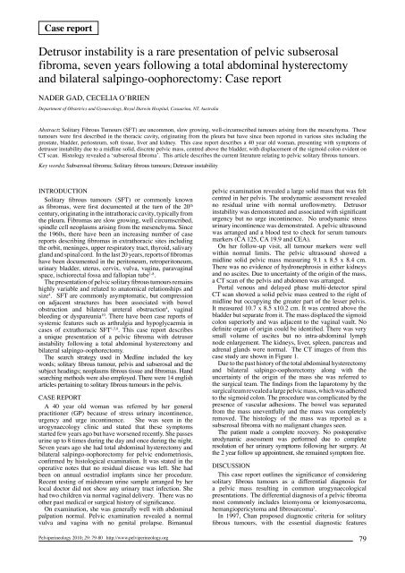
Fig. 1. – Is a sagittal section from the portal venous and delayed phase<br />
spiral computer<strong>is</strong>ed tomography <strong>of</strong> the patient in th<strong>is</strong> case study. Th<strong>is</strong><br />
figure illustrates clearly the subserosal solitary fibrous tumour in<br />
the paravaginal space, centered above and compressing the bladder<br />
along with d<strong>is</strong>placing the sigmoid colon. The mass measures 10.7<br />
cm anterioposteriorly x 8.5 cm wide x 10.2 cm craniocaudally (SC:<br />
sigmoid colon, B: bladder, V: vagina and F: fibroma).<br />
including circumscription, hypercellular and hypocellular<br />
foci, short spindly with scanty and poorly defined<br />
cytoplasm, haphazard, storiform or fascicular arrangement<br />
<strong>of</strong> spindle cells, interwining <strong>of</strong> thin or thick collagen and<br />
CD 34 positive. Immunoh<strong>is</strong>tochemical markers such as CD<br />
34 are used to further differentiate solitary fibrous tumours<br />
from my<strong>of</strong>ibromas, neur<strong>of</strong>ibromas, nodular fasciit<strong>is</strong>,<br />
dermat<strong>of</strong>ibroma and dermat<strong>of</strong>ibrosacroma protuberans 10 .<br />
The CD 34 antigen <strong>is</strong> a transmembrane glycoprotein on<br />
the cell surface that <strong>is</strong> widely used as a vascular marker 12 .<br />
Positive markers for solitary fibrous tumours include bcl-2,<br />
CD 99 and CD 34.<br />
The majority <strong>of</strong> extrathoracic solitary fibrous tumours<br />
follow a benign course. Chan and colleagues found that<br />
malignancy was documented in13% <strong>of</strong> extrathoracic SFT,<br />
compared with the 23% <strong>of</strong> intrathoracic tumours. A review<br />
article examining the h<strong>is</strong>tological features and outcome <strong>of</strong> 7<br />
cases <strong>of</strong> retroperitoneal solitary fibrous tumours found there<br />
was local recurrence in one case, associated with incomplete<br />
resection 3 . Chan believes th<strong>is</strong> observed difference <strong>is</strong> due to<br />
the more recent recognition and reporting <strong>of</strong> the extrathoracic<br />
solitary fibrous tumours, rather than a lower malignant<br />
potential. Therefore, h<strong>is</strong>topathology can not always predict<br />
the clinical behaviour, and follow up <strong>is</strong> essential 12 .<br />
The importance <strong>of</strong> imaging <strong>of</strong> palpable <strong>pelvic</strong> masses<br />
<strong>is</strong> essential in the diagnos<strong>is</strong> and planning for surgical<br />
management (see Figure 1 and 2). MR imaging <strong>of</strong> the <strong>pelvic</strong><br />
region may be useful in further d<strong>is</strong>tingu<strong>is</strong>hing the t<strong>is</strong>sue <strong>of</strong><br />
origin and has an important role in staging <strong>of</strong> <strong>pelvic</strong> tumours,<br />
although imaging modalities have not been specifically<br />
defined for solitary fibrous tumours 13 . Colour or Power<br />
Doppler ultrasound and MRI have been shown to further<br />
differentiate subserosal myomas from extrauterine tumours 14 .<br />
Interestingly, one case report describes central malignant<br />
degeneration <strong>of</strong> an extrathoracic SFT in the pelv<strong>is</strong> <strong>of</strong> a 61<br />
year old man, which was detected on CT and MRI 15 .<br />
In th<strong>is</strong> case study, the significance <strong>of</strong> the previous total<br />
abdominal hysterectomy and bilateral salpingo-oophorectomy<br />
7 years prior to th<strong>is</strong> <strong>presentation</strong> <strong>is</strong> most probably unrelated to<br />
the subserosal fibroma but contributed to a more complicated<br />
surgical resection. There has been only one case report in the<br />
literature that described a benign solitary uterine leiomyoma<br />
on the <strong>pelvic</strong> peritoneum following a hysterectomy for<br />
subserosal fibroids 5 years previously 16 .<br />
The ability to resect the extrathoracic solitary fibrous<br />
tumours <strong>is</strong> the major determinant <strong>of</strong> prognos<strong>is</strong> and ongoing<br />
surveillance <strong>is</strong> essential 12 . While the recognition <strong>of</strong><br />
extrathoracic solitary fibrous tumours has evolved in the last<br />
20 years, the epidemiology, pathophysiology and long term<br />
prognos<strong>is</strong> <strong>of</strong> th<strong>is</strong> mesenchymal tumour remains uncertain.<br />
Most cases <strong>of</strong> detrusor <strong>instability</strong> are idiopathic in nature.<br />
References<br />
1) Trastour C, Bafghi A, Checchi-Guichard C, Bongain A and<br />
Pichie M. Solitary Fibrous tumours <strong>of</strong> the pelv<strong>is</strong> [Letter to the<br />
Editor]. European Journal <strong>of</strong> Obstetrics & Gynecology and<br />
Reproductive Biology 2006; 124: 254-259.<br />
2) Goodland JR, Fletcher CD. Solitary fibrous tumour ar<strong>is</strong>ing at unusual<br />
sites: Analys<strong>is</strong> <strong>of</strong> series. H<strong>is</strong>topathology 1991; 19:515-522.<br />
3) Madhuvrata P, Jayachandran MC, Edmonds DK, Agarwal S and<br />
El-Bahrawy M. Retroperitoneal solitary fibrous tumour ar<strong>is</strong>ing<br />
from the pelv<strong>is</strong> in women – A case report and review <strong>of</strong> the<br />
literature. Journal <strong>of</strong> Obstetrics and Gynaecology 2005; 25(2):<br />
189-192.<br />
4) Yi B, Bewtra C, Yussef K and Silva E. Giant <strong>pelvic</strong> solitary<br />
fibrous tumour obstructing intestinal and urinary tract: a case<br />
report and literature review. American Surgeon 2007; 73(5);<br />
478-80.<br />
5) Dudkiewicz M, Deschenes JL, Bloom C and Gordon PH.<br />
Solitary Fibrous Tumor <strong>of</strong> the Ishioanal Fossa: Report <strong>of</strong> a<br />
Case and Review <strong>of</strong> the Literature. D<strong>is</strong>eases <strong>of</strong> the Colon and<br />
Rectum 2004; 47: 535-7.<br />
6) Tzelepi V, Zolota V, Bat<strong>is</strong>tatou A and Fokaefs E. Solitary<br />
fibrous tumor <strong>of</strong> the urinary bladder: report <strong>of</strong> a case with longterm<br />
follow-up and review <strong>of</strong> the literature. European Review<br />
for Medical and Pharmacological Sciences 2007; 11: 101-6.<br />
7) Thaung C, Shanks J, Eyden B and Fitzmaurice R. Solitary<br />
fibrous tumour <strong>of</strong> the uterus [Correspondence]. H<strong>is</strong>topathology<br />
2006; 49: 198-216.<br />
8) Wakami K, Tateyama H, Kawashima H, Matsuno T, Kamiya<br />
Y, Jin-no Y, Kimura G and Eimoto T. Solitary Fibrous Tumor<br />
<strong>of</strong> the Uterus Producing High-Molecular-Weight Insulin-like<br />
Growth Factor II and Associated With Hypoglycemia.[Report]<br />
International Journal <strong>of</strong> Gynecological Pathology 2005;<br />
24(1):79-84.<br />
9) Michael CA. Pelvic fibroma causing recurrent attacks <strong>of</strong><br />
hypoglycaemia. Journal <strong>of</strong> Obstetrics & Gynaecology <strong>of</strong> the<br />
Brit<strong>is</strong>h Commonwealth, 1967; 74(2): 301-3.<br />
10) Daskalak<strong>is</strong> GJ, Karabinas CD, Parantoniou NE, Papaspyrou<br />
IA and Antaskl<strong>is</strong> AJ. Vaginal Fibroma. Case Report. European<br />
Journal <strong>of</strong> Gynaecological Oncology 2002; 575-6.<br />
11) Vadmal MS and Pellegrini AE. Solitary Fibrous Tumor <strong>of</strong> the<br />
Vagina. The American Journal <strong>of</strong> Dermatopathology 2000;<br />
22(1): 83-86.<br />
12) Chan, JKC. Solitary fibrous tumour – everywhere, and a<br />
diagnos<strong>is</strong> in vogue. H<strong>is</strong>topathology 1997; 31: 568-576.<br />
13) Olson MN, Posniak HV, Tempany CM, Dudiak CM. MR<br />
imaging <strong>of</strong> the female <strong>pelvic</strong> region. A Review Publication <strong>of</strong><br />
the The Radiological Society <strong>of</strong> North America 1992; 12(3):<br />
445-65.<br />
14) Kim SH, Sim JS and Seong CK. Interface Vessels on Color/<br />
Power Doppler US and MRI: A Clue to Differientate Subserosal<br />
Uterine Myomas from Extrauterine Tumors. Journal <strong>of</strong><br />
Computer Ass<strong>is</strong>ted Tomography 2001; 25(1) 36-42.<br />
15) Vossough A, Torigian DA, Zhang PJ, Siegelman ES, Banner M.<br />
Extrathoracic solitary fibrous tumour <strong>of</strong> the <strong>pelvic</strong> peritoneum<br />
with central malignant degeneration on CT and MRI. Journal <strong>of</strong><br />
Magnetic Resonance Imaging 2005; 22(5): 684-686.<br />
16) Shukunami K, N<strong>is</strong>hijima K, Kurokawa T, Or<strong>is</strong>aka M, Yoshida Y<br />
and Kotsuji F. A benign solitary uterine leiomyoma on the pelv<strong>is</strong><br />
peritoneum detected long after the hysterectomy for fibroids.<br />
Journal <strong>of</strong> Obstetrics and Gynaecology 2006; 26(6): 589.<br />
Correspondence to:<br />
Dr Nader Gad<br />
MBChB, MChGO, FRCOG, FRANZCOG<br />
Consultant and Senior Lecturer - Obstetrician and Gynaecolog<strong>is</strong>t<br />
Department <strong>of</strong> Obstetrics and Gynaecology<br />
PO Box 41326, Casuarina, NT 0811<br />
Email: drnadergad@hotmail.com<br />
80