This Issue Complete PDF - Pelviperineology
This Issue Complete PDF - Pelviperineology
This Issue Complete PDF - Pelviperineology

Create successful ePaper yourself
Turn your PDF publications into a flip-book with our unique Google optimized e-Paper software.
Posterior intravaginal slingplasty: Feasibility and preliminary results in a prospective observational study of 108 cases<br />
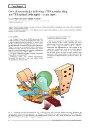
the perforations in the pelvic fascia and laid flat against<br />
the posterior surface of the pubis using the forefinger and<br />
a dissection forceps with no grasping function. Adhesion<br />
to the pubis is sufficient to ensure reliable and sturdy anterior<br />
anchorage. The other end of the anterior prosthesis is<br />
fixed to the uterine isthmus using two stitches of resorbable<br />
suture. When there is no uterus, this end is fixed to the vaginal<br />
vault. A check is made that there are no sharp edges and<br />
that it is not placed under tension. Anterior colporrhaphy<br />
using rapid resorption suture material to close the entire<br />
thickness of the vagina (both mucosa and fascia) is carried<br />
out without colpectomy. Insertion of the PIVS mesh, and<br />
treatment of any existing rectocele requires standard sagittal<br />
posterior colpotomy, without incising the perineum in<br />
order to keep pain to a minimum. The top of the incision<br />
reaches the neck of the uterus or the vaginal vault when<br />
there has been a hysterectomy. The recto-vaginal plane and<br />
enterocoele pouch are dissected. The two para-rectal fossa<br />
are opened using the finger and blunt-tipped scissors. The<br />
landmarks on each side are the ischial spine, the sacrospinous<br />
ligament and the levator ani muscles (iliococcygeal<br />
fasciculus). Upwards, the uterine isthmus and its junction<br />
with the utero-sacral ligaments are visible. <strong>This</strong> classic dissection<br />
is carried out without any retractors. A 5 millimetre<br />
incision is made 3 centimetres lateral and inferior to the anal<br />
margin on each side. The IVS Tunneller ® (Tyco Healthcare,<br />
USA) is inserted via this buttock incision in the ischio-rectal<br />
fossa, separated from the rectum by the levator ani muscles<br />
and the surgeon’s finger which is inserted via the para-rectal<br />
fossa. <strong>This</strong> finger is used to keep a check on movement of<br />
the tunneller through the muscle layers. The blunt tip of the<br />
tunneller is maneuvered to a position where it is in contact<br />
with the sacrospinous ligament, and 2 centimetres medial to<br />
the ischial spine. The muscle is then perforated at this level<br />
by the blunt tip that comes into contact with the surgeon’s<br />
finger. Thus covered and protected from any contact with<br />
the rectum, the blunt tip of the tunneller is taken out of the<br />
colpotomy area. The polypropylene tape is taken through the<br />
tunneller using the plastic stylette, and then the tunneller is<br />
removed. The tape is fixed to the utero-sacral ligaments, the<br />
uterine isthmus and the vaginal vault using two resorbable<br />
sutures. If there is a rectocele, a polypropylene recto-vaginal<br />
interposition prosthesis (Surgipro ® , TYCO Healthcare,<br />
USA) measuring 8 centimetres long and 4 centimetres wide<br />
is used. Like the anterior prosthesis, its corners are rounded.<br />
The aim is to cover and reinforce the recto-vaginal septum<br />
in order to correct the rectocele. To the top it is fixed to the<br />
PIVS tape by two stitches of resorbable suture, and at the<br />
bottom, its point of fixation is to the central fibrous core<br />
of the perineum on each side of the anus, again using two<br />
stitches of resorbable suture. The prosthesis must lie flat<br />
against the rectum, with no large creases. It is pulled up<br />
into the sacral concavity at the same time as the vaginal<br />
vault or uterus, together with the vesico-vaginal prosthesis<br />
which acts integrally with the uterine isthmus or vaginal<br />
vault when the system is placed under tension. No colpectomy<br />
is used here either. The posterior colpotomy is closed<br />
with rapid resorption suture prior to pulling on the two<br />
external ends of the PIVS mesh. A vaginal pack is inserted<br />
into the vagina for 24 hours in order to ensure that the vaginal<br />
walls are properly in contact with the prostheses and the<br />
dissection planes. A bladder catheter is inserted for the same<br />
period of time. 21<br />
RESULTS<br />
The PIVS operation was performed as planned in all 108<br />
cases. Thirty three patients had a past history of hysterectomy<br />
or surgery for prolapse of the upper or posterior<br />
compartment (27 hysterectomies and 19 rectocoele repairs).<br />
From a functional point of view, all the patients had previously<br />
complained of a dragging sensation in the pelvis and<br />
the uncomfortable presence of a protruding mass. Twenty<br />
seven patients had also complained of stress urinary incontinence,<br />
10 of stubborn constipation that worsened concomitant<br />
with the prolapse, 2 of anal pain at defecation and<br />
one of anal incontinence. All the prolapses included descent<br />
of upper compartment organs (vaginal vault, hysterocoele,<br />
enterocoele) with a point C > 0 cm according to the POP-Q<br />
classification. 20 Associated with this was a cystocoele (point<br />
Ba > 0 cm) in 73 cases, and a rectocoele (point Bp > 0 cm)<br />
in 87 cases. Nineteen hysterectomies, 22 amputations of the<br />
cervix and 49 urinary incontinence repairs using a sub-urethral<br />
sling (Anterior IVS) were carried out as detailed in the<br />
previous section.<br />
Group 1 comprised 19 patients who underwent hysterectomy<br />
during the same anaesthesia, whatever the other<br />
associated procedures (PIVS in every case, and sometimes<br />
correction of cystocoele or rectocoele). Group 2 comprised<br />
31 patients with installation of PIVS and in some cases<br />
recto-vaginal prosthesis and/or a sub-urethral sling for stress<br />
incontinence (excluding any other procedure). Group 3<br />
included 58 patients in whom a vesico-vaginal interposition<br />
prosthesis was installed (associated with any other procedure<br />
except hysterectomy).<br />
The intra-operative complications (9 cases) were essentially<br />
bladder injuries (7 cases), either during dissection of<br />
the cystocoele (4 cases), or during passage of the sub-urethral<br />
sling insertion device (3 cases). One low rectal injury<br />
occurred during dissection of the rectocoele, and one case of<br />
bleeding from the Cave of Retzius during treatment of urinary<br />
incontinence was controlled by simple pressure (using<br />
a vaginal pack on the full bladder), for which the subsequent<br />
history was uncomplicated apart from anaemia at 9.5<br />
g/dl. The post-operative complications consisted of anaemia<br />
(loss of more than 2 g/dl of haemoglobin) in 7 cases (6.5%),<br />
with a trend that did not reach significant level (p = 0.14)<br />
between the hysterectomy group 1 (3 cases or 15.8%) and<br />
the cystocoele (2 cases or 3.4%) and PIVS (2 cases or 6.4%)<br />
groups. Two cases of haematoma of the Cave of Retzius<br />
were observed, which had no further consequences for the<br />
patients. With respect to the cystocoele repair 2 vaginal<br />
erosions occurred at 2 and 18 months, that were resolved<br />
by simple excision of the exposed mesh under local anaesthesia.<br />
For the treatment of the upper and posterior compartments<br />
there were 2 infections of the prosthetic material<br />
which had to be completely removed, with one case occurring<br />
with a haematoma of the para-rectal fossa (on day 15)<br />
and the other on a vaginal erosion at 5 months. Finally, there<br />
were 6 cases of simple post-operative urinary infection and<br />
5 cases of isolated fever, which resolved without complications<br />
in every case. The average hospital stay was 4.8 days<br />
(ranging from 2 to 10 days). No immediate re-operation was<br />
necessary. Note that the stays were significantly longer (p<br />
< 0.001) for Group 1 (hysterectomy) (5.4 days) and Group<br />
3 (cystocoele) (4.9 days) compared with Group 2 (Posterior<br />
IVS) (4.1 days). The mean follow-up of the patients who<br />
were seen again was 19 months (ranging from 9 to 31<br />
months). Six patients were lost to follow-up. They had<br />
had no intra-operative complication and their characteristics<br />
(age, past history, type of operation) were similar to those of<br />
the total cohort.<br />
From an anatomical perspective, the presence of a prolapse<br />
at the first post-operative consultation at 6 weeks was<br />
considered as a failure, whilst if the same was found later,<br />
this was considered as a recurrence. With regard to correction<br />
of the upper and posterior compartments (assessment of<br />
13