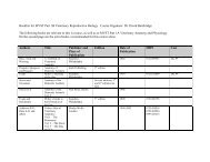
FRIDAY MORNING, 20 MAY 2005 REGENCY E, 8:30 A.M. TO 12:00 ...
FRIDAY MORNING, 20 MAY 2005 REGENCY E, 8:30 A.M. TO 12:00 ...
FRIDAY MORNING, 20 MAY 2005 REGENCY E, 8:30 A.M. TO 12:00 ...

Create successful ePaper yourself
Turn your PDF publications into a flip-book with our unique Google optimized e-Paper software.
9:<strong>20</strong><br />
5aBBb4. Shear wave interferometry, an application of sonoelastography. Clark Z. Wu and Kevin J. Parker Hopeman <strong>20</strong>4 ECE<br />
Dept., Univ. of Rochester, Rochester, NY 14623, wuzhe@ece.rochester.edu<br />
Sonoelastography is an ultrasound imaging technique where low-amplitude, low-frequency LF vibration is detected and displayed<br />
via real-time Doppler techniques. When multiple coherent shear wave sources exist, shear wave interference patterns appear.<br />
Two shear wave sources at the same frequency create hyperbolic shaped interference patterns in homogeneous, isotropic elastic media.<br />
Shear wave speed can be estimated from the fringe separation and the source frequency. If the two sources are driven at slightly<br />
different sinusoidal frequencies, the interference patterns no longer remain stationary. It is proven that the apparent velocity of the<br />
fringes is approximately proportional to the local shear wave velocity. With this approach, local shear wave speed in elastic media can<br />
be estimated. In addition, with a single shear wave source at frequency f and the ultrasound probe externally vibrated at frequency<br />
f f , a novel type of moving interference between the shear waves and the frame of reference motion is created. The moving<br />
interference fringes represent the shape of shear wave wavefronts while traveling at a much slower speed. This approach provides a<br />
real-time visualization of shear wave propagation and local wave speed estimation from which local stiffness is inferred. Work<br />
supported by NIH.<br />
9:45<br />
5aBBb5. Magnetic resonance elastography. Richard Ehman and Armando Manduca Depts. of Radiol. and Bioengineering, Mayo<br />
Clinic, Rochester, MN 55905<br />
The goal of our research is to develop MRI-based methods for assessing the mechanical properties of tissues in vivo. We have<br />
focused on a novel MRI technique for visualizing propagating acoustic shear waves Science 269, 1854–1857 1995. Suitable<br />
dynamic shear stress for Magnetic Resonance Elastography MRE can be generated by surface drivers, inertial effects, acoustic<br />
radiation pressure, or endogenous physiologic mechanisms. The MRE acquisition sequence is capable of visualizing cyclic tissue<br />
motion of less than 1 micron in displacement amplitude, with imaging times ranging from 1<strong>00</strong> ms to several minutes. Inversion<br />
algorithms based on continuum mechanics are used to process the acquired data to generate maps of mechanical properties such as<br />
depict stiffness, viscosity, attenuation, and anisotropic behavior. We have applied MRE to assess specimens of a variety of tissues,<br />
ranging in stiffness from lung to cartilage. Human studies have demonstrated that it is feasible to apply MRE to quantitatively image<br />
the mechanical properties of skeletal muscles, gray and white matter in the brain, thyroid, kidney, liver, and skin. Our preliminary<br />
clinical studies have to date applied MRE to observe changes in tissue mechanical properties in patients with breast, brain, and thyroid<br />
tumors, liver fibrosis, and diffuse diseases of skeletal muscle.<br />
10:10–10:25 Break<br />
Contributed Papers<br />
10:25<br />
5aBBb6. Microscopic dynamic magnetic resonance elastography.<br />
Shadi F. Othman, Thomas J. Royston, and Richard L. Magin Univ. of<br />
Illinois at Chicago, 842 W. Taylor St. MC 251, Chicago, IL 60607<br />
Microscopic magnetic resonance elastography uMRE is a high resolution<br />
imaging technique for measuring the viscoelastic properties of small<br />
synthetic and biological samples. Mechanical shear waves, typically with<br />
amplitudes of less than 1<strong>00</strong> m and frequencies of 5<strong>00</strong>–6<strong>00</strong> Hz, are induced<br />
using a piezoelectric oscillator directly coupled to the region of<br />
interest. By using multiple phase offsets and motion encoding gradients<br />
we acquire data that allows the generation of images that depict shear<br />
wave motion and the calculation of local values of the tissue viscoelastic<br />
properties. Recent MRE investigations are increasingly being conducted at<br />
higher spatial resolution to establish histological correlations between<br />
elasticity maps and tissue structures; such microscopic MRE studies require<br />
stronger static fields, stronger magnetic field gradients, higher performance<br />
RF coils, and more compact, higher frequency mechanical actuators.<br />
Microscopic MRE experiments were conducted at 11.74 T in a 54<br />
mm diameter vertical bore magnet with a 10 mm diameter25 mm length<br />
cylindrical space available for imaging. The field-of-view ranged from 4 to<br />
14 mm. The study was conducted on agarose gel phantoms of different<br />
concentrations ranging from 025%–1% w. Different biological samples,<br />
including frog oocytes and tissue engineered constructs, were also tested.<br />
10:40<br />
5aBBb7. Coupled vibration and sound radiation from a fluid-filled<br />
and submerged or embedded vascular tube with internal turbulent<br />
flow due to a constriction. Yigit Yazicioglu, Thomas J. Royston, Todd<br />
Spohnholtz, Bryn Martin Univ. of Illinois at Chicago, 842 W. Taylor St.<br />
MC 251, Chicago, IL 60607, Francis Loth Univ. of Illinois at Chicago,<br />
Chicago, IL 60607, and Hisham Bassiouny Univ. of Chicago, Chicago,<br />
IL 60637<br />
The vibration of a thin-walled cylindrical, compliant viscoelastic tube<br />
with internal flow and an axisymmetric constriction that results in turbulent<br />
fluid flow is studied theoretically and experimentally. Vibration of the<br />
tube is considered with internal fluid-coupling only and with coupling to<br />
internal flowing fluid and external stagnant fluid or external tissue-like<br />
viscoelastic material. The theoretical analysis includes the adaptation of a<br />
model for turbulence in the internal fluid and its vibratory excitation of<br />
and interaction with the tube wall and surrounding viscoelastic medium.<br />
Theoretical predictions are compared with experimental measurements<br />
conducted on a flow model system using laser Doppler vibrometry to<br />
measure tube vibration and the vibration of the surrounding viscoelastic<br />
medium, as well as miniature hydrophones to measure fluid pressure in the<br />
tube. Discrepancies between theory and experiment and the coupled nature<br />
of the fluid-structure interaction are highlighted. This study is relevant to<br />
and may provide further incite into vascular patency and mechanisms of<br />
5a FRI. AM<br />
2587 J. Acoust. Soc. Am., Vol. 117, No. 4, Pt. 2, April <strong>20</strong>05 149th Meeting: Acoustical Society of America 2587