CHAPTER 3 Tumours of the Stomach - Pathology Outlines
CHAPTER 3 Tumours of the Stomach - Pathology Outlines
CHAPTER 3 Tumours of the Stomach - Pathology Outlines

You also want an ePaper? Increase the reach of your titles
YUMPU automatically turns print PDFs into web optimized ePapers that Google loves.
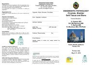
A<br />
B<br />
A<br />
C<br />
Fig. 3.06 Endoscopic views <strong>of</strong> gastric cancer (A, C) and corresponding images with dye enhancement (B, D).<br />
A, B Depressed early gastric cancer. C, D Deep ulcer scar surrounded by superficial early gastric cancer infiltrating<br />
<strong>the</strong> mucosa and submucosa.<br />
D<br />
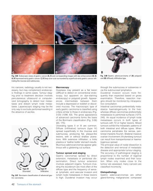
B<br />
Fig. 3.08 Gastric adenocarcinoma <strong>of</strong> (A) polypoid<br />
and (B) diffusely infiltrative type.<br />
tric cancers, radiology usually is not necessary,<br />
but may complement endoscopic<br />
findings in some cases. Tumour staging<br />
prior to treatment decision involves<br />
percutaneous ultrasound or computerized<br />
tomography to detect liver metastases<br />
and distant lymph node metastases.<br />
Laparoscopic staging may be <strong>the</strong><br />
only way to exclude peritoneal seeding in<br />
<strong>the</strong> absence <strong>of</strong> ascites.<br />
Type I<br />
Polypoid<br />
Type III<br />
Ulcerated<br />
Type II<br />
Fungating<br />
Type IV<br />
Infiltrative<br />
Fig. 3.07 Borrmann classification <strong>of</strong> advanced gastric<br />
carcinoma.<br />
Macroscopy<br />
Dysplasia may present as a flat lesion<br />
(difficult to detect on conventional endoscopy,<br />
but apparent on dye-staining<br />
endoscopy) or polypoid growth. Appearances<br />
intermediate between <strong>the</strong>m<br />
include a depressed or reddish or discolored<br />
mucosa. The macroscopic type <strong>of</strong><br />
early gastric carcinoma is classified using<br />
critera similar to those in endoscopy (Fig.<br />
3.03) {1298, 63}. The gross appearance<br />
<strong>of</strong> advanced carcinoma forms <strong>the</strong> basis<br />
<strong>of</strong> <strong>the</strong> Borrmann classification (Fig. 3.06)<br />
{63, 175}.<br />
Ulcerating types II or III are common.<br />
Diffuse (infiltrative) tumours (type IV)<br />
spread superficially in <strong>the</strong> mucosa and<br />
submucosa, producing flat, plaque-like<br />
lesions, with or without shallow ulcerations.<br />
With extensive infiltration, a linitis<br />
plastica or ‘lea<strong>the</strong>r bottle’ stomach results.<br />
Mucinous adenocarcinomas appear gelatinous<br />
with a glistening cut surface.<br />
Tumour spread and staging<br />
Gastric carcinomas spread by direct<br />
extension, metastasis or peritoneal dissemination.<br />
Direct tumour extension<br />
involves adjacent organs. <strong>Tumours</strong> invading<br />
<strong>the</strong> duodenum are most <strong>of</strong>ten <strong>of</strong> <strong>the</strong><br />
diffuse type and <strong>the</strong> frequency <strong>of</strong> serosal,<br />
lymphatic, and vascular invasion and<br />
lymph node metastases in <strong>the</strong>se lesions<br />
is high. Duodenal invasion may occur<br />
through <strong>the</strong> submucosa or subserosa or<br />
via <strong>the</strong> submucosal lymphatics.<br />
Duodenal invasion occurs more frequently<br />
than expected based on gross<br />
examination. Therefore, resection margins<br />
should be monitored by intraoperative<br />
consultation.<br />
Intestinal carcinomas preferentially metastasize<br />
haematogenously to <strong>the</strong> liver,<br />
whereas diffuse carcinomas preferentially<br />
metastasize to peritoneal surfaces {1273,<br />
245}. An equal incidence <strong>of</strong> lymph node<br />
metastases occurs in both types <strong>of</strong><br />
tumours with T2 or higher lesions. Mixed<br />
tumours exhibit <strong>the</strong> metastatic patterns <strong>of</strong><br />
both intestinal and diffuse types. When<br />
carcinoma penetrates <strong>the</strong> serosa, peritoneal<br />
implants flourish. Bilateral massive<br />
ovarian involvement (Krukenberg tumour)<br />
can result from transperitoneal or haematogenous<br />
spread.<br />
The principal value <strong>of</strong> nodal dissection is<br />
<strong>the</strong> detection and removal <strong>of</strong> metastatic<br />
disease and appropriate tumour staging.<br />
The accuracy <strong>of</strong> pathological staging is<br />
proportional to <strong>the</strong> number <strong>of</strong> regional<br />
lymph nodes examined and <strong>the</strong>ir location.<br />
When only nodes close to <strong>the</strong><br />
tumour are assessed, many cancers are<br />
classified incorrectly.<br />
Histopathology<br />
Gastric adenocarcinomas are ei<strong>the</strong>r<br />
gland-forming malignancies composed<br />
42 <strong>Tumours</strong> <strong>of</strong> <strong>the</strong> stomach