CHAPTER 3 Tumours of the Stomach - Pathology Outlines
CHAPTER 3 Tumours of the Stomach - Pathology Outlines
CHAPTER 3 Tumours of the Stomach - Pathology Outlines

You also want an ePaper? Increase the reach of your titles
YUMPU automatically turns print PDFs into web optimized ePapers that Google loves.
890, 178}. The organism has been shown<br />
to be present in 90% <strong>of</strong> cases limited to<br />
<strong>the</strong> mucosa and submucosa, falling to<br />
76% when deep submucosa is involved,<br />
and is present in only 48% <strong>of</strong> cases with<br />
extension beyond <strong>the</strong> submucosa<br />
{1316}. It has been shown that <strong>the</strong> infection<br />
by H. pylori precedes <strong>the</strong> development<br />
<strong>of</strong> lymphoma, both by sequential<br />
serological studies {1474} and by retrospective<br />
studies <strong>of</strong> archival gastric biopsy<br />
material {2211, 1314}.<br />
There is some controversy surrounding<br />
<strong>the</strong> role <strong>of</strong> <strong>the</strong> organism’s genetic features<br />
and <strong>the</strong> risk <strong>of</strong> lymphoma development.<br />
Studies <strong>of</strong> <strong>the</strong> association between<br />
MALT lymphoma and cagA bearing<br />
H. pylori strains have produced conflicting<br />
results, ranging from a lack <strong>of</strong> association<br />
between cagA and lymphoma<br />
{1492, 384} to a strong association {441}.<br />
One study claimed no association with<br />
low-grade lymphoma but a high frequency<br />
<strong>of</strong> cagA strains in high-grade lesions<br />
{1492}. Recently, a truncated form <strong>of</strong> an<br />
H. pylori associated protein, fldA, has<br />
been shown to be closely associated<br />
with gastric MALT lymphoma. All strains<br />
<strong>of</strong> H. pylori associated with MALT lymphoma<br />
showed a nucleotide G insertion<br />
at position 481 <strong>of</strong> <strong>the</strong> fldA gene, compared<br />
to 6/17 stains unassociated with<br />
lymphoma. This mutation causes a short<br />
truncation in <strong>the</strong> protein and antibodies<br />
to this truncated protein could be detected<br />
in 70% <strong>of</strong> <strong>the</strong> patients studied with<br />
MALT lymphoma, compared to 17% <strong>of</strong><br />
control patients {274}.<br />
Immunosuppression<br />
Lymphomas may arise or involve <strong>the</strong><br />
stomach in patients with both congenital<br />
and acquired immunodeficiencies. In<br />
general, <strong>the</strong> incidence, clinical features<br />
and <strong>the</strong> histology <strong>of</strong> <strong>the</strong> lesions is indistinguishable<br />
from those that develop outside<br />
<strong>the</strong> stomach. Up to 23% <strong>of</strong> gastrointestinal<br />
tract non-Hodgkin lymphomas<br />
arising in HIV infected patients occur in<br />
<strong>the</strong> stomach and <strong>the</strong> vast majority <strong>of</strong><br />
<strong>the</strong>se are large B-cell or Burkitt/Burkittlike<br />
lymphomas, {122} although occasional<br />
low-grade MALT lymphomas are<br />
described {2132}.<br />
Clinical features<br />
Symptoms and signs<br />
Patients with low-grade lymphomas <strong>of</strong>ten<br />
present with a long history <strong>of</strong> non-specific<br />
symptoms, including dyspepsia, nausea<br />
and vomiting. High-grade lesions<br />
may appear as a palpable mass in <strong>the</strong><br />
epigastrium and can cause severe<br />
symptoms, including weight loss.<br />
Imaging<br />
Low-grade MALT lymphomas present as<br />
intragastric nodularity with preferential<br />
location in <strong>the</strong> antrum {2180}. A more<br />
precise assessment is obtained with spiral<br />
CT, particularly if this is used in conjunction<br />
with distension <strong>of</strong> <strong>the</strong> stomach<br />
by water. This technique can identify up<br />
to 88% <strong>of</strong> cases, most <strong>of</strong> which have<br />
nodularity or enlarged rugal folds, and it<br />
can assess <strong>the</strong> submucosal extent <strong>of</strong> <strong>the</strong><br />
tumour {1493}. High-grade lymphomas<br />
are usually larger and more frequently<br />
associated with <strong>the</strong> presence <strong>of</strong> a mass<br />
and with ulceration. In some cases, <strong>the</strong><br />
radiological features may mimic diffuse<br />
adenocarcinoma {1059}. Endoscopic<br />
ultrasound is emerging as <strong>the</strong> investigation<br />
<strong>of</strong> choice in <strong>the</strong> assessment <strong>of</strong> <strong>the</strong><br />
extent <strong>of</strong> lymphoma infiltration through<br />
<strong>the</strong> gastric wall. Local lymph node<br />
involvement can also be assessed by<br />
this technique.<br />
Endoscopy<br />
Some cases show enlarged gastric folds,<br />
gastritis, superficial erosions or ulceration.<br />
In <strong>the</strong>se cases <strong>the</strong> surrounding normal<br />
appearing gastric mucosa may harbour<br />
lymphoma, and accurate mapping<br />
<strong>of</strong> <strong>the</strong> lesion requires multiple biopsies<br />
from all sites including areas appearing<br />
macroscopically normal. In a proportion<br />
<strong>of</strong> cases, endoscopic examination shows<br />
very minor changes such as hyperaemia<br />
and in a few cases random biopsies <strong>of</strong><br />
apparently entirely normal mucosa may<br />
reveal lymphoma. High-grade lymphoma<br />
is usually associated with more florid<br />
lesions, ulcers and masses. It is <strong>of</strong>ten<br />
impossible to distinguish lymphoma from<br />
carcinoma endoscopically.<br />
MALT lymphomas<br />
Pathogenesis<br />
The normal gastric mucosa contains<br />
scattered lymphocytes and plasma cells<br />
but is devoid <strong>of</strong> organised lymphoid tissue.<br />
The initial step in <strong>the</strong> development <strong>of</strong><br />
primary gastric lymphoma is <strong>the</strong> acquisition<br />
<strong>of</strong> organised lymphoid tissue from<br />
within which <strong>the</strong> lymphoma can develop.<br />
In most cases, this is associated with<br />
infection by H. pylori {572}, although it<br />
has also been seen following infection by<br />
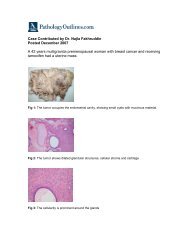
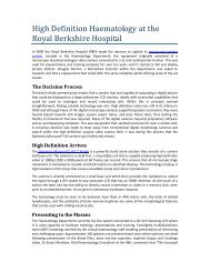
Fig. 3.38 Multifocal malignant lymphoma <strong>of</strong> <strong>the</strong><br />
stomach. The two larger lesions are centrally ulcerated.<br />
Helicobacter heilmannii {1842} and in<br />
association with coeliac disease {227}.<br />
This organised lymphoid tissue shows all<br />
<strong>the</strong> features <strong>of</strong> MALT, including <strong>the</strong> infiltration<br />
<strong>of</strong> <strong>the</strong> epi<strong>the</strong>lium by B-lymphocytes<br />
reminiscent <strong>of</strong> <strong>the</strong> lymphoepi<strong>the</strong>lium<br />
seen in Peyer patches {2135}.<br />
The cellular basis <strong>of</strong> <strong>the</strong> interaction<br />
between H. pylori and MALT lymphoma<br />
cells has been studied in detail. When<br />
unseparated cells isolated from lowgrade<br />
gastric MALT lymphomas are incubated<br />
in vitro with heat treated whole cell<br />
preparations from H. pylori, <strong>the</strong> tumour<br />
cells proliferate while those cultured in<br />
<strong>the</strong> absence <strong>of</strong> <strong>the</strong> organism or stimulating<br />
chemical mitogen rapidly die {768}.<br />
The proliferative response appeared to<br />
be strain specific for individual tumours<br />
but varied between tumours from different<br />
patients {768}. When T-cells were<br />
removed from <strong>the</strong> culture system <strong>the</strong> proliferative<br />
response was not seen and this<br />
could not be induced if <strong>the</strong> T-cells were<br />
replaced by supernatant from o<strong>the</strong>r cultures<br />
containing unseparated tumour<br />
derived cells {769}. Toge<strong>the</strong>r <strong>the</strong>se studies<br />
show that <strong>the</strong> proliferation <strong>of</strong> <strong>the</strong><br />
MALT lymphoma is driven by <strong>the</strong> presence<br />
<strong>of</strong> <strong>the</strong> H. pylori but that this, ra<strong>the</strong>r<br />
than being a direct effect on <strong>the</strong> tumour<br />
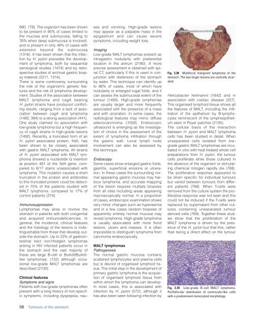
Fig. 3.39 Low-grade B-cell MALT lymphoma.<br />
Perifollicular distribution <strong>of</strong> centrocyte-like cells<br />
with a predominant monocytoid morphology.<br />
58 <strong>Tumours</strong> <strong>of</strong> <strong>the</strong> stomach