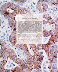
contributed by Dr. Najla Fakruddin - Pathology Outlines
contributed by Dr. Najla Fakruddin - Pathology Outlines
contributed by Dr. Najla Fakruddin - Pathology Outlines

Create successful ePaper yourself
Turn your PDF publications into a flip-book with our unique Google optimized e-Paper software.
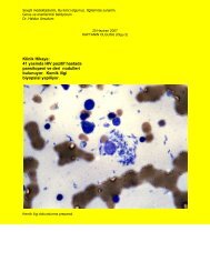
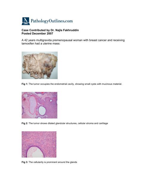
Case Contributed <strong>by</strong> <strong>Dr</strong>. <strong>Najla</strong> Fakhruddin<br />
Posted December 2007<br />
A 42 years multigravida premenopausal woman with breast cancer and receiving<br />
tamoxifen had a uterine mass:<br />
Fig 1: The tumor occupies the endometrial cavity, showing small cysts with mucinous material.<br />
Fig 2: The tumor shows dilated glandular structures, cellular stroma and cartilage<br />
Fig 3: The cellularity is prominent around the glands
Fig 4: Sarcomatous areas are present with bizarre cells and mitotic activity.<br />
A<br />
B<br />
C<br />
D<br />
Fig 5: Immunohistochemical stains for estrogen and progesterone receptors show focal (30%)<br />
positivity (fig A and B). Smooth muscle actin is positive (fig C). Proliferative index (MIB-1) is high<br />
(fig D).<br />
Diagnosis: Adenosarcoma