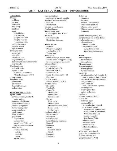
Unit 4 – LAB STRUCTURE LIST – Nervous System
Unit 4 – LAB STRUCTURE LIST – Nervous System
Unit 4 – LAB STRUCTURE LIST – Nervous System

Create successful ePaper yourself
Turn your PDF publications into a flip-book with our unique Google optimized e-Paper software.
PHYSO 2A <strong>LAB</strong> SG-4 Cara Burres-Jones, PCC<br />
<strong>Unit</strong> 4 <strong>–</strong> <strong>LAB</strong> <strong>STRUCTURE</strong> <strong>LIST</strong> <strong>–</strong> <strong>Nervous</strong> <strong>System</strong><br />
Tissue Level<br />
Neuron<br />
cell body<br />
nucleus<br />
neurofibril<br />
Nissl body<br />
dendrites<br />
axon<br />
axonal hillock<br />
axon terminal<br />
synaptic knob/bulb<br />
synaptic vesicles<br />
neurotransmitter<br />
multipolar neuron<br />
unipolar neuron<br />
bipolar neuron<br />
Neuroglial cells<br />
astrocytes<br />
microglia<br />
ependymal cells<br />
oligodendrocytes<br />
Schwanncells/neurolemmocytes<br />
satellite cells<br />
Myelinated axon<br />
node of Ranvier<br />
myelin sheath<br />
•Schwann Cells in PNS<br />
•Oligodendrocytes in CNS<br />
neurilemma<br />
Peripheral nerve<br />
epineurium<br />
perineurium<br />
fascicle (fasiculus)<br />
endoneurium<br />
Gross Anatomical Level:<br />
Spinal Cord:<br />
cervical, thoracic, lumbar &<br />
sacral regions<br />
anterior medial fissure<br />
posterior median sulcus<br />
cervical enlargement<br />
lumbar enlargement<br />
conus medullaris<br />
cauda equina<br />
filum terminale<br />
gray matter<br />
anterior horn<br />
posterior horn<br />
lateral horn (T1-L2 only)<br />
grey commissure w/canal<br />
white matter<br />
columns (funiculi)<br />
anterior<br />
posterior<br />
lateral<br />
tracts (fasciculi)<br />
Descending tracts<br />
corticospinal tracts/pyramidal<br />
Meninges (meninx=singular)<br />
Dura mater<br />
Dural sheath<br />
Epidural space (fat, etc.)<br />
Arachnoid mater<br />
Subarachnoid space<br />
Cerebrospinal fluid (CSF)<br />
Pia mater<br />
Filum terminale<br />
Denticulate ligamnets<br />
Spinal Nerves:<br />
Dorsal root<br />
Dorsal root ganglion<br />
w/Satellite cells<br />
Ventral root<br />
Spinal nerve<br />
Dorsal ramus (to epaxial body)<br />
Ventral ramus (to hypaxial body)<br />
(communicating rami=autonomic)<br />
Intercostal nerve<br />
Nerve plexuses<br />
Cervical C1-C4 (C5)<br />
Brachial (C4) C5-T1<br />
Lumbar L1-L5<br />
Sacral (Lumbosacral) S1-S5<br />
Coccygeal<br />
Cervical plexus<br />
Phrenic nerve (C3, 4 & 5)<br />
Brachial plexus<br />
Roots (5)<br />
Trunks (3)<br />
Cords (3)<br />
Medial cord<br />
Lateral cord<br />
Posterior cord<br />
Nerves<br />
Ulnar nerve<br />
Median nerve<br />
Musculocutaneous nerve<br />
Radial nerve<br />
Axillary nerve<br />
Lumbosacral plexus<br />
Iliohypogastric<br />
Ilioinquinal<br />
Genitofemoral<br />
Lateral femoral cutaneous<br />
Femoral<br />
Obturator<br />
Scitatic<br />
Common fibular<br />
Tibial<br />
Pudendal<br />
Dermatomes<br />
Reflex arc<br />
(stimulus)<br />
Receptor<br />
Afferent sensory neuron<br />
Interneuron(s) in CNS<br />
Efferent motor neuron<br />
Effector (skeletal muscle)<br />
(response)<br />
central nervous system (CNS)<br />
peripheral nervous system (PNS)<br />
afferent (sensory)<br />
efferent (motor)<br />
somatic division<br />
autonomic division<br />
sympathetic system<br />
parasympathetic system<br />
Brain:<br />
Procencephalon<br />
Telencephalon<br />
Diencephalon<br />
Mesencephalon<br />
Rhombencephalon<br />
Metencephalon<br />
Myelencelphalon<br />
ventricles<br />
lateral ventricles (left=1, right=2)<br />
-anterior, posterior, inferior horns<br />
interventricular foramen (ofMunro)<br />
cerebral aqueduct<br />
fourth ventricle<br />
lateral (2) and median (1) apertures<br />
choroid plexus<br />
cerebrospinal fluid<br />
septum pellucidum<br />
meninges:<br />
epidural space (cord only)<br />
dura mater<br />
periosteal layer<br />
meningeal layer<br />
falx cerebri, falx cerebelli<br />
dural venous sinuses<br />
superior & Inf. sagittal sinuses<br />
straight sinus & confluence<br />
transverse sinus (to)<br />
sigmoid sinus (to)<br />
(Int. Jugular Vein)<br />
subdural space<br />
arachnoid mater<br />
arachnoid villi<br />
subarachnoid space<br />
pia mater<br />
white mater (myelinated)<br />
gray mater (unmyelinated)<br />
nucleus & tract (CNS)<br />
ganglion & nerve (PNS)<br />
t<br />
1
PHYSO 2A <strong>LAB</strong> SG-4 Cara Burres-Jones, PCC<br />
Telencephalon:<br />
Epithalamus<br />
Papillae<br />
Cerebrum<br />
Pineal gland (epiphysis of)<br />
Taste buds<br />
Lateral ventricles<br />
Olfactory epithelium<br />
anterior horn<br />
“Brainstem”<br />
Olfactory neurons<br />
lateral horn<br />
Mesencephalon:<br />
Cribriform plate of ethmoid<br />
posterior horn<br />
Mesencephalon (or midbrain)<br />
Filaments of Olfactory Nerve<br />
Cranial nerve I<br />
Cerebral aqueduct<br />
Olfactory Bulb<br />
Frontal lobe<br />
Cerebral peduncles<br />
Olfactory Tract<br />
Precentral gyrus<br />
Corpora quadrigemina<br />
Central sulcus<br />
Superior colliculi<br />
Eye:<br />
Parietal lobe<br />
Inferior colliculi<br />
Sclera<br />
Postcentral gyrus<br />
Cranial n. III (Occulomotor N.) Cornea<br />
Parieto-occipital sulcus<br />
Cranial n. IV (Trochlear N.)<br />
Scleral venous sinus<br />
Occipital lobe<br />
Metencephalon:<br />
Uvea<br />
Temporal lobe<br />
Pons<br />
Choroid layer<br />
Lateral sulcus<br />
4 th Ventricle (upper portion)<br />
Ciliary body<br />
Insula lobe<br />
Grey matter nuclei<br />
Ciliary muscle<br />
Circular sulcus<br />
Cranial n. V (Trigeminal N.)<br />
Ciliary processes<br />
White matter:<br />
Cranial n. VI (Abducens N.)<br />
Ciliary zonule (Suspensory<br />
Projection tracts<br />
Cranial n. VII (Facial N.)<br />
ligaments of lens)<br />
Internal capsule<br />
Cranial n. VIII (Vestibulocochlear)<br />
Ora serrata retinae<br />
Corona radiata<br />
Cerebellum<br />
Iris<br />
Commissural tracts<br />
Roof over 4 th Ventricle<br />
Pupil<br />
Association tracts<br />
Cerebellar hemispheres<br />
Retina<br />
Basal Nuclei:<br />
Vermis<br />
Macula lutea<br />
caudate nucleus<br />
Folia<br />
Fovea centralis<br />
lentiform nucleus<br />
Arbor vitae<br />
Optic disk=blind spot<br />
putamen<br />
Cerebellar peduncles<br />
Central artery and vein of the retina<br />
globus pallidus<br />
Inferior, Middle & Superior<br />
Anterior segment<br />
Limbic <strong>System</strong>:<br />
Myelencephalon:<br />
Anterior chamber<br />
cingulate gyrus<br />
Medulla Oblongata<br />
Posterior chamber<br />
fornix<br />
4 th Ventricle (lower portion)<br />
(both w/Aqueous humor)<br />
hippocampus<br />
Pyramids<br />
Posterior segment<br />
amygdaloid nucleus<br />
Corticospinal tracts<br />
w/Vitreous humor<br />
olfactory cortex and tracts<br />
Anterior median fissure<br />
mammillary bodies<br />
Olive (olivary nucleus)<br />
Ear:<br />
Functional Areas of Cortex<br />
Cranial n. IX (Glossopharyngeal) Outer ear<br />
primary motor area<br />
Cranial n. X (Vagus N.)<br />
Pinna/Auricle<br />
pre-motor area<br />
Cranial n. XI (Spinal Accessory) External auditory meatus & canal<br />
prefrontal area<br />
Cranial n. XII (Hypoglossal N.)<br />
Tympanum (=“eardrum”)<br />
Broca’s area (motor speech area)<br />
Middle ear<br />
primary somatosensory area<br />
Autonomic <strong>Nervous</strong> <strong>System</strong><br />
Malleus<br />
somatosensory association area Sympathetic Division<br />
Incus<br />
gustatory area<br />
thoracolumbar outflow (T1-L2)<br />
Stapes<br />
auditory area<br />
white ramus communicans<br />
Oval Window<br />
auditory association area<br />
gray ramus communicans<br />
Round Window<br />
visual area<br />
sympathetic trunk=paravertebral<br />
Pharyngotympanic Tube<br />
visual association area<br />
adrenal medulla<br />
Inner ear<br />
Wernicke’s area<br />
Parasympathetic Division<br />
Perilymph<br />
olfactory area<br />
Craniosacral outflow<br />
Endolymph<br />
Diencephalon:<br />
III, VII, IX, X & S1-S4<br />
Semicircular canals (rotation)<br />
Thalamus<br />
-Ampule w/crista ampularis<br />
Third ventricle<br />
Peripheral Motor Ending:<br />
Vestibule (linear acceleration)<br />
Massa intermedia<br />
Neuromuscular junction<br />
-Utricle<br />
Hypothalamus<br />
Neuroglandular junction<br />
-Saccule<br />
Infundibulum<br />
Unencapsulated Nerve Endings:<br />
Cochlea<br />
Pituitary Gland (hypophysis of) -Free nerve endings<br />
Scala Vestibuli<br />
Posterior & Anterior<br />
-Tactile (Merkel) discs<br />
Scala Media = cochlear duct<br />
Optic chiasma<br />
-Hair recepors (root hair plexus)<br />
Organ of Corti<br />
Mammilary bodies<br />
Encapsulated Nerve Endings<br />
Basilar membrane<br />
Cranial Nerve II<br />
-Tactile (Meissner) corpuscles<br />
Scala Tympani<br />
-Lamellated (pacinian) corpuscles<br />
2