Greater Occipital Nerve Stimulation via the Bion ... - Pain Physician
Greater Occipital Nerve Stimulation via the Bion ... - Pain Physician
Greater Occipital Nerve Stimulation via the Bion ... - Pain Physician

You also want an ePaper? Increase the reach of your titles
YUMPU automatically turns print PDFs into web optimized ePapers that Google loves.
<strong>Pain</strong> <strong>Physician</strong> 2009; 12:621-628 • ISSN 1533-3159<br />
Clinical Report<br />
<strong>Greater</strong> <strong>Occipital</strong> <strong>Nerve</strong> <strong>Stimulation</strong> <strong>via</strong> <strong>the</strong><br />
<strong>Bion</strong> ® Microstimulator: Implantation Technique<br />
and <strong>Stimulation</strong> Parameters<br />
Clinical Trial: NCT00205894<br />
Terrence L. Trentman, MD 1 , David M. Rosenfeld, MD 1 , Bert B. Vargas, MD 2 ,<br />
Todd J. Schwedt, MD 3 , Richard S. Zimmerman, MD 4 , and David W. Dodick, MD 5<br />
From: Departments of<br />
1<br />
Anes<strong>the</strong>siology, 5 Neurology,<br />
and 4 Neurosurgery, Mayo Clinic<br />
Arizona, Scottsdale, AZ, 2 Center<br />
for Neurosciences, Tucson, AZ,<br />
and 3 Department of Neurology,<br />
Washington University School of<br />
Medicine, St Louis, MO.<br />
Address correspondence:<br />
Terrence L. Trentman, MD<br />
Mayo Clinic<br />
Department of Anes<strong>the</strong>siology<br />
5777 E Mayo Blvd<br />
Phoenix, AZ 85054<br />
E-mail:<br />
trentman.terrence@mayo.edu<br />
Disclaimer: This study was supported<br />
by funding from Boston Scientific<br />
Neuromodulation Corporation<br />
(formerly known as Advanced<br />
<strong>Bion</strong>ics® Corporation), Valencia, CA.<br />
<strong>Bion</strong> usage, baseline programming,<br />
and MIDAS disability scores were<br />
collected as part of a study supported<br />
by funding from Boston Scientific<br />
Neuromodulation Corporation.<br />
Additional data, such as headache<br />
locations and stimulation thresholds,<br />
were collected by <strong>the</strong> authors,<br />
independent of <strong>the</strong> sponsored study.<br />
Conflict of interest: Drs. Trentman,<br />
Dodick, and Zimmerman have<br />
received research support from<br />
Boston Scientific Neuromodulation<br />
Corporation. Drs. Trentman and<br />
Zimmerman have received consulting<br />
fees from Boston Scientific<br />
Neuromodulation Corporation.<br />
Manuscript received: 07/20/2008<br />
Revised manuscript received:<br />
10/29/2008<br />
Accepted for publication: 10/31/2008<br />
Background: Millions of patients suffer from medically refractory and disabling<br />
primary headache disorders. This problem has led to a search for new and innovative<br />
treatment modalities, including neuromodulation of <strong>the</strong> occipital nerves.<br />
Objectives: The primary aim of this study is to describe an implantation technique<br />
for <strong>the</strong> <strong>Bion</strong> ® microstimulator and document stimulation parameters and stimulation<br />
maps after <strong>Bion</strong> placement adjacent to <strong>the</strong> greater occipital nerve. The secondary aim<br />
is to document outcome measures one year post-implant.<br />
Design: Prospective, observational feasibility study.<br />
Methods: Nine patients with medically refractory primary headache disorders<br />
participated in this study. Approximately 6 months after <strong>Bion</strong> insertion, stimulation<br />
parameters and maps were documented for all patients. At one year, outcome<br />
measures were collected including <strong>the</strong> Migraine Disability Assessment Score.<br />
Results: At 6 months, <strong>the</strong> mean perception threshold was 0.47 mA, while <strong>the</strong><br />
mean discomfort threshold was 6.8 mA (stimulation range 0.47 – 6.8 mA). The mean<br />
pares<strong>the</strong>sia threshold was 1.64 mA and <strong>the</strong> mean usage range was 16.0. There were<br />
no major complications reported such as device migration, infection, or erosion. One<br />
patient stopped using her <strong>Bion</strong> before <strong>the</strong> 12-month follow-up visit. At one year, 7<br />
of <strong>the</strong> 8 patients were judged as having obtained fair or better results in terms of<br />
reduction of disability; 5 patients had greater than a 90% reduction in disability.<br />
Limitations: Small, heterogeneous patient population without control group. Not<br />
blinded or randomized.<br />
Conclusion: The <strong>Bion</strong> can be successfully inserted adjacent to <strong>the</strong> greater occipital<br />
nerve in an effort to treat refractory primary headache disorders. This microstimulator<br />
may provide effective occipital stimulation and headache control while minimizing <strong>the</strong><br />
risks associated with percutaneous or paddle leads implanted subcutaneously in <strong>the</strong><br />
occipital region.<br />
Key words: Chronic headache, migraine, cluster headache, peripheral nerve<br />
stimulation<br />
<strong>Pain</strong> <strong>Physician</strong> 2009; 12:621-628<br />
Free full manuscript:<br />
www.painphysicianjournal.com<br />
www.painphysicianjournal.com
<strong>Pain</strong> <strong>Physician</strong>: May/June 2009: 12:621-628<br />
Migraine is <strong>the</strong> most common form of<br />
disabling primary headache, affecting<br />
12% of Caucasian populations (1). Cluster<br />
headache and hemicrania continua, although much<br />
less common, also have a significant negative impact<br />
on quality of life (2,3). Subcutaneous occipital nerve<br />
stimulation (ONS) has been reported to effectively<br />
treat medically refractory primary headache disorders.<br />
A number of recent studies have documented efficacy<br />
outcomes and stimulation parameters associated with<br />
ONS (4-10). These studies document off-label use of<br />
spinal cord stimulation technology to stimulate <strong>the</strong><br />
distal branches of <strong>the</strong> C1-3 nerve roots. Prospective,<br />
multicenter studies are underway to determine <strong>the</strong><br />
safety and efficacy of this modality (11).<br />
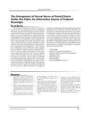
The implantable <strong>Bion</strong> microstimulator was initially<br />
developed as a radiofrequency (RF) powered<br />
functional electrical stimulator (12). However, <strong>the</strong><br />
<strong>Bion</strong> microstimulator (from Boston Scientific Neuromodulation<br />
Corporation, Valencia, CA) (Fig. 1) used in<br />
this study is <strong>the</strong> only battery powered microstimulator<br />
of its type and as such does not require an external<br />
RF power source. It includes a single cathode on one<br />
end and a single anode on <strong>the</strong> o<strong>the</strong>r. Currently an<br />
investigational device, <strong>the</strong> <strong>Bion</strong> contains a programmable<br />
microchip, stimulating electrodes, telemetry<br />
capability, and a transcutaneously rechargeable (3<br />
milliamp hours) lithium ion battery (13). It is expected<br />
that <strong>the</strong> battery will lose no more than 30% of its capacity<br />
after 500 cycles of full charge and discharge.<br />
Fig. 1. The <strong>Bion</strong> microstimulator.<br />
The <strong>Bion</strong> microstimulator’s small size (27.5 mm x 3.2<br />
mm) allows implantation adjacent to nerves <strong>via</strong> a less<br />
invasive technique than utilizing spinal cord stimulation<br />
technology. Previous studies have evaluated <strong>Bion</strong><br />
implantation for pudendal nerve neuromodulation in<br />
<strong>the</strong> setting of refractory detrusor overactivity incontinence<br />
(14,15). At our institution, we have implanted 9<br />
<strong>Bion</strong> microstimulators adjacent to <strong>the</strong> greater occipital<br />
nerve (GON) in an effort to treat refractory headache<br />
disorders.<br />
The primary aim of this feasibility study is to describe<br />
an implantation technique for <strong>the</strong> <strong>Bion</strong> microstimulator<br />
and document stimulation parameters and<br />
stimulation maps after <strong>Bion</strong> placement adjacent to<br />
<strong>the</strong> GON. The secondary aim is to document outcome<br />
measures one year post-implant.<br />
Methods<br />
After <strong>the</strong> United States Food and Drug Administration<br />
(IDE G030225) and Institutional Review Board<br />
approval of this feasibility study, 9 patients diagnosed<br />
with chronic migraine or chronic cluster headache<br />
presenting to our clinic were screened for inclusion<br />
and exclusion. All 9 patients met <strong>the</strong> inclusion and exclusion<br />
criteria and all agreed to participate. Written<br />
informed consent was obtained from each. Inclusion<br />
criteria included 18 years of age or older, 12 or more<br />
months of chronic migraine or chronic cluster headache,<br />
refractory to at least 4 preventative medications<br />
used at adequate dosage for adequate duration of<br />
time, willingness to maintain current pain medication<br />
regimen during <strong>the</strong> study, and willingness and<br />
ability to maintain a headache diary for <strong>the</strong> duration<br />
of <strong>the</strong> study. Exclusion criteria included pregnancy or<br />
planned pregnancy, previous surgery in <strong>the</strong> occipital<br />
region, and participation in a device or drug trial within<br />
<strong>the</strong> previous 30 days.<br />
All patients underwent a detailed neurologic<br />
exam and were assigned a diagnosis based on <strong>the</strong> International<br />
Classification of Headache Disorders – II<br />
(16). Six patients had chronic cluster headache including<br />
one with migraines and one with hemicrainia continua,<br />
and 3 patients had chronic migraine only (Table<br />
1). Each patient underwent a psychiatric evaluation<br />
to determine <strong>the</strong>ir psychological stability to undergo<br />
<strong>the</strong> procedure. The patients did not undergo a trial of<br />
stimulation or occipital nerve block before implantation<br />
of <strong>the</strong> <strong>Bion</strong>.<br />
For <strong>the</strong> purposes of this study, and consistent with<br />
our previous study on occipital stimulation mapping<br />
622 www.painphysicianjournal.com
<strong>Occipital</strong> <strong>Stimulation</strong> <strong>via</strong> <strong>the</strong> <strong>Bion</strong><br />
Table 1. Patient demographics, 6-month <strong>Bion</strong> microstimulator usage and Migraine Disability Assessment Scores (MIDAS).<br />
Patient Diagnosis Stimulator Usage<br />
Pulse<br />
Width<br />
(us)<br />
Rate (PPS)<br />
Baseline<br />
MIDAS**<br />
1 – 72f† Migraine 0.5-1.5 hrs/day 250 60 130-85-4<br />
2 – 39f Migraine<br />
3 – 44f Cluster<br />
0.5 hrs,<br />
2-3 days/wk<br />
0.5-0.75<br />
hrs/2 wks<br />
1 year<br />
MIDAS**<br />
Response*<br />
Did not complete<br />
study<br />
300 55 235-80-4 5-5-5 Excellent<br />
350 60 270-90-6 260-87-6 Poor<br />
4 – 66f Cluster 5-6 hrs/day 200 45 147-82-7 8-40-7 Excellent<br />
5 – 46m Cluster 22 hrs/day 250 55 225-90-7 130-80-5 Fair<br />
6 – 44f<br />
Cluster / hemicrania<br />
continua<br />
16 hrs/day 350 45 87-90-5 8-25-4 Excellent<br />
7 – 60m Migraine 18 hrs/day 250 45 108-90-6 6-90-4 Excellent<br />
8 – 44m Cluster 24 hrs/day 300 60 120-88-6 10-85-3 Excellent<br />
9 – 35m Cluster/Migraine 20 hrs/day 200 45 110-90-7 70-60-7 Fair<br />
*Response key:<br />
PPS = pulse per second<br />
> 90% reduction in disability = excellent †4 month visit<br />
70–90% reduction in disability = very good<br />
50–69% reduction in disability = good<br />
25–49% reduction in disability = fair<br />
< 25% reduction in disability = poor<br />
** The first number of each 3 digit series is <strong>the</strong> Migraine Disability Assessment Score, <strong>the</strong> second number is <strong>the</strong> number of headache days<br />
over a 3-month period, and <strong>the</strong> third number is <strong>the</strong> average severity of each headache.<br />
(9), <strong>the</strong> following definitions were used: perception<br />
threshold is <strong>the</strong> lowest current amplitude that elicits<br />
sensation. Perception threshold is assumed to represent<br />
local tissue stimulation, while <strong>the</strong> upper end of<br />
<strong>the</strong> stimulation range (discomfort threshold) is defined<br />
as <strong>the</strong> current amplitude where patients feel<br />
stimulation strongly and do not wish <strong>the</strong> stimulation<br />
to be increased any fur<strong>the</strong>r. The stimulation range<br />
(perception through discomfort threshold) represents<br />
<strong>the</strong> useful amplitudes for any given electrode combination<br />
while <strong>the</strong> usage range (discomfort threshold<br />
divided by perception threshold) “represents <strong>the</strong> relative<br />
size of <strong>the</strong> <strong>the</strong>rapeutic stimulating window” (17).<br />
Pares<strong>the</strong>sia threshold is <strong>the</strong> current amplitude where<br />
<strong>the</strong> patient first noted stimulation traveling toward<br />
<strong>the</strong> vertex of his head, suggesting direct GON stimulation.<br />
The maximum stimulation tested during <strong>the</strong><br />
study was 10 milliamps (mA).<br />
The <strong>Bion</strong> implantation procedure was carried out<br />
under monitored anes<strong>the</strong>sia care in <strong>the</strong> prone position.<br />
Antibiotic prophylaxis was given to each patient before<br />
incision. The goal of each implant was to position <strong>the</strong><br />
<strong>Bion</strong> microstimulator subcutaneously in <strong>the</strong> occipital<br />
region at a right angle to <strong>the</strong> GON, with <strong>the</strong> cathode<br />
immediately adjacent to <strong>the</strong> nerve. The anatomy of this<br />
region has recently been reviewed (8,18), and a cadaver<br />
study noted that in 10 specimens (20 nerves), <strong>the</strong> GON<br />
ascended between 5 and 28 mm from <strong>the</strong> midline at<br />
<strong>the</strong> level of <strong>the</strong> intermastoid line (19).<br />
First, using fluoroscopic guidance, a line was<br />
drawn between <strong>the</strong> tips of <strong>the</strong> mastoid processes (intermastoid<br />
line) and in <strong>the</strong> midline. After sterile prep<br />
and injection of local anes<strong>the</strong>tic for skin wheal, a small<br />
(< 1 cm) incision was made 3 cm contralateral to <strong>the</strong><br />
side of intended GON stimulation. Next, a 20-gauge, 15<br />
cm insulated stimulating needle was inserted through<br />
<strong>the</strong> incision and across <strong>the</strong> midline toward <strong>the</strong> side to<br />
be stimulated. The ideal depth was estimated to be<br />
below <strong>the</strong> dermis but superficial to <strong>the</strong> fascia, in <strong>the</strong><br />
subcutaneous fat layer. The location of <strong>the</strong> GON was<br />
marked as <strong>the</strong> point where <strong>the</strong> patient experienced<br />
maximal stimulation induced pares<strong>the</strong>sia towards <strong>the</strong><br />
vertex of <strong>the</strong>ir head, at least to <strong>the</strong> level of <strong>the</strong> top of<br />
<strong>the</strong> ear.<br />
www.painphysicianjournal.com 623
<strong>Pain</strong> <strong>Physician</strong>: May/June 2009: 12:621-628<br />
Next, <strong>the</strong> stimulating needle was removed and<br />
<strong>the</strong> <strong>Bion</strong> introducer with <strong>the</strong> dissector/stimulator was<br />
inserted through <strong>the</strong> incision. The <strong>Bion</strong> dissector/stimulator<br />
was used to confirm <strong>the</strong> location of <strong>the</strong> GON,<br />
after which <strong>the</strong> <strong>Bion</strong> was deployed adjacent to <strong>the</strong><br />
GON using <strong>the</strong> <strong>Bion</strong> placement tool and holder (Figs.<br />
2,3).<br />
Post implantation management included device<br />
activation 7–10 days postoperatively in 7 of <strong>the</strong> patients<br />
and activation on <strong>the</strong> day of implant in 2 patients.<br />
The timing of device activation was based on<br />
patient preference and travel considerations. The patients<br />
were initially given a radiofrequency “pillow”<br />
charger; a smaller “butterfly” charger subsequently<br />
became available that could be attached to a hat,<br />
allowing <strong>the</strong> patient to recharge while upright. The<br />
patients were instructed to initially use <strong>the</strong> <strong>Bion</strong> constantly<br />
at low amperage and <strong>the</strong>n to increase <strong>the</strong> amperage<br />
as needed to treat intermittent headache exacerbations.<br />
However, <strong>the</strong>y were given <strong>the</strong> prerogative<br />
to adjust use to comfort and effect. Patients turned<br />
stimulation on or off, and adjusted <strong>the</strong> amplitude of<br />
stimulation, <strong>via</strong> a wireless remote control.<br />
Approximately 6 months after implantation, <strong>the</strong><br />
following data were ga<strong>the</strong>red in addition to <strong>the</strong> data<br />
collected as part of <strong>the</strong> sponsored study: headache location,<br />
average number of hours of <strong>Bion</strong> use per day,<br />
frequency of recharging, pulse width and rate, perception<br />
threshold with associated pares<strong>the</strong>sia map,<br />
discomfort threshold with associated pares<strong>the</strong>sia map,<br />
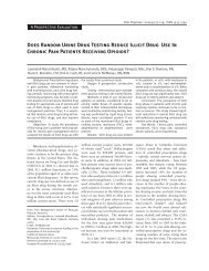
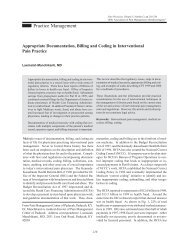
and pares<strong>the</strong>sia threshold. The patients were given a<br />
map of <strong>the</strong> head (Fig. 4) to allow <strong>the</strong>m to identify <strong>the</strong><br />
location of <strong>the</strong>ir baseline headaches and areas where<br />
<strong>the</strong>y perceived stimulation. Mean values with standard<br />
de<strong>via</strong>tion (SD) were used to summarize <strong>the</strong> data.<br />
Migraine Disability Assessment Scores (MIDAS)<br />
were obtained prior to and at 12 months after implantation<br />
of <strong>the</strong> stimulator. The MIDAS questionnaire is a<br />
validated headache-related disability instrument that<br />
is increasingly used as a surrogate measure of outcome<br />
in episodic and chronic migraine trials (20). The 5-question<br />
instrument quantifies time lost due to headache<br />
from work, school, household work, and social/family<br />
or leisure activities over <strong>the</strong> preceding 3 months. The<br />
score is typically reported in a 3-digit format (e.g. 106-<br />
75-7): The first number is <strong>the</strong> MIDAS (greater than 20<br />
is considered severe), <strong>the</strong> second number is <strong>the</strong> number<br />
of days in <strong>the</strong> past 3 months that <strong>the</strong> patient had a<br />
headache (max 90), and <strong>the</strong> third number is <strong>the</strong> average<br />
severity of each headache (0–10 scale).<br />
Fig. 2. The <strong>Bion</strong> placement tools and holder.<br />
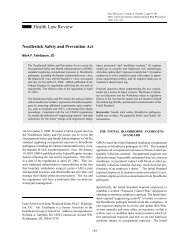
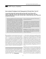
Fig. 3. AP skull film showing <strong>Bion</strong> after insertion. The<br />
device is subcutaneous in <strong>the</strong> occipital region.<br />
624 www.painphysicianjournal.com
<strong>Occipital</strong> <strong>Stimulation</strong> <strong>via</strong> <strong>the</strong> <strong>Bion</strong><br />
Fig. 4. Head maps used to identify areas of baseline headache, perception threshold, and discomfort threshold. Used with permission.<br />
Trentman et al (9) and <strong>the</strong> International Neuromodulation Society.<br />
Results<br />
All 9 patients invited to participate signed informed<br />
consent. One patient completed <strong>the</strong> headache<br />
maps approximately 4 months after implant. She<br />
subsequently did not appear for her 6-month followup<br />
and stopped using <strong>the</strong> <strong>Bion</strong> before study completion<br />
at 12 months. She stated <strong>the</strong> battery recharging<br />
schedule was too demanding, specifically that she was<br />
spending 1.5 hours recharging her <strong>Bion</strong> for every 1.5<br />
hours of use. Patient demographics, baseline usage<br />
data, and MIDAS are summarized in Table 1. The mean<br />
decrease in number of days with headache for <strong>the</strong> 8<br />
patients who completed <strong>the</strong> study was 28.5 (SD 29.6),<br />
while <strong>the</strong> average headache severity score decreased<br />
by 0.88 (SD 1.36). More detailed outcome data continues<br />
to be analyzed and will be presented separately.<br />
Self-reported stimulator usage ranged from 30<br />
minutes every 2 weeks to 24 hours/day (mean 12.2<br />
hours/day, median 16 hours/day), and recharging frequency<br />
ranged from 35 minutes per week to 4 hours<br />
per day (mean 1.67 hours/day, median 1.5 hours/day).<br />
Distribution of baseline headache and stimulation<br />
thresholds (perception and discomfort) are shown in<br />
Table 2 (see Fig. 4 for maps). The zones of stimulation<br />
as noted in Table 2 are not meant to imply that<br />
<strong>the</strong> patients felt pares<strong>the</strong>sia throughout <strong>the</strong> entire<br />
zone; ra<strong>the</strong>r, <strong>the</strong>y recorded stimulation in some part<br />
of each area noted. Table 3 summarizes stimulation<br />
parameters, including a mean perception threshold of<br />
0.47 mA and a mean discomfort threshold of 6.8 mA<br />
(stimulation range 0.47 – 6.8 mA). The mean pares<strong>the</strong>sia<br />
threshold was 1.64 mA, and <strong>the</strong> mean usage<br />
range was 16.0. The average tissue impedance was<br />
1.34 kilo-ohms.<br />
No patients reported major device-related complications<br />
during <strong>the</strong> 12-month duration of <strong>the</strong> study<br />
Table 2. Headache and <strong>Stimulation</strong> Distribution (See Fig. 4).<br />
This data was obtained at <strong>the</strong> 6-month follow-up visit (except<br />
where noted) in addition to <strong>the</strong> data collected as part of <strong>the</strong><br />
sponsored study.<br />
Patient<br />
Location<br />
of Baseline<br />
Headache<br />
Location of<br />
Perception<br />
Threshold<br />
Location of<br />
Discomfort<br />
Threshold<br />
1† 1,2,10,11 7,9,18 7<br />
2 5 – 7,9 9 5,7,9<br />
3 10 – 12,14 – 16 16,18 16,18<br />
4 2 – 7, 9,18 7,9 5 – 7,9<br />
5 10,16 16 14 – 16<br />
6 10 – 12,14,16 16 7,14,16<br />
7 10,12,15 18 14,16,18<br />
8 10 – 12 18 7,9,16 – 18<br />
9 1,5,6 1,5 – 7, 9 1,5 – 7,9<br />
†Four month visit.<br />
www.painphysicianjournal.com 625
<strong>Pain</strong> <strong>Physician</strong>: May/June 2009: 12:621-628<br />
Table 3. <strong>Bion</strong> Tested <strong>Stimulation</strong> Parameters. This data was obtained at <strong>the</strong> 6-month follow-up visit (except where noted) in addition<br />
to <strong>the</strong> data collected as part of <strong>the</strong> sponsored study.<br />
Patient<br />
Perception Threshold<br />
(mA)<br />
Discomfort Threshold<br />
(mA)<br />
Pares<strong>the</strong>sia Threshold<br />
(mA)<br />
Usage Range*<br />
1† 0.6 10.0 1.8 16.67<br />
2 0.2 5.4 1.6 27.0<br />
3 0.4 1.6 1.2 4.0<br />
4 0.2 2.2 0.6 11.0<br />
5 0.8 10.0 1.6 12.5<br />
6 0.4 10.0 4.0 25.0<br />
7 0.4 6.2 1.6 15.5<br />
8 0.8 5.8 1.6 7.25<br />
9 0.4 10.0 0.8 25.0<br />
Mean (SD) 0.47 (0.22) 6.8 (3.4) 1.64 (0.97) 16.0(8.2)<br />
Median 0.4 6.2 1.6 15.5<br />
*Usage range = discomfort threshold divided by perception threshold. †Four month visit.<br />
such as infection, migration, or erosion. Adverse events<br />
reported after <strong>the</strong> study was complete at 12 months<br />
included loss of stimulation (N = 1). The patient’s <strong>Bion</strong><br />
had malfunctioned and was unrechargeable, requiring<br />
replacement. This patient subsequently experienced<br />
an infection necessitating explant and reimplant of a<br />
third <strong>Bion</strong>. Minor adverse events included 2 patients<br />
who complained of muscle stimulation at high amplitudes,<br />
and one patient who complained of pain near<br />
<strong>the</strong> implant site.<br />
Discusson<br />
Millions of patients suffer from primary headache<br />
disorders; a portion of <strong>the</strong>m, like <strong>the</strong> patients in this<br />
study, endure severe, medically refractory pain. This<br />
problem has led to a search for new and innovative<br />
treatment modalities, including neuromodulation of<br />
<strong>the</strong> occipital nerves.<br />
A number of recent studies have suggested that<br />
stimulation of <strong>the</strong> distal branches of <strong>the</strong> C1-2-3 nerve<br />
roots can produce pain relief in patients with o<strong>the</strong>rwise<br />
refractory headache disorders (21-24). The mechanism<br />
of analgesia may be due to inhibition of nociceptive<br />
specific neurons in <strong>the</strong> trigeminal-cervical complex.<br />
Electrical stimulation of <strong>the</strong> GON may also result in mobilization<br />
of central pain modulatory centers (25,26).<br />
Of note, we previously reported pain relief despite<br />
persistent cranial autonomic activity (lacrimation, rhinorrhoea,<br />
conjunctival injection) in 2 of <strong>the</strong>se patients<br />
implanted with <strong>Bion</strong>s (27). One patient was diagnosed<br />
with cluster headache, while <strong>the</strong> o<strong>the</strong>r was diagnosed<br />
with hemicrania continua. This separation of autonomic<br />
signs from analgesia suggests that <strong>the</strong> autonomic features<br />
and first division pain are dissociated and separately<br />
controlled from a supranuclear generator.<br />
While previous occipital stimulation techniques<br />
have resulted in diffuse distal C1-2-3 stimulation <strong>via</strong><br />
cylindrical (percutaneous) or paddle (surgical) spinal<br />
cord stimulator (SCS) leads (4-7), <strong>the</strong> <strong>Bion</strong> was used<br />
here to stimulate a limited area and a specific nerve<br />
(GON). Our approach assumed that GON stimulation<br />
will have <strong>the</strong> same central analgesic affect as more<br />
diffuse C1-2-3 stimulation <strong>via</strong> percutaneous stimulator<br />
leads. If this assumption is correct, clinicians may be<br />
able to achieve <strong>the</strong> benefits of occipital stimulation<br />
<strong>via</strong> a microstimulator while circumventing <strong>the</strong> technical<br />
problems associated with occipital percutaneous<br />
leads and remote power sources.<br />
In terms of stimulation parameters and <strong>the</strong> <strong>Bion</strong>,<br />
all of <strong>the</strong> patients had a sensory threshold of less than<br />
one milliamp, suggesting local tissue stimulation,<br />
while several patients had discomfort thresholds at<br />
<strong>the</strong> maximum tested amplitude of 10 mA. The mean<br />
pares<strong>the</strong>sia threshold of 1.64 mA suggests that at this<br />
amperage <strong>the</strong> GON was being stimulated directly. The<br />
large usage range (16.0, SD 8.2) indicates wide variation<br />
between <strong>the</strong>se patients in terms of <strong>the</strong> size of<br />
<strong>the</strong>ir <strong>the</strong>rapeutic stimulating windows. Lengthy daily<br />
usage time and high rates of stimulation (pulses per<br />
second) will increase recharging frequency.<br />
626 www.painphysicianjournal.com
<strong>Occipital</strong> <strong>Stimulation</strong> <strong>via</strong> <strong>the</strong> <strong>Bion</strong><br />
Our previous study on occipital pares<strong>the</strong>sia mapping<br />
in patients with subcutaneously implanted SCS<br />
leads provides several points for comparison (9). In that<br />
study, <strong>the</strong> mean perception threshold was higher (1.07<br />
V), while <strong>the</strong> discomfort threshold (3.63 V) was lower<br />
than <strong>the</strong> <strong>Bion</strong> (Table 3). The average tissue impedance<br />
in this study (1.34 kilo-ohms) is close to 1.0 kilo-ohms,<br />
allowing us to assume that <strong>the</strong> mA recorded for <strong>the</strong><br />
<strong>Bion</strong> are roughly equivalent to <strong>the</strong> volts recorded in<br />
our previous study of SCS leads. The <strong>Bion</strong>’s proximity to<br />
<strong>the</strong> GON may explain its lower perception threshold,<br />
while <strong>the</strong> reason for <strong>the</strong> higher discomfort threshold<br />
for <strong>the</strong> <strong>Bion</strong> is more obscure. Diffuse tissue stimulation<br />
produced by SCS leads may increase <strong>the</strong> likelihood of<br />
patient discomfort.<br />
In terms of pares<strong>the</strong>sia mapping, it is difficult to<br />
determine if a correlation exists between distribution<br />
of pares<strong>the</strong>sia (Table 2 and Fig. 4) and outcome, but<br />
a pattern did not appear to emerge. Despite <strong>the</strong> <strong>Bion</strong>’s<br />
small size, we were able to produce pares<strong>the</strong>sia<br />
in remote areas of <strong>the</strong> head, including at least one<br />
patient who noted trigeminal distribution pares<strong>the</strong>sia.<br />
It is unknown if outcome is improved by covering<br />
patient’s baseline headache regions with pares<strong>the</strong>sia,<br />
analogous to spinal cord stimulation.<br />
The MIDAS was used as a measure of outcome in<br />
this study. At one year, 7 of <strong>the</strong> 8 patients were judged<br />
as having obtained fair or better results in terms of<br />
reduction of disability; 5 patients had greater than<br />
90% reduction in disability. All patients with excellent<br />
outcomes experienced a > 50% reduction in headache<br />
days and/or 30–50% reduction in average headache severity,<br />
while patients with a fair response experienced<br />
a reduction in headache days or severity of 25–50%.<br />
The <strong>Bion</strong>’s small size and low profile may help<br />
minimize or eliminate device displacement and lead<br />
breakage problems associated with SCS equipment<br />
implanted subcutaneously in <strong>the</strong> occipital region. As<br />
recently reviewed (10), complications of SCS systems<br />
implanted in <strong>the</strong> occipital region can occur frequently<br />
with lead migration rates as high as 100%. O<strong>the</strong>r reported<br />
complications include lead fracture or disconnection,<br />
infection, erosion, and allergic reaction.<br />
The <strong>Bion</strong> microstimulator requires no anchoring<br />
or tunneling of extensions to remote power sources;<br />
as such, much of <strong>the</strong> mechanical stress on <strong>the</strong> occipital<br />
stimulator system is eliminated. However, it is possible<br />
that a foreign body such as <strong>the</strong> <strong>Bion</strong> can move<br />
within tissue planes or become encapsulated, hence<br />
increasing <strong>the</strong> energy required to stimulate <strong>the</strong> occipital<br />
nerve. As a current controlled device, <strong>the</strong> <strong>Bion</strong><br />
has <strong>the</strong> (limited) ability to automatically adjust <strong>the</strong><br />
voltage to maintain <strong>the</strong> current amplitude despite<br />
encapsulation.<br />
Disadvantages of a microstimulator system include<br />
<strong>the</strong> need for frequent recharging and limited choices<br />
in terms of electrode combinations. Future versions of<br />
this device may include a larger battery and multiple<br />
electrodes.<br />
Limitations of this feasibility study include its small<br />
and heterogeneous patient population without a control<br />
group. It was nei<strong>the</strong>r randomized nor blinded. This<br />
however was a pilot study to ascertain <strong>the</strong> feasibility<br />
of <strong>the</strong> technique and potential for this modality in <strong>the</strong><br />
treatment of refractory primary headache disorders.<br />
Thus far, <strong>the</strong>re are no randomized controlled studies<br />
published on <strong>the</strong> safety or efficacy of any occipital<br />
nerve stimulation device for <strong>the</strong> treatment of primary<br />
headache disorders. The results of this feasibility study<br />
would support randomized controlled trials with <strong>the</strong><br />
<strong>Bion</strong> microstimulator in this patient population. Potentially,<br />
a blinded study could be carried out wherein<br />
<strong>Bion</strong> microstimulators would be inserted in both a<br />
treatment and a placebo group. The treatment group<br />
would receive stimulation immediately after implant,<br />
while <strong>the</strong> placebo group would not have <strong>the</strong>ir <strong>Bion</strong><br />
microstimulators activated for several months. A<br />
blinded, head to head comparison of <strong>the</strong> <strong>Bion</strong> microstimulator<br />
to o<strong>the</strong>r ONS systems that use spinal cord<br />
stimulation equipment could be more difficult to carry<br />
out, as <strong>the</strong> spinal cord stimulators systems require a<br />
remote battery implant. In this scenario, it would not<br />
be possible to “blind” <strong>the</strong> patient as to which system<br />
was implanted.<br />
Conclusion<br />
In conclusion, we have documented pares<strong>the</strong>sia<br />
maps and stimulation parameters for 9 patients after<br />
permanent implantation of a <strong>Bion</strong> microstimulator,<br />
with one year outcome data. There were no major adverse<br />
events during <strong>the</strong> study period, including device<br />
migration or infection. One patient did not complete<br />
<strong>the</strong> study. Fur<strong>the</strong>r studies are needed to evaluate <strong>the</strong><br />
safety and efficacy of subcutaneous C1-2-3 stimulation<br />
for headache disorders, including <strong>the</strong> use of microstimulators<br />
to stimulate specific nerves. If this novel<br />
microstimulator is shown to be effective in randomized<br />
trials, it may be possible to achieve headache<br />
control <strong>via</strong> neurostimulation with a low incidence of<br />
long-term complications.<br />
www.painphysicianjournal.com 627
<strong>Pain</strong> <strong>Physician</strong>: May/June 2009: 12:621-628<br />
References<br />
1. Goadsby PJ. Migraine: Diagnosis and<br />
management. Internal Medicine Journal<br />
2003; 33:436-42.<br />
2. Steiner TJ, Fontebasso M. Headache.<br />
BMJ (Clinical research ed.) 2002;<br />
325:881-886.<br />
3. Bigal ME, Lipton RB, Tepper SJ, Rapoport<br />
AM, Sheftell FD. Primary chronic<br />
daily headache and its subtypes in adolescents<br />
and adults. Neurology 2004;<br />
63:843-847.<br />
4. Weiner RL, Reed KL. Peripheral neurostimulation<br />
for <strong>the</strong> control of intractable<br />
occipital neuralgia. Neuromodulation<br />
1999; 2:217-221.<br />
5. Popeney CA, Alo KM. Peripheral neurostimulation<br />
for <strong>the</strong> treatment of chronic,<br />
disabling transformed migraine. Headache<br />
2003; 43:369-375.<br />
6. Oh MY, Ortega J, Bellotte JB, Whiting<br />
DM, Alo KM. Peripheral nerve stimulation<br />
for <strong>the</strong> treatment of occipital neuralgia<br />
and transformed migraine using a<br />
c1-2-3 subcutaneous paddle style electrode:<br />
A technical report. Neuromodulation<br />
2004; 7:103-112.<br />
7. Kapural L, Mekhail N, Hayek SM, Stanton-Hicks<br />
M, Malak O. <strong>Occipital</strong> nerve<br />
electrical stimulation <strong>via</strong> <strong>the</strong> midline<br />
approach and subcutaneous surgical<br />
leads for treatment of severe occipital<br />
neuralgia: A pilot study. Anesth Analg<br />
2005; 101:171-174.<br />
8. Johnstone CS, Sundaraj R. <strong>Occipital</strong><br />
nerve stimulation for <strong>the</strong> treatment of<br />
occipital neuralgia — eight case studies.<br />
Neuromodulation 2006; 9:41-47.<br />
9. Trentman TL, Zimmerman RS, Seth N,<br />
Hentz J, Dodick DW. <strong>Stimulation</strong> ranges,<br />
usage ranges, and pares<strong>the</strong>sia mapping<br />
during occipital nerve stimulation.<br />
Neuromodulation 2008; 11:56-61.<br />
10. Jasper JF, Hayek SM. Implanted occipital<br />
nerve stimulators. <strong>Pain</strong> <strong>Physician</strong> 2008;<br />
11:187-200.<br />
11. From U.S. National Institutes of Health<br />
http://clinicaltrials.gov/ct2/results?<br />
term=occipital+nerve+stimulation. Accessed:<br />
10-27-08.<br />
12. Salter ACD, Bagg SD, Creasy JL, Romano<br />
C, Romano D, Richmond FJR, Loeb<br />
GE. First clinical experience with BION<br />
implants for <strong>the</strong>rapeutic electrical stimulation.<br />
Neuromodulation 2004; 7:38-<br />
47.<br />
13. Carbunaru R, Whitehurst T, Jaax K, Koff J,<br />
JM. Rechargeable batter-powered <strong>Bion</strong><br />
microstimulator for neuromodulation.<br />
26th Annual International Conference<br />
of <strong>the</strong> Engineering in Medicine and Biology<br />
Society 2004; 2:4193-4196.<br />
14. Bosch JL. The <strong>Bion</strong> device: A minimally<br />
invasive implantable ministimulator for<br />
pudendal nerve neuromodulation in patients<br />
with detrusor overactivity incontinence.<br />
The Urologic Clinics of North<br />
America 2005; 32:109-112.<br />
15. Groen J, Amiel C, Bosch JL. Chronic pudendal<br />
nerve neuromodulation in women<br />
with idiopathic refractory detrusor<br />
overactivity incontinence: Results of a<br />
pilot study with a novel minimally invasive<br />
implantable mini-stimulator.<br />
Neurourology and Urodynamics 2005;<br />
24:226-230.<br />
16. The International Classification of Headache<br />
Disorders: 2nd edition. Cephalalgia<br />
2004; 24 Suppl 1:9-160.<br />
17. Wesselink WA, Holsheimer J, King GW,<br />
Torgerson NA, Boom HBK. Quantitative<br />
aspects of <strong>the</strong> clinical performance of<br />
transverse tripolar spinal cord stimulation.<br />
Neuromodulation 1999; 2:5-14.<br />
18. Natsis K, Baraliakos X, Appell HJ, Tsikaras<br />
P, Gigis I, Koebke J. The course of<br />
<strong>the</strong> greater occipital nerve in <strong>the</strong> suboccipital<br />
region: A proposal for setting<br />
landmarks for local anes<strong>the</strong>sia in patients<br />
with occipital neuralgia. Clinical<br />
Anatomy 2006;19:332-336.<br />
19. Becser N, Bovim G, Sjaastad O. Extracranial<br />
nerves in <strong>the</strong> posterior part of<br />
<strong>the</strong> head. Anatomic variations and <strong>the</strong>ir<br />
possible clinical significance. Spine<br />
1998; 23:1435-1441.<br />
20. Stewart WF, Lipton RB, Whyte J, Dowson<br />
A, Kolodner K, Liberman JN, Sawyer<br />
J. An international study to assess<br />
reliability of <strong>the</strong> Migraine Disability Assessment<br />
(MIDAS) score. Neurology<br />
1999; 53:988-994.<br />
21. Burns B, Watkins L, Goadsby PJ. Treatment<br />
of medically intractable cluster<br />
headache by occipital nerve stimulation:<br />
Long-term follow-up of eight patients.<br />
Lancet 2007; 369:1099-1106.<br />
22. Magis D, Allena M, Bolla M, De Pasqua<br />
V, Remacle JM, Schoenen J. <strong>Occipital</strong><br />
nerve stimulation for drug-resistant<br />
chronic cluster headache: A prospective<br />
pilot study. Lancet Neurol 2007;<br />
6:314-321.<br />
23. Melvin EA, Jr., Jordan FR, Weiner RL,<br />
Primm D. Using peripheral stimulation<br />
to reduce <strong>the</strong> pain of c2-mediated occipital<br />
headaches: A preliminary report.<br />
<strong>Pain</strong> <strong>Physician</strong> 2007; 10:453-460.<br />
24. Schwedt TJ, Dodick DW, Hentz J, Trentman<br />
TL, Zimmerman RS. <strong>Occipital</strong><br />
nerve stimulation for chronic headache<br />
—long-term safety and efficacy. Cephalalgia<br />
2007; 27:153-157.<br />
25. Vincent MB, Ekman R, Edvinsson L,<br />
Sand T, Sjaastad O. Reduction of calcitonin<br />
gene-related peptide in jugular<br />
blood following electrical stimulation<br />
of rat greater occipital nerve. Cephalalgia<br />
1992; 12:275-279.<br />
26. Matharu MS, Bartsch T, Ward N, Frackowiak<br />
RS, Weiner R, Goadsby PJ. Central<br />
neuromodulation in chronic migraine<br />
patients with suboccipital stimulators:<br />
A PET study. Brain 2004; 127:220-230.<br />
27. Schwedt TJ, Dodick DW, Trentman TL,<br />
Zimmerman RS. <strong>Occipital</strong> nerve stimulation<br />
for chronic cluster headache<br />
and hemicrania continua: <strong>Pain</strong> relief<br />
and persistence of autonomic features.<br />
Cephalalgia 2006; 26:1025-1027.<br />
628 www.painphysicianjournal.com