Percutaneous Osteoplasty for the Treatment of a ... - Pain Physician
Percutaneous Osteoplasty for the Treatment of a ... - Pain Physician
Percutaneous Osteoplasty for the Treatment of a ... - Pain Physician

Create successful ePaper yourself
Turn your PDF publications into a flip-book with our unique Google optimized e-Paper software.
<strong>Pain</strong> <strong>Physician</strong> 2012; 15:E743-E748 • ISSN 2150-1149<br />
Case Report<br />
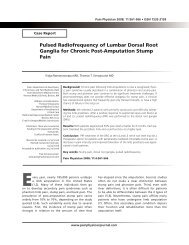
<strong>Percutaneous</strong> <strong>Osteoplasty</strong> <strong>for</strong> <strong>the</strong> <strong>Treatment</strong> <strong>of</strong><br />
a <strong>Pain</strong>ful Osteochondral Lesion <strong>of</strong> <strong>the</strong> Talus: A<br />
Case Report and Literature Review<br />
Sung-Suk Seo, MD, Joo-Yeon Park, MD, Hae-Jin Kim, MD, Ji-Wook Yoon, MD, Sang-Hyun<br />
Park, MD, PhD, and Kyung-Hoon Kim, MD, PhD<br />
From: Department <strong>of</strong><br />
Anes<strong>the</strong>sia and <strong>Pain</strong> Medicine,<br />
School <strong>of</strong> Medicine, Pusan<br />
National University Korea<br />
Address correspondence:<br />
Kyung-Hoon Kim, MD, PhD<br />
Pr<strong>of</strong>essor<br />
Department <strong>of</strong> Anes<strong>the</strong>sia and<br />
<strong>Pain</strong> Medicine<br />
School <strong>of</strong> Medicine<br />
Pusan National University,<br />
Pusan, Korea<br />
<strong>Pain</strong> Clinic, Pusan National<br />
University Yangsan Hospital,<br />
Bumeuri, Mulgeumup,<br />
Yangsan, Kyungnam, 626-770,<br />
Korea<br />
E-mail: pain@pusan.ac.kr<br />
Disclaimer: There was no external<br />
funding in <strong>the</strong> preparation <strong>of</strong> this<br />
manuscript.<br />
Conflict <strong>of</strong> interest: None.<br />
Manuscript received: 02/21/2012<br />
Revised manuscript received:<br />
03/21/2012<br />
Accepted <strong>for</strong> publication:<br />
05/25/2012<br />
Free full manuscript:<br />
www.painphysicianjournal.com<br />
An osteochondral lesion <strong>of</strong> <strong>the</strong> talus (OLT) is a lesion involving <strong>the</strong> talar articular cartilage<br />
and its subchondral bone. OLT is a known cause <strong>of</strong> chronic ankle pain after ankle sprains<br />
in <strong>the</strong> active population. The lesion causes deep ankle pain associated with weight-bearing,<br />
impaired function, limited range <strong>of</strong> motion, stiffness, catching, locking, and swelling. There<br />
are 2 common patterns <strong>of</strong> OLTs. Anterolateral talar dome lesions result from inversion and<br />
dorsiflexion injuries <strong>of</strong> <strong>the</strong> ankle at <strong>the</strong> area impacting against <strong>the</strong> fibula. Posteromedial lesions<br />
result from inversion, plantar flexion, and external rotation injuries <strong>of</strong> <strong>the</strong> ankle at <strong>the</strong> area<br />
impacting against <strong>the</strong> tibial ceiling <strong>of</strong> <strong>the</strong> ankle joint. Early diagnosis <strong>of</strong> an OLT is particularly<br />
important because <strong>the</strong> tibiotalar joint is exposed to more compressive load per unit area than<br />
any o<strong>the</strong>r joint in <strong>the</strong> body. Failure <strong>of</strong> diagnosis can lead to <strong>the</strong> evolution <strong>of</strong> a small, stable lesion<br />
into a larger lesion or an unstable fragment, which can result in chronic pain, joint instability,<br />
and premature osteoarthritis.<br />
A 43-year-old man, with a history <strong>of</strong> ankle sprain one year previously, visited our pain clinic<br />
<strong>for</strong> continuous right ankle pain after walking or standing <strong>for</strong> more than 30 minutes. There<br />
was a focal tenderness on <strong>the</strong> posteromedial area <strong>of</strong> <strong>the</strong> right talus. Imaging studies revealed<br />
a posteromedial OLT classified as having a geode <strong>for</strong>m according to <strong>the</strong> FOG (fractures,<br />
osteonecroses, geodes) radiological classification and categorized as a stage 2a lesion on<br />
magnetic resonance imaging.<br />
The patient was scheduled <strong>for</strong> aspiration and osteoplasty with hydroxyapatite under arthroscopic<br />
and fluoroscopic guidance. A 26-gauge needle was inserted to infiltrate local anes<strong>the</strong>tics into<br />
<strong>the</strong> skin over <strong>the</strong> cyst and ankle joint. An arthroscope was placed into <strong>the</strong> joint to approach <strong>the</strong><br />
OLT. The arthroscopic view showed that <strong>the</strong>re was no connection between <strong>the</strong> OLT and <strong>the</strong> cyst<br />
<strong>of</strong> <strong>the</strong> talus body. A 13-gauge bone biopsy needle was inserted into <strong>the</strong> cyst, and aspiration was<br />
per<strong>for</strong>med. Aspirated fluid from <strong>the</strong> cyst was originally white and clear; however, it changed to<br />
a blood-tinged, reddish color due to mixing with <strong>the</strong> incisional blood. After aspiration, contrast<br />
medium was injected, and <strong>the</strong> shape <strong>of</strong> <strong>the</strong> spread was observed. Bone cement comprising<br />
hydroxyapatite was injected to fill <strong>the</strong> bone defect <strong>of</strong> <strong>the</strong> cyst. A 1.5 mL volume <strong>of</strong> cement was<br />
injected into <strong>the</strong> talus under vigilant fluoroscopic and arthroscopic monitoring to prevent its<br />
dissemination into <strong>the</strong> joint. There was no cement leakage into <strong>the</strong> vessels or articular space.<br />
Postoperative fluoroscopy and computed tomography images showed bone cement filling <strong>of</strong><br />
<strong>the</strong> defect.<br />
In <strong>the</strong> present case, arthroscopic and fluoroscopic guidance was used <strong>for</strong> aspiration <strong>of</strong> an OLT<br />
and <strong>for</strong> per<strong>for</strong>ming percutaneous osteoplasty with hydroxyapatite <strong>for</strong> one defect; this treatment<br />
decreased pain upon weight bearing and enabled a return to work without any restrictions one<br />
week after <strong>the</strong> procedure. The purpose <strong>of</strong> this report was to highlight <strong>the</strong> presence <strong>of</strong> OLT in<br />
chronic ankle pain and to review its management strategies.<br />
Key words: Ankle, bone fracture, cartilage fracture, calcium polyacrylate-hydroxyapatite<br />
cement, cementoplasty, endoscopy, osteochondritis dissecans, pain, sprain, talus.<br />
<strong>Pain</strong> <strong>Physician</strong> 2012; 15:E743-E748<br />
www.painphysicianjournal.com
<strong>Pain</strong> <strong>Physician</strong>: September/October 2012; 15:E743-E748<br />
An osteochondral lesion <strong>of</strong> <strong>the</strong> talus (OLT) is <strong>the</strong><br />
collective term <strong>for</strong> a focal lesion involving <strong>the</strong> talar<br />
hyaline cartilage and its underlying subchondral bone,<br />
mostly caused by a single or multiple traumatic events<br />
and leading to partial or complete detachment <strong>of</strong> <strong>the</strong><br />
fragment. The lesion causes deep ankle pain associated<br />
with weight-bearing, impaired function, limited<br />
range <strong>of</strong> motion, stiffness, catching, locking, and swelling.<br />
These symptoms may adversely affect <strong>the</strong> ability to<br />
walk, work, and participate in sports at risk (1,2).<br />
There are 2 common patterns <strong>of</strong> OLTs. Anterolateral<br />
talar dome lesions result from inversion and dorsiflexion<br />
injuries <strong>of</strong> <strong>the</strong> ankle at <strong>the</strong> area impacting<br />
against <strong>the</strong> fibula. Posteromedial lesions result from<br />
inversion, plantar flexion, and external rotation injuries<br />
<strong>of</strong> <strong>the</strong> ankle at <strong>the</strong> area impacting against <strong>the</strong> tibial<br />
ceiling <strong>of</strong> <strong>the</strong> ankle joint (3,4).<br />
Early diagnosis <strong>of</strong> an OLT is particularly important<br />
because <strong>the</strong> tibiotalar joint is exposed to more compressive<br />
load per unit area than any o<strong>the</strong>r joint in <strong>the</strong><br />
body. Failure <strong>of</strong> diagnosis can lead to <strong>the</strong> evolution <strong>of</strong><br />
a small, stable lesion into a larger lesion or an unstable<br />
fragment, which can result in chronic pain, joint instability,<br />
and premature osteoarthritis (5).<br />
The purpose <strong>of</strong> this report was to emphasize <strong>the</strong><br />
importance <strong>of</strong> considering an OLT as a cause <strong>of</strong> chronic<br />
ankle pain and to review its management strategies.<br />
Case Report<br />
A 43-year-old man with a history <strong>of</strong> ankle sprain<br />
one year previously visited our pain clinic <strong>for</strong> continuous<br />
right ankle pain after walking or standing <strong>for</strong> more<br />
than 30 minutes. He was unresponsive to conservative<br />
treatments, including immobilization, restricted<br />
weight-bearing, nonsteroidal anti-inflammatory drugs,<br />
and rehabilitation. There was a focal tenderness on <strong>the</strong><br />
posteromedial area <strong>of</strong> <strong>the</strong> right talus with no limitation<br />
<strong>of</strong> joint motion on physical examination. Imaging studies<br />
revealed a posteromedial OLT classified as having a<br />
geode <strong>for</strong>m according to <strong>the</strong> FOG (fractures, osteonecroses,<br />
geodes) radiological classification and categorized<br />
as a stage 2a lesion according to Anderson and<br />
Crichton’s classification <strong>of</strong> <strong>the</strong> magnetic resonance image<br />
(MRI) (6). Bone scans showed an active bone lesion<br />
<strong>of</strong> <strong>the</strong> right talus (Fig. 1).<br />
The patient was scheduled <strong>for</strong> aspiration and osteoplasty<br />
with hydroxyapatite under arthroscopic and<br />
fluoroscopic guidance. In<strong>for</strong>med consent was obtained<br />
after explaining <strong>the</strong> risks <strong>of</strong> extravasation <strong>of</strong> bone ce-<br />
ment into <strong>the</strong> ankle joint, infection, bleeding, and joint<br />
stiffness or locking due to arthroscopy.<br />
Basic noninvasive monitoring <strong>of</strong> parameters such<br />
as noninvasive blood pressure, electrocardiography,<br />
and pulse oximetry was per<strong>for</strong>med throughout <strong>the</strong><br />
procedure. One milligram <strong>of</strong> cefazolin was infused intravenously<br />
30 minutes be<strong>for</strong>ehand. To reduce intraoperative<br />
pain, we also injected 30 mg <strong>of</strong> ketorolac and<br />
50 μg <strong>of</strong> fentanyl intravenously be<strong>for</strong>e <strong>the</strong> operation.<br />
The patient was placed in a supine position, and<br />
<strong>the</strong> skin was prepared and draped in a sterile fashion.<br />
All procedures were per<strong>for</strong>med under fluoroscopic<br />
guidance. A line representing <strong>the</strong> tibiotalar joint and a<br />
circle representing <strong>the</strong> cyst were drawn on <strong>the</strong> right ankle.<br />
A 26-gauge needle was inserted to infiltrate local<br />
anes<strong>the</strong>tics into <strong>the</strong> skin over <strong>the</strong> cyst and ankle joint.<br />
An arthroscope was placed into <strong>the</strong> joint to approach<br />
<strong>the</strong> OLT. The arthroscopic view showed that <strong>the</strong>re was<br />
no connection between <strong>the</strong> OLT and <strong>the</strong> cyst <strong>of</strong> <strong>the</strong> talus<br />
body. A 13-gauge bone biopsy needle was inserted<br />
into <strong>the</strong> cyst, and aspiration was per<strong>for</strong>med. Aspirated<br />
fluid from <strong>the</strong> cyst was originally white and clear; however,<br />
it changed to a blood-tinged, reddish color due to<br />
mixing with <strong>the</strong> incisional blood. After aspiration, contrast<br />
medium was injected, and <strong>the</strong> shape <strong>of</strong> <strong>the</strong> spread<br />
was observed. Bone cement comprising hydroxyapatite<br />
(Spine-Fix, Teknimed SAS, Vic en Bigorre, France) was<br />
injected to fill <strong>the</strong> bone defect <strong>of</strong> <strong>the</strong> cyst. A 1.5 mL<br />
volume <strong>of</strong> cement was injected into <strong>the</strong> talus under<br />
vigilant fluoroscopic and arthroscopic monitoring to<br />
prevent its dissemination into <strong>the</strong> joint. There was no<br />
cement leakage into <strong>the</strong> vessels or articular space (Fig.<br />
2). Postoperative fluoroscopy and computed tomography<br />
(CT) images showed bone cement filling <strong>of</strong> <strong>the</strong> defect<br />
(Fig. 3).<br />
The patient was discharged one day after <strong>the</strong> procedure<br />
with postoperative pain fairly controlled. At <strong>the</strong><br />
one-week and one-month postoperative follow-ups,<br />
no significant complications were detected. There was<br />
no complaint <strong>of</strong> pain or limitation <strong>of</strong> range <strong>of</strong> motion.<br />
One week after <strong>the</strong> operation, <strong>the</strong> patient returned to<br />
work without any restrictions and was fairly satisfied<br />
with <strong>the</strong> results. He was scheduled to revisit <strong>the</strong> clinic at<br />
3 and 6 months postoperatively.<br />
Discussion<br />
The present patient experienced chronic pain after<br />
an ankle sprain; this pain was unresponsive to conservative<br />
treatment. A focal tenderness <strong>of</strong> <strong>the</strong> talus was<br />
E744<br />
www.painphysicianjournal.com
<strong>Percutaneous</strong> <strong>Osteoplasty</strong> at Osteochondral Lesion <strong>of</strong> <strong>the</strong> Talus<br />
Fig. 1. Preoperative images. (A) Finding a definite lesion <strong>of</strong> <strong>the</strong> right ankle in <strong>the</strong> radiographic images (anteroposterior, Mortise,<br />
and lateral view from left to right) was difficult; (B) Magnetic resonance images revealed an abnormal high signal area<br />
surrounded with marrow edema (6.57 mm x 12.21 mm x 12.31 mm) in <strong>the</strong> posteromedial dome <strong>of</strong> <strong>the</strong> right talus; (C) Bone<br />
scans showing an active bone lesion.<br />
identified on physical examination without limitation<br />
<strong>of</strong> range <strong>of</strong> motion, and MRI was per<strong>for</strong>med as an OLT<br />
was suspected. Aspiration <strong>of</strong> <strong>the</strong> talar cyst and osteoplasty<br />
<strong>of</strong> <strong>the</strong> relatively large bone defect with hydroxyapatite<br />
was per<strong>for</strong>med under arthroscopic and fluoroscopic<br />
guidance. The patient was discharged one day<br />
after <strong>the</strong> procedure without significant complications,<br />
and he returned to work without any restrictions after<br />
one week.<br />
A patient with acute ankle sprain and no evidence<br />
<strong>of</strong> fracture or complete ligament injury is prone to be<br />
left without medical attention. The most common<br />
mechanism <strong>of</strong> injury in ankle sprains is a combination<br />
<strong>of</strong> plantar flexion and inversion. The lateral stabilizing<br />
ligaments, which include <strong>the</strong> anterior tal<strong>of</strong>ibular, calcane<strong>of</strong>ibular,<br />
and posterior tal<strong>of</strong>ibular ligaments, are<br />
most <strong>of</strong>ten damaged. The anterior tal<strong>of</strong>ibular ligament<br />
is <strong>the</strong> most easily injured structure. Concomitant injury<br />
to this ligament and <strong>the</strong> calcane<strong>of</strong>ibular ligament can<br />
result in appreciable instability (7).<br />
Commonly missed diagnoses in patients with<br />
chronic ankle pain include ei<strong>the</strong>r fractures <strong>of</strong> <strong>the</strong><br />
www.painphysicianjournal.com<br />
E745
<strong>Pain</strong> <strong>Physician</strong>: September/October 2012; 15:E743-E748<br />
Fig. 2. Intraoperative images. (A) An imaginary line <strong>of</strong> <strong>the</strong> tibiotalar joint and an imaginary circle <strong>of</strong> <strong>the</strong> cyst were drawn on<br />
<strong>the</strong> right ankle under fluoroscopy. A 26-gauge needle was inserted to infiltrate local anes<strong>the</strong>tics into <strong>the</strong> skin over <strong>the</strong> cyst and ankle<br />
joint under fluoroscopy. There was no connection from <strong>the</strong> OLT into <strong>the</strong> cyst <strong>of</strong> <strong>the</strong> talus body in <strong>the</strong> arthroscopic view. (B) A<br />
13-gauge bone biopsy needle was inserted into <strong>the</strong> cyst and aspiration was per<strong>for</strong>med. Aspirated fluid from <strong>the</strong> cyst was originally<br />
white and clear; however, it changed to a blood-tinged, reddish color when mixed with <strong>the</strong> incisional blood. (C) After aspiration<br />
(left), contrast medium was injected to observe <strong>the</strong> spread (middle). Hydroxyapatite bone cement was injected to fill <strong>the</strong> bone defect<br />
<strong>of</strong> <strong>the</strong> cyst. A 1.5 ml volume <strong>of</strong> cement was injected into <strong>the</strong> talus under vigilant monitoring <strong>of</strong> arthroscopy to prevent its dissemination<br />
into <strong>the</strong> joint. There was no cement (dark black one within a red circle) leakage into <strong>the</strong> vessels or articular space (right).<br />
Fig. 3. Postoperative images showed<br />
filling <strong>of</strong> bone cement in <strong>the</strong> defect. (A)<br />
Fluoroscopic Mortise view. (B) Computed<br />
tomography.<br />
E746<br />
www.painphysicianjournal.com
<strong>Percutaneous</strong> <strong>Osteoplasty</strong> at Osteochondral Lesion <strong>of</strong> <strong>the</strong> Talus<br />
osteochondral talar dome, lateral talar process, calcaneal<br />
anterior process, lateral malleolar, posterolateral<br />
distal fibular flake, fifth metatarsal base, and <strong>the</strong><br />
navicular bone or injuries <strong>of</strong> <strong>the</strong> Achilles, peroneal,<br />
posterior tibial, anterior tibial, and flexor hallucis longus<br />
tendons. If <strong>the</strong> primary problem is ankle pain, a<br />
concentrated ef<strong>for</strong>t should be made to rule out occult<br />
fracture <strong>of</strong> <strong>the</strong> foot or ankle. A bone scan is an excellent<br />
screening test to rule out occult fractures and to<br />
guide fur<strong>the</strong>r treatment. If <strong>the</strong> bone scan reveals increased<br />
uptake in a discrete area, a spot radiograph or<br />
CT scan is useful to fur<strong>the</strong>r identify <strong>the</strong> exact location<br />
<strong>of</strong> <strong>the</strong> fracture. Occult or associated injuries <strong>of</strong> <strong>the</strong> tendons<br />
<strong>of</strong> <strong>the</strong> foot and ankle should also be considered,<br />
and MRI is <strong>the</strong> most useful exam to identify and confirm<br />
<strong>the</strong>se injuries (8). Both CT and MRI are useful <strong>for</strong><br />
<strong>the</strong> diagnosis <strong>of</strong> OLT. However, bone analysis is more<br />
precise with CT, and articular analysis can be superior<br />
with MRI (6).<br />
The updated term “osteochondral lesion” replaces<br />
<strong>the</strong> terms osteochondritis, osteonecrosis, and subchondral<br />
fracture. Traditionally, <strong>the</strong> Bernt and Harty classification<br />
(9) <strong>of</strong> OLT was used, based on anatomic and<br />
evolving variations <strong>of</strong> an injury lesion. Our case falls in<br />
stage I (subchondral impaction) according to <strong>the</strong>ir classification<br />
and stage V (cystic lesions) according to <strong>the</strong><br />
revised classification (10). The Anderson and Crichton<br />
classification based on MRI revealed a stage 2a lesion<br />
(subchondral cysts) (11). FOG radiological classification<br />
revealed a geode <strong>for</strong>m based on <strong>the</strong> lesional aspect and<br />
<strong>the</strong> relation <strong>of</strong> <strong>the</strong> lesion with <strong>the</strong> talus body (situation<br />
in relation to <strong>the</strong> surface, condensation around <strong>the</strong><br />
fragment), as proposed by Doré and Rosset (6).<br />
When symptoms persist after a 6-month trial period<br />
<strong>of</strong> conservative treatment <strong>for</strong> OLTs ≤ 15 mm in size,<br />
debridement and drilling/micr<strong>of</strong>racturing is recommended.<br />
When <strong>the</strong> lesion is cystic and > 15 mm in size,<br />
cancellous bone grafting, use <strong>of</strong> <strong>the</strong> osteochondral autograft<br />
transfer system procedure, or autologous chondrocyte<br />
implantation with cancellous bone should also<br />
be undertaken (2). In <strong>the</strong> current study, preoperative<br />
MRI revealed an abnormal high signal area surrounded<br />
with marrow edema (6.57 mm x 12.21 mm x 12.31<br />
mm). Although <strong>the</strong> size <strong>of</strong> <strong>the</strong> lesion was < 15 mm in<br />
diameter, bone cement insertion after aspiration <strong>of</strong> <strong>the</strong><br />
bone cyst was considered mandatory given <strong>the</strong> continuous<br />
pain felt on weight bearing. Large bone defects remaining<br />
after resection and curettage <strong>of</strong> benign bone<br />
tumors are traditionally filled with a substitute as soon<br />
as possible. When <strong>the</strong> length <strong>of</strong> <strong>the</strong> cortical defect exceeds<br />
75% <strong>of</strong> <strong>the</strong> cortical diameter, <strong>the</strong> “open-section<br />
effect” results, and this can greatly reduce <strong>the</strong> load-carrying<br />
capacity <strong>of</strong> <strong>the</strong> cortical bone, particularly under<br />
torsional loading. Because bone substitutes can act as a<br />
scaffold <strong>for</strong> new bone <strong>for</strong>mation, bone filled with substitute<br />
can gradually become stronger and withstand<br />
more mechanical stress than a bone defect that is left<br />
empty (12).<br />
Many would favor <strong>the</strong> use <strong>of</strong> osteochondral autografts<br />
<strong>for</strong> <strong>the</strong> treatment <strong>of</strong> large osteochondral defects.<br />
Autologous bone grafts have <strong>the</strong> advantages <strong>of</strong> a low<br />
risk <strong>for</strong> disease transmission and good osteoinduction.<br />
However, it may be difficult to obtain <strong>the</strong> quantity <strong>of</strong><br />
bone graft required, particularly when <strong>the</strong> tumor is<br />
large or if <strong>the</strong> patient is a child. In addition, <strong>the</strong> surface<br />
area that can be covered is limited, leaving <strong>the</strong> dead<br />
space between grafts to be filled by fibrocartilage.<br />
There may also be irregularities <strong>of</strong> <strong>the</strong> surface due to<br />
differing thickness <strong>of</strong> <strong>the</strong> cartilage transplant. The risk<br />
<strong>of</strong> pain, infection, and nerve damage at <strong>the</strong> donor site<br />
are also potential problems (12).<br />
Alternative choices include cortico-cancellous autograft,<br />
cortical or cancellous allograft, composite<br />
graft, ceramics, polymethyl methacrylate bone cement,<br />
bone marrow, and demineralized bone matrix. The<br />
ideal bone substitute must be easily fabricated and preserved,<br />
biocompatible, and biodegradable. Hydroxyapatite<br />
was chosen in this case, because porous and<br />
granular hydroxyapatite implanted into bony defects<br />
has been proven to provide a suitable framework <strong>for</strong><br />
osteogenesis, and compares well with allografts and<br />
xenografts. Both short- and long-term follow-up studies<br />
have demonstrated <strong>the</strong> biological and mechanical<br />
benefits <strong>of</strong> implanted hydroxyapatite (12,13).<br />
Conclusion<br />
When chronic ankle pain and focal tenderness on<br />
physical examination persist after various conservative<br />
treatments have failed, an OLT should be ruled out<br />
through imaging studies such as bone scans, CT, and<br />
MRI. In <strong>the</strong> present case, arthroscopic and fluoroscopic<br />
guidance was used <strong>for</strong> aspiration <strong>of</strong> an OLT and <strong>for</strong> per<strong>for</strong>ming<br />
percutaneous osteoplasty with hydroxyapatite<br />
<strong>for</strong> <strong>the</strong> defect; this treatment decreased pain on weight<br />
bearing and enabled a return to work without any restrictions<br />
one week after <strong>the</strong> procedure.<br />
www.painphysicianjournal.com<br />
E747
<strong>Pain</strong> <strong>Physician</strong>: September/October2012; 15:<br />
References<br />
1. Zengerink M, Struijs PA, Tol JL, van Dijk<br />
CN. <strong>Treatment</strong> <strong>of</strong> osteochondral lesions<br />
<strong>of</strong> <strong>the</strong> talus: A systematic review. Knee<br />
Surg Sports Traumatol Arthrosc 2010;<br />
18:238-246.<br />
2. van Bergen CJ, de Leeuw PA, van Dijk<br />
CN. <strong>Treatment</strong> <strong>of</strong> osteochondral defects<br />
<strong>of</strong> <strong>the</strong> talus. Rev Chir Orthop Reparatrice<br />
Appar Mot 2008; 94:398-408.<br />
3. Kreuz PC, Steinwachs M, Erggelet C,<br />
Lahm A, Henle P, Niemeyer P. Mosaicplasty<br />
with autogenous talar autograft<br />
<strong>for</strong> osteochondral lesions <strong>of</strong> <strong>the</strong> talus after<br />
failed primary arthroscopic management:<br />
A prospective study with a 4-year<br />
follow-up. Am J Sports Med 2006; 34:55-<br />
63.<br />
4. Chew KTL, Tay E, Wong YS. Osteochondral<br />
Lesions <strong>of</strong> <strong>the</strong> talus. Ann Acad Med<br />
Singapore 2008; 37:63-68.<br />
5. Naran KN, Zoga AC. Osteochondral lesions<br />
about <strong>the</strong> ankle. Radiol Clin North<br />
Am 2008; 46:995-1002.<br />
6. Laffenêtre O. Osteochondral lesions <strong>of</strong><br />
<strong>the</strong> talus: Current concept. Orthop Traumatol<br />
Surg Res 2010; 96:554-566.<br />
7. Wolfe MW, Uhl TL, Mattacola CG, Mc-<br />
Cluskey LC. Management <strong>of</strong> ankle<br />
sprains. Am Fam <strong>Physician</strong> 2001; 63:93-<br />
104.<br />
8. Hockenbury RT, Sammarco GJ. Evaluation<br />
and treatment <strong>of</strong> ankle sprains:<br />
Clinical recommendations <strong>for</strong> a positive<br />
outcome. Phys Sportsmed 2001; 29:57-64.<br />
9. Berndt L, Harty M. Transchondral fractures<br />
(osteochondritis dissecans <strong>of</strong> <strong>the</strong><br />
talus). J Bone Joint Surg Am 1959; 41:988-<br />
1020.<br />
10. Loomer R, Fischer C. Osteochondral lesions<br />
<strong>of</strong> <strong>the</strong> talus. Am J Sports Med 1993;<br />
21: 13-9.<br />
11. Anderson BF, Crichton KJ. Osteochondral<br />
fractures <strong>of</strong> <strong>the</strong> dome <strong>of</strong> <strong>the</strong> talus. J<br />
Bone Joint Surg 1989; 62:1143-1152.<br />
12. Matsumine A, Myoui A, Kusuzaki K,<br />
Araki N, Seto M, Yoshikawa H, uchida<br />
A. Calcium hydroxyapatite ceramic implants<br />
in bone tumour surgery. A longterm<br />
follow-up study. J Bone Joint Surg<br />
Br 2004; 86:719-725.<br />
13. Yamamoto T, Onga T, Mariu T, Mizuno<br />
K. Use <strong>of</strong> hydroxyapatite to fill cavities<br />
after excision <strong>of</strong> benign bone tumours:<br />
Clinical results. J Bone Joint Surg 2000;<br />
82:1117-1120.<br />
E748<br />
www.painphysicianjournal.com