

Memoir cover 0.tif - Ohio University College of Osteopathic Medicine
Memoir cover 0.tif - Ohio University College of Osteopathic Medicine
Memoir cover 0.tif - Ohio University College of Osteopathic Medicine

Create successful ePaper yourself
Turn your PDF publications into a flip-book with our unique Google optimized e-Paper software.
WZTMER-ANTORBITAL CAVITY OF ARCHOSAURS 5 1<br />
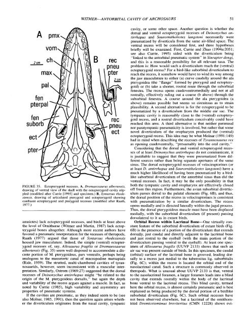
FIGURE 35. Ectopterygoid recesses. A, Dromaeosaurus albertensis,<br />
drawing <strong>of</strong> ventral view <strong>of</strong> the skull with the ectopterygoid cavity stippled<br />
(modified after Currie [I9951 and specimen.) B, Syntarsus rhodesiensis,<br />
drawing <strong>of</strong> articulated pterygoid and ectopterygoid showing<br />
confluent ectopterygoid and pterygoid recesses (modified after Raath,<br />
1977.)<br />
amniotes) lack ectopterygoid recesses, and birds at least above<br />
the level <strong>of</strong> Ornithurae (Witmer and Martin, 1987) lack ectopterygoid<br />
bones altogether. Although more recent authors have<br />
favored a pneumatic interpretation for the recesses <strong>of</strong> theropods,<br />
Raath (1977) argued that those <strong>of</strong> Syntarsus rhodesiensis<br />
housed jaw musculature. Indeed, the simple (ventral) ectopterygoid<br />
recesses <strong>of</strong>, say, Allosaurus fragilis or Dromaeosaurus<br />
albertensis (Fig. 35) seem well disposed to accommodate a discrete<br />
portion <strong>of</strong> M. pterygoideus, pars ventralis, perhaps being<br />
analogous to the masseteric canal <strong>of</strong> macropodine marsupials<br />
(Ride, 1959). The strutted, multi-chambered cavities <strong>of</strong> tyrannosaurids,<br />
however, seem inconsistent with a muscular interpretation.<br />
Similarly, Ostrom (1969:27) suggested that the dorsal<br />
recesses <strong>of</strong> Deinonychus antirrhopus might "be related to the<br />
origin <strong>of</strong> the M. pterygoideus dorsalis," but again the depth<br />
and variability <strong>of</strong> the recess argues against a muscle. In fact, as<br />
noted by Cume (1985), high variability and asymmetry are<br />
properties <strong>of</strong> pneumatic systems.<br />
If the cavities are pneumatic, as they indeed seem to be (see<br />
also Molnar, 1985, 1991), then the question again arises whether<br />
the diverticulum originates from the nasal cavity, tympanic<br />
cavity, or some other space. Another question is whether the<br />
dorsal and ventral ectopterygoid recesses <strong>of</strong> Deinonychus antirrhopus<br />
and Saurornitholestes langstoni necessarily were<br />
pneumatized by diverticula from the same air-filled space. The<br />
ventral recess will be considered first, and three hypotheses<br />
briefly will be examined. First, Cunie and Zhao (1994a:2051;<br />
see also Cume, 1995) sided with the diverticulum being<br />
"linked to the antorbital pneumatic system" in Sinraptor dongi,<br />
and this is a reasonable possibility for all relevant taxa. The<br />
problem is: How would such a diverticulum reach the (ventral)<br />
ectopterygoid recess? For a bird-like suborbital diverticulum to<br />
reach the recess, it somehow would have to wind its way among<br />
the jaw musculature to either (a) curve caudally around the ala<br />
pterygoidea (the "flange" formed by pterygoid and ectopterygoid)<br />
or (b) take a shorter, rostral route through the suborbital<br />
fenestra. The recess opens caudoventromedially and not at all<br />
rostrally, effectively ruling out a course (b above) through the<br />
suborbital fenestra. A course around the ala pterygoidea (a<br />
above) remains possible but seems so circuitous as to strain<br />
plausibility. A second alternative is for the ectopterygoid to be<br />
pneumatized by a diverticulum from the middle ear sac. The<br />
tympanic cavity is reasonably close to the (ventral) ectopterygoid<br />
recess, and a rostral diverticulum conceivably could have<br />
reached this area. A third alternative is that neither paranasal<br />
nor paratympanic pneumaticity is involved, but rather that some<br />
novel diverticulum <strong>of</strong> the oropharynx produced the (ventral)<br />
ectopterygoid recess. This idea may be what Molnar (1991: 149)<br />
had in mind when describing the recesses <strong>of</strong> Tyrannosaurus rex<br />
as opening caudoventrally, "presumably into the oral cavity."<br />
Considering that the dorsal and ventral ectopterygoid recesses<br />
<strong>of</strong> at least Deinonychus antirrhopus do not communicate, it<br />
is justifiable to suggest that they were pneumatized from different<br />
sources rather than being separate apertures <strong>of</strong> the same<br />
sinus. The dorsal ectopterygoid recesses <strong>of</strong> velociraptorines (or<br />
at least D. antirrhopus and Saurornitholestes langstoni) have a<br />
much higher likelihood <strong>of</strong> having been pneumatized by a birdlike<br />
suborbital diverticulum <strong>of</strong> the antorbital sinus than did the<br />
ventral recesses. In fact, it may be the only possibility in that<br />
both the tympanic cavity and oropharynx are effectively closed<br />
<strong>of</strong>f from this region. Furthermore, the avian suborbital diverticulum<br />
passes dorsal to the palatal elements (Fig. 6B), and thus,<br />
the dorsal position <strong>of</strong> the recess in velociraptorines is consistent<br />
with pneumatization by a similar diverticulum. The recess<br />
opens medially and is directed laterally within the jugal process.<br />
Thus, the dorsal pterygoideus muscle must have been displaced<br />
medially, with the suborbital diverticulum (if present) passing<br />
dorsolateral to it as in extant birds.<br />
Orbital Recess within Lacrimal Bone-One virtually constant<br />
feature <strong>of</strong> the suborbital diverticulum <strong>of</strong> extant birds (Fig.<br />
6B) is the presence <strong>of</strong> a portion <strong>of</strong> the diverticulum that extends<br />
dorsally, just caudal and directly adjacent to the lacrimal bone<br />
and just rostral to the eyeball (with the main portion <strong>of</strong> the<br />
diverticulum passing ventral to the eyeball). At least one specimen<br />
<strong>of</strong> Allosaurus fragilis (UUVP 2133) shows that such an<br />
air sac was present outside <strong>of</strong> birds. In this specimen, the caudal<br />
(orbital) surface <strong>of</strong> the lacrimal bone is grooved, leading dorsally<br />
to a recess just medial to the tuberositas lig. suborbitalis<br />
(Fig. 6C); within the recess is located the orbital end <strong>of</strong> the<br />
nasolacrimal canal. Such a structure is commonly observed in<br />
theropods. What is unusual about UUVP 2133 is that, ventral<br />
to the nasolacrimal foramen, a larger foramen leads into a blind<br />
cavity that extends rostrally within the body <strong>of</strong> the lacrimal<br />
bone ventral to the lacrimal recess. This blind cavity, termed<br />
here the orbital recess, is almost certainly pneumatic and is best<br />
explained as being produced by the dorsal portion <strong>of</strong> a birdlike<br />
suborbital diverticulum (Fig. 6C). Such orbital recesses have<br />
not been observed elsewhere, but a lacrimal <strong>of</strong> the ornithomimid<br />
Dromiceiomimus brevitertius (CMN 12228) shows evi-


