

Memoir cover 0.tif - Ohio University College of Osteopathic Medicine
Memoir cover 0.tif - Ohio University College of Osteopathic Medicine
Memoir cover 0.tif - Ohio University College of Osteopathic Medicine

You also want an ePaper? Increase the reach of your titles
YUMPU automatically turns print PDFs into web optimized ePapers that Google loves.
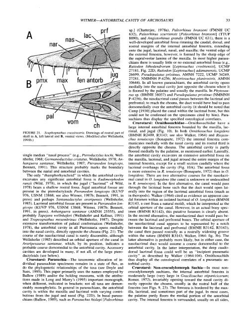
WITMER-ANTORBITAL CAVITY OF ARCHOSAURS 33<br />
FIGURE 21. Scaphognathus crassirostris. Drawings <strong>of</strong> rostral part <strong>of</strong><br />
skull in A, left lateral and B, ventral views. (Modified after Wellnh<strong>of</strong>er,<br />
1991b.)<br />
single median "nasal process" (e.g., Pterodactylus kochi, Wellnh<strong>of</strong>er,<br />
1968; Germanodactylus cristatus, Wellnh<strong>of</strong>er, 1978; Anhanguera<br />
santanae, Wellnh<strong>of</strong>er, 1987; Pteranodon longiceps,<br />
Bennett, 1991). This structure probably marks the boundary<br />
between the narial and antorbital cavities.<br />
The only "rhamphorhynchoid" in which the antorbital cavity<br />
excavates any significant antorbital fossa is Eudimorphodon<br />
ranzii (Wild, 1978), in which the jugal ("lacrimal" <strong>of</strong> Wild,<br />
1978) bears a shallow rostral fossa. Jugal antorbital fossae are<br />
present in the pterodactyloids Pteranodon longiceps (KUVP<br />
976, USNM 13868; see also Witmer, 1987b; Bennett, 1991, in<br />
press) and perhaps Santanadactylus araripensis (Wellnh<strong>of</strong>er,<br />
1985). Lacrimal antorbital fossae are present in Pteranodon longiceps<br />
(KUVP 976, USNM 13868; Witmer, 1987b; Bennett,<br />
1991, in press), Anhanguera santanae (AMNH 25555), and<br />
probably Tapejara wellnh<strong>of</strong>eri (Wellnh<strong>of</strong>er and Kellner, 1991)<br />
and Tropeognathus mesembrinus (Wellnh<strong>of</strong>er, 1987). Despite<br />
extensive transformation <strong>of</strong> palatal conformation (Wellnh<strong>of</strong>er,<br />
1978), the antorbital cavity in all Pterosauria opens medially<br />
into the nasal cavity, directly opposite the choana (Fig. 21). The<br />
course <strong>of</strong> the nasolacrimal canal is rarely discernible, although<br />
Wellnh<strong>of</strong>er (1985) described an orbital aperture <strong>of</strong> the canal in<br />
Araripesaurus santanae, which, by its position, indicates a<br />
probable course dorsomedial to the antorbital cavity. Accessory<br />
cavities are developed in many, if not all, <strong>of</strong> the large pterodactyloids<br />
(see below).<br />
Crurotarsi: Parasuchia-The taxonomic allocation <strong>of</strong> individual<br />
parasuchian specimens remains in a state <strong>of</strong> flux, as<br />
do the phylogenetic relationships <strong>of</strong> the taxa (see Doyle and<br />
Sues, 1995). This paper primarily uses the names employed by<br />
Ballew (1989) andlor the holding museums, with the attributions<br />
made in Long and Murry's (1995) important monograph,<br />
when different, indicated in brackets; not all taxa are demonstrably<br />
monophyletic. In general in parasuchians, the antorbital<br />
cavity is within the maxilla and lacrimal with varying contributions<br />
from the jugal and nasal (Fig. 22D). In basal parasuchians<br />
(Ballew, 1989), such as Parasuchus hislopi [Paleorhinus<br />
sp.] (Chatterjee, 1978a), Paleorhinus bransoni (FMNH UC<br />
632), Paleorhinus scurriensis [Paleorhinus bransoni] (TTUP<br />
8090), and Angistorhinus grandis (FMNH UC 631), there is a<br />
well-developed antorbital fossa rimming the caudal, dorsal, and<br />
rostral margins <strong>of</strong> the internal antorbital fenestra, extending<br />
onto the jugal, lacrimal, nasal, and maxilla; the ventral edge <strong>of</strong><br />
the external fenestra, however, is formed by the sharp edge <strong>of</strong><br />
the supralveolar lamina <strong>of</strong> the maxilla. In most higher parasuchians<br />
there is usually little or no external antorbital fossa (e.g.,<br />
Rutiodon lithodendrorum [Leptosuchus crosbiensis], UCMP<br />
27181, Fig. 22D; Rutiodon [Leptosuchus] adamanensis, UCMP<br />
26699; Pseudopalatus pristinus, AMNH 7222, UCMP 34249,<br />
27281, NMMNH P-4256; Mystriosuchus planirostris, AMNH<br />
10644). In all known parasuchians, the antorbital cavity opens<br />
medially into the nasal cavity just opposite the choana where it<br />
is floored by the palatine and usually the maxilla. In Phytosaurus<br />
sp. (BMNH 38037) and Pseudopalatus pristinus (NMMNH<br />
P-4256), the nasolacrimal canal passes between the lacrimal and<br />
prefrontal; to reach the choana, the duct would have had to pass<br />
dorsomedially over the antorbital cavity (it should be noted that<br />
Camp [I9301 placed the canal within the lacrimal bone, but this<br />
could not be confirmed on the specimens cited by him). Parasuchians<br />
thus display the specified osteological correlates.<br />
Crurotarsi: Ornithosuchidae-Ornithosuchidae have a<br />
large internal antorbital fenestra bounded by the maxilla, lacrimal,<br />
and jugal (Fig. 10). In both Ornithosuchus longidens<br />
(BMNH R2409, R3143; see also Walker, 1964) and Riojasuchus<br />
tenuisceps (Bonaparte, 1972) the internal fenestra communicates<br />
medially with the nasal cavity and its rostral third is<br />
directly opposite the choana. The antorbital cavity is partly<br />
floored medially by the palatine, as mentioned earlier. Laterally,<br />
the antorbital cavity excavates an extensive antorbital fossa on<br />
the maxilla, lacrimal, and jugal around the entire margin <strong>of</strong> the<br />
internal fenestra, except for a small section caudally where the<br />
lacrimal overhangs the cavity (Fig. IOA). The antorbital fossa<br />
is more extensive in R. tenuisceps (Bonaparte, 1972) than in 0.<br />
longidens. There are two alternative courses for the nasolacrima1<br />
canal in 0. longidens (the canal is unknown or undescribed<br />
in R. tenuisceps). In the first alternative, the canal would pass<br />
through the lacrimal bone such that the duct would open laterally<br />
into the region <strong>of</strong> the lacrimal antorbital fossa (much as<br />
in theropods). Walker (1964) cited as evidence an apparent caudal<br />
foramen within an isolated lacrimal <strong>of</strong> 0. longidens (BMNH<br />
R3143, a cast from a natural mold), which he interpreted as the<br />
orbital aperture <strong>of</strong> the canal. However, in an actual bony specimen<br />
(BMNH R3142), this putative foramen is clearly absent.<br />
In the second alternative, the nasolacrimal duct would pass between<br />
the lacrimal and prefrontal bones. The orbital aperture <strong>of</strong><br />
the nasolacrimal canal appears to be represented by a notch<br />
between the lacrimal and prefrontal (BMNH R3142, R3143);<br />
the canal then passed rostrally as a rostrally widening groove<br />
within the suture (BMNH R3143; Walker, 1964: fig. 3b). The<br />
latter alternative is probably more likely, but in either case, the<br />
nasolacrimal duct would assume a course dorsomedial to the<br />
antorbital cavity. In the latter interpretation, the deep caudodorsal<br />
lacrimal fossa could well be an "incipient pneumatic<br />
cavity" as described by Walker (1964:104). Ornithosuchids<br />
thus display all the osteological correlates <strong>of</strong> a pneumatic antorbital<br />
cavity.<br />
Crurotarsi: Non-crocodylomorph Suchia-In most noncrocodylomorph<br />
suchians, the internal antorbital fenestra is<br />
moderately large (very large in Gracilisuchus stipanicicorum;<br />
Romer, 1972), invariably opening toward the nasal cavity directly<br />
opposite the choana, usually in the rostral half <strong>of</strong> the<br />
fenestra (see Figs. 9, 23). The fenestra is bordered by the maxilla,<br />
lacrimal, and sometimes part <strong>of</strong> the jugal. In most cases,<br />
the palatine partly floors the medial portion <strong>of</strong> the antorbital<br />
cavity. The internal fenestra is surrounded, usually on all sides,


