

Memoir cover 0.tif - Ohio University College of Osteopathic Medicine
Memoir cover 0.tif - Ohio University College of Osteopathic Medicine
Memoir cover 0.tif - Ohio University College of Osteopathic Medicine

You also want an ePaper? Increase the reach of your titles
YUMPU automatically turns print PDFs into web optimized ePapers that Google loves.
26 SOCIETY OF VERTEBRATE PALEONTOLOGY, MEMOIR 3<br />
/ / I<br />
sin proc jug mes<br />
rnax pal<br />
fen IQP<br />
tub<br />
antorb ,<br />
promax max<br />
lug<br />
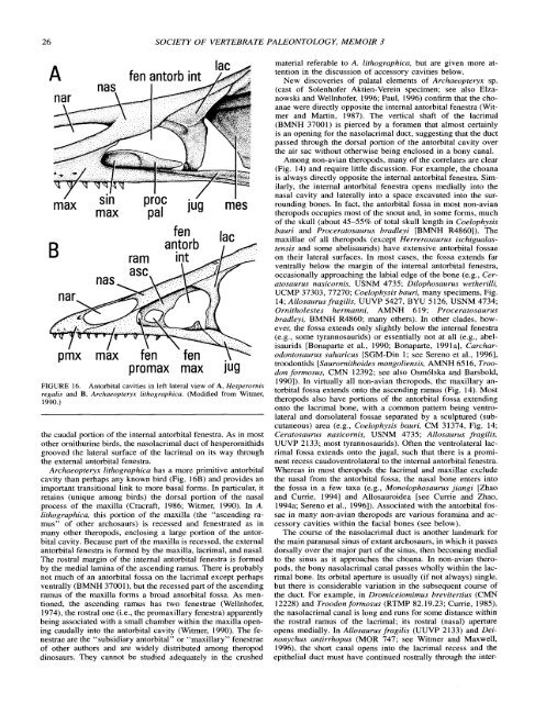
FIGURE 16. Antorbital cavities in left lateral view <strong>of</strong> A, Hesperornis<br />
regalis and B, Archaeopteryx lithographica. (Modified from Witmer,<br />
1990.)<br />
the caudal portion <strong>of</strong> the internal antorbital fenestra. As in most<br />
other ornithurine birds, the nasolacrimal duct <strong>of</strong> hesperornithids<br />
grooved the lateral surface <strong>of</strong> the lacrimal on its way through<br />
the external antorbital fenestra.<br />
Archaeopteryx lithographica has a more primitive antorbital<br />
cavity than perhaps any known bird (Fig. 16B) and provides an<br />
important transitional link to more basal forms. In particular, it<br />
retains (unique among birds) the dorsal portion <strong>of</strong> the nasal<br />
process <strong>of</strong> the maxilla (Cracraft, 1986; Witmer, 1990). In A.<br />
lithographica, this portion <strong>of</strong> the maxilla (the "ascending ,ramus"<br />
<strong>of</strong> other archosaurs) is recessed and fenestrated as in<br />
many other theropods, enclosing a large portion <strong>of</strong> the antorbital<br />
cavity. Because part <strong>of</strong> the maxilla is recessed, the external<br />
antorbital fenestra is formed by the maxilla, lacrimal, and nasal.<br />
The rostral margin <strong>of</strong> the internal antorbital fenestra is formed<br />
by the medial lamina <strong>of</strong> the ascending ramus. There is probably<br />
not much <strong>of</strong> an antorbital fossa on the lacrimal except perhaps<br />
ventrally (BMNH 37001), but the recessed part <strong>of</strong> the ascending<br />
ramus <strong>of</strong> the maxilla forms a broad antorbital fossa. As mentioned,<br />
the ascending ramus has two fenestrae (Wellnh<strong>of</strong>er,<br />
1974), the rostral one (i.e., the promaxillary fenestra) apparently<br />
being associated with a small chamber within the maxilla opening<br />
caudally into the antorbital cavity (Witmer, 1990). The fenestrae<br />
are the "subsidiary antorbital" or "maxillary" fenestrae<br />
<strong>of</strong> other authors and are widely distributed among theropod<br />
dinosaurs. They cannot be studied adequately in the crushed<br />
material referable to A. lithographica, but are given more attention<br />
in the discussion <strong>of</strong> accessory cavities below.<br />
New dis<strong>cover</strong>ies <strong>of</strong> palatal elements <strong>of</strong> Archaeopteryx sp.<br />
(cast <strong>of</strong> Solenh<strong>of</strong>er Aktien-Verein specimen; see also Elzanowski<br />
and Wellnh<strong>of</strong>er, 1996; Paul, 1996) confirm that the choanae<br />
were directly opposite the internal antorbital fenestra (Witmer<br />
and Martin, 1987). The vertical shaft <strong>of</strong> the lacrimal<br />
(BMNH 37001) is pierced by a foramen that almost certainly<br />
is an opening for the nasolacrimal duct, suggesting that the duct<br />
passed through the dorsal portion <strong>of</strong> the antorbital cavity over<br />
the air sac without otherwise being enclosed in a bony canal.<br />
Among non-avian theropods, many <strong>of</strong> the correlates are clear<br />
(Fig. 14) and require little discussion. For example, the choana<br />
is always directly opposite the internal antorbital fenestra. Similarly,<br />
the internal antorbital fenestra opens medially into the<br />
nasal cavity and laterally into a space excavated into the surrounding<br />
bones. In fact, the antorbital fossa in most non-avian<br />
theropods occupies most <strong>of</strong> the snout and, in some forms, much<br />
<strong>of</strong> the skull (about 45-55% <strong>of</strong> total skull length in Coelophysis<br />
bauri and Proceratosaurus bradleyi [BMNH R48601). The<br />
maxillae <strong>of</strong> all theropods (except Herrerasaurus ischigualastensis<br />
and some abelisaurids) have extensive antorbital fossae<br />
on their lateral surfaces. In most cases, the fossa extends far<br />
ventrally below the margin <strong>of</strong> the internal antorbital fenestra,<br />
occasionally approaching the labial edge <strong>of</strong> the bone (e.g., Ceratosaurus<br />
nasicornis, USNM 4735; Dilophosaurus wetherilli,<br />
UCMP 37303, 77270; Coelophysis bauri, many specimens, Fig.<br />
14; Allosaurus fragilis, UUVP 5427, BYU 5126, USNM 4734;<br />
Ornitholestes hermanni, AMNH 619; Proceratosaurus<br />
bradleyi, BMNH R4860; many others). In other clades, however,<br />
the fossa extends only slightly below the internal fenestra<br />
(e.g., some tyrannosaurids) or essentially not at all (e.g., abelisaurids<br />
[Bonaparte et al., 1990; Bonaparte, 1991 a], Carcharodontosaurus<br />
saharicus [SGM-Din 1; see Sereno et al., 19961,<br />
troodontids [Saurornithoides mongoliensis, AMNH 65 16, Troodon<br />
formosus, CMN 12392; see also Osmdlska and Barsbold,<br />
19901). In virtually all non-avian theropods, the maxillary antorbital<br />
fossa extends onto the ascending ramus (Fig. 14). Most<br />
theropods also have portions <strong>of</strong> the antorbital fossa extending<br />
onto the lacrimal bone, with a common pattern being ventrolateral<br />
and dorsolateral fossae separated by a sculptured (subcutaneous)<br />
area (e.g., Coelophysis bauri, CM 31374, Fig. 14;<br />
Ceratosaurus nasicornis, USNM 4735; Allosaurus fragilis,<br />
UUVP 2133; most tyrannosaurids). Often the ventrolateral lacrimal<br />
fossa extends onto the jugal, such that there is a prorninent<br />
recess caudoventrolateral to the internal antorbital fenestra.<br />
Whereas in most theropods the lacrimal and maxillae exclude<br />
the nasal from the antorbital fossa, the nasal bone enters into<br />
the fossa in a few taxa (e.g., Monolophosaurus jiangi [Zhao<br />
and Cume, 19941 and Allosauroidea [see Cume and Zhao,<br />
1994a; Sereno et al., 19961). Associated with the antorbital fossae<br />
in many non-avian theropods are various foramina and accessory<br />
cavities within the facial bones (see below).<br />
The course <strong>of</strong> the nasolacrimal duct is another landmark for<br />
the main paranasal sinus <strong>of</strong> extant archosaurs, in which it passes<br />
dorsally over the major part <strong>of</strong> the sinus, then becoming medial<br />
to the sinus as it approaches the choana. In non-avian theropods,<br />
the bony nasolacrimal canal passes wholly within the lacrimal<br />
bone. Its orbital aperture is usually (if not always) single,<br />
but there is considerable variation in the subsequent course <strong>of</strong><br />
the duct. For example, in Dromiceiomimus brevitertius (CMN<br />
12228) and Troodon formosus (RTMP 82.19.23; Cume, 1985),<br />
the nasolacrimal canal is long and runs for some distance within<br />
the rostral ramus <strong>of</strong> the lacrimal; its rostral (nasal) aperture<br />
opens medially. In Allosaurus fragilis (UUVP 2133) and Deinonychus<br />
antirrhopus (MOR 747; see Witmer and Maxwell,<br />
1996), the short canal opens into the lacrimal recess and the<br />
epithelial duct must have continued rostrally through the inter-


