

The Nature of the Antorbital Fossa of Archosaurs - Ohio University ...
The Nature of the Antorbital Fossa of Archosaurs - Ohio University ...
The Nature of the Antorbital Fossa of Archosaurs - Ohio University ...

Create successful ePaper yourself
Turn your PDF publications into a flip-book with our unique Google optimized e-Paper software.
Fourth Symposium on Mesozoic Terrestrial Ecosystems, Short Papers Ed. by P.J. Currie and EH. Koster.<br />
<strong>The</strong> <strong>Nature</strong> <strong>of</strong> <strong>the</strong> <strong>Antorbital</strong><br />
(Baltimore, Maryland).<br />
<strong>Fossa</strong> <strong>of</strong> <strong>Archosaurs</strong>: Shifting <strong>the</strong> Null Hypo<strong>the</strong>sis. L.M. Witrner<br />
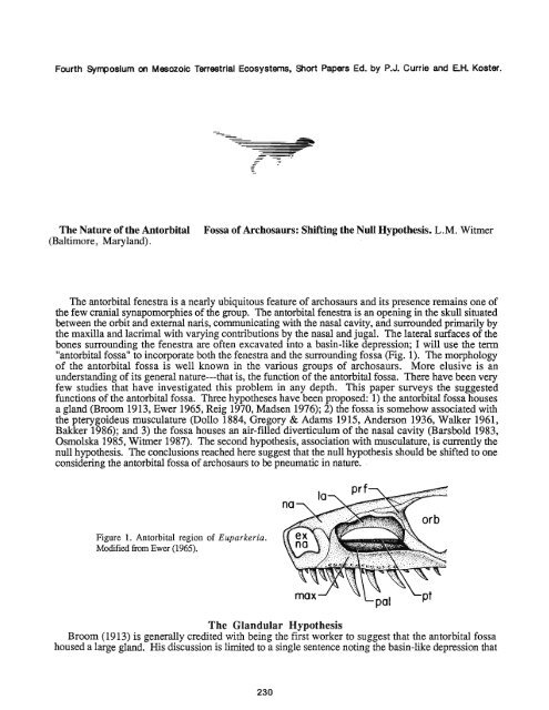
<strong>The</strong> antorbital fenestra is a nearly ubiquitous feature <strong>of</strong> archosaurs and its presence remains one <strong>of</strong><br />
<strong>the</strong> few cranial synapomorphies <strong>of</strong> <strong>the</strong> group. <strong>The</strong> antorbital fenestra is an opening in <strong>the</strong> skull situated<br />
between <strong>the</strong> orbit and external naris, communicating with <strong>the</strong> nasal cavity, and surrounded primarily by<br />
<strong>the</strong> maxilla and lacrimal with varying contributions by <strong>the</strong> nasal and jugal. <strong>The</strong> lateral surfaces <strong>of</strong> <strong>the</strong><br />
bones surrounding <strong>the</strong> fenestra are <strong>of</strong>ten excavated into a basin-like depression; I will use <strong>the</strong> term<br />
"antorbital fossa" to incorporate both <strong>the</strong> fenestra and <strong>the</strong> surrounding fossa (Fig. 1). <strong>The</strong> morphology<br />
<strong>of</strong> <strong>the</strong> antorbital fossa is well known in <strong>the</strong> various groups <strong>of</strong> archosaurs. More elusive is an<br />
understanding <strong>of</strong> its general nature---that is, <strong>the</strong> function <strong>of</strong> <strong>the</strong> antorbital fossa. <strong>The</strong>re have been very<br />
few studies that have investigated this problem in any depth. This paper surveys <strong>the</strong> suggested<br />
functions <strong>of</strong> <strong>the</strong> antorbital fossa. Three hypo<strong>the</strong>ses have been proposed: 1) <strong>the</strong> antorbital fossa houses<br />
a gland (Broom 1913, Ewer 1965, Reig 1970, Madsen 1976); 2) <strong>the</strong> fossa is somehow associated with<br />
<strong>the</strong> pterygoideus musculature (Dollo 1884, Gregory & Adams 19 15, Anderson 1936, Walker 1961,<br />
Bakker 1986); and 3) <strong>the</strong> fossa houses an air-filled diverticulum <strong>of</strong> <strong>the</strong> nasal cavity (Barsbold 1983,<br />
Osmolska 1985, Witrner 1987). <strong>The</strong> second hypo<strong>the</strong>sis, association with musculature, is currently <strong>the</strong><br />
null hypo<strong>the</strong>sis. <strong>The</strong> conclusions reached here suggest that <strong>the</strong> null hypo<strong>the</strong>sis should be shifted to one<br />
considering <strong>the</strong> antorbital fossa <strong>of</strong> archosaurs to be pneumatic in nature.<br />
Figure 1. <strong>Antorbital</strong> region <strong>of</strong> Euparkeria.<br />
Modified fiom Ewer (1965).<br />
<strong>The</strong> Glandular Hypo<strong>the</strong>sis<br />
Broom (1913) is generally credited with being <strong>the</strong> first worker to suggest that <strong>the</strong> antorbital fossa<br />
housed a large gland. His discussion is limited to a single sentence noting <strong>the</strong> basin-like depression that
constituted <strong>the</strong> antorbital fossa <strong>of</strong> Euparkeria (Fig. 1). Ewer (1965), responding to <strong>the</strong> same antorbital<br />
morphology <strong>of</strong> Euparkeria, agreed with Broom that it likely housed a gland and fur<strong>the</strong>r suggested that it<br />
was probably a nasal salt gland. Ewer considered <strong>the</strong> primitive function <strong>of</strong> <strong>the</strong> fossa to be <strong>the</strong> housing<br />
<strong>of</strong> this gland; she refuted <strong>the</strong> muscular hypo<strong>the</strong>sis---but only for <strong>the</strong> earliest archosaurs, arguing that<br />
later in archosaur phylogeny <strong>the</strong> "antorbital gland" was lost as <strong>the</strong> pterygoideus musculature moved<br />
anteriorly to occupy <strong>the</strong> fossa. Thus Ewer argued for both <strong>the</strong> glandular and <strong>the</strong> muscular hypo<strong>the</strong>ses.<br />
More recently, Madsen (1976) suggested that <strong>the</strong> large cavity in <strong>the</strong> body <strong>of</strong> <strong>the</strong> lacrimal <strong>of</strong> Allosm<br />
(Fig. 4C) may have housed a gland.<br />
<strong>The</strong> only worker to <strong>of</strong>fer evidence o<strong>the</strong>r than <strong>the</strong> general morphology <strong>of</strong> <strong>the</strong> antorbital fossa in<br />
support <strong>of</strong> <strong>the</strong> glandular hypo<strong>the</strong>sis was Reig (1970). His argument is based on <strong>the</strong> assumption that<br />
archosaurs were derived from varanopsid pelycosaurs. According to Reig, because mammals are<br />
ureotelic, <strong>the</strong>ir precursors---<strong>the</strong> pelycosaurs---were also ureotelic; thus, because wchosaurs were<br />
derived from pelycosaurs, ancestral archosaurs were also ureotelic. As archosaurs were considered<br />
primarily upland forms, Reig suggested that <strong>the</strong>y required some form <strong>of</strong> extrarenal salt excretion---a salt<br />
gland in <strong>the</strong> antorbital fossa. Through this circuitous and ra<strong>the</strong>r tenuous chain <strong>of</strong> reasoning, Reig<br />
concluded that <strong>the</strong> archosaurian antorbital fossa functioned as a housing for a nasal salt gland.<br />
While it is generally agreed that archosaurs are not related to pelycosaurs and instead are members <strong>of</strong><br />
a monophyletic group, <strong>the</strong> Diapsida (invalidating Reig's basic assumption), <strong>the</strong> glandular hypo<strong>the</strong>sis is<br />
a valid hypo<strong>the</strong>sis that deserves discussion. Most proponents <strong>of</strong> <strong>the</strong> hypo<strong>the</strong>sis have suggested that if a<br />
gland were present it was a site <strong>of</strong> extrarenal salt excretion. In extant diapsids such as squamates<br />
(Parsons 1970) and birds (Fange, et al. 1958) extrarenal salt excretion takes place via <strong>the</strong> lateral nasal<br />
gland. <strong>The</strong> question is whe<strong>the</strong>r or not <strong>the</strong> lateral nasal gland was housed in <strong>the</strong>,antorbital fossa <strong>of</strong> fossil<br />
archosaurs. Comparisons with extant diapsids suggest that it was not. In squamates <strong>the</strong> lateral nasal<br />
gland is situated in <strong>the</strong> anterior portion <strong>of</strong> <strong>the</strong> preorbital region <strong>of</strong> <strong>the</strong> head and lies dorsal or lateral to<br />
<strong>the</strong> nasal capsule (Parson 1970). Crocodilians exhibit a similar situation with <strong>the</strong> gland lying dorsal to<br />
<strong>the</strong> nasal capsule in <strong>the</strong> vicinity <strong>of</strong> <strong>the</strong> juncture <strong>of</strong> <strong>the</strong> premaxilla, maxilla, and nasal (Parsons 1970). In<br />
birds <strong>the</strong> lateral nasal gland is generally located in <strong>the</strong> orbital region (Marples 1932).<br />
Thus, although fossil archosaurs may have had lateral nasal glands, <strong>the</strong>y almost certainly did not<br />
reside in <strong>the</strong> antorbital fossa. Osmolska (1979) likewise rejected <strong>the</strong> antorbital fossa as a site <strong>of</strong> <strong>the</strong><br />
lateral nasal gland and suggested that <strong>the</strong> gland in some dinosaurs may have had a more primitive<br />
position in <strong>the</strong> vicinity <strong>of</strong> <strong>the</strong> external naris. <strong>The</strong> possibility will always remain that some glandular<br />
structure not known in modern organisms may have been housed in <strong>the</strong> antorbital fossa. But such an<br />
assertion will never be supported by much evidence and is not testable. Indeed, <strong>the</strong> morphology that<br />
originally led Broom to suggest <strong>the</strong> glandular hypo<strong>the</strong>sis---<strong>the</strong> basin-like morphology <strong>of</strong> <strong>the</strong> antorbital<br />
fossa---obtains in a group <strong>of</strong> archosaurs that show demonstrable pneumatic features in <strong>the</strong>ir antorbital<br />
fossae; many <strong>the</strong>ropods show very similar depressions in <strong>the</strong> maxilla and lacrimal (Fig. 2C). As is<br />
argued below, one <strong>of</strong> <strong>the</strong> best cases for a pneumatic antorbital fossa can be made for <strong>the</strong>ropod<br />
dinosaurs.<br />
<strong>The</strong> Muscular Hypo<strong>the</strong>sis<br />
<strong>The</strong> most widely accepted hypo<strong>the</strong>sis is that <strong>the</strong> antorbital fossa <strong>of</strong> archosaurs is closely associated<br />
with <strong>the</strong> pterygoideus musculature and I consider this <strong>the</strong> current "null hypo<strong>the</strong>sis." In a sense, <strong>the</strong><br />
fossa has been considered to be in <strong>the</strong> same general class as <strong>the</strong> temporal fenestrae (Gregory & Adams<br />
1915); that is, M. pterygoideus anterior originated from or bulged into <strong>the</strong> antorbital fossa. Evidence<br />
supporting this hypo<strong>the</strong>sis does not come from direct observation <strong>of</strong> <strong>the</strong> morphology <strong>of</strong> antorbital<br />
fossae. Ra<strong>the</strong>r, evidence derives from two sources: 1) biomechanical considerations "demanding" a<br />
large M. pterygoideus anterior, and 2) that extant crocodilians have a large M. pterygoideus anterior<br />
originating from <strong>the</strong> maxilla in <strong>the</strong> region <strong>of</strong> <strong>the</strong> skull where <strong>the</strong> antorbital fossa exists in o<strong>the</strong>r<br />
archosaurs.<br />
Walker's (1961) treatment <strong>of</strong> <strong>the</strong> jaw mechanics <strong>of</strong> early archosaurs represents <strong>the</strong> most cogent<br />
argument for <strong>the</strong> muscular hypo<strong>the</strong>sis. His biomechanical discussions are largely <strong>the</strong>oretical. Walker<br />
(1961) noted that in carnivorous archosaurs like Ornithosuchus and <strong>the</strong>ropods <strong>the</strong> gape must have been<br />
very large. Because <strong>the</strong> temporal muscles are <strong>the</strong> most powerful adductors when <strong>the</strong> jaws are almost<br />
closed (Anderson 1936), Walker (1961) suggested that a large M. pterygoideus anterior must have been<br />
present to act when <strong>the</strong> gape was wide. In his view, <strong>the</strong> large M. pterygoideus anterior originated from<br />
<strong>the</strong> antorbital fossa. This powerful adductor occupying much <strong>of</strong> <strong>the</strong> preorbital region would have
provided "an exceedingly rapid 'snap"' (Walker 1961). For <strong>the</strong> same functional reasons, Bakker<br />
(1986) postulated a huge M. pterygoideus anterior within <strong>the</strong> antorbital fossae <strong>of</strong> Coelophysis and<br />
Dimorphodon.<br />
Figure 2. Attachments <strong>of</strong> M. pterygoideus anterior in A, a<br />
crocodilian (Caiman); B, a bird (Gavia); and C, a <strong>the</strong>ropod<br />
(Coelophysis). Portions <strong>of</strong> <strong>the</strong> maxilla, jugal, and lower<br />
jaw have been cut away in A and B; <strong>the</strong>se bones are<br />
transparent in C.<br />
Support for this view was found in extant crocodilians which have a wide gape and a large M.<br />
pterygoideus anterior. In modem crocodilians M. pterygoideus anterior does indeed originate from <strong>the</strong><br />
maxilla (in addition to o<strong>the</strong>r bones; Schumacher 1973). <strong>The</strong> muscle passes over <strong>the</strong> dorsal surface <strong>of</strong><br />
<strong>the</strong> palate and inserts on <strong>the</strong> medial surface <strong>of</strong> <strong>the</strong> articulare and angular and also on <strong>the</strong> "intermuscular<br />
tendon" (Fig. 2A; Anderson 1936, Schumacher 1973). <strong>The</strong> paradox <strong>of</strong> using extant crocodilians as<br />
evidence for <strong>the</strong> muscular hypo<strong>the</strong>sis is that, despite <strong>the</strong> massive development <strong>of</strong> M. pterygoideus<br />
anterior, eusuchian crocodilians lack <strong>the</strong> antorbital fossa. Ewer (1965) and Osmolska (1985)<br />
recognized this paradox and Osmolska considered <strong>the</strong> muscular hypo<strong>the</strong>sis untenable.<br />
<strong>The</strong> o<strong>the</strong>r group <strong>of</strong> extant archosaurs, <strong>the</strong> birds, retain <strong>the</strong> antorbital fossa and also have a large<br />
anterior division <strong>of</strong> M. pterygoideus (M. pterygoideus lateralis or M. pterygoideus dorsalis <strong>of</strong> avian<br />
anatomy; this portion <strong>of</strong> M. pterygoideus is certainly homologous to M. pterygoideus anterior <strong>of</strong><br />
crocodilians). In birds, however, <strong>the</strong> pterygoideus musculature, although <strong>of</strong>ten <strong>the</strong> largest adductor,<br />
never arises from <strong>the</strong> bones forming <strong>the</strong> antorbital fossa. Instead, <strong>the</strong> muscle originates from <strong>the</strong><br />
dorsolateral surface <strong>of</strong> <strong>the</strong> palatine bone medial to <strong>the</strong> antorbital fossa (Fig. 2B). Thus, birds are like<br />
crocodilians in having a large anterior division <strong>of</strong> M. pterygoideus; but in birds <strong>the</strong> muscle nei<strong>the</strong>r<br />
originates from <strong>the</strong> antorbital fossa nor "bulges" into <strong>the</strong> fossa. Postulation <strong>of</strong> a large M. pterygoideus<br />
anterior on biomechanical grounds is insufficient evidence to support <strong>the</strong> association <strong>of</strong> this muscle<br />
with <strong>the</strong> antorbital fossa.<br />
But <strong>the</strong> functional arguments <strong>of</strong> Anderson (1936) and Walker (1961) suggesting a large M.<br />
pterygoideus anterior in fossil archosaurs are persuasive. I suggest, however, that <strong>the</strong> origin <strong>of</strong> this<br />
muscle was not <strong>the</strong> antorbital fossa itself but ra<strong>the</strong>r <strong>the</strong> bones medial to <strong>the</strong> fossa---<strong>the</strong> pterygoid and<br />
palatine. In many fossil archosaurs <strong>the</strong> pterygoids and especially <strong>the</strong> palatines were more or less<br />
vertically-oriented plates at <strong>the</strong>ir anterior extremities. It seems likely that in most fossil archosaurs, as<br />
in birds, M. pterygoideus anterior originated from <strong>the</strong> palatine and pterygoid and not <strong>the</strong> bones forming<br />
<strong>the</strong> antorbital fossa (Fig. 2C).<br />
<strong>The</strong> Pneumatic Hypo<strong>the</strong>sis<br />
In <strong>the</strong> course <strong>of</strong> my studies <strong>of</strong> <strong>the</strong> cranial air sac system <strong>of</strong> fossil and recent birds (Witmer 1987) it<br />
became apparent that <strong>the</strong> null hypo<strong>the</strong>sis <strong>of</strong> a muscular antorbital fossa had to be rejected for birds. In<br />
birds <strong>the</strong> antorbital fossa houses a large air-filled diverticulum <strong>of</strong> <strong>the</strong> nasal cavity---<strong>the</strong> antorbital sinus<br />
(Fig. 3). <strong>The</strong>re are several subsidiary diverticula <strong>of</strong> <strong>the</strong> antorbital sinus that may pneumatize <strong>the</strong><br />
maxilla, lacrimal, premaxilla, palatine, and o<strong>the</strong>r bones (Witmer 1987). Osmolska (1985) in a<br />
necessarily brief but highly insightful paper suggested that <strong>the</strong> antorbital fossae <strong>of</strong> some fossil<br />
archosaurs were also associated in some way with <strong>the</strong> airway.
Figure 3. <strong>Antorbital</strong> sinus in a golden eagle<br />
(Aquila chrysaetos). Only <strong>the</strong> premaxillary and<br />
lacrimal diverticula are figured.<br />
Birds are not alone among archosaurs in exhibiting pneumatic features in <strong>the</strong> antorbital region.<br />
Although eusuchian crocodilians lack antorbital fenestrae, <strong>the</strong>y none<strong>the</strong>less resemble birds in<br />
possessing air-filled diverticula <strong>of</strong> <strong>the</strong> nasal cavity. But whereas birds have only a single diverticulum,<br />
<strong>the</strong> antorbital sinus, crocodilians have four (Parsons 1970); which <strong>of</strong> <strong>the</strong>se, if any, are homologous to<br />
<strong>the</strong> avian antorbital sinus is presently under study. <strong>The</strong>se diverticula produce vast pneumatic cavities in<br />
<strong>the</strong> maxillae and palatines <strong>of</strong> crocodilians (Fig. 4A; Wegner 1957).<br />
<strong>The</strong>ropod dinosaurs also exhibit unequivocal pneumatic features in <strong>the</strong> bones surrounding <strong>the</strong><br />
antorbital fossa. <strong>The</strong> most striking example is Oviraptor (Osmolska 1976, Barsbold 1983) in which all<br />
<strong>of</strong> <strong>the</strong> bones surrounding <strong>the</strong> fossa are produced into a lattice <strong>of</strong> pneumatic foramina that in some<br />
individuals result in an air-filled bony crest (Fig. 4B). Less extravagant manifestations are found in<br />
o<strong>the</strong>r <strong>the</strong>ropods. <strong>The</strong> most common pneumatic features are <strong>the</strong> distinctive chambering <strong>of</strong> <strong>the</strong> maxilla<br />
and lacrimal (Fig. 4C) in all but <strong>the</strong> more primitive ceratosaurians (Coelophysis and Syntarsus). <strong>The</strong><br />
nasal bones <strong>of</strong> Allosaurus (Fig. 4C) and Ceratosaurus also are pierced by pneumatic foramina<br />
communicating with chambers inside <strong>the</strong> bone. Additionally, some carnosaurs exhibit pneumatic<br />
formamina into <strong>the</strong> jugal bone. <strong>The</strong> muscular hypo<strong>the</strong>sis cannot accomodate <strong>the</strong>se observations as it is<br />
very unlikely that muscle fibers would enter bones such as <strong>the</strong> lacrimal, nasal, and jugal through small<br />
foramina and <strong>the</strong>n expand within <strong>the</strong> body <strong>of</strong> <strong>the</strong> bone. Likewise, <strong>the</strong> thin struts <strong>of</strong> bone forming <strong>the</strong><br />
maxillary sinuses (Madsen 1976) are too fragile to serve as area <strong>of</strong> muscle attachment.<br />
rla<br />
Figure 4. Pneumatic cavities in skulls <strong>of</strong> A, Caiman (cross section; after Wegner 1957); B, Oviraptor (after Barsbold<br />
1983); C, Allosaurus (after Madsen 1976); and D, Pteranodon (partly after Wellnh<strong>of</strong>er 1978).
Ornithischian dinosaurs tend to close <strong>the</strong> external wall <strong>of</strong> <strong>the</strong> antorbital fossa and form a cavity within<br />
<strong>the</strong> maxilla (Osmolska 1985). Ankylosaurids have large pneumatic cavities in <strong>the</strong>ir maxillae formed by<br />
diverticula <strong>of</strong> <strong>the</strong> nasal cavity (Maryanska 1977). Thus, some ornithischians are like crocodilians in<br />
having closed <strong>the</strong> antorbital fenestra yet retaining pneurnaticity in <strong>the</strong> region.<br />
Pterosaurs are ano<strong>the</strong>r group in which diverticula <strong>of</strong> an antorbital air space can be inferred. Not<br />
having examined rhamphorhynchoids, I can only comment on pterydactyloids and in particular<br />
Pteranodon. In large pterydactyloids <strong>the</strong> external naris and antorbital fossa become confluent<br />
suggesting a relationship between <strong>the</strong> fossa and <strong>the</strong> nasal cavity. Fur<strong>the</strong>rmore, specimens <strong>of</strong><br />
Pteranodon exhibit a large pneumatic foramen at <strong>the</strong> posterodorsal comer <strong>of</strong> <strong>the</strong> antorbital fossa in <strong>the</strong><br />
same position as <strong>the</strong> pneumatic foramen <strong>of</strong> <strong>the</strong>ropods and birds (Fig. 4D); this foramen does not<br />
communicate with <strong>the</strong> orbit and cannot be for <strong>the</strong> nasolacximal duct.<br />
Thus, in recent and many fossil archosaurs <strong>the</strong> antorbital fossa shows many features indicative <strong>of</strong><br />
pneumaticity, and even if <strong>the</strong> fossa is closed externally, pneumaticity is retained. It should be noted that<br />
while <strong>the</strong> function <strong>of</strong> <strong>the</strong> fossa was to house an air sac, <strong>the</strong> function <strong>of</strong> <strong>the</strong> air sac itself is a separate<br />
matter and is not considered here.<br />
Discussion<br />
A relevant test <strong>of</strong> <strong>the</strong> hypo<strong>the</strong>sized function <strong>of</strong> a structure is <strong>the</strong> observation <strong>of</strong> <strong>the</strong> situation when <strong>the</strong><br />
structure is absent. Two different hypo<strong>the</strong>ses have been proposed for <strong>the</strong> loss <strong>of</strong> <strong>the</strong> antorbital fossa.<br />
Dollo (1884) noted that in dinosaurs that grind plant food, <strong>the</strong> temporal fossae are enlarged and <strong>the</strong><br />
antorbital fossa is reduced; he correlated this reduction <strong>of</strong> <strong>the</strong> antorbital fossa with reduction <strong>of</strong> <strong>the</strong><br />
pterygoideus musculature. Walker (1961), following Dollo, suggested that in herbivores <strong>the</strong> "speedy<br />
closure <strong>of</strong> <strong>the</strong> jaws" provided by M. pterygoideus anterior was "no longer essential." This correlation<br />
is real but <strong>the</strong> argument fails on consideration <strong>of</strong> modern crocodilians which have prominent<br />
pterygoideus muscles but lack <strong>the</strong> fossa.<br />
Osmolska (1985) advanced a perceptive alternative hypo<strong>the</strong>sis. Osmolska noted <strong>the</strong> "coincidence"<br />
between <strong>the</strong> positions <strong>of</strong> <strong>the</strong> antorbital fossa and <strong>the</strong> choana (that is, <strong>the</strong>y are in <strong>the</strong> same coronal plane)<br />
and cited this as evidence that <strong>the</strong> fossa was somehow associated with <strong>the</strong> nasal passage. Forms with<br />
large fossae retain this coincidence, but taxa that exhibit modifications <strong>of</strong> <strong>the</strong> nasal passage<br />
(ankylosaurids, hadrosaurids, ceratopsids) show reduction or loss <strong>of</strong> <strong>the</strong> antorbital fossa (Osmolska<br />
1985). An additional example are crocodilians. Primitive crocodilians retain <strong>the</strong> coincidence <strong>of</strong> fossa<br />
and choana, but with <strong>the</strong> modification <strong>of</strong> <strong>the</strong> nasal passage associated with <strong>the</strong>ir extensive secondary<br />
palates, derived crocodilians lost <strong>the</strong> antorbital fossa. Thus, despite <strong>the</strong>ir perceived biomechanical<br />
"need" <strong>of</strong> an antorbital fossa, crocodilians are like some ornithischians in loss <strong>of</strong> <strong>the</strong> fossa---not due to<br />
changes in muscle attachment but due to changes in <strong>the</strong> nasal cavity.<br />
A final consideration is whe<strong>the</strong>r or not <strong>the</strong> antorbital fossa originated in response to a diverticulum <strong>of</strong><br />
<strong>the</strong> nasal cavity. Unfortunately, this question may never have a satisfactory answer. It seems likely<br />
that <strong>the</strong> antorbital fossae <strong>of</strong> "<strong>the</strong>codonts" such as ornithosuchids, rauisuchids, stagonolepidids,<br />
phytosaurs, and proterochampsids housed an air-filled diverticulum; indeed, <strong>the</strong> antorbital morphology<br />
<strong>of</strong> some <strong>of</strong> <strong>the</strong>se taxa is strikingly similar to that <strong>of</strong> ceratosaurian <strong>the</strong>ropods such as Coelophysis. <strong>The</strong><br />
point is whe<strong>the</strong>r or not this diverticulum could have produced <strong>the</strong> fenestration. By way <strong>of</strong> analogy it<br />
may be noted that some mammals (many cervids, some fossil equids, lagomorphs) exhibit fenestrate<br />
preorbital regions due to <strong>the</strong> agency <strong>of</strong> air-filled diverticula; <strong>the</strong> morphology <strong>of</strong> <strong>the</strong> lagomorph<br />
Ochotona, in fact, is remarkably similar to <strong>the</strong> antorbital fossa <strong>of</strong> many archosaurs.<br />
In conclusion it may be stated that <strong>the</strong> pneumatic hypo<strong>the</strong>sis for <strong>the</strong> archosaurian antorbital fossa is<br />
<strong>the</strong> most strongly suphorted by evidenci. It involves direct comparisons with living forms, direct<br />
observation <strong>of</strong> morphology, explains all relevant data, and makes no recourse to <strong>the</strong> perceived "needs"<br />
<strong>of</strong> extinct organisms. As such, <strong>the</strong> pneumatic hypo<strong>the</strong>sis should replace <strong>the</strong> muscular hypo<strong>the</strong>sis as <strong>the</strong><br />
"null hypo<strong>the</strong>sis" for <strong>the</strong> nature <strong>of</strong> <strong>the</strong> antorbital fossa <strong>of</strong> archosaurs.<br />
Acknowledgements<br />
For help in various ways I would like to thank S. C. Bennett, M. D. Gottfried, J. A. McAllister, Dr. L. D. Martin,<br />
Dr. H.-P. Schultze, and Dr. D. B. Weishampel. Funding was provided by <strong>the</strong> Johns Hopkins <strong>University</strong> School <strong>of</strong><br />
Medicine.
Abbreviations<br />
antorb sin, antorbital sinus; ec, ectopterygoid; ex na, extemal naris; ex na - ao fos, confluent extemal naris and antorbital<br />
fossa; ju, jugal; la, lacrimal; la div, lacrimal diverticulum; mx, maxilla; na, nasal; nas cav, nasal cavity; orb, orbit; pal,<br />
palatine; pmx, premaxilla; pmx div, premaxillary diverticulum; pneu cav, pneumatic cavity; prf, prefkontal; pt, pterygoid.<br />
Literature Cited<br />
Anderson, H. T. 1936. <strong>The</strong> jaw musculature <strong>of</strong> <strong>the</strong> phytosaur Machaeroprosopus. Journal <strong>of</strong> Morphology, 59, pp.<br />
549-587.<br />
Bakker, R. T. 1986. <strong>The</strong> Dinosaur Heresies. William Morrow & Co., Inc., New York.<br />
Barsbold, R. 1983. Carnivorous dinosaurs from <strong>the</strong> Cretaceous <strong>of</strong> Mongolia. Joint Soviet-Mongolian Paleontological<br />
Expedition, Transactions, 19, pp. 5-120. (In Russian.)<br />
Broom, R. 1913. On <strong>the</strong> South African pseudosuchian Euparkeria and allied genera. Proceedings <strong>of</strong> <strong>the</strong> Zoological Society<br />
<strong>of</strong> London, pp. 619-633.<br />
Dollo, L. 1884. Cinquikme note sur les Dinosauriens de Bemissart. Bulletin du Musee Royal d'Histoire <strong>Nature</strong>lle de<br />
Belgique, 3, pp. 129-146.<br />
Ewer, R. F. 1965. <strong>The</strong> anatomy <strong>of</strong> <strong>the</strong> <strong>the</strong>codont reptile Euparkeria capensis Broom. Philosophical Transactions <strong>of</strong> <strong>the</strong><br />
Royal Society <strong>of</strong> London, Series B, 248, pp. 379-435.<br />
Fiinge, R., Schmidt-Nielsen, K., and Osaki, H. 1958. <strong>The</strong> salt gland <strong>of</strong> <strong>the</strong> herring gull (Larus argentatus). Biological<br />
Bulletin, 115, pp. 162-171.<br />
Gregory, W. K. and Adams, L. A. 1915. <strong>The</strong> temporal fossae <strong>of</strong> vertebrates in relation to <strong>the</strong> jaw muscles. Science, 41,<br />
pp. 763-765.<br />
Madsen, J. H., Jr. 1976. Allosaurus fragilis: a revised osteology. Utah Geological and Mineral Survey, Bulletin 109, pp.<br />
1-163.<br />
Marples, B. J. 1932. <strong>The</strong> structure and development <strong>of</strong> <strong>the</strong> nasal glands <strong>of</strong> birds. Proceedings <strong>of</strong> <strong>the</strong> Zoological Society <strong>of</strong><br />
London, pp. 829-844.<br />
Maryanska, T. 1977. Ankylosauridae (Dinosauria) from Mongolia. Palaeontologia Polonica, 37, pp. 85-151.<br />
Osmolska, H. 1976. New light on <strong>the</strong> skull anatomy and systematic position <strong>of</strong> Oviraptor. <strong>Nature</strong>, 262, pp. 683-684.<br />
--------- 1979. Nasal salt gland in dinosaurs. Acta Palaeontologia Polonica, 24, pp. 205-215.<br />
--------- 1985. <strong>Antorbital</strong> fenestra <strong>of</strong> archosaurs and its suggested function. In Vertebrate Morphology. Edited by H.-R.<br />
Dunclcer and G. Fleischer. Gustav Fischer Verlag, New York, pp. 159-162.<br />
Parsons, T. S. 1970. <strong>The</strong> nose and Jacobson's organ. In Biology <strong>of</strong> <strong>the</strong> Reptilia. Vol. 2. Morphology B. Edited by C.<br />
Gans. Academic Press, New York, pp. 99-191.<br />
Reig, 0. A. 1970. <strong>The</strong> Proterosuchia and <strong>the</strong> early evolution <strong>of</strong> <strong>the</strong> archosaurs; an essay about <strong>the</strong> origin <strong>of</strong> a major<br />
taxon. Bulletin <strong>of</strong> <strong>the</strong> Museum <strong>of</strong> Comparative Zoology, 139, pp. 229-292.<br />
Schumacher, G.-H. 1973. <strong>The</strong> head muscles and hyolaryngeal skeleton <strong>of</strong> turtles and crocodilians. In Biology <strong>of</strong> <strong>the</strong><br />
Reptilia. Vol. 4. Morphology D. Edited by C. Gans and T. S. Parsons. Academic Press, New York, pp. 101-199.<br />
Walker, A. D. 1961. Triassic reptiles from <strong>the</strong> Elgin area: Stagonolepis, Dasygnathus and <strong>the</strong>ir allies. Philosophical<br />
Transactions <strong>of</strong> <strong>the</strong> Royal Society <strong>of</strong> London, Series B, 244, pp. 103-204.<br />
Wegner, R. N. 1957. Die NebenhBhlen der Nase bei den Krokodilen. Wissenschaftliche Zeitschrift der Ernst<br />
Moritz-Universitilt Greifswald, 7, pp. 1-39.<br />
Wellnh<strong>of</strong>er, P. 1978. Pterosauria. In Handbuch der Palbherpetologie. Part 19. Edited by 0. Kuhn. Gustav Fischer Verlag,<br />
New York, pp. 1-82.<br />
Witmer, L. M. 1987. <strong>The</strong> cranial air sac system <strong>of</strong> Mesozoic birds. Unpublished M.A. <strong>the</strong>sis, <strong>University</strong> <strong>of</strong> Kansas.<br />
Museum <strong>of</strong> Natural History, <strong>The</strong> <strong>University</strong> <strong>of</strong> Kansas<br />
currently:<br />
Department <strong>of</strong> Cell Biology and Anatomy<br />
<strong>The</strong> Johns Hopkins <strong>University</strong> School <strong>of</strong> Medicine<br />
Baltimore, Maryland 21205 USA


