T2 Ankle Arthrodesis Nail - Stryker
T2 Ankle Arthrodesis Nail - Stryker T2 Ankle Arthrodesis Nail - Stryker
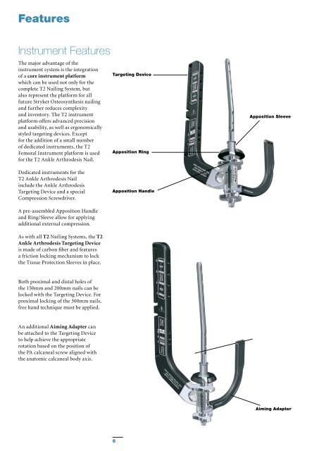
Features Instrument Features The major advantage of the instrument system is the integration of a core instrument platform which can be used not only for the complete T2 Nailing System, but also represent the platform for all future Stryker Osteosynthesis nailing and further reduces complexity and inventory. The T2 instrument platform offers advanced precision and usability, as well as ergonomically styled targeting devices. Except for the addition of a small number of dedicated instruments, the T2 Femoral Instrument platform is used for the T2 Ankle Arthrodesis Nail. Targeting Device Apposition Ring Apposition Sleeve Dedicated instruments for the T2 Ankle Arthrodesis Nail include the Ankle Arthrodesis Targeting Device and a special Compression Screwdriver. Apposition Handle A pre-assembled Apposition Handle and Ring/Sleeve allow for applying additional external compression. As with all T2 Nailing Systems, the T2 Ankle Arthrodesis Targeting Device is made of carbon fiber and features a friction locking mechanism to lock the Tissue Protection Sleeves in place. Both proximal and distal holes of the 150mm and 200mm nails can be locked with the Targeting Device. For proximal locking of the 300mm nails, free hand technique must be applied. An additional Aiming Adapter can be attached to the Targeting Device to help achieve the appropriate rotation based on the position of the PA calcaneal screw aligned with the anatomic calcaneal body axis. Aiming Adapter 6
Relative Indications & Contraindications Relative Indications and Contraindications The T2 Ankle Arthrodesis Nail may be used for: • Posttraumatic and primary Arthrosis • Neuromuscular deformity • Revision of Failed Ankle Arthrodesis • Failed Total Ankle Replacement • Avascular Necrosis of the Talus (requiring tibiocalcaneal arthrodesis) • Neuroarthropathy (Charcot) • Rheumatoid Arthritis with severe deformity • Osteoarthritis • Pseudarthrosis The T2 Ankle Arthrodesis Nail should NOT be used if following conditions are present: • Tibial malalignment of > 10˚ in any plane • Severe vascular deficiency • Osteomyelitis or soft tissue infection Pre-operative Planning Preoperative clinical and radiological assessments are very important for the surgical outcome. • Clinical assessment comprises: evaluation of pain, quality and viability of soft tissue at the surgical site, neurological and vascular status. • Radiological assessment of the ankle includes: weight bearing anteroposterior and lateral views. A lateral hindfoot and Broden’s view are useful in evaluating the subtalar and transverse tarsal joints. • Appropriate implant size can be selected with the T2 Ankle X-Ray Template (1806-3217). Locking Options Based on the clinical and radiological assessment, different locking options can be used to obtain the Tibiotalocalcaneal fusion: Apposition/Compression Locking Mode: - Tibio-talo internal compression with or without additional talocalcaneal external compression (static locking proximal) - Tibio-talo-calcaneal external compression (static locking proximal and distal) Static Locking Mode: - Talo-calcaneal static locking with proximal static locking Dynamic Locking Mode: - The proximal oblong hole allows for secondary dynamization Note: Please see package insert for warnings, precautions, adverse effects and other essential product information. 7
- Page 1 and 2: T2 Ankle Arthrodesis Nail Operative
- Page 3 and 4: Contents Page 1. Introduction 4 Imp
- Page 5: Features Technical Details - T2 Ank
- Page 9 and 10: Operative Technique Joint Preparati
- Page 11 and 12: Operative Technique Reaming Insert
- Page 13 and 14: Operative Technique Nail Insertion
- Page 15 and 16: Operative Technique • The Trocar
- Page 17 and 18: Operative Technique • The Trocar
- Page 19 and 20: Operative Technique Step 4 (optiona
- Page 21 and 22: Operative Technique • After the T
- Page 23 and 24: Operative Technique Freehand Proxim
- Page 25 and 26: Operative Technique Nail Removal Na
- Page 27 and 28: References 1. Tibiotalocalcaneal fu
- Page 29 and 30: Ordering Information - Instruments
- Page 31 and 32: Notes 31
Features<br />
Instrument Features<br />
The major advantage of the<br />
instrument system is the integration<br />
of a core instrument platform<br />
which can be used not only for the<br />
complete <strong>T2</strong> <strong>Nail</strong>ing System, but<br />
also represent the platform for all<br />
future <strong>Stryker</strong> Osteosynthesis nailing<br />
and further reduces complexity<br />
and inventory. The <strong>T2</strong> instrument<br />
platform offers advanced precision<br />
and usability, as well as ergonomically<br />
styled targeting devices. Except<br />
for the addition of a small number<br />
of dedicated instruments, the <strong>T2</strong><br />
Femoral Instrument platform is used<br />
for the <strong>T2</strong> <strong>Ankle</strong> <strong>Arthrodesis</strong> <strong>Nail</strong>.<br />
Targeting Device<br />
Apposition Ring<br />
Apposition Sleeve<br />
Dedicated instruments for the<br />
<strong>T2</strong> <strong>Ankle</strong> <strong>Arthrodesis</strong> <strong>Nail</strong><br />
include the <strong>Ankle</strong> <strong>Arthrodesis</strong><br />
Targeting Device and a special<br />
Compression Screwdriver.<br />
Apposition Handle<br />
A pre-assembled Apposition Handle<br />
and Ring/Sleeve allow for applying<br />
additional external compression.<br />
As with all <strong>T2</strong> <strong>Nail</strong>ing Systems, the <strong>T2</strong><br />
<strong>Ankle</strong> <strong>Arthrodesis</strong> Targeting Device<br />
is made of carbon fiber and features<br />
a friction locking mechanism to lock<br />
the Tissue Protection Sleeves in place.<br />
Both proximal and distal holes of<br />
the 150mm and 200mm nails can be<br />
locked with the Targeting Device. For<br />
proximal locking of the 300mm nails,<br />
free hand technique must be applied.<br />
An additional Aiming Adapter can<br />
be attached to the Targeting Device<br />
to help achieve the appropriate<br />
rotation based on the position of<br />
the PA calcaneal screw aligned with<br />
the anatomic calcaneal body axis.<br />
Aiming Adapter<br />
6


