Untitled
Untitled
Untitled

Create successful ePaper yourself
Turn your PDF publications into a flip-book with our unique Google optimized e-Paper software.
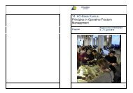
DOSÅrsmøde2011<br />
Abstract-onlineversion
Onlinevejledning<br />
Luk“oversigt”<br />
Fuldsøgbart<br />
program ogabstracts<br />
Funktions-menu<br />
Mulighedfor“fuldskærm”ogdownload(pdf)
Program >>
Session 1: Hip/knee<br />
Onsdag 26. oktober<br />
09:00 – 10:30<br />
Lokale: A<br />
Chairmen:<br />
Micheal Ulrich-Vinter, Rune Dueholm Bech<br />
1. Shoulder and hip patients´pain, mental health and quality of life<br />
Randi Bilberg, Birgitte Noergaard, Kirsten Kaya Roessler, Soeren<br />
Overgaard<br />
2. Continuous evaluation of the metabolism in the femoral head<br />
and neck after Resurfacing Hip Arthroplasty. A randomized controlled<br />
clinical trial<br />
Nina Dyrberg Lorenzen, Michael Ulrich-Vinther, Maiken Stilling, Kjeld<br />
Søballe, Hanne Birke-Sørensen<br />
3. Patient satisfaction, expectations, and quality of life following<br />
periacetabular osteotomy: An 8-12 year follow-up study<br />
Jakob Klit , Charlotte Hartig Andreasen , Steffen Jacobsen, Kjeld<br />
Søballe, Anders Troelsen<br />
4. Cup anteroversion appears to be dependent on surgical approach<br />
– do surgeons overcompensate to prevent dislocation?<br />
Peter Gebuhr, Henrik Palm, Audrey K Nebergall, Meridith E Greene ,<br />
Henrik Malchau<br />
5. The genetic influence on symptomatic osteoarthritis of the hip. A<br />
nationwide population and register based study in Danish twins<br />
Søren Glud Skousgaard, Axel Skytthe, Jacob Hjelmborg, Lars Brandt,<br />
Søren Overgaard<br />
6. Survival of Total Hip Arthroplasty with Ceramic-on-Ceramic<br />
Bearings - Results from Danish Hip Arthroplasty Registry<br />
Claus Varnum, Alma B. Pedersen, Per Kjærsgaard-Andersen, Søren<br />
Overgaard
7. Neuromuscular exercise improves functional performance in<br />
patients with severe osteoarthritis<br />
Allan Villadsen, Søren Overgaard, Anders Holsgaard-Larsen, Ewa<br />
Roos<br />
8. BMD changes in the femoral neck, Gruen zones and the acetabulum<br />
following total and resurfacing hip arthroplasty. 2 year results<br />
from a RCT.<br />
Jeannette Penny, Kim Brixen, Ole Ovesen, Jens-Erik Varmarken, Søren<br />
Overgaard<br />
9. Compaction vs. Broaching Surgical Technique with the Cementless<br />
Bi-Metric Femoral Stem. A 5 year RCT evaluating implant<br />
fixation and clinical outcome<br />
Maiken Stilling, Kjeld Soballe, Poul Torben Nielsen, Poul Hedevang<br />
Christensen, Soren Overgaard, Soren Kold<br />
10 Number of patients needed to discriminate between subgroups<br />
in patient reported outcome measures<br />
Aksel Paulsen, Alma B. Pedersen, Søren Overgaard, Ewa M. Roos
Session 2: Foot/ankle + hand/wrist<br />
Onsdag 26. oktober<br />
09:00 – 10:30<br />
Lokale: B<br />
Chairmen: Inge Lunding Kjær, Bo Munk<br />
11. Lowest occlusion pressure technique for bloodless field in foot<br />
and ankle surgery<br />
Frank Linde, Tine Benzen, Niels Chr. Jensen, Hanne Mainz, Kristian<br />
Kibak Nielsen<br />
12. Short-term outcome of arthroplasty of the first metatarsophalangeal<br />
joint with a non-cemented, three-part prosthesis<br />
Mette Grøndahl Rosenstand, Otto Langhoff<br />
13. Diabetes does not increase short term mortality after nontraumatic<br />
unilateral lower limb amputation<br />
Klaus Kirketerp-Møller, Gitte Holm, Michael Krasheninnikoff, Morten<br />
Tange Kristensen<br />
14. Electra prosthesis for trapezio-metacarpal osteoarthritis: a follow-up<br />
of 39 consecutive cases<br />
Anders Klahn, Marianne Nygaard, Robert Gvozdenovic, Michel E H<br />
Boeckstyns<br />
15. SRTM-MCP Arthroplasties. A prospective consecutive study<br />
for 1 month to five years.<br />
Allan Ibsen Sørensen<br />
16. SRTM-PIP Arthroplasty. A prospective study for 1 month to<br />
six years.<br />
Allan Ibsen Sørensen<br />
17. Acu-Loc volar distal radius plate outcome. A retrospective casestudy.<br />
Mathias Bjerring Ho
18. Concomitant Injury to the Wrist with Severe Distal Radius<br />
Fracture<br />
Hans Tromborg<br />
19. A Three Dimensional Analysis of Osteoarthritic Changes in the<br />
Trapeziometacarpal Joint<br />
Sepp de Raedt, Maiken Stilling, Martijn van de Giessen, Geert J.<br />
Streekstra, Frans M. Vos, Torben B. Hansen<br />
20. An online registry for total wrist replacement<br />
Michel Boeckstyns, Guillaume Herzberg, Allan Ibsen Sørensen, Peter<br />
Axelsson, Karsten Krøner
Session 3: Pediatrics + shoulder/elbow<br />
Onsdag 26. oktober<br />
09:00 – 10:30<br />
Lokale:C<br />
Chairmen:<br />
Hans Viggo Johannsen, Klaus Hindsø<br />
21. The roll of Fat Pad sign in diagnosing occult elbow fractures in<br />
the pediatric patient: A prospective MRI study<br />
Zaid Al-Aubaidi, Trine Torfing, Torben Stryhn, Niels Wisbech Pedersen<br />
22. CPOP (Cerebral palsy follow-up program) in The Region of<br />
Southern Denmark<br />
Niels Wisbech Pedersen, Helle Mätzke Rasmussen, Ulrike Dunkhase-<br />
Heinl<br />
23. Is reversible total epiphysiodesis using 8-plates a safe practice?<br />
Martin Gottliebsen, Bjarne Møller-Madsen, Hans Stødkilde-Jørgensen,<br />
Ole Rahbek<br />
24. Surgical treatment of distal biceps tendon ruptures.<br />
Rasmus Elsøe, Thomas Falstie Jensen, Janne Ovesen<br />
25. Low failure rate after primary and revision arthroscopic Bankart<br />
repair with a knotless anchor<br />
Klaus Bak<br />
26. Reconstruction of the chronic, unstable acromio-clavicular<br />
joint. A prospective case control study comparing the modified<br />
Weaver-Dunn procedure with Anatomical reconstruction of the<br />
coraco- and acromio-clavicular ligaments.<br />
Klaus Bak<br />
27. The Danish Shoulder Arthroplasty Registry<br />
Jeppe Rasmussen, John Jakobsen, Stig Brorson, Bo Sanderhoff Olsen
28. The sternoclavicular joint: Biomechanical effects of arthroscopic<br />
procedures in a cadaveric model<br />
Mads H. Okholm, Jørgen Tranum-Jensen, Andreas Knudsen, Martin W.<br />
Rathcke, Dariush Radi, Michael R. Krogsgaard<br />
29. Revision Total Elbow Arthroplasty with the Semi Constrained<br />
Coonrad-Morrey Total Elbow Arthroplasty<br />
Hans Christian Plaschke, Theis Muncholm Thillemann, Anne-Kathrine<br />
Belling-Sørensen, Bo Sanderhoff Olsen<br />
30. Hemiarthroplasty for fractures of the distal humerus<br />
Lars Henrik Frich, Peter Gaster, Søren Skødt Kristensen
Session 4: Experimental + hip/knee<br />
Torsdag 27. oktober<br />
13:30 – 15:00<br />
Lokale:A<br />
Chairmen:<br />
Henrik Husted, Anders Troelsen<br />
31. Topical Bisphosphonate Treatment of Cemented Prosthesis<br />
Fixation – An Animal Study<br />
Andreas West, Kasra Zainali, Jørgen Baas, Kjeld Søballe<br />
32. Zoledronate-treated Allograft Improves the Fixation of Orthopaedic<br />
Revision Implants<br />
Mette Sørensen, Jørgen Baas, Jeppe Barckman, Joan E. Bechtold,<br />
Kjeld Søballe<br />
33. Cortical-Marrow Ratio: A revised method to detect low bone<br />
mineral density in plain x-rays of the hip<br />
Bjarke Viberg, Frederik SG Harbo, Jens Lauritsen, Søren Overgaard,<br />
Ole Ovesen<br />
34. Have operative indications weakened in primary TKA? An<br />
evaluation of preoperative osteoarthritis and SF-36 scores in 2004<br />
and 2009.<br />
Morten G. Thomsen, Henrik Husted, Kristian S. Otte, Thue Ørsnes,<br />
Anders Troelsen<br />
35. Why in hospital after fast-track hip and knee arthroplasty?<br />
Henrik Husted, Troels H Lunn, Anders Troelsen, Lissi Gaarn-Larsen,<br />
Billy B Kristensen, Henrik Kehlet<br />
36. Migration, bone quality and clinical performance of PFC Sigma<br />
TKA, fixed bearing vs. mobile bearing<br />
Michael Tjørnild, Kjeld Søballe, Maiken Stilling
37. What is the importance of cortical fissures of the femur and<br />
tibia following stemmed revision TKA?<br />
Mads Vinding, Henrik Husted, Kristian S. Otte, Thue Ørsnæs, Anders<br />
Troelsen<br />
38. Increased Periprosthetic Stress Shielding with an I-Beam compared<br />
with a Finned Tibial Component Stem Design. An RCT with<br />
2-years Follow-up by RSA and DXA<br />
Maiken Stilling, Anders Odgaard, Ole Rahbek, Niels Trolle Andersen,<br />
Kjeld Søballe, Frank Madsen<br />
39. Survival study of 298 open-wedge osteotomies in patients with<br />
unilateral gonarthrosis; Region North in the period 2000 to 2010<br />
Anders Christian Laursen, Poul Torben Nielsen, Mogens Berg Laursen<br />
40. The Pegged Trabecular Metal Tibial Plateau Remain Superior<br />
to Screw-fixed Pegged Porous Titanium Fiber-mesh Tibial Plateaus<br />
at 5 years: A randomized clinical RSA study of cementless tibial<br />
components.<br />
Maiken Stilling, Frank Madsen, Anders Odgaard, Claus Fink Jepsen,<br />
Lone Romer, Kjeld Soballe
Session 5: Trauma<br />
Torsdag 27. oktober<br />
13:30 – 15:00<br />
Lokale: B<br />
Chairmen:<br />
Henrik Eckardt, Michael Brix<br />
41. Circumferential wires as a supplement to intramedullary nailing<br />
in inter- and subtrochanteric hip fractures<br />
Lasse Birkelund, Ilija Ban, Henrik Palm, Michael Brix, Anders Troelsen<br />
42. Surgeons agree more on treatment recommendations than on<br />
classification of proximal humeral fractures<br />
Stig Brorson, Bo Sanderhoff Olsen, Lars Henrik Frich, Steen Lund<br />
Jensen, Anne Kathrine Belling Sørensen, Michael Krogsgaard<br />
43. Complications associated with supra- and intracondylar distal<br />
humerus fractures treated with contoured locking plates<br />
Ann Gillberg, Jakob Klit, Paul Hjortberg, Anders Troelsen<br />
44. Local infiltration analgesia in the management of pain after<br />
osteosynthesis of extracapsular hip fracture: A randomized, placebo-controlled,<br />
double-blind clinical trial<br />
Rune Dueholm Bech, Ole Ovesen, Jens Lauritsen, Claus Emmeluth,<br />
Peter Lindholm, Søren Overgaard<br />
45. Secondary hyperparathyroidism and mortality in hip fracture<br />
patients<br />
Christian Medom Madsen, Henrik Løvendahl Jørgensen, Bent Lind,<br />
Troels Riis, Benn Rønnow Duus, Jes Bruun Lauritzen<br />
46. Complications and reoperations following locked plate osteosynthesis<br />
of periprosthetic fractures of the distal femur: A review<br />
of 41 cases from two centers.<br />
Anders W. Paulsen , Michael Brix, Kim Holck, Morten S. Larsen, Anders<br />
Troelsen
47. Periprosthetic fractures around hip replacements treated by<br />
locking plate osteosynthesis: Follow up on 63 consecutive Vancouver<br />
type B1 and C fractures<br />
Lonnie Froberg, Michael Brix, Carsten Fladmose Madsen, Morten<br />
Schultz Larsen<br />
48. Supracondylar Humeral Fractures in Children: Open vs.<br />
Closed Reduction<br />
Morten Søholt Wad, Peter Søndergaard, Louise Jørgensen, Karen Lisbeth<br />
Dirksen<br />
49. Results of 44 bone lengthening procedures with Fitbone for<br />
posttraumatic malunion.<br />
Søren Kold, Kristian Guldbæk Bundgaard, Knud Stenild Christensen<br />
50. X-ray follow-up of surgically treated fractures of the adult distal<br />
forearm<br />
Sune Jauffred, Mitra Sepehri, Susanne Mallet
Session 6:<br />
Pediatrics + sports medicine/arthroscopy<br />
Torsdag 27. oktober<br />
13:30 – 15:00<br />
Lokale: C<br />
Chairmen:<br />
Bjarne Møller Madsen, Michael Krogsgaard<br />
51. Anterior tibial hemiepiphysiodysis for the treatment of recurrent<br />
equinus deformity after surgical treatment of club feet<br />
Zaid Al-Aubaidi, Niels Wisbech Pedersen, Bjarne Lundgaard<br />
52. Long-term outcome after delayed surgery for Bado type 1 radial<br />
head dislocation<br />
Ole Rahbek, Søren Deutch, Søren Kold, Søjbjerg Jens Ole , Bjarne<br />
Møller-Madsen<br />
53. Is radiological screening for hip dysplasia necessary for breech<br />
infants?<br />
Ann-Sofie Broundal Ibfelt, Bjarne Møller-Madsen, Ivan Hvid, Ole Rahbek,<br />
Michel Bach Hellfritzsch<br />
54. Three-Dimensional Kinetic and Kinematic Analysis of Knee<br />
Rotational stability in ACL-deficient patients.<br />
Marie B Bohn, Mette K Petersen, Dennis Nielsen , Henrik Sørensen,<br />
Kjeld Søballe, Martin Lind<br />
55. Incidence and outcome after revision ACL reconstructions.<br />
Results from the Danish registry for knee ligament reconstructions<br />
with 5 years follow-up<br />
Martin Lind, Frank Mehnert, Alma Pedersen<br />
56. Correlation between different patella alta measurements in<br />
patients with and without patella-femoral instability.<br />
Sinan Said, Svend Erik Christainsen, Peter Faunø, Bent Lund, Martin<br />
Lind
57. Tunnel bone density after allogenic bone chip and bone cylinder<br />
transplantation in staged revision anterior cruciate ligament reconstruction<br />
Sinan Said, Katriina B. Puhakka, Svend Erik Christainsen, Peter<br />
Faunø, Bent Lund, Martin Lind<br />
58. Reducing donor site morbidity after after ACL reconstruction<br />
with hamstring tendons<br />
Peter Faunø, Svend Erik Christiansen, Bent Lund, Marc Strauss, Martin<br />
Lind<br />
59. Outcome after re-revision ACL reconstruction in 17 patients<br />
from a single institution. 2-15 years follow-up<br />
Marc Jacob Strauss, Martin Lind, Bent Lund, Svend Erik Kristiansen,<br />
Peter Faunø<br />
60. Arthroscopy of the sternoclavicular joint. Establishing portals,<br />
anatomy, structures at risk and possible arthroscopic procedures<br />
using fresh frozen cadavers.<br />
Martin Rathcke, Jørgen Tranum-Jensen, Dariush Radi, Andreas Knudsen,<br />
Mads Okholm, Michael Krogsgaard
Session 7: Hip/knee<br />
Fredag 28. oktober<br />
09:00 – 10:30<br />
Lokale: A<br />
Chairmen:<br />
Ole Ovesen og Maiken Stilling<br />
61. Peri-Acetabular Osteotomy Improves Sport Performance in<br />
Patients with Dysplasia of the Hip<br />
Michael Ulrich, Kjeld Soballe<br />
62. Blood Perfusion and Bone Formation before and after minimally<br />
invasive periacetabular osteotomy analysed PET combined with<br />
CT<br />
Inger Mechlenburg, Flemming Hermansen, Theis Thillemann, Kjeld<br />
Søballe<br />
63. Evaluation of a Verbal Rating Scale for post-operative pain in<br />
hip fracture patients<br />
Bech Rune Dueholm, Jens Lauritsen, Ole Ovesen, Søren Overgaard<br />
64. Clinical and radiographic outcome 10-17 years following surgical<br />
treatment of a slipped capital femoral epiphysis.<br />
Jakob Klit, Kasper Gosvig, Steffen Jacobsen, Kjeld Søballe, Anders<br />
Troelsen<br />
65. Do RSA measurements of femoral head penetration and acetabular<br />
cup migration in THR depend upon shell and liner segment<br />
assignment?<br />
Karl Tobias Haak, Henrik Palm, Meridith E Greene, Henrik Malchau,<br />
Peter Gebuhr<br />
66. Multicenter RSA Evaluation of In Vivo Wear of Vitamin E Stabilized<br />
Highly Cross-linked Polyethylene<br />
Henrik Palm, Meridith E Greene, Poul Torben Nielsen, Mogens Berg<br />
Laursen, Karl Tobias Haak, Henrik Malchau
67. Increased wear of polyethylene in first generation modular<br />
metal-backed cups with ETO sterilized acetabular components. A<br />
minimum 11 years follow-up and clinical assessment.<br />
DM Hengst, PB Thomsen, TB Hansen, M Homilius, M Stilling<br />
68. Correlation between conventional radiographs and computed<br />
tomography in acetabular retroversion<br />
Christoffer Skovkjær Jensen, Søren Overgaard , Trine Torfing, Ole<br />
Ovesen<br />
69. Two year Metal ions and lymphocyte counts. An RCT between<br />
the ASR Resurfacing system (RHA) and ceramic on poly Total Hip<br />
Arthroplasty (THA)<br />
Jeannette Penny, Christian Nielsen, Ole Ovesen, Jens-Erik Varmarken,<br />
Søren Overgaard<br />
70. Early micro motion of the ASRTM femoral component. 2 year<br />
radiostereometry (RSA) results<br />
Jeannette Penny, Ming Ding, Jens-Erik Varmarken, Ole Ovesen, Søren<br />
Overgaard
Session 8:<br />
Experimental + rehabilitation + tumors<br />
Fredag 28. oktober<br />
09:00 – 10:30<br />
Lokale: B<br />
Chairmen:<br />
Michael Mørk Petersen, Peter Holmberg Jførgensen<br />
71. Topical Bisphosphonate Augments Fixation of Bone-grafted<br />
Implants, BMP-2 causes Resorption-based Instability<br />
Jorgen Baas, Marianne Vestermark, Thomas Jakobsen, Thomas Jensen,<br />
Joan Bechtold, Kjeld Soballe<br />
72. ANTIBIOTIC IMPREGNATION OF ALLOGRAFT BONE<br />
AND THE EFFECT ON IMPLANT FIXATION<br />
Jeppe Barckman, Jørgen Baas, Mette Sørensen, Jeppe Lange, Joan<br />
Bechtold, Kjeld Søballe<br />
73. Active and passive immunization against Staphylococcus aureus<br />
periprosthetic osteomyelitis in rats<br />
Niels H. Søe, Nina Vendel Jensen, Steen Seier Poulsen, Frank Espersen,<br />
Gerald Pier, Helle Krogh Johansen<br />
74. Hypoxia Enhances Chondrogenic Differentiation of Human<br />
Cord Blood Multilineage Progenitor Cells Seeded on a Novel Scaffold<br />
of Freeze Dried Polycaprolactone<br />
Samir Munir, Ryan Figueroa, Thomas Gadegaard Koch, Dang Quang<br />
Svend Le, Jens Vinge Nygaard, Michael Ulrich-Vinther<br />
75. Why do non-traumatic lower limb amputee patients stay in<br />
hospital and where are they discharged to?<br />
Morten Tange Kristensen, Gitte Holm, Klaus Kirketerp-Møller , Peter<br />
Gebuhr<br />
76. Setting up an orthogeriatric unit, seen from an orthopaedic<br />
point of view<br />
Christian Tollund, Benn Duus, Susanne Van Der Mark
77. Optimizing Multidrug Resistance Protein 1 silencing in osteoand<br />
chondrosarcoma<br />
Sarah Stammose Freund, Michael Bendtsen, Akmal Safwat, Peter<br />
Holmberg Jørgensen<br />
78. Reverse shoulder prosthesis after removal of the proximal humerus<br />
due to tumour<br />
Anne Kathrine Kaa, Hans Viggo Johannsen, Johnny Østergaard Østergaard<br />
Keller, Bjarne Hauge Hansen, Thomas Einer Baad-Hansen,<br />
Peter Holmberg Jørgensen<br />
79. The Diagnostic Value of 18F-FDG PET/CT in Staging of Highgrade<br />
Bone and Soft Tissue Sarcomas<br />
Hanna Maria Fuglø, Simon Møller Jørgensen, Dorrit Hovgaard, Annika<br />
Loft, Michael Mørk Petersen<br />
80. The Early Experience with the GMRS Mega-prosthesis<br />
Michala Skovlund Sørensen, Henrik Schrøder, Michael Mørk Petersen
Session 9: Spine + trauma<br />
Fredag 28. oktober<br />
09:00 – 10:30<br />
Lokale: C<br />
Chairmen:<br />
Benny Dahl, Morten Schultz Larsen<br />
81. A prospective randomized trail between Transforaminal (TLIF)<br />
and instrumented posterolateral fusion (PLF) a two year followup.<br />
Kristian Høy, Bent Niederman, Peter Helmig, Ebbe Stender Hansen,<br />
Thomas Andersen, Cody Bünger<br />
82. A retrospective study of infectious spondylodiscitis managed at<br />
the Department of Orthopedic Surgery, Spine unit, Aarhus University<br />
Hospital between 2000 and 2010.<br />
Kestutis Valancius, Ebbe Stender Hansen, Kristian Høy, Peter Helmig,<br />
Bent Niedermann, Cody Bünger<br />
83. Long-term results after short-segment pedicle fixation of thoracolumbar<br />
fractures<br />
Michael Mølmer, Tobias Aasvang, Martin Gehrchen, Benny Dahl<br />
84. Routine Blood Tests as Predictors of Mortality in Hip Fracture<br />
Patients, A Literature-Based Meta-Analysis<br />
Anne Sofie Laulund, Jes Bruun Lauritzen, Løvendahl Jørgensen Henrik<br />
85. Arthroscopy as an aid in treating tibial plateau fractures<br />
Steffen Skov Jensen, Flemming Hansen, Janni Stroem<br />
86. What to expect following locked plate osteosynthesis of midshaft<br />
clavicle fractures? Complications, reoperations and patient<br />
reported outcome.<br />
Ulrik Branner, Ilija Ban, John Kloth Petersen, Anders Troelsen
87. In-hospital mortality following hip fracture- before and after<br />
orthogeriatrics<br />
Anna Gaki Lindestrand, Claes Carson, Susanne Van der Mark, Benn<br />
Duus<br />
88. Does the postoperative regime influence complications and reoperations<br />
following locking plate osteosynthesis of clavicle fractures?<br />
Marie Fridberg-Tobiassen, Zaid Issa, Ilija Ban, Michael Krasheninnikoff,<br />
Anders Troelsen<br />
89. Mobilization inhibited by Post operative pain predicted by fracture<br />
type and type of surgery<br />
Troels Riis, Henning Ogarrio, Henrik Jorgensen, Jes Lauritzen, Susanne<br />
van der Mark, Benn Duus<br />
90. Municipal cost and impact on home aid utilisation and nursing<br />
home services following hip fracture: A population-based study<br />
Martin L. W. Engeland, Gunnar F. Hvattum, Lars E. Matzen, Søren<br />
Overgaard, Jan Sørensen, Jesper Ryg
Foredragskonkurrence:<br />
Fredag 28. oktober<br />
13.30 – 14:30<br />
Lokale: A<br />
Chairmen:<br />
Jeannette Østergaard Penny, Martin Lind<br />
91. Cemented or Uncemented Fixation of Proximal Interphalangeal<br />
Joint Implants? A 2 year RCT of implant fixation, periprosthetic<br />
bone density and finger function.<br />
Maiken Stilling, Karsten Krøner, Bo Munk, Ole Rahbek, Inge Helleberg,<br />
Kjeld Soballe<br />
92. A prospective randomized clinical trial using an uncemented<br />
and screw fixated porous coated cup with or without hydroxyapatite<br />
in revision total hip arthroplasty<br />
Ole Ovesen, Ming Ding, Søren Overgaard<br />
93. The effect of local infiltration analgesia in the management of<br />
pain after periacetabular osteotomy: A randomized, placebocontrolled,<br />
double-blind clinical trial<br />
Rune Dueholm Bech, Ole Ovesen, Peter Lindholm, Søren Overgaard<br />
94. Acetabular cup position is better using the direct anterior approach<br />
with fluoroscopy compared to the postero- and anterolateral<br />
approaches<br />
Henrik Palm, Meridith E Greene, Roger H Emerson, James Huddleston,<br />
Henrik Malchau , Peter Gebuhr<br />
95. Factors predicting failure following periacetabular osteotomy:<br />
A 2-12 years follow-up study of 406 periacetabular osteotomies<br />
Charlotte Hartig-Andreasen, Anders Troelsen, Michael Ulrich-Vinther,<br />
Theis Thilleman, Kjeld Søballe
Poster Udstilling<br />
Lokale: Foyer<br />
96. Strontium-substitution of Hydroxyapatite Coating did not Improve<br />
Osseointegration and Implant Fixation<br />
Marianne Toft Vestermark, Ellen-Margrethe Hauge, Joan Elizabeth<br />
Bechtold, Thomas Jakobsen, Heiko Gruner, Baas Søballe<br />
97. Telos ankle stress test for lateral ankle instability,Should the<br />
test be performed with the patient awake or during general anaesthesia?<br />
Kristian Kibak Nielsen, Frank Linde, Niels Chr. Jensen<br />
98. Quality assurance in the surgical treatment of distal radius<br />
fractures: Initial results from a new database in the Hand Surgery<br />
Unit, Department of Orthopaedic Surgery, Aarhus University Hospital.<br />
Ann Sobrón, Anders Ditlev Jensen<br />
99. Results of Thumb Trapeziometacarpal Joint Replacements in<br />
Young Patients<br />
Hans Tromborg, Anders Peter Højlund, Anders Lorentsen<br />
100. Localized Statistical Shape Model for Detection of Osteoarthritis<br />
in the Trapeziometacarpal Joint<br />
Sepp de Raedt, Martijn van de Giessen, Maiken Stilling, Torben B.<br />
Hansen, van Vliet Lucas J., Frans M. Vos<br />
101. Promising preliminary results from the CORIHA study (Cementless<br />
One-stage Revision of chronic Infected Hip Arthroplasty)<br />
Jeppe Lange, Anders Troelsen, Kjeld Søballe, Kristian Otte, Søren Solgaard,<br />
Martin La On behalf of CORIHA research group<br />
102. The lateral approach to the hip in hemiarthroplasty surgery:<br />
Results of a “Danish experience”.<br />
Ulrik Knudsen, Lief Broeng, Anders Troelsen
103. Rehabilitation after THR: Telephone interview and individual<br />
support versus visits in outpatient clinic.<br />
Britta Hørdam, Linda Koldsgaard, Preben U Pedersen, Kjeld Søballe<br />
104. Are there any benefits of preoperative methylprednisolone in<br />
primary TKA surgery when pain treatment is opioid based?<br />
Amir Pasha Attarzadeh, Jesper Fabrin, Klaus Poulsen, Jens Kurt Johansen,<br />
Anders Troelsen<br />
105. Physical activity levels measured in patients prior to total joint<br />
replacement compared with healthy controls<br />
Anders Holsgaard-Larsen, Ewa M. Roos<br />
106. Total hip arthroplasty with the Thrust Plate Prosthesis. Late<br />
results in a pilot series.<br />
Janus D. Christiansen, Poul T. Nielsen, Mogens B. Lauersen<br />
107. Incidence of THA after acetabular fracture<br />
Dennis Hallager Nielsen, Søren Lange Rasmussen, Søren Peter<br />
Eiskjær, Poul Torben Nielsen, Sten Rasmussen<br />
108. Effect of continuous Adductor-Canal-Blockade on pain and<br />
mobilization after total knee arthroplasty<br />
Pia Jæger, Morten T. Jenstrup, Jørgen Lund, Søren Bache, Tommy K.<br />
Larsen, Jonna S. Fomsgaard<br />
109. Hemiarthroplasty with the Aequalis fracture stem in the<br />
treatment of comminuted fractures of proximal humerus<br />
Thomas Jensen, Otto Falster<br />
110. Septic Arthritis in the Sternoclavicular Region. Clinical and<br />
Radiological Aspects.<br />
Tina Boedker, Mikkel Tøttrup, Klaus Kjær Petersen, Anne Grete Jurik<br />
111. Impact of different graft types after ACL reconstruction, result<br />
from the Danish registry of Knee ligament reconstruction<br />
Lene Rahr Wagner, Theis M Thillemann, Alma B Pedersen, Martin<br />
Lind
112. Use of an ambulatory infusion pump after harvesting a semitendinosis<br />
and gracilis graft to manage postoperative pain, in outpatient<br />
having ACL reconstructive surgery<br />
Karl Bertil Jöhnk, Steffen Skov Jensen<br />
113. Delirium assessment in orthopedic surgery<br />
Peter Kjær Nielsen, Benn Duus, Henrik Løvendahl Jørgensen, Susanne<br />
van der Mark<br />
114. Reproducibility of goniometric measurements of passive hip<br />
mobility and hand-held dynamometry of hip muscle strength in<br />
patients with hip osteoarthritis – an intra- and inter-rater study<br />
Erik Poulsen, Henrik Wulff Christensen, Jeannette Østergaard Penny,<br />
Søren Overgaard, Werner Vach, Jan Hartvigsen<br />
116. Epiphysiodesis Made with Radio Frequency Ablation: First<br />
Results From a Pilot Study<br />
Juan Manuel Shiguetomi-Medina, Ole Rahbek, Hans Stødkilde-<br />
Jørgensen, Bjarne Møller-Madsen<br />
117. Precision of Radiological Methods novel in relation to Resurfacing<br />
Humeral Head Implants: Assessment by radiostereometric<br />
analysis, DXA, and Geometrical Analysis.<br />
Inger Mechlenburg, Anette Liljensøe, Maiken Stilling, Anders Amstrup,<br />
Kjeld Søballe , Thomas Klebe<br />
118. Minimally invasive dynamic hip screw for fixation of hip fractures<br />
Zaid al-Saadi<br />
119. Non-union rate in humeral shaft fractures treated with a functional<br />
brace<br />
Karl Bertil Jöhnk, Michael Toft Væsel
Poster session I:<br />
Torsdag 27. oktober<br />
11.30 – 12.30<br />
Lokale: A<br />
Chairmen:<br />
Jens Erik Varmarken, Jeannette Østergaard Penny<br />
120. Metal ion levels in the blood of patients with metal-on-metal<br />
(MoM) hip joint articulations. Does storage of blood samples matter?<br />
Martin Lindberg-Larsen, Jeannette Ø. Penny<br />
121. Quality of Life and Hip-Function is improved during the first<br />
month after Total Hip Arthroplasty<br />
Peter Uhrbrand, Kjeld Søballe, Michael Urich-Vinther<br />
122. ECCENTRIC HIP ABDUCTOR WEAKNESS IN PATIENTS<br />
WITH SYMPTOMATIC EXTERNAL SNAPPING HIP - A<br />
CROSS-SECTIONAL STUDY<br />
Julie Sandell Jacobsen, Kristian Thorborg, Kjeld Søballe, Michael<br />
Ulrich-Vinther<br />
123. Survival of the hip after periacetabular osteotomy.<br />
Line Borreskov Dahl, Michael Mørk Petersen, Jens Stürup<br />
124. Revision Total Knee Arthroplasty with the use of Trabecular<br />
Metal Cones.<br />
Claus L. Jensen, Michael M. Petersen, Henrik M. Schrøder, Gunnar<br />
Flivik, Bjarne Lund<br />
125. Stem alignments influence of migration of cemented femoral<br />
component in THA<br />
Jens Trærup, Juozas Petruskevicius, Poul Torben Nielsen, Mogens<br />
Berg Laursen
126. The Incidence of stem fracture of monoblock and modular<br />
revision stems in Denmark 1995-2008<br />
Jamal Bech Bouknaitir, Søren Overgaard, Jane Schwartz Leonhardt ,<br />
Alma Becic Pedersen, Per Kjaersgaard-Andersen<br />
127. The predictive values of perioperative biopsies in periprosthetic<br />
infections<br />
Mikkel Rathsach Andersen, Stine Hangaard, Benn Duus<br />
128. Monitoring patients with metal-on-metal hip implants<br />
Arne Borgwardt, Søren Ribel-Madsen, Tine Bertelsen, Sandra Fabricius<br />
129. A proof-of-concept evaluation of a new semi-quantitative urine<br />
test to exclude ongoing coagulation activity after THA and TKA<br />
Lars Borris, Michael Lassen, Akos Pap<br />
130. Catastrophic results after the use of a selfretaining running<br />
suture in total hip replacement<br />
Søren Solgaard, Anne Grete Kjersgaard<br />
131. Periacetabular osteotomy in young adults with Calvé-Legg-<br />
Perthes disease.<br />
Jens Lindhardt Palm, Ole Ovesen, Søren Overgaard
Poster session II:<br />
Torsdag 27. oktober<br />
11.30 – 12.30<br />
Lokale: B<br />
Chairmen:<br />
Sten Rasmussen, Claus Munk Jensen<br />
132. Forensic finite element simulation of skull fracture<br />
Tina Lercke Skytte, Lars Pilgaard Mikkelsen<br />
133. Bispebjerg (Hvolris) Orthopadic PreSurgery Score (HOPSS)<br />
Jesper Hvolris, Kenneth Jensen, Jens Børglum<br />
134. Twenty-nine ankle arthrodesis with the Calandruccio external<br />
compression fixator. Is the rate of bony union affected by preoperative<br />
bone stock?<br />
Niels Chr. Jensen, Claus Sundsatrup, Frank Linde, Kristian Kibak<br />
Nielsen<br />
135. Parametric study of a drop foot brace<br />
Ragnhild í Skorini, Lars P. Mikkelsen, Tom L. Andersen, Christian<br />
Wong<br />
136. The Elektra prosthesis for total replacement of the first CMC-<br />
Join: Prospective study with follow-up of one to six years.<br />
Allan Ibsen Sørensen<br />
137. The role of MRI in the diagnosis of wrist fractures<br />
Wail Ahmad, Merete König, Ole Holm, Annette Sylvest<br />
138. No differences in growth plate zone fractions in a comparative<br />
porcine study on 8-plates versus staples<br />
Martin Gottliebsen, Bjarne Møller-Madsen, Hanne Damgaard Poulsen,<br />
Ole Rahbek
139. Guided growth for the abnormal physis. Experience with the<br />
8-plate technique in non-idiopathic cases<br />
Martin Gottliebsen, Manoj Ramachandran, Mark Paterson, Matthew<br />
Barry<br />
140. A Retrospective Examination of the Copeland Resurfacing<br />
Humeral Head Implant with respect to Overstuffing<br />
Inger Mechlenburg, Anders Amstrup, Thomas Klebe, Stig Storgaard<br />
Jacobsen, Gerhardt Teichert, Maiken Stilling<br />
141. Comparison of Two Humeral Head Resurfacing Implants. 1<br />
year Results of a Randomized Controlled Clinical Trial.<br />
Inger Mechlenburg, Maiken Stilling, Kaj Døssing, Kjeld Soballe, Thomas<br />
Klebe<br />
142. Dose optimization of O-arm paediatric 3D spine protocol<br />
Asger Greval Petersen, Søren Eiskjær, Jon Kaspersen, Karen Petra<br />
Weigert, Sten Rasmussen<br />
143. The feasibility of prospective quality of life measurement in<br />
metastatic spinal cord compression<br />
Søren Schmidt Morgen, Rikke Søgaard, Cody Bünger , Claus Falck<br />
Larsen , Benny Dahl
Poster session III:<br />
Torsdag 27. oktober<br />
11.30 – 12.30<br />
Lokale: C<br />
Chairmen: Frank Farsø Nielsen, Steen Lund Jensen<br />
144. Anterior Distal Femoral Epiphysiodesis for the treatment of<br />
fixed knee flexion contractures<br />
Zaid Al-Aubaidi, Bjarne Lundgaard, Niels Wisbech Pedersen<br />
145. Osseointegrated prosthesis for the femoral amputees<br />
Peter Holmberg Jørgensen, Klaus Kjær Petersen, Jens Ulrik Pedersen,<br />
Maiken Stilling, Inger Krog-Mikkelsen, Kjeld Søballe<br />
146. Task shifting in orthopaedic out-patients – an audit of shoulder<br />
patients<br />
Jette W Vobbe, Sven T D Lausen, Thomas Soerensen, Lilli Soerensen<br />
147. Nerve injury after hip arthroscopy with labral repair, a prospective<br />
cohort study - is there a learning curve?<br />
Crhistian Dippmann, Otto Kraemer, Søren Winge<br />
148. The myotendinous junction in humans: A method to study its<br />
structure and reaction to exercise and immobilisation.<br />
Andreas Knudsen, Mathias Hjort, Karin Kjær-Hansen, Abigail Mackey,<br />
Michael Kjær, Michael Rindom Krogsgaard<br />
149. Experience with surgical skin Closure System within ortopeadic<br />
surgery<br />
Flemming Hansen, Janni Stroem, Louise Hosbond, Steffen Skov Jensen<br />
150. High incidence of long term chronic complications after ankle<br />
sprain<br />
Per Hviid Gundtoft, Sten Rasmussen
151. Per-operative fluid challenge in hip fracture patients<br />
Viktoria Oline Barrios Poulsen<br />
152. Initial results of treating femoral trochanteric fractures with a<br />
double screw nail<br />
Steffen Skov Jensen, Flemming Hansen, Maj-Britt Brinkmann, Mikael<br />
Skov Nielsen, Rasmus Stokholm<br />
153. Benefits and harms of locking plate osteosynthesis in intraarticular<br />
(OTA Type C) fractures of the proximal humerus: a systematic<br />
review<br />
Stig Brorson, Jeppe Vejlgaard Rasmussen, Lars Henrik Frich, Bo Sanderhoff<br />
Olsen, Asbjørn Hróbjartsson<br />
154. Self-administrated home intravenous antibiotic therapy using<br />
elastomeric infusion pumps in orthopaedic patients<br />
Jens ER Svendsson, Hilde Omestad, Klaus Kjær Petersen<br />
155. Work up of sarcomas in The North Denmark Region before<br />
and after implemention of National Integrated Cancer Pathways<br />
(“Pakkeforløb for sarkomer i knogle og bløddele”)<br />
Lene Dremstrup, Poul Verner Madsen
Abstracts>>
1.<br />
Shoulder and hip patients´pain, mental health and quality of life<br />
Randi Bilberg, Birgitte Noergaard, Kirsten Kaya Roessler, Soeren Overgaard<br />
The Department of Orthopedic Surgery Kolding Hospital, a part of Lilltebaelt<br />
Hospital, Denmark; Emergency department, Kolding Hospital, a part of<br />
Lilltebaelt Hospital, Denmark; 4Institute of Sports Science and Clinical<br />
Biomechanics, University of Southern Denmark, Odense; Department of<br />
Orthopaedics and Traumatology, Odense University Hospital, Denmark<br />
Background: Studies have shown a significant reduction in pain after<br />
operation of hip patients, but report not the same effect on shoulder patients,<br />
which might be explained by different preoperative mental health and healthrelated<br />
quality of life.<br />
Purpose / Aim of Study: The aim of this study is to investigate preoperatively<br />
difference in depression, anxiety, concern, somatisation and quality of life in<br />
patients with hip and shoulder pain.<br />
Materials and Methods: The cross sectional study included 49 patients (age:<br />
69 CI: 67-71) with hip pain scheduled for THA and 223 patients (age: 52 CI:<br />
50 – 54) with shoulder pain referred from a general practise to an orthopaedic<br />
department. The patients filled in the questionnaire assessing mental health<br />
CMD-SQ (Common Mental Disorders – Screening Questionnaire), healthrelated<br />
quality of life SF-12 (Short form – 12) and EQ-5D (Euroqol 5 items)<br />
and the disease specific questionnaire. OHS (Oxford Hip Score) or OSS<br />
(Oxford Shoulder Score). There were no significant differences in gender, civil<br />
status, education and BMI between the two samples.<br />
Findings / Results: The patients with shoulder pain were significantly more<br />
depressed, concerned and more somatised than the patients with hip pain<br />
(p
2.<br />
Continuous evaluation of the metabolism in the femoral head and neck<br />
after Resurfacing Hip Arthroplasty. A randomized controlled clinical trial<br />
Nina Dyrberg Lorenzen, Michael Ulrich-Vinther, Maiken Stilling, Kjeld<br />
Søballe, Hanne Birke-Sørensen<br />
Department of Orthopaedic Surgery Orthopaedic Research, Aarhus University<br />
Hospital, Tage-Hansens Gade; Department of Orthopaedic Surgery,<br />
Orthopaedic Research, Aarhus University Hospital, Tage-Hansens Gade;<br />
Department of Orthopaedic Surgery, Orthopaedic Research, Aarhus University<br />
Hospital, Tage-Hansens Gade; Department of Orthopaedic Surgery,<br />
Orthopaedic Research, Aarhus University Hospital, Tage-Hansens Gade;<br />
Orthopaedic Research, Aarhus University Hospital, Tage-Hansens Gade<br />
Background: Resurfacing hip arthroplasty (RHA) is a recognized choice for<br />
younger patients, though associated with two specific complications; avascular<br />
necrosis of the femoral head and femoral neck fracture. These complications<br />
may be initiated during surgery, due to interruption of the arterial supply.<br />
Purpose / Aim of Study: The aim was to investigate the effect of the surgical<br />
approach on the metabolism in the femoral head and neck in RHA. Our<br />
hypothesis was that the blood flow to the femoral head would be preserved and<br />
metabolism restored using antero-lateral compared to posterior surgical<br />
approach. This was investigated using Laser Doppler Flowmetry (LDF) to<br />
monitor blood flow during surgery and microdialysis (MD) to monitor the<br />
metabolism continuously in the femoral head and neck after surgery.<br />
Materials and Methods: 38 patients were allocated to one of two treatments;<br />
posterior approach (Post) or antero-lateral approach (AntLat). LDF<br />
measurements were performed before and after the implant was inserted. MD<br />
was performed after surgery until the patient was discharged. The MD samples<br />
were collected and analyzed for the content of: glucose, lactate, pyruvate and<br />
glycerol. The lactate/pyruvate ratio (L/P) was calculated as an indicator of<br />
ischemia.<br />
Findings / Results: The mean reduction in blood flow was 122.8 Flux Units<br />
(FU) (Post) and 73.1 FU (AntLat). There was no difference between groups<br />
(p=0.814). At 44-50 hours after surgery the mean L/P was higher in the Post<br />
group; 198.5(40.9) in Post and 128.0(27.2) in AntLat (p=0.021). There was no<br />
difference between groups regarding glucose, lactate, pyruvate and glycerol.<br />
Conclusions: RHA surgery results in ischemia in the femoral head and neck in<br />
both groups, but the degree of ischemia is higher in the Post group. The longterm<br />
consequences of these findings are currently unknown, but will be<br />
investigated.
3.<br />
Patient satisfaction, expectations, and quality of life following<br />
periacetabular osteotomy: An 8-12 year follow-up study<br />
Jakob Klit , Charlotte Hartig Andreasen , Steffen Jacobsen, Kjeld Søballe,<br />
Anders Troelsen<br />
Orthopaedic Hvidovre Hospital; Orthopaedic, Aarhus University Hospital;<br />
Orthopaedic, Hvidovre Hospital; Orthopaedic, Aarhus University Hospital;<br />
Orthopaedic, Hvidovre Hospital<br />
Background: Periacetabular osteotomy (PAO) is performed in patients with<br />
hip dysplasia to prevent joint degeneration. The focus of previous studies has<br />
been improvements in surgical technique, hip joint survival and function.<br />
However, little is known about patient satisfaction and quality of life after<br />
PAO.<br />
Purpose / Aim of Study: The purpose of this study was to assess patient<br />
satisfaction and quality of life 8-12 years after PAO.<br />
Materials and Methods: Included patients were 73 preserved hip joints (55<br />
patients) operated between 1999-2002 previously seen for medium- term<br />
functional and radiographic follow- up. A questionnaire, developed and<br />
evaluated by the authors, regarding various aspects of patient reported outcome<br />
were administered and returned by 49 of 55 patients (64 hips). The study<br />
population (70% women) had a mean age of 31 years. Follow-up ranged from<br />
8-12 years. Preoperative grade of osteoarthritis, and pre- and postoperative<br />
center-edge angles were evaluated.<br />
Findings / Results: Median preoperative CE-angle was 13º (-29º-+30º) and<br />
postoperative CE-angle was 29º (+4º-+43º). Preoperative Tönnis grade was 0-1<br />
in 83%. Median satisfaction at follow-up was 4 (scale 1- 5). Forty-four of<br />
forty-nine patients were willing to undergo PAO again. Significant<br />
improvement between the preoperative and follow-up status were seen in;<br />
quality of life, ability to do sports, ability to participate in social activities, and<br />
in the patients’ sex-life (all p
4.<br />
Cup anteroversion appears to be dependent on surgical approach – do<br />
surgeons overcompensate to prevent dislocation?<br />
Peter Gebuhr, Henrik Palm, Audrey K Nebergall, Meridith E Greene , Henrik<br />
Malchau<br />
Department of Orthopaedics Hvidovre University Hospital; Harris Orthopaedic<br />
Laboratory, Massachusetts General Hospital, Harvard Medical School, Boston<br />
MA, USA; Harris Orthopaedic Laboratory, Massachusetts General Hospital,<br />
Harvard Medical School, Boston MA, USA; Harris Orthopaedic Laboratory,<br />
Massachusetts General Hospital, Harvard Medical School, Boston MA, USA;<br />
Harris Orthopaedic Laboratory, Massachusetts General Hospital, Harvard<br />
Medical School, Boston MA, USA<br />
Background: In Denmark, most total hip replacements are inserted through<br />
the posterolateral and less frequently the anterolateral approach. Subsequent<br />
dislocations most often occur in the approach direction. Cup anteversion is<br />
however increasingly appreciated among surgeons as a key factor for<br />
preventing dislocations<br />
Purpose / Aim of Study: The purpose of this study was to establish if the cup<br />
anteversion differed between posterolateral and anterolateral inserted cups<br />
Materials and Methods: 15 European and North American centers are<br />
participating in a 10-year prospective randomized study of acetabular<br />
component survivorship and polyethylene wear. Data collected included<br />
surgical information, approach and cup position measured on standardized<br />
anterior-posterior pelvic radiographs using the Martell technique. Mann-<br />
Whitney range sum test was used for comparison<br />
Findings / Results: From a series of 524 hips, 308 were performed through<br />
the posterolateral and 216 through the anterolateral approach. Posterolateral<br />
procedures had less blood loss (398 vs. 440 ml, p=0.02), less skin to skin time<br />
(75 vs. 99 min, p
5.<br />
The genetic influence on symptomatic osteoarthritis of the hip. A<br />
nationwide population and register based study in Danish twins<br />
Søren Glud Skousgaard, Axel Skytthe, Jacob Hjelmborg, Lars Brandt, Søren<br />
Overgaard<br />
Dept. of Occupational and Environmental Medicine and Dept. of Orthopedic<br />
Surgery and Research Unit Odense University Hospital, 5000 Odense C; The<br />
Danish Twin Registry, University of Southern Denmark, 5000 Odense C; The<br />
Danish Twin Registry, University of Southern Denmark, 5000 Odense C;<br />
Dept. of Occupational and Environmental Medicine, Odense University<br />
Hospital, 5000 Odense C; Dept. of Orthopedic Surgery and Research Unit,<br />
Odense University Hospital, 5000 Odense C<br />
Background: Primary hip osteoarthritis (OA) is a frequent form of OA<br />
encountered in the western world. However, little is known about the genetic<br />
influence on symptomatic hip OA.<br />
Purpose / Aim of Study: To investigate the genetic influence on symptomatic<br />
primary hip OA in men and women.<br />
Materials and Methods: The Danish Twin Register is a nationwide<br />
population based register and comprises app. 80.000 twin pairs. The Danish<br />
National Health Register holds information on every hospital admission and<br />
discharge including the proper diagnosis covering the hospital stay. In 2007<br />
these two registries were merged. Data from the 1931-82 cohort in The Danish<br />
Twin Register and hospital discharges in The Danish National Health Register<br />
for the period of 1977-2003 were sampled and analysed.<br />
Findings / Results: The study population comprised 267 discordant and 13<br />
concordant twin pairs. Mean age was 55.4 years (95% CI: 53.6-57.1) and 53.8<br />
years (95%CI: 52.2-55.3) for women and men respectively (p=0.19). Overall<br />
concordance rates were 0.204 (0.106-0.332) for MZ and 0.031 (0.064-0.086)<br />
for DZ twin pairs (p=0.001). Concordance rates for males were 0.160 (0.047-<br />
0.344) for MZ and 0.018 (0.005-0.093) for DZ (p=0.012). Concordance rates<br />
for females were 0.250 (0.102-0.444) for MZ and 0.049 (0.006-0.161) for DZ<br />
(p=0.012). Tetrachoric correlations for males were 0.595 for MZ and 0.105 for<br />
DZ. Tetrachoric correlations for females were 0.714 for MZ and 0.306 for DZ.<br />
The AE model had the best fit with 0.69 (95%CI: 0.49-0.80) accounting for an<br />
additive genetic effect. There was no significant effect of gender.<br />
Conclusions: Additive genetic factors have a highly significant contribution to<br />
symptomatic primary hip OA in men and women and accounts for 69% of the<br />
variation in the population liability to the disease. The effect of age and sex<br />
differences are presently under study.
6.<br />
Survival of Total Hip Arthroplasty with Ceramic-on-Ceramic Bearings -<br />
Results from Danish Hip Arthroplasty Registry<br />
Claus Varnum, Alma B. Pedersen, Per Kjærsgaard-Andersen, Søren<br />
Overgaard<br />
Orthopaedic Surgery Vejle Hospital / Odense University Hospital; Clinical<br />
Epidemiology, Aarhus University Hospital; Orthopaedic Surgery, Vejle<br />
Hospital; Orthopaedic Surgery and Traumatology, Odense University Hospital<br />
Background: Ceramic bearings were introduced to reduce wear debris, as<br />
polyethylene wear can result in osteolysis and loosening of implants. Ceramic<br />
bearings have been associated with early complications such as implant<br />
fracture and squeaking.<br />
Purpose / Aim of Study: In this population-based follow-up study we wanted<br />
to study medium-term survival of total hip arthroplasties (THAs) with<br />
ceramic- on-ceramic (CoC) bearings and to investigate the revision causes.<br />
Materials and Methods: From the Danish Hip Arthroplasty Registry 51.412<br />
primary THAs operated from 2002 to 2009 were identified. 2.129 THAs had<br />
CoC bearings, 8.519 ceramic-on-polyethylene (CoP) bearings, 25.384 metalon-<br />
polyethylene (MoP) bearings, and 1.393 metal-on-metal (MoM) bearings.<br />
Total 13.987 THAs were not registered with an unambiguous couple of<br />
bearings and therefore excluded.<br />
Findings / Results: The 8.7 years survival for uncemented THAs was 95.2%<br />
(95% CI, 93.8-96.3%) for CoC bearings, 96.1% (95% CI, 95.5- 96.7%) for<br />
CoP bearings, 95.1% (95% CI, 94.5-95.6) for MoP bearings, and 96.8% (95%<br />
CI, 95.6-97.7%) for MoM bearings. The relative risk (RR) for any revision of<br />
CoP THA was significant lower (adjusted RR=0.84, 95% CI: 0.73- 0.96)<br />
during complete follow-up compared to MoP THAs, but no difference in RR<br />
for any revision were found when comparing CoC and MoM THAs to MoP<br />
THAs. 1.317 (3.5%) revisions were identified in the study population. CoC<br />
THA had significantly higher rate of revision because of component failure,<br />
pain, and other revision causes in total than other bearings, and the incidence<br />
of ceramic head fracture was 0.28% for CoC bearings.<br />
Conclusions: We have not found any difference in survival and RR for any<br />
revision of CoC THA at medium-term follow-up, but reasons for revision<br />
differ between CoC and other bearings. RR for any revision of CoP THA was<br />
lower compared to the ”standard” MoP THA.
7.<br />
NEUROMUSCULAR EXERCISE IMPROVES FUNCTIONAL<br />
PERFORMANCE IN PATIENTS WITH SEVERE HIP<br />
OSTEOARTHRITIS<br />
Allan Villadsen, Søren Overgaard, Anders Holsgaard-Larsen, Ewa Roos<br />
Unit for Musculoskeletal Function and Physiotherapy Institute for Sports<br />
Sciences and Clinical Biomechanics, University of Southern Denmark;<br />
Orthopaedic Research Unit, Department of Orthopaedics and Traumatology,<br />
Odense University Hospital, Institute of Clinical Research, University of<br />
Southern Denmark, Denmark; Orthopaedic Research Unit, Department of<br />
Orthopaedics and Traumatology, Odense University Hospital, Institute of<br />
Clinical Research, University of Southern Denmark, Denmark; Unit for<br />
Musculoskeletal Function and Physiotherapy, Institute for Sports Sciences and<br />
Clinical Biomechanics, University of Southern Denmark<br />
Background: Exercise is regarded a cornerstone in the treatment of mild to<br />
moderate osteoarthritis (OA). However, little is known of the effects in<br />
patients with advanced and end-stage OA.<br />
Purpose / Aim of Study: The purpose was to evaluate the effect of<br />
neuromuscular exercise in patients with severe hip OA.<br />
Materials and Methods: Design. Randomized controlled trial<br />
(Clinicaltrials.gov identifier: NCT01003756). 84 patients, 51% female, mean<br />
age 68.6±7.8 years, BMI 28.7±4.7 scheduled for total hip replacement at<br />
Svendborg Community Hospital, Odense University Hospital, Denmark were<br />
included. Intervention. Participants were randomized to an eight-week<br />
neuromuscular exercise (NEMEX-TJR) intervention or care-as-usual (verbal<br />
and written preoperative information). Intervention was supervised and offered<br />
twice a week with each session lasting one hour. The program is considered<br />
feasible and safe in this patient group and previously described in detail.<br />
Assessments were carried out at baseline and within one week after the<br />
intervention. Outcomes. Functional performance: 20-m walk at maximal pace<br />
and 5 repeated chair stands timed. Muscle power: Unilateral multi-joint leg<br />
extension power and unilateral single- joint knee extension power evaluated<br />
with a leg extension press and a seated knee extension machine (Oemmebi,<br />
Moglia, Italy) adapted with a linear encoder, respectively.<br />
Findings / Results: On average the intervention group attended 13±4 sessions<br />
(Table 1). In favor of the intervention group, the between-group difference was<br />
significant for 20-m walk (2.2 seconds, p=.009), chair stands (1.7 seconds,<br />
p=.022) and leg extension for the non operated leg (.17 W/kg, p=.049)<br />
Conclusions: Eight weeks neuromuscular exercise according to the NEMEX-<br />
TJR program improves functional performance and leg extension power in<br />
patients with severe OA of the hip joint.
8.<br />
BMD changes in the femoral neck, Gruen zones and the acetabulum<br />
following total and resurfacing hip arthroplasty. 2 year results from a<br />
RCT.<br />
Jeannette Penny, Kim Brixen, Ole Ovesen, Jens-Erik Varmarken, Søren<br />
Overgaard<br />
Dept. of Orthopedic Surgery and Traumatology Næstved Hospital and Odense<br />
University Hospital, Institute of Clinical Research, University of South; Dept.<br />
of Endocrinlogy, Odense University Hospital, Institute of Clinical Research,<br />
University of Southern Denmark; Dept. of Orthopedic Surgery and<br />
Traumatology, Odense University Hospital, Institute of Clinical Research,<br />
University of Southern Denmark; Dept. of Orthopedic Surgery , Næstved<br />
Hospital; Dept. of Orthopedic Surgery and Traumatology, Odense University<br />
Hospital, Institute of Clinical Research, University of Southern Denmark<br />
Background: The hypothesis of Resurfacing Hip Arthroplasties (RHA) is that<br />
it may prevent femoral stress shielding and maintain bone stock. Due to the<br />
bearings the load transferred and stresses might differ between RHA and Total<br />
Hip Arthroplasty (THA) resulting in differential bone loss.<br />
Purpose / Aim of Study: To compare femoral and acetabular Bone Mineral<br />
Density (BMD) in RHA to THA within the first 2 years.<br />
Materials and Methods: 38 osteoarthrosis patients were randomised to RHA<br />
or THA. Cups placed in press fit. DXA scans postoperatively, at 8 weeks, 1<br />
and 2 years. BMD was measured in 4 acetabular (W1-4) and 7 femoral<br />
(Gruen1-7) zones and for RHA only, in 6 femoral neck zones (M1-3/L1-3).<br />
Findings / Results: Results reported as mean (95%CI). During the first two<br />
years, the net BMD of all acetabular regions in the RHA group dropped to<br />
96(94- 99) % (p=0.03) and for the THA is 98(94-102) % (p=0.68). A clear<br />
decrease to 90( 81- 99)% (RHA) and 91( 86- 96)% (THA) (p
9.<br />
Compaction vs. Broaching Surgical Technique with the Cementless Bi-<br />
Metric Femoral Stem. A 5 year RCT evaluating implant fixation and<br />
clinical outcome<br />
Maiken Stilling, Kjeld Soballe, Poul Torben Nielsen, Poul Hedevang<br />
Christensen, Soren Overgaard, Soren Kold<br />
Department of Orthopaedics Aarhus University Hospital, THG; Department of<br />
Orthopaedics, Aarhus University Hospital, THG; Department of Orthopaedics,<br />
Aalborg University Hospital; Department of Orthopaedics, Aalborg University<br />
Hospital; Department of Orthopaedics, Odense University Hospital;<br />
Department of Orthopaedics, Aalborg University Hospital<br />
Background: Bone compaction(C), as opposed to traditional broaching(B),<br />
retains the cancellous bone and increase mechanical implant stability in shortterm<br />
dog studies. Reservation for clinical implementation of C was related to<br />
concern for peri-prosthetic fractures.<br />
Purpose / Aim of Study: The purpose of this study was to compare implant<br />
fixation and fracture risk with C versus B preparation of the femoral canal.<br />
Materials and Methods: 44 patients (19 males) were randomly treated with<br />
either C by a smooth tamp or B by a broach in preparation for cementless HAcoated<br />
Bi-Metric stems (Biomet Inc). Patients were operated in two sites<br />
(Århus, Farsø) but were all followed in Aarhus with radiostereometry (RSA)<br />
through 5 years of follow-up. Complications throughout the observation period<br />
were recorded. Supplementary clinical outcomes were collected at a mean 7<br />
years (range 5- 8.8) follow-up.<br />
Findings / Results: All migration-data between the groups were comparable at<br />
all follow-ups. No progressive migration was seen. 5 year total translation was<br />
0.75mm (SD 1.2) for B (n=19) and 0.84mm (SD 0.64) for C (n=19) (p=0.19).<br />
Early (6 weeks) stem- subsidence was 0.01mm (sd 0.19) for B and 0.27mm (sd<br />
0.99) for C (p=0.24). Rotation in anteversion was a mean 0.7¢ª(SD 1.5). One<br />
patient with B had a peri-prosthetic fracture. Male gender, young age, patient<br />
satisfaction, and pain were not different between the two groups and did not<br />
correlate to implant migration.<br />
Conclusions: RSA showed similarly well-fixed Bi- Metric stems throughout 5<br />
years observation regardless of compaction or broaching technique. No periprosthetic<br />
fractures were seen with bone- compaction, and clinical outcomes<br />
were similar. Long-term follow-up and bone density studies are needed to<br />
clarify if a compacted and tighter sealing of the bone-implant interface can<br />
prevent particle-disease, osteolysis and loosening.
10.<br />
NUMBER OF PATIENTS NEEDED TO DISCREMINATE BETWEEN<br />
SUBGROUPS IN PATIENT REPORTED OUTCOME MEASURES<br />
Aksel Paulsen, Alma B. Pedersen, Søren Overgaard, Ewa M. Roos<br />
Department of Orthopaedic Surgery and Traumatology Odense University<br />
Hospital; Department of Clinical Epidemiology, Aarhus University Hospital;<br />
Department of Orthopaedic Surgery and Traumatology, Odense University<br />
Hospital; Research Unit for Musculoskeletal Function and Physiotherapy,<br />
Institute of Sports Science and Clinic, University of Southern Denmark<br />
Background: Patient reported outcome-measures (PROs) are increasingly<br />
used in orthopedics. Information on number of patients needed in different<br />
settings is warranted.<br />
Purpose / Aim of Study: To assess the number of patients needed for different<br />
PROs to discriminate between subgroups of age, gender, and diagnosis.<br />
Materials and Methods: 5777 primary THA patients, operated 1-2, 5-6, and<br />
10-11 years ago. SF-12 Health Survey (SF-12), EQ-5D, Oxford 12--item Hip<br />
Score (OHS), and Hip dysfunction and Osteoarthritis Outcome Score (HOOS)<br />
were included. The different PRO subscales abilities to discriminate between<br />
groups were studied using analysis of variance. The hypothetical number of<br />
subjects needed to find the significant difference in PRO mean value between<br />
groups (assuming a significance level of 5 % and a power of 85 % to detect<br />
differences between the actual groups in our current study) was estimated for<br />
each PRO subscales with sample size calculations or by power calculations<br />
and simulated ANOVA F tests, depending on the number of groups.<br />
Findings / Results: To discriminate between gender, the least number needed<br />
to find a statistically significant difference in mean sum score in each group<br />
was 298 (OHS) while HOOS QoL required the most number of subjects (760<br />
in each group). PCS had the least number needed in relation to diagnoses (51<br />
patients per group needed), while HOOS Pain required the most (116 patients<br />
per group needed). Concerning age, the least number needed was 270 (EQ-<br />
VAS), and OHS required the most (1566 in each group).<br />
Conclusions: Subgroup analyses in a hip registry setting require group-sizes<br />
from 51 to 1566 dependent on descriptive factor and choice of PRO which<br />
seems feasible in registry context.
11.<br />
Lowest occlusion pressure technique for bloodless field in foot and ankle<br />
surgery<br />
Frank Linde, Tine Benzen, Niels Chr. Jensen, Hanne Mainz, Kristian Kibak<br />
Nielsen<br />
Foot and Ankle section, Department of Orthopaedics University Hospital of<br />
Aarhus; Foot and Ankle section, Department of Orthopaedics, University<br />
Hospital of Aarhus; Foot and Ankle section, Department of Orthopaedics,<br />
University Hospital of Aarhus; Foot and Ankle section, Department of<br />
Orthopaedics, University Hospital of Aarhus; Foot and Ankle section,<br />
Department of Orthopaedics, University Hospital of Aarhus<br />
Background: The thigh cuff pressure for bloodless field during major foot and<br />
ankle surgery is usually set empirically to 300-350 mmHg by the surgeon. The<br />
cuff pressure is potentially harmful to the muscles and nerves.<br />
Purpose / Aim of Study: An automated limp occlusion measurement system<br />
was tested in a series of foot and ankle operations in order obtain effective<br />
bloddless field with a minimum cuff pressure.<br />
Materials and Methods: 126 patients who underwent major foot and ankle<br />
surgery (34 total joint replacements, 23 ankle arthrodeses, 25 hind foot<br />
arthrodeses, 44 others) were included. The patients were operated in general or<br />
spinal anesthesia. The limp occlusion pressure (LOP) was measured during<br />
anesthesia by plethysmography on the second toe. The cuff pressure during<br />
operation was selected as the measured LOP + 50 mmHg in the LOP-interval<br />
90-130 mmHg, + 75 mmHg in the interval 131-190 mmHg and + 100 mmHg<br />
in the interval 191-300 mmHg.<br />
Findings / Results: The cuff pressure during operation was determined to<br />
between 150 and 200 mmHg in 52 patients, between 201 and 250 mmHg in 49<br />
patients, between 251 and 300 mmHg in 14 patients, between 301 and 350<br />
mmHg in 3 patients, and above 350 mmHg in 3 patients. The highest LOP was<br />
268 mmHg. In 5 cases the LOP could not be measured. The cuff pressure was<br />
adjusted in 12 cases (raised in 11 cases and lowered in 1 case). In 7 cases the<br />
surgeon judged the bloodless field as poor. Overall 80% of the patient were<br />
operated using a thigh cuff pressure below 250 mmHg and more than 40%<br />
were operated with a cuff pressure below 200 mmHg.<br />
Conclusions: The thigh cuff pressure can be reduced significantly during<br />
operation in most cases by using a plethysmographic limp occlusion<br />
measurement system compared with empirically set cuff pressure of 300-350<br />
mmHg.
12.<br />
Short-term outcome of arthroplasty of the first metatarsophalangeal joint<br />
with a non-cemented, three-part prosthesis<br />
Mette Grøndahl Rosenstand, Otto Langhoff<br />
Orthopaedic department Odense University Hospital; Orthopaedic Department,<br />
Regionshospitalet Horsens<br />
Background: Even though 1st Metatarsophalangeal (MP) joint arthroplasties<br />
have been inserted for several years, there is still some controversy about their<br />
use and there are only few reports on the long-term results of this operative<br />
procedure.<br />
Purpose / Aim of Study: Our aim is to evaluate the clinical and radiological<br />
results after insertion of the Rotoglide three-part prosthesis.<br />
Materials and Methods: A series of 35 Rotoglide arthroplasties in 33 patients<br />
were retrospectively reviewed after a mean follow-up of 40 months (13-64<br />
months). The operative results were assessed according to the American<br />
Orthopaedic Foot and Ankle Society (AOFAS) score and standard radiographs<br />
were examined for signs of loosening and implant dislocation. The patients<br />
were also asked for their subjective satisfaction with the result.<br />
Findings / Results: The mean flexion-extension range of motion at follow-up<br />
was 31° (from 15- 45°). The mean AOFAS score was 79.5 of 100 possible. 24<br />
patients were subjectively satisfied and would undergo the procedure again, 7<br />
patients would not. The AOFAS score was markedly higher among the<br />
satisfied patients. All of the dissatisfied patients scored 20 or below in the pain<br />
category in the AOFAS score (of 40 possible). 75% of all x-rays showed<br />
radiological signs of bone resorption around the prosthesis, supposedly<br />
because of stress shielding. Complications included 1 case of slow postoperative<br />
infection, which resulted in removal of the prosthesis. 1 patient had a<br />
supplementary wedge resection and 1 patient had a bunion removed.<br />
Conclusions: Despite some success in relieving pain, the achieved movement<br />
in the MP-joint is not as great as expected. We have found a disturbingly high<br />
degree of radiological changes around the prostheses. We advise further<br />
investigations to establish whether these changes are progressive.
13.<br />
Diabetes does not increase short term mortality after non-traumatic<br />
unilateral lower limb amputation<br />
Klaus Kirketerp-Møller, Gitte Holm, Michael Krasheninnikoff, Morten Tange<br />
Kristensen<br />
Department of Orthopaedic Surgery Hvidovre Hospital; Department of<br />
Orthopaedic Surgery , Hvidovre Hospital; Department of Orthopaedic Surgery<br />
, Hvidovre Hospital; Department of Physiotherapy, Hvidovre Hospital<br />
Background: Diabetes is a risk-factor in most surgical procedures and a risk<br />
factor for being amputated.<br />
Purpose / Aim of Study: To study the influence of diabetes to short-term<br />
mortality in patients who had a non-traumatic unilateral lower limb<br />
amputation.<br />
Materials and Methods: Ninety-three consecutive non- traumatic unilateral<br />
lower limb amputations (16% toe/foot, 33% trans- tibial, 9% through knee, and<br />
42% trans- femoral amputations) who were amputated in the year 2009, in an<br />
acute Orthopaedic ward, were studied. Mean (SD) age was 75.8 (11.4); 55<br />
(59%) male and 18 (19%) had a previous amputation of the contra lateral limb.<br />
Amputations were performed due to diabetes (n=39) or peripheral artery<br />
diseases (PAD) (n=54). Co-morbidities were grouped as 0-1, 2-3, or 4-5 for<br />
analysis.<br />
Findings / Results: Thirty days mortality was 30% with no significant (P=0.5)<br />
difference between diabetic (33%) vs. non-diabetic (28%) patients. Factors<br />
independently associated with 30-day mortality in univariate analysis (P
14.<br />
ELECTRA PROSTHESIS FOR TRAPEZIO METACARPAL<br />
OSTEOARTHRITIS: A FOLLOW UP OF 39 CONSECUTIVE CASES<br />
Anders Klahn, Marianne Nygaard, Robert Gvozdenovic, Michel E H<br />
Boeckstyns<br />
Håndkirurgisk sektion, Afd. Z Gentofte; Håndkirurgisk sektion, Afd. Z,<br />
Gentofte; Håndkirurgisk sektion, Afd. Z, Gentofte; Håndkirurgisk sektion,<br />
Afd. Z, Gentofte<br />
Background: The early results of trapeziometacarpal osteoarthritis treated<br />
with Electra prosthesis were encouraging with a fast recovery, good mobility<br />
and grip strength, and a revision rate of 15% after 53 months. Since then very<br />
little has been presented on long term follow up.<br />
Purpose / Aim of Study: Prospective clinical results and a long term follow<br />
up are not quite elucidated. For this reason we wish to contribute with our<br />
clinical results in a prospective long term follow up of 39 Electra prosthesis<br />
implanted in the TMC joint.<br />
Materials and Methods: We present a prospective follow up of 39 Electra<br />
prostheses in 37 patients, 32 women and 5 men mean age 56,5 years. There<br />
were 36 patients with osteoarthritis and 3 with rheumatoid arthritis. Patients<br />
were followed by clinical examination including measurement of VAS,<br />
mobility and strength after at 6, 12, 26 and 52 weeks and after that annually.<br />
Radiological examination was done preoperatively, after 6, 26, 52 weeks and<br />
annually. Mean follow up time was 48 months (range 3-91).<br />
Findings / Results: We found a fast recovery, maintenance of mobility, and a<br />
gradual increase in grip strength. However we experienced a need for revision<br />
with a revision rate of 18% after 48 months increasing to 43% after 84 months.<br />
Conclusions: The main reason for revision was loosening of the trapezium<br />
component and the biomechanical properties of the trapezium may be the key<br />
problem in treating trapeziometacarpal osteoarthritis by total prosthesis.
15.<br />
SRTM-MCP Arthroplasties. A prospective consecutive study for 1 month<br />
to five years.<br />
Allan Ibsen Sørensen<br />
Clinic of Handsurgery University Hospital Sahlgrenska<br />
Background: A resurfacing of the MCP joint has been an option for years to<br />
the Golden Standard of flexible of the MCP joint.<br />
Purpose / Aim of Study: There has only been presented few reports of Avanta<br />
SR-MCP with long time follow up. Results of a prospective study is presented.<br />
Materials and Methods: This prospective study includes 105 SRTM-MCP<br />
arthroplasties with a follow up from 1 month to 5 years (74 with a minimum of<br />
1 year). Median age 59 years (29-86). Main indications are Reumatoid<br />
Arthritis 88 joints and 14 Osteoarthritis. In the reumatoid group, severe<br />
conditions with volar sub- or volar dislocation also are included. In 7 joints<br />
Tupper arthroplasty and 2 joints flexible Silicone athroplasties has been<br />
performed.<br />
Findings / Results: Results: Preop. ROM (median) 42 degrees (0–88) and 1<br />
year follow up ROM was 46 degrees (16–90). VAS preop. 35mm (0-99) and at<br />
1 year 0 mm (0-100). Grip strength preop. 14 kgF (2-60) and at 1 year 14 kgF<br />
(2-60). Pinch grip preop. 15 KPa (0-38) and at 1 year 24 Kpa (0-70).<br />
Complications: No infections. Loosening around the stem 1 prostheses at 3<br />
years follow up. Eight early postoperatively joint dislocations due to<br />
insufficient collaterale ligament and these collaterale ligaments were<br />
reconstructed and did not have any influence on the postop. ROM, VAS, and<br />
grip strength.<br />
Conclusions: SRTM-MCP arthroplasty is a god alternative to the flexible<br />
silicone prostheses in rheumatoid and osteoarthritic patients with not displaced<br />
joints and there are few complications postoperatively. And SR- MCP is an<br />
option in patients with more severe rheumatoid changes if ligaments are to be<br />
reconstructed.
16.<br />
SRTM-PIP Arthroplasty. A prospective study for 1 month to six years.<br />
Allan Ibsen Sørensen<br />
Clinic of Handsurgery University Hospital Sahlgrenska<br />
Background: : A resurfacing of the PIP joint has been an option for years to<br />
flexible Prostheses and arthrodeses of the joint.<br />
Purpose / Aim of Study: The aim of this study is to report results of<br />
uncemented SRTM-PIP arthroplasties.<br />
Materials and Methods: This prospective study includes 78 SRTM PIP<br />
arthroplasties with a follow up from 1 month to 6 years (55 with a minimum of<br />
1 year). Median age 62,3 years (25-83). Main indications are Reumatoid<br />
Arthritis 18 joints, 45 Osteoarthritic joints and 15 other indications. In the<br />
reumatoid group few cases with severe conditions of Boutonnière deformity or<br />
MCP arthroplasty in the same finger were included.<br />
Findings / Results: Results: Preoperative ROM (median) 36 degrees (0–75)<br />
and at 1 year follow up ROM was increased to 42 degrees (0- 102). VAS<br />
preoperative 62 mm (0-100) and at 1 year 2 mm (0-98). Grip strength<br />
preoperative 16 KgF (2-51) and at 1 year 18 kgF (1-34). Pinch grip<br />
preoperative 20 KPa (0-52) and at 1 year 29 Kpa (8-52). The increase of ROM,<br />
strength and decrease in VAS are unchanged 6 years postoperatively.<br />
Complications: No infections. Due to loosening around the stem of 8<br />
prostheses were revised to new SRTM PIP prostheses. 20 patients reoperated<br />
one or more than one time. Five arthrodesis and three of these due to<br />
dislocation in severe rheumatoid conditions. Tenolyses more than once in 7<br />
joints caused by adhesions the extensor tendon. One stem was repositioned<br />
postoperatively due to incorrect position. Three FDS tenodeses performed due<br />
to hyperextension.<br />
Conclusions: SRTM PIP increases ROM and strength and a decreases pain,<br />
but are related to soft tissue problems postoperatively as reported in other<br />
papers. 11,8% loosening of the prostheses and revision. Cementing or HA<br />
coating of the stem should be considered. Severe rheumatoid cases including<br />
Boutoniere deformity should be avoided.
17.<br />
Acu-Loc volar distal radius plate outcome. A retrospective case-study.<br />
Mathias Bjerring Ho<br />
Horsens Regionshospital<br />
Background: Surgical management of unstable distal radius fractures is<br />
changing towards modern low-profile anatomical locking- plates. Introducing<br />
new surgical techniques and equipment demands careful monitoring of the<br />
clinical and radiological outcome.<br />
Purpose / Aim of Study: Our aim was to evaluate clinical and radiological<br />
outcome of fractures treated with the Acu-Loc plate.<br />
Materials and Methods: 81 patients were treated from June 2007 to April<br />
2009. 65 (80%) patients were available for interview and examination. Mean<br />
age were 60.1 and A, B, and C fractures according the AO classification were<br />
all present. Outcome-parameters: DASH score. Range of movement evaluated<br />
in both fractured and non-fractured wrist at one- year follow-up. Primary,<br />
postoperative, and one-year follow-up X-rays were evaluated (radio- ulnar<br />
length, volar tilt, radial inclination, AO-classification). Complication was<br />
defined as where secondary surgery was necessary.<br />
Findings / Results: Mean DASH was 18.6 at one-year follow- up. 85 % were<br />
excellent/good, 12 % fair, 3 % poor. No loss of fixation or significant change<br />
in radiology was recorded. Fractures was fixated with a mean volar tilt of 3° a<br />
radial inclination of 22° and a radio-ulnar length of 0 mm. ROM showed a<br />
relative decrease in wrist flexion (82%) and grip-strength (74 %) when<br />
compared to the non-fractured side (worse for the B & C type fractures). 65<br />
(82 %) had no or minor complications. 14 (18 %) had secondary surgery, 11<br />
due to medianus-nerve symptoms, 3 due to tendon-ruptures. No statistically<br />
significant difference was found regarding seniority of the surgeon and<br />
outcome- parameters.<br />
Conclusions: The Acu-Loc plate provides a very reliable and stable method of<br />
maintaining reduction of unstable distal radius fractures. An overall good<br />
functional outcome after 1 year. High rate of secondary surgery, however,<br />
remains an issue.
18.<br />
Concomitant Injury to the Wrist with Severe Distal Radius Fracture<br />
Hans Tromborg<br />
Hand Surgery, Ortopaedics Odense University Hospital<br />
Background: The use of arthroscopic guidance (ASKG) during reduction and<br />
fixation of distal radius fractures is still controversial. There is, however, good<br />
evidence that unsatisfactory reduction of the joint surface will produce arthritic<br />
changes and pain (Ghormley, Frykmann, Knirk). There is good evidence that<br />
simple X-ray is nor always adequate to evaluate the fracture properly<br />
compared to CT (Dahlen 2004).<br />
Purpose / Aim of Study: To document the frequency of concomitant injuries<br />
to the wrist with a distal radius fracture<br />
Materials and Methods: 31 consecutive young patients (age 16 to 60 years)<br />
with severe distal radius fractures (AO C-type). All x-rayed in ER before and<br />
after primary reduction and subsequent casting. All patients had CT scan of<br />
distal radius, ulna and carpus. All patients were booked for ASKR and fixation<br />
of their distal radius fracture (but without any scientific protocol for inclusion).<br />
No patients had preoperative diagnosed concomitant injuries. We have no<br />
clinical outcome data.<br />
Findings / Results: Age (range 19-59 mean 58 years), male/female = 27/4)<br />
Lesions found (several patients had more than one injury): • 23 TFCC Lesions<br />
• 8 Scaphoid fractures • 12 SL-ruptures • 4 major cartilage injuries (Capitate,<br />
Hamate) • 16 minor cartilage injuries of scaphoid, lunate or triquetrum radiocarpal.)<br />
Conclusions: The author recommends taking steps to diagnose concomitant<br />
injuries of the wrist in young patients with severe distal radius fractures (AO<br />
type C). Discussion: We do not know if some of the minor injuries benefitted<br />
from our treatment. Most of the major injuries would be expected to influence<br />
the outcome if they had not been treated. Some injuries could not be treated<br />
even though they were expected to seriously influence the outcome.
19.<br />
A Three Dimensional Analysis of Osteoarthritic Changes in the<br />
Trapeziometacarpal Joint<br />
Sepp de Raedt, Maiken Stilling, Martijn van de Giessen, Geert J. Streekstra,<br />
Frans M. Vos, Torben B. Hansen<br />
Quantitative Imaging Group Delft University of Technology; Orthopaedic<br />
Research Unit, Regionshospitalet Holstebro; Division of Image Processing,<br />
Leiden University Medical Center; Dept. of Biomed. Engineering and Physics,<br />
AMC Amsterdam; Dept. of Radiology, AMC Amsterdam; Orthopaedic<br />
Research Unit, Regionshospitalet Holstebro<br />
Background: Osteoarthritis (OA) is a disabling disease commonly affecting<br />
the trapeziometacarpal (TMC) joint. In clinical practice, TMC OA is verified<br />
on conventional radiographs using the Eaton Glickel (EG) classification.<br />
However, the inherent overprojection of carpal bones in standard radiographs<br />
make quantitative measurement of OA indicators difficult.<br />
Purpose / Aim of Study: The purpose of this study was to obtain a better<br />
understanding of OA changes in the TMC joint because they may affect the<br />
biomechanics and outcome of surgical interventions such as joint<br />
replacements.<br />
Materials and Methods: A total of 90 wrists (45 controls and 45 OA) were<br />
included after EG classification by experts. Automated novel measurement<br />
methods were developed and used on CT scans. The carpal bones in the CT<br />
slices were segmented and characteristic points were automatically determined<br />
on the scaphoid, trapezium, and first metacarpal. The trapezium and scaphoid<br />
of each wrist was registered allowing measurements of the subluxation of the<br />
first metacarpal with respect to the trapezium. The bone-gap width was<br />
measured between the metacarpal and the trapezium points. The scaphoidmetacarpal<br />
height was measured between the scaphoid and metacarpal points.<br />
Findings / Results: In comparison with the normal wrists the OA wrists had a<br />
radial-dorsal metacarpal subluxation of 2.45mm (P
20.<br />
An online registry for total wrist replacement<br />
Michel Boeckstyns, Guillaume Herzberg, Allan Ibsen Sørensen, Peter<br />
Axelsson, Karsten Krøner<br />
Hand Surgery Gentofte Hospital; Hand Surgery, University Clinic Lyon; Hand<br />
Surgery, Sahlgrenska, Gothenburg; Sahlgrenska, Gothenburg; Hand Surgery,<br />
Århus University Hospital<br />
Background: 1.Single centers can only obtain data on a restricted number of<br />
cases. An international database offers the possibility to create and analyze<br />
data on larger number of cases 2. An online registry with instant calculation of<br />
statistics and generation of reports is a new tool in clinical research concerning<br />
total wrist arthroplasty<br />
Purpose / Aim of Study: 1.To create an international webbased registry 2. To<br />
analyze statistics concerning a larger number of cases<br />
Materials and Methods: To this date, 168 cases from 11 centers in 6<br />
European countries have been registered, but as the number of cases and<br />
registered centers increases continuously and the data are analyzed instantly<br />
and automatically when new data are added, updated data and analyses will be<br />
presented. Analysis comprises life tables for implant survival and follow-up<br />
results at regular intervals.<br />
Findings / Results: E.g.: at this moment a 2-year follow is available on 59<br />
cases, a 4-year follow- up on 23 cases and a 5-year follow-up on 18 cases. The<br />
follow-up data show, that pain - measured on a visual analogue scale - was<br />
reduced efficiently. Function - evaluated by the QuickDASH - was improved,<br />
in almost all cases. Grip strength increased in average by 70 percent, dorsal<br />
flexion by 20 percent, ulnar flexion by 25 percent and forearm rotation by 13<br />
percent, while volar and radial flexion remained essentially unchanged. The 7-<br />
year survival rate of the implants was 95 percent.<br />
Conclusions: Conclusion: The registry offers the possibility continuously to<br />
analyze a larger number of cases than any single center would be able to.<br />
Based on the analyses, the results obtained with the Remotion total wrist<br />
implants are very favorable.
21.<br />
The roll of Fat Pad sign in diagnosing occult elbow fractures in the<br />
pediatric patient; A prospective MRI study<br />
Zaid Al-Aubaidi, Trine Torfing, Torben Stryhn, Niels Wisbech Pedersen<br />
Orthopaedic Odense University Hospital; Radiological Departmetn, Odense<br />
University Hospital; Orthopaedic, Svendborg Hospital; Orthopaedic, Odense<br />
University Hospital<br />
Background: Elbow joint injuries encounter a high percent in the pediatric<br />
skeletal injuries. In 1954, Norell described "Fat pad sign" for the first time.<br />
This stands simply for the radiological visualization of the intracapsular but<br />
extrasynovial posterior and anterior fatty tissue<br />
Purpose / Aim of Study: To clarify the relation between the presence of<br />
positive fat pad sign on the lateral X-ray and the type of injury verified on MRI<br />
Materials and Methods: 31 children were diagnosed primarily by the<br />
attendant emergency physician with positive fat pad sign. An above the elbow<br />
cast was applied and all patients were referred to a sub acute MRI<br />
Findings / Results: 5 patients found to have negative fat pad sign and were<br />
excluded. 26 patients, 10 males and 16 females, with a mean age of 10 ± 2.62<br />
years. Time between the injury and initial radiological examination was 0,8 ±<br />
0,27 days and the MRI was obtained on an average of 6,6 ± 3,84 days. 12<br />
patients had injury of the left and 14 of the right side. The MRI showed<br />
posterior positive sign in all but 5 cases and 6 occult fractures (23%). 19<br />
patients (73%) had bone bruise. 13 cases had soft tissue injury. All patients but<br />
one had normal range of movement with no pain on the last clinical<br />
examination after 2-3 weeks. Finding these fractures on MRI did not alter the<br />
treatment. All fractures were judged to be stable and therefore no follow up<br />
radiographs were taken<br />
Conclusions: The presence of positive fad pad sign is not a synonymous with<br />
occult fractures, but should alert the treating physician for the possibility of<br />
intraarticular fractures or intraarticular diseases. Finding occult fractures on<br />
MRI does not alter the final treatment of these patients.
22.<br />
CPOP (Cerebral palsy follow-up program) in The Region of Southern<br />
Denmark<br />
Niels Wisbech Pedersen, Helle Mätzke Rasmussen, Ulrike Dunkhase-Heinl<br />
Department of Orthopedic Surgery O Odense University Hospital; Coordinator<br />
Physiotherapist , CPOP; Pediatric Department, Kolding SLB<br />
Background: Children with cerebral palsy need a regular and standardized<br />
follow-up. In Sweden it was shown that such a follow-up succeeded by<br />
relevant treatment reduced the incidence of hip dislocation, severe contractures<br />
and severe scoliosis. CPOP was established in Southern Denmark in 2009. In<br />
2010, it was approved by the National Board of Health as a Regional Clinical<br />
Quality Database for children with Cerebral Palsy (CP)<br />
Purpose / Aim of Study: To evaluate the quality of CPOP<br />
Materials and Methods: Depending on the GMFCS (Gross Motor Function<br />
Classifikation Scale) registration of data is performed regularly based on 4<br />
protocols (physiotherapeutic, neuropediatric, radiographic and surgical record).<br />
Registrations have been performed on children born in 2003 or later who are<br />
enrolled in CPOP. The quality of the database was assessed with regard to<br />
number of children enrolled, 4 process indicators and 9 outcome indicators.<br />
Findings / Results: The percentage of registred children in CPOP born after<br />
2003 is estimated to be 88% of the CP population. The quality requirement<br />
was set at 90%. Process indicators: o Hip X-ray protocols have been completed<br />
in 62% of the target group (75% required) o Assessment of gross motor<br />
function has been completed in 90% (80 % required) o Assessment of<br />
functional mobility is completed in 87% (75% required) o Description of<br />
physiotherapy is completed in 98% (90% required) Outcome indicators: o<br />
Subluxation of the hip was found in 20% (should be below 15%) o Dislocation<br />
of the hip was found in 2% (should be below 10%) o Frequency of other<br />
outcome indicators can not be determined at present<br />
Conclusions: The CPOP database is still in its early phase of implementation.<br />
The degree of coverage and the data completeness is not fully satisfactory.<br />
Therefor the data should be considered preliminary and used with caution
23.<br />
Is reversible totalepiphysiodesis using 8-plates a safe practice?<br />
Martin Gottliebsen, Bjarne Møller-Madsen, Hans Stødkilde-Jørgensen, Ole<br />
Rahbek<br />
Department of Children`s Orthopaedics Aarhus University Hospital;<br />
Department of Children`s Orthopaedics, Aarhus University Hospital; MR-<br />
Research Center, Aarhus University Hospital; Department of Children`s<br />
Orthopaedics, Aarhus University Hospital<br />
Background: Permanent fusion of the growth plate (epiphysiodesis) is an<br />
established method to correct leg length discrepancy (LLD) between 2 and 5<br />
cm. The 8-plate technique is widely used for reversible hemiepiphysiodesis<br />
and may be used to obtain total reversible epiphysiodesis in treatment of LLD<br />
thus avoiding the need for timing of the procedure. No clinical or experimental<br />
reports on the efficiency and safety of this practice has yet been published.<br />
Purpose / Aim of Study: To study the ability of 8-plate epiphysiodesis to<br />
control longitudinal bone growth in a paired randomised animal study.<br />
Materials and Methods: 40 kg pigs (n=5) were randomised to either total<br />
epiphysiodesis on right proximal tibia with medial and lateral 8- plates or no<br />
treatment. The left side received the opposite treatment. Implant removal after<br />
10 weeks treatment and euthanasia after additional 5 weeks of housing. A 1.5<br />
Tesla clinical magnetic resonance (MR) scanner was used to examine both<br />
tibias at baseline, after 10 weeks and 15 weeks. The distance between the distal<br />
and proximal growth plate (interphyseal distance) was measured on T1 whole<br />
tibiae MR images using a standardized method.<br />
Findings / Results: At baseline the interphyseal distances were equal in all<br />
animals left and right legs. We measured a mean discrepancy in interphyseal<br />
distance of 11.1 mm after 10 weeks and 9.3 mm after 15 weeks with the<br />
treated leg being shortest in all cases (P = 0.031). Reversibility of growth was<br />
noted. Coronal slices indicate deformation of the tibial plateau due to<br />
uninhibited central growth. No implant failure or infection were observed.<br />
Conclusions: Total reversible epiphysiodesis can be obtained in this animal<br />
model. Using longer screws may increase the stability of growth arrest.<br />
Adjacent joint line deformation might be a concern. Further research is needed<br />
before clinical application.
24.<br />
Surgical treatment of distal biceps tendon ruptures.<br />
Rasmus Elsøe, Thomas Falstie Jensen, Janne Ovesen<br />
Department of Orthopaedic Surgery Randers Hospital; Department of<br />
Orthopaedic Surgery, Randers Hospital; Department of Orthopaedic Surgery,<br />
Randers Hospital<br />
Background: Injuries to the distal biceps brachii, account for 3-10 % of all<br />
biceps injuries. It is virtually unknown in females and usually occurs in the<br />
dominant extremity of middle-aged males engaged in heavy lifting activities<br />
Purpose / Aim of Study: The purpose of this study was to measure the<br />
clinical outcome of an anatomic repair of the distal biceps tendon rupture using<br />
a 2-incision technique with a limited dorsal muscle splitting incision for<br />
exposure of the bicipital tuberosity using implantable bone suture anchors<br />
Materials and Methods: Patients with a distal biceps tendon rupture who<br />
were presented to the shoulder- and elbow clinic at Randers Hospital were<br />
selected. All patients underwent a standardized evaluation before surgery that<br />
included a detailed interview, radiological and clinical examination. Between<br />
2004 and 2011, 24 patients (all males), with a mean age at surgery of 43 years<br />
(range, 24 to 58 years) underwent surgery. There were 12 right and 13 left<br />
ruptures. One patient had a bilateral rupture At a mean follow-up of 13.7<br />
months, all elbows were assessed with the Mayo Elbow Performance Score<br />
and a modified functional elbow score (Constant)<br />
Findings / Results: 20 patients (83.3 %) had no reduction in range of motion.<br />
By use of the MEPS, the nonoperative elbows had a mean score of 99.8 points,<br />
whereas the operated elbows had a mean score of 88.1 points. The functional<br />
elbow score showed a mean difference of 6.6 points between the operated and<br />
nonoperative elbows. One patient was reoperated because the suture anchors<br />
were placed wrong, one patient developed heterotopic bone and one patient<br />
had a superficial infection. No deep infection or neurologic deficit developed<br />
after surgery in any case<br />
Conclusions: Distal biceps tendon repair with a double-incision approach is a<br />
reliable and safe technique to provide satisfactory clinical results
25.<br />
Low failure rate after primary and revision arthroscopic Bankart repair<br />
with a knotless anchor<br />
Klaus Bak<br />
Dept of Shoulder Services Parkens Privathospital<br />
Background: With increasing evolution of techniques, implant strength,<br />
suture quality, and proper patient selection, the results of arthroscopic Bankart<br />
repair are more promising.<br />
Purpose / Aim of Study: In this material we present the results of both<br />
primary and revision arthroscopic Bankart repairs in an active population.<br />
Materials and Methods: Thirty-seven consecutive patients underwent<br />
arthroscopic Bankart repair from 2008-2010. There were 26 males and 11<br />
females (median age 31 (16-58) years). Duration of symptoms was median 25<br />
(1-1156) months. Twenty- seven were primary repairs while 10 were revision<br />
cases (median 3 previous procedures (1-4)). There were 22 athletes and 15<br />
non-athletes. Boileau’s Instability Severity Index Score was median 2 (0-6)<br />
preoperatively. A knotless anchor (Pushlock, Arthrex) was used together with<br />
a FibreWire #2 (Arthrex). The median number of anchors used was 2 (1-3).<br />
One surgeon performed all operations. Follow-up evaluation was done by the<br />
Western Ontario Instability Score (WOSI), Rowe score and a telephone<br />
interview at median 20 (12-30) months after the operation.<br />
Findings / Results: One patient (2.7 %) had a redislocation, and three others<br />
reported single episodes of subluxation, of which one reported the shoulder to<br />
be unstable at follow-up (failure rate 5.4 %). The WOSI-score improved from<br />
median 38 (10-56) to median 77 (41- 97) % at follow-up. The Rowe score was<br />
median 80 (25-100) at follow-up. None of the 10 revision cases had<br />
recurrence, while one redislocation occurred among primary repairs. The<br />
recurrence rate was 2/22 (9.1 %) in the athletes, while no non-athletes suffered<br />
of recurrence.<br />
Conclusions: The short-term results of arthroscopic Bankart repair with<br />
knotless Pushlock anchor show good results with regards to stability and<br />
improvement of function.
26.<br />
Reconstruction of the chronic, unstable acromio-clavicular joint. A<br />
prospective case control study comparing the modified Weaver-Dunn<br />
procedure with Anatomical reconstruction of the coraco- and acromioclavicular<br />
ligaments.<br />
Klaus Bak<br />
Dept of Shoulder Services Parkens Privathospital<br />
Background: The chronic unstable AC-joint may cause prolonged symptoms<br />
and dysfunction in selected individuals. The number of different surgical<br />
techniques is in contrast to the limited number of series with acceptable high<br />
numbers of patients and controls.<br />
Purpose / Aim of Study: The purpose of the study is to compare two<br />
prospective series of two different AC-joint reconstructive procedures.<br />
Materials and Methods: From 2003-2009 51 consecutive patients underwent<br />
modified open Weaver-Dunn (WD) repair (n=19, 3 F and 16 M) (2003- 2007)<br />
or anatomical coraco-clavicular and acromioclavicular (ACCAC) tendon<br />
autograft reconstruction (n=32, 4 F, 28 M) (2007-2009). One in the WD-group<br />
and three in the ACCAC-group were revisions from previous failed<br />
procedures. The mean age at surgery was 40 (18-70) years for the W-D group<br />
and 44 (18-73) years for the ACCAC group.. The patients were evaluated with<br />
the Western Ontario Shoulder Instability (WOSI) score preoperatively and at<br />
follow-up median 60 (36-84) months (WD) and 24 (12-36) months (ACCAC)<br />
after the operation.<br />
Findings / Results: The WOSI scores improved from median 58 (24-75) % to<br />
median 85 (49-97) % in the WD-group, and from median 49 (37-78) % to<br />
median 86 (47-96) % in the ACCAC- group (NS between groups). There were<br />
2 (10.5%) failures in the WD-group and 4 (12.5 %) failures in the ACCAC<br />
group (NS). The patients graded their result as good or excellent in 90 % and<br />
92 % respectively. The mean satisfaction (0-10, 10 best) was median 8 (3-10)<br />
in both groups. All patients would undergo the same procedure again if<br />
needed.<br />
Conclusions: In this case control series there was no difference in medium to<br />
long term outcome comparing the non-anatomical Weaver Dunn and the<br />
anatomical coraco- and acromioclavicular reconstruction. Both groups<br />
exhibited significantly improved function
27.<br />
The Danish Shoulder Arthroplasty Registry<br />
Jeppe Rasmussen, John Jakobsen, Stig Brorson, Bo Sanderhoff Olsen<br />
Department og Orthopedic Surgery Herlev Hospital; , ARTROS Private<br />
Hospital; Department og Orthopedic Surgery, Herlev Hospital; Department og<br />
Orthopedic Surgery, Herlev Hospital<br />
Background: The Danish Shoulder Arthroplasty Registry (DSR) was<br />
established with the purpose of registering and improving the results after<br />
shoulder replacement in Denmark.<br />
Purpose / Aim of Study: We present the DSR and report demographic data,<br />
short-term outcome and implant survival.<br />
Materials and Methods: The DSR was established in 2004. All Danish<br />
hospitals performing arthroplasty surgery are reporting. Data are collected<br />
electronically by the operating surgeon providing demographic and operative<br />
data. Follow-up results are collected 10- 14 months post-operatively using<br />
Western Ontario Osteoarthritis of the Shoulder index (WOOS). We use<br />
descriptive statistics for presentation and the Kaplan-Meier statistics for<br />
survival data.<br />
Findings / Results: 2139 primary shoulder arthroplasties were reported<br />
between January 2006 and December 2008. There were 70.3% women. Mean<br />
age was 69.4 (SD 12.4). 53.8% patients were diagnosed with a proximal<br />
humeral fracture; 29.8% with osteoarthritis; 4.3% with arthritis; 6.5% with<br />
rotator cuff arthropathy; and 3.3% with caput necrosis. Median WOOS for all<br />
diagnoses was 59.1, interquartile range 36.9-81.6. WOOS for patients with<br />
arthritis, osteoarthritis, fractures, rotator cuff arthropathy and caput necrosis<br />
was 59.3; 68.1; 54.3; 65.6; and 49.4 respectively. 107 (5.0%) primary<br />
arthroplasties had been revised by the end of June 2010. The most frequent<br />
indications of revision were dislocation (n = 26) and glenoid attrition (n=18).<br />
The cumulative revision rate was lowest for patients diagnosed with a<br />
proximal humeral fracture or caput necrosis and highest for patients diagnosed<br />
with rheumatoid arthritis.<br />
Conclusions: We have presented the funding, data collection and organization<br />
of the DSR. As data increases, DSR will become a valuable tool for obtaining<br />
knowledge on risk factors, functional outcome and implant survival.
28.<br />
The sternoclavicular joint: Biomechanical effects of arthroscopic<br />
procedures in a cadaveric model<br />
Mads H. Okholm, Jørgen Tranum-Jensen, Andreas Knudsen, Martin W.<br />
Rathcke, Dariush Radi, Michael R. Krogsgaard<br />
Medical student University of Copenhagen; Department of Cellular and<br />
Molecular Medicine, University of Copenhagen; Medical student, University<br />
of Copenhagen; Department of Sports Surgery, Bispebjerg Hospital;<br />
Department of Sports Surgery, Bispebjerg Hospital; Department of Sports<br />
Surgery, Bispebjerg Hospital<br />
Background: Arthroscopic procedures of the sternoclavicular joint (SCJ) are<br />
not described in literature. In inflammatory, degenerative or traumatic disease,<br />
arthroscopic synovectomy or resection of the intraarticular disc (RD) and the<br />
medial clavicular joint surface (RMC) could be treatment options. It is<br />
unknown if these procedures will influence SCJ stability.<br />
Purpose / Aim of Study: To establish whether RD, RMC, resection of the<br />
superior, inferior and anterior joint capsule (RJC) or the costoclavicular<br />
ligament (RCCL) causes joint laxity in the anterior- posterior (A-P) and<br />
caudal-cranial (C-C) directions of the SCJ.<br />
Materials and Methods: Twenty SCJs in 10 cadaveric torsos were divided<br />
into 4 groups, each group treated with either RD (n=6), RMC (n=6), RJC (n=5)<br />
or RCCL (n=3). Before and after each procedure a calliper gauge was used to<br />
measure SCJ instability while applying 5.7- 6.0 kgs of traction on the clavicle<br />
4 cm from the SCJ.<br />
Findings / Results: Significantly increased A-P laxity was only found after<br />
RD (before/after ratio: 1.84 (0.80-3.00), p = 0.046), as the mean laxity<br />
increased from 3,6 mm to 6,3 mm. Significant increase in C-C laxity was<br />
found after RMC (ratio 1.75 (0.42-2.32), p = 0.046) and RD (ratio 1.33 (1.00-<br />
2.59), p = 0.043). Surprisingly, neither RCCL nor RJC resulted in any<br />
significant instability.<br />
Conclusions: Arthroscopic resection of the intact intraarticular disc (RD) of<br />
the SCJ causes a significant increase in joint C-C and A-P laxity (33 and 84%,<br />
respectively), and trimming of a torn disc might therefore be better than<br />
complete resection. RMC was shown only to cause a significant increase in C-<br />
C laxity (75%), and RJC and RCCL did not result in significant instability.
29.<br />
Revision Total Elbow Arthroplasty with the Semi Constrained Coonrad-<br />
Morrey Total Elbow Arthroplasty<br />
Hans Christian Plaschke, Theis Muncholm Thillemann, Anne-Kathrine<br />
Belling-Sørensen, Bo Sanderhoff Olsen<br />
Orthopaedic Herlev Hospital; Orthopaedic, Aarhus Hospital; Orthopaedic,<br />
Herlev Hospital; Orthopaedic, Herlev Hospital<br />
Background: Revision after total elbow arthroplasty remains a challenge. In<br />
this context we evaluated the short- to medium-term results after Coonrad-<br />
Morrey revision total elbow arthroplasty (TEA).<br />
Purpose / Aim of Study: To provide a report on the results after revision TEA<br />
with the Coonrad-Morrey prosthesis.<br />
Materials and Methods: We retrospectively included a consecutive series of<br />
revision TEAs performed at our institution from 2004- 2010. At mean followup<br />
of 29 months patients were clinically evaluated using the Mayo Elbow<br />
Performance Score (MEPS ), and radiographically we evaluated standard<br />
elbow anterior- posterior and lateral radiographs. Patient reported outcome<br />
measures were performed using the Oxford Elbow Score (OES) and the<br />
Disabilities of the Arm, Shoulder and Hand questionnaire (DASH).<br />
Findings / Results: 18 primary TEAs and 2 revision TEAs were revised in 19<br />
patients. Median time of implant survival for primary prosthesis TEA was 115<br />
months. Revisions were performed due to infection(1), loosening (15),<br />
periprosthetic fracture(2), mechanical failure(1) and maltraction(1). Mean<br />
postoperatively MEPS was 79. Mean DASH at follow-up was 48 and mean<br />
OES was 58, 66 and 53 for function, pain and psychosocial dimension<br />
respectively. Mean preoperatively flexion/extension range of motion (ROM)<br />
was 79 degrees and improved to 111 degrees at follow-up. There were two<br />
cases of radiolucent line, however, none of the implants were clinically loose.<br />
In one case deep infection led to a new revision. Five patients had ulnar nerve<br />
paraesthesia.<br />
Conclusions: Results after revision TEA using the Coonrad-Morrey prosthesis<br />
are acceptable with low short to midterm failure rate. Revision improves both<br />
range of motion and pain scores. Two cases of radiolucent line are of concern<br />
and needs attention. The treatment can be recommended as a good option for<br />
failed TEA.
30.<br />
Hemiarthroplasty for fractures of the distal humerus<br />
Lars Henrik Frich, Peter Gaster, Søren Skødt Kristensen<br />
Orthopaedic Odense University Hospital; Orthopaedic, Odense University<br />
Hospital; Othopaedic, Odense University Hospital<br />
Background: Distal humerus fractures can successfully be treated with open<br />
reduction and double plate fixation. However, in cases of severe comminution<br />
and poor bone stock as seen in elderly results are often hampered by stiffness<br />
and non-union.<br />
Purpose / Aim of Study: The purpose of this study was to introduce a new<br />
treatment modality and to report the results of 4 cases.<br />
Materials and Methods: 3 patients aged 68, 78 and 81 with complex distal<br />
humerus fractures were treated with a hemiarthroplasty. In all cases the<br />
fractures was considered impossible to treat with a stable internal fixation (AO<br />
type C and/or Dubberly type III). We used the Latitude humeral implant<br />
(Tornier). All patients were evaluated according to the Mayo Elbow<br />
Performance Score and the Oxford Elbow Score. Follow-up period was 5- 22<br />
months<br />
Findings / Results: The joint fulcrum and joint stability was restored in all<br />
patients. All 4 patients had excellent scores according to the mayo Score and<br />
the Oxford Elbow Score. All patients reported no pain. No complications were<br />
observed. At follow-up radiologic evidence of heeling of the collateral<br />
ligaments around the prosthesis was seen in all cases.<br />
Conclusions: This study on 4 patients indicates that primary hemiarthroplasty<br />
seems to be a safe procedure for the treatment of distal humerus fractures in<br />
elderly. Joint congruity and stability can effectively be restored. This modality<br />
demands further investigation. We have therefore initiated a protocolled Intra-<br />
Scandinavian randomised study in order to evaluate traditional plaiting against<br />
hemiarhroplasty.
31.<br />
Topical Bisphosphonate Treatment of Cemented Prosthesis Fixation – An<br />
Animal Study<br />
Andreas West, Kasra Zainali, Jørgen Baas, Kjeld Søballe<br />
Department of Orthopaedics Aarhus University Hospital; Department of<br />
Orthopaedics, Aarhus University Hospital; Department of Orthopaedics,<br />
Aarhus University Hospital; Department of Orthopaedics, Aarhus University<br />
Hospital<br />
Background: The long-term success of cemented joint replacements relies on<br />
a stable interdigitation of cement and bone. Cementation is known to be<br />
followed by remodeling of the bone stock surrounding the cement, which may<br />
lead to instability in the bone-cement-interface.<br />
Purpose / Aim of Study: We hypothesized that soaking bone with<br />
bisphosphonate prior to cementation decelerates bone remodeling, which<br />
would increase the stability of the interdigitation.<br />
Materials and Methods: The design chosen was a paired animal study in 12<br />
dogs. In the proximal tibia of each dog, we cemented one extra-articular drill<br />
hole. Prior to cementation the drill holes were soaked in either bisphosphonate<br />
or placebo. Equal pressurization of cement was achieved by volume<br />
displacement. After 4 weeks of observation, bones were harvested and the<br />
tissue surrounding the cement was evaluated by histomorphometrical analysis.<br />
Findings / Results: Stereological histomorphometry showed that drill holes<br />
treated with bisphosphonate prior to cementation had significantly higher<br />
volumes of retained lamellar bone and significantly lower volumes of osteoid,<br />
bone marrow cells, and fibrous tissue surrounding the cement. These bone<br />
adaptations are consistent with a deceleration of the bone remodeling in<br />
bisphosphonate treated drill holes.<br />
Conclusions: Topical bisphosphonate treatment may be a valuable adjuvant<br />
therapy for cemented implant fixation, as it reduces the transient loss of<br />
mechanical support around the cement in the early phases of remodeling where<br />
lamellar bone is replaced by immature woven bone.
32.<br />
Zoledronate-treated Allograft Improves the Fixation of Orthopaedic<br />
Revision Implants<br />
Mette Sørensen, Jørgen Baas, Jeppe Barckman, Joan E. Bechtold, Kjeld<br />
Søballe<br />
Department of Orthopaedic Surgery Aarhus University Hospital; Department<br />
of Orthopaedic Surgery, Aarhus University Hospital; Department of<br />
Orthopaedic Surgery, Aarhus University Hospital; Minneapolis Medical<br />
Research Foundations, Minneapolis, MN USA; Department of Orthopaedic<br />
Surgery, Aarhus University Hospital<br />
Background: Aseptic loosening of a total joint replacement is a common<br />
cause of revision. Quality of bone stock may be reduced and it can be<br />
necessary to impact morcelized bone allograft around the implant. Bone<br />
allograft is subject to resorption, which may reduce early implant fixation.<br />
Purpose / Aim of Study: The overall purpose of the present study was to<br />
improve early fixation of bone-grafted revision implants. We hypothesized that<br />
soaking morcelized bone allograft in bisphospohnate would improve the<br />
fixation of orthopaedic implants following revision.<br />
Materials and Methods: Twenty-four micromotion implants were inserted<br />
bilaterally into the knees of 12 dogs according to our revision protocol,<br />
allowing formation of a standardized revision cavity (loose implant, fibrous<br />
tissue, and sclerotic bone rim). Eight weeks later revision surgery was<br />
performed with removal of the fibrous capsule and reaming of the endosteal<br />
neocortex. This left a 1.1 mm gap around the stable, plasma-sprayed titanium<br />
revision-implant. The gap was impacted with morcelized allograft. On the<br />
intervention side the allograft was soaked in zoledronate (0.005mg/ml) for 3<br />
minutes and rinsed while the allograft for the control side was soaked in saline<br />
following the same procedure. Observation period after revision was 4 weeks.<br />
Data was evaluated by paired t-test. P- values
33.<br />
Cortical-Marrow Ratio: A revised method to detect low bone mineral<br />
density in plain x-rays of the hip<br />
Bjarke Viberg, Frederik SG Harbo, Jens Lauritsen, Søren Overgaard, Ole<br />
Ovesen<br />
Dept. of Orthopaedic Surgery and Traumatology, Odense University Hospital<br />
Inst. of Clinical Research, University of Southern Denmark; Department of<br />
Diagnostic Radiology, Odense University Hospital; Dept. of Orthopaedic<br />
Surgery and Traumatology, Odense University Hospital , Dept. of<br />
Biostatistics, University of Southern Denmark; Dept. of Orthopaedic Surgery<br />
and Traumatology, Odense University Hospital , Inst. of Clinical Research,<br />
University of Southern Denmark; Dept. of Orthopaedic Surgery and<br />
Traumatology, Odense University Hospital<br />
Background: Osteoporosis is associated with hip fractures. The gold standard<br />
to assess osteoporosis is to measure bone mineral density (BMD) by dualenergy<br />
x-ray absorptiometri, which is quick and reliable but not available in<br />
most orthopaedic departments. A fast and feasible way to assess bone quality<br />
is using plain x-rays but previous attempts to correlate plain x-rays to<br />
osteoporosis are inconclusive.<br />
Purpose / Aim of Study: Evaluate the reliability of the Cortex-Marrow Ratio<br />
(CMR) measure and precision for CMR as a screening test for osteoporosis<br />
Materials and Methods: From a previous study we extracted all consecutive<br />
patients with femoral neck fractures referred for BMD measurements, n=133<br />
patients (mean 78.1 years, 100 female). On preoperative digital AP hip x-rays<br />
two observers measured the outer cortex diameter just below the minor<br />
trochanter (TM) on a 20 inch screen or larger with maximal possible zoom.<br />
Two diameters distal to TM the outer and inner cortex distance were measured.<br />
The outer cortex divided by the inner cortex is defined as the CMR. One<br />
observer measured twice, 3 months apart. Intra- and interrater reliability was<br />
assessed with Intra Class Correlation (ICC) and smallest detectable change<br />
(SDC). Osteoporosis was defined as a Total-hip T-score below -2.5, which<br />
53.5 % of the patients had.<br />
Findings / Results: Mean CMR 1.81 (1.76;1.87), range 1.19-2.52. Intrarater<br />
ICC 0.97 (0.96;0.98) and interrater ICC 0.84 (0.78;0.88). SDC was 0.16 and<br />
0.10. No indication of systematic bias (Bland-Altman Plot). With a cutpoint at<br />
CMR≤1.5 specificity is 95.5 % for having osteoporosis (16.5 % of the<br />
patients), at CMR≥2.05 specificity is 95.5 % for not having osteoporosis (20.3<br />
% of the patients).<br />
Conclusions: CMR is a reliable measure and osteoporosis status is settled for<br />
36.8 % of patients of having either osteoporosis or not. Further exploration of<br />
CMR is needed.
34.<br />
Have operative indications weakened in primary TKA? An evaluation of<br />
preoperative osteoarthritis and SF-36 scores in 2004 and 2009.<br />
Morten G. Thomsen, Henrik Husted, Kristian S. Otte, Thue Ørsnes, Anders<br />
Troelsen<br />
Dept. of Orthopedics University Hospital of Hvidovre, Denmark; Dept. of<br />
Orthopedics, University Hospital of Hvidovre, Denmark; Dept. of<br />
Orthopedics, University Hospital of Hvidovre, Denmark; Dept. of<br />
Orthopedics, University Hospital of Hvidovre, Denmark; Dept. of<br />
Orthopedics, University Hospital of Hvidovre, Denmark<br />
Background: Between 2004 and 2009 the incidence of primary total knee<br />
arthroplasty (TKA) in Denmark has almost doubled. It has been speculated<br />
that this increase is a result of patients being operated based on weaker<br />
indications, in terms of less osteoarthritis and less affection of general health<br />
related quality of life.<br />
Purpose / Aim of Study: To compare preoperative degrees of osteoarthritis<br />
and health related quality of life in patients receiving primary TKA in 04 and<br />
09. A change in these indices may indicate a change in operative indications.<br />
Materials and Methods: Through database search we identified 154 and 369<br />
primary TKAs inserted at our institution in 04 and 09, respectively. Patients<br />
had been asked to complete the SF-36 questionnaire preoperatively. Two<br />
groups of patients, representative with regard to age and gender, were<br />
randomly sampled and compared (n=44 in 04 vs. n=106 in 09). The Kellgren-<br />
Lawrence (K-L) grade of osteoarthritis was assessed on preoperative<br />
radiographs in all patients and compared between groups together with patient<br />
demographics.<br />
Findings / Results: We found no significant differences in gender distribution<br />
and mean age at surgery comparing 04 and 09. The K-L grades did not differ<br />
significantly, with K-L grades 2-4 in 83.0% vs. 85.3% of patients in 04 and 09<br />
(p=0.52). Mean SF-36 physical component scores were 32.6 and 33.7 in 04<br />
and 09 (p=0.44). Mean SF-36 mental component scores were 43.0 and 49.3<br />
leaving the score 6.3 points higher in 09 (p=0.003).<br />
Conclusions: Preoperative degrees of osteoarthritis and physical health related<br />
quality of life have not changed in primary TKA. Thus, it seems that operative<br />
indications have not weakened. The increased SF-36 mental component score,<br />
however, may indicate that patients receive treatment earlier and thus suffer<br />
less negative affection of mental health related quality of life.
35.<br />
Why in hospital after fast-track hip and knee arthroplasty?<br />
Henrik Husted, Troels H Lunn, Anders Troelsen, Lissi Gaarn-Larsen, Billy B<br />
Kristensen, Henrik Kehlet<br />
Ortopedic dep. Hvidovre Hospital; Anesthesiologic dep, Hvidovre Hospital;<br />
Orthopedic dep., Hvidovre Hospital; Anesthesiologic dep., Hvidovre Hospital;<br />
Anesthesilogic dep., Hvidovre Hospital; Section for Surgical Pathophysiology,<br />
Rigshospitalet<br />
Background: Length of stay following total hip and knee arthroplasty (THA<br />
and TKA) has been reduced to about 3 days in fast- track set-ups with<br />
functional discharge criteria. Earlier studies have identified patient<br />
characteristics predicting LOS, but little is known on specific reasons for being<br />
hospitalised following fast- track THA and TKA.<br />
Purpose / Aim of Study: To evaluate clinical and logistical factors detaining<br />
patients in hospital for the first postoperative 24-72 hours following fast track<br />
THA and TKA.<br />
Materials and Methods: A cohort-study of consecutive, unselected patients<br />
operated with unilateral primary THA (n=98) or TKA (n=109) with a median<br />
length of stay of 2 days were studied. Patients were operated in spinal<br />
anaesthesia and received multimodal analgesia with paracetamol, a COX-2<br />
inhibitor and gabapentin and opioid on request only. Fulfillment of functional<br />
discharge criteria was assessed twice daily and specified reasons for not doing<br />
so were registered.<br />
Findings / Results: Pain, dizziness and general weakness were the main<br />
clinical reasons for being hospitalized at 24 and 48 hours postoperatively while<br />
nausea, vomiting, confusion and sedation had minimal influence to delay<br />
discharge. Logistical challenges were early upstart of physiotherapy (day of<br />
surgery) to establish ambulation, and transfusion of blood when required.<br />
Conclusions: Future efforts to enhance recovery and reduce length of stay<br />
after THA and TKA should focus on analgesia, orthostatic and muscle<br />
function.
36.<br />
Migration, bone quality and clinical performance of PFC Sigma TKA,<br />
fixed bearing vs. mobile bearing<br />
Michael Tjørnild, Kjeld Søballe, Maiken Stilling<br />
Center for Planned Surgery Silkeborg Regional Hospital; Department of<br />
Orthopedic Surgery, Aarhus University Hospital; Department of Orthopedic<br />
Surgery, Aarhus University Hospital<br />
Background: Mobile bearing (MB) total knee arthroplasty (TKA) was<br />
recommended for lower risk of tibial implant loosening, lower wear rate, and<br />
better clinical performance. Current literature shows limited or no<br />
improvement with mobile bearings compared with fixed bearings (FB).<br />
Purpose / Aim of Study: To facilitate evidence based information to our<br />
patients when deciding between FB and MB TKA a randomized controlled<br />
trial evaluating tibial implant migration, periprosthetic bone mineral density<br />
(BMD) changes, and patient satisfaction was initiated.<br />
Materials and Methods: 50 patients with osteoarthritis were included from<br />
2007 to 2010 (26 FB/24 MB). PFC Sigma (DePuy) implants were used.<br />
Implant migration was assessed by model based Radiostereometric Analysis.<br />
The post-operative stereo- radiograph was used as baseline, and migration was<br />
calculated at 3, 6 and 12 months follow-up (mfu). Peri-prosthetic BMD<br />
changes in 3 regions of interest (ROI) around the stem was assessed by DXA<br />
between post- op and 12 mfu.<br />
Findings / Results: Clinical performance was evaluated by Oxford Knee<br />
Score (OKS) and American Knee Society Score (AKSS) at baseline, at 6- and<br />
12 mfu. Implant migration was higher for FB implants at all follow-ups when<br />
comparing the total translation (p=0.037 at 1 year). Total periprosthetic BMD<br />
decreased (range 2.81-11.04%) in both implant groups between baseline and 1<br />
year (p
37.<br />
What is the importance of cortical fissures of the femur and tibia<br />
following stemmed revision TKA?<br />
Mads Vinding, Henrik Husted, Kristian S. Otte, Thue Ørsnæs, Anders Troelsen<br />
Orthopedic Hvidovre Hospital; Orthopedic, Hvidovre Hospital; Orthopedic,<br />
Hvidovre Hospital; Orthopedic, Hvidovre Hospital; Orthopedic, Hvidovre<br />
Hospital<br />
Background: Cortical fissures of the femur and tibia are known complications<br />
to press fit stemmed revisions of total knee arthroplasties (TKA). The<br />
progression of fissures into actual fractures is feared. However, little it is<br />
known about the importance of these cortical fissures during the postoperative<br />
course of revision knee surgery.<br />
Purpose / Aim of Study: To investigate complications and reoperations<br />
following the occurrence of cortical fissures in stemmed revision TKA, and to<br />
assess if allowance of weight bearing postoperatively is a viable solution.<br />
Materials and Methods: Through database search we identified all revision<br />
TKAs performed at our arthroplasty section since Jan. 2000 (n=120). The<br />
radiographic material and patient files were assessed to identify 20 patients<br />
with cortical fissures of the femur and/or tibia on postoperative radiographs.<br />
Two were lost to follow- up. Thus, the study group of 18 patients had a median<br />
age of 72 yrs, 13 females, and 16 with ASA score 1-2. We retrospectively<br />
assessed complications and reoperations. All patients were allowed full<br />
weightbearing postoperatively.<br />
Findings / Results: Two patients were reoperated: 1 had a locking plate<br />
osteosynthesis of a periprosthetic tibial fracture following a fall; 1 was revised<br />
due to infection. No patient experienced complications directly related to the<br />
occurrence of cortical fissures, however, it can not be ruled out that the fissure<br />
may play a role in the patient sustaining a fall and a full scale periprosthetic<br />
fracture. No patient had died during the follow-up ranging from 6 months to 5<br />
years.<br />
Conclusions: We found no complications or reoperations directly related to<br />
cortical fissures of the femur and tibia. It seems that allowance of full<br />
weightbearing is a viable solution when cortical fissures present following<br />
stemmed revision TKA.
38.<br />
Increased Periprosthetic Stress Shielding with an I-Beam compared with<br />
a Finned Tibial Component Stem Design. An RCT with 2-years Follow-up<br />
by RSA and DXA<br />
Maiken Stilling, Anders Odgaard, Ole Rahbek, Niels Trolle Andersen, Kjeld<br />
Søballe, Frank Madsen<br />
Department of Orthopaedics Aarhus University Hospital, THG; Department of<br />
Orthopaedics, Aarhus University Hospital, THG; Department of Orthopaedics,<br />
Aarhus University Hospital, THG; Department of Biostatistics, Aarhus<br />
University; Department of Orthopaedics, Aarhus University Hospital, THG;<br />
Department of Orthopaedics, Aarhus University Hospital, THG<br />
Background: The tibial component often has a stem to aid fixation in the<br />
tibia.<br />
Purpose / Aim of Study: The purpose of this study was to compare implant<br />
fixation, periprosthetic bone change, and clinical outcome of tibial components<br />
with different stem design.<br />
Materials and Methods: 54 patients/knees (15 males) with knee osteoarthritis<br />
at a mean age of 77 years (70 – 90) were randomly allocated to receive tibial<br />
components with either an I- Beam stem (n=27) or a finned stem (n=27) (CoCr<br />
modular Tibial Tray Interlok, Biomet Inc). The tibial component was<br />
cemented on the cut surface (Palacos R bone cement) but not around the stems.<br />
Five patients (I-Beam stems) were lost to follow-up. Implant migration<br />
(Model- Based RSA), periprosthetic bone mineral density (BMD), and<br />
American Knee Society Score (AKSS) was evaluated through 2 years followup.<br />
Findings / Results: At 2 years follow-up, total translation (p=0.24) was<br />
0.70mm (SD 0.66) and 0.47mm (SD 0.42), and total rotation (p=0.45) was<br />
1.11¢ª (SD 0.85) and 0.88¢ª (SD 0.52), for the I-beam stem and the finned<br />
stem tibial components, respectively. Migration (MTPM) between 1 and 2<br />
years was less than 0.2 mm and all implants were considered stable. Between<br />
baseline and 1 year the peri- prosthetic BMD on AP scans decreased 10%<br />
(0.09 g/cm2) around I-beam and 2% (0.02 g/cm2) around the finned stem<br />
components (p=0.02). In the tibia below the stem BMD decreased by 6% and<br />
increased by 3% (p=0.01) at 1 year for the I-beam and finned stem<br />
components, respectively. At 2 years BMD loss progressed in general in both<br />
groups. Knee score, function score, pain, and satisfaction were similar.<br />
Conclusions: RSA showed similar stability of the tibial components with I-<br />
Beam and finned stems at 1 and 2 years follow-up. The heterogeneous BMD<br />
changes raise concerns of potential component subsidence or loosening for the<br />
I-Beam stem at longer-term.
39.<br />
Survival study of 298 open-wedge osteotomies in patients with unilateral<br />
gonarthrosis; Region North in the period 2000 to 2010<br />
Anders Christian Laursen, Poul Torben Nielsen, Mogens Berg Laursen<br />
Orthopedic Surgery Aalborg Hospital; Orthopedic Surgery, Aalborg Hospital;<br />
Orthopedic Surgery, Aalborg Hospital<br />
Background: Open-wedge osteotomy (OWO) as treatment of unilateral<br />
gonarthrosis with malalignment is quite well established, and the efficacy of<br />
the procedure proven in a number of studies. Even so, issues are still debated<br />
and studies with large patient numbers and long follow-up periods are very<br />
rare.<br />
Purpose / Aim of Study: The purpose of the study was to investigate survival<br />
of our OWO's. Survival analysis was performed on all osteotomies, using reosteotomy,<br />
total knee arthroplasty (TKA) and death as endpoints. Modes of<br />
failure were also recorded, if applicable.<br />
Materials and Methods: In the period 2000 to 2010, 298 osteotomies were<br />
performed in Region North on patients with unilateral knee osteoarthrosis and<br />
malalignment. Surgery types including mainly open-wedge high tibial<br />
osteotomy (HTO) and few femur osteotomies. There were 190 male and 108<br />
female patients with a median age of 51 and 48 years, respectively. At the time<br />
of investigation 107 patients were available for 5-year survival analysis.<br />
Findings / Results: In the entire follow-up period for all 298 osteotomies, 7<br />
patients underwent re- osteotomy, 16 TKA, 1 re-osteotomy and TKA, and 3<br />
died. We recorded a 5-year survival of 94%. In no cases did the primary<br />
osteotomy compromise re-osteotomy or TKA.<br />
Conclusions: Alignment correcting osteotomy, especially HTO, is a good<br />
option for patients with unilateral knee arthrosis and malalignment, with good<br />
survival rate.
40.<br />
The Pegged Trabecular Metal Tibial Plateau Remain Superior to Screwfixed<br />
Pegged Porous Titanium Fiber-mesh Tibial Plateaus at 5 years: A<br />
randomized clinical RSA study of cementless tibial components.<br />
Maiken Stilling, Frank Madsen, Anders Odgaard, Claus Fink Jepsen, Lone<br />
Romer, Kjeld Soballe<br />
Department of Orthopaedics Aarhus University Hospital, THG; Department of<br />
Orthopaedics, Aarhus University Hospital, THG; Department of Orthopaedics,<br />
Aarhus University Hospital, THG; Department of Orthopaedics, Aarhus<br />
University Hospital, THG; Department of Radiology, Aarhus University<br />
Hospital, THG; Department of Orthopaedics, Aarhus University Hospital,<br />
THG<br />
Background: Trabecular-metal is highly biocompatible and has a porous<br />
structure similar to bone. Recently, we showed superior fixation of trabecular<br />
metal (TM) tibial monoblocs in comparison with modular titanium (Ti) fibermesh<br />
tibial metal- backings as measured by radio- stereometric analysis (RSA)<br />
at 1 and 2 years follow-up.<br />
Purpose / Aim of Study: The purpose of this study was to continue follow-up<br />
of radiological and clinical outcomes at 5 years.<br />
Materials and Methods: 41 of the total 50 included patients remained for a 5-<br />
year analysis. They had randomly received treatment by cementless NexGen<br />
components (Zimmer Inc, Warzaw); either a porous Ti fiber-mesh metalbacking<br />
fixed by 4 titanium screws/4 short pegs, or a trabecular metal (TM)<br />
monobloc fixed press-fit by two hexagonal pegs. Evaluation of implant<br />
migration, radiolucent lines (RLL), polyethylene wear (loaded RSA set-up),<br />
and clinical outcomes (AKSS and OKS) was performed.<br />
Findings / Results: The TM implant (n=21) migrated less (total translation)<br />
than the Ti implants (n=20) at 5 years (p=0.001). 10/20 Ti and 0/21 TM<br />
implants had RLL of 1-2 mm below the baseplate between the pegs. Knee- and<br />
function-scores and OKS were similar between the groups. 1 Ti tibial<br />
component was revised at 2½ years. 2 patients with a TM implant reported<br />
constant pain, low function (knee_score < 40) and dissatisfaction with the<br />
treatment in spite of a stable and low TT throughout 5 years follow- up.<br />
Analysis of polyethylene wear is currently ongoing.<br />
Conclusions: Superior fixation of the TM monobloc tibial plateau continued at<br />
5 years in comparison with the porous pegged Ti fiber-mesh tibial metalbacking;<br />
yet, the clinical outcomes were similar. Half of the Ti metal-backings<br />
had radiolucent lines whereas the TM monoblocs were radiologically fully<br />
osseous-integrated. We expect better long-term survival with the TM tibial<br />
tray.
41.<br />
Circumferential wires as a supplement to intramedullary nailing in interand<br />
subtrochanteric hip fractures<br />
Lasse Birkelund, Ilija Ban, Henrik Palm, Michael Brix, Anders Troelsen<br />
Department of Orthopaedics Aabenraa Hospital; Clinical Orthopaedic<br />
Research, Department of Orthopaedics, University Hospital of Hvidovre;<br />
Clinical Orthopaedic Research, Department of Orthopaedics, University<br />
Hospital of Hvidovre; Department of Orthopaedics, Odense University<br />
Hospital; Clinical Orthopaedic Research, Department of Orthopaedics,<br />
University Hospital of Hvidovre<br />
Background: The use of cerclages remains controversial because of the<br />
traditional view that a circumferential cerclage compromise the blood supply<br />
to the underlying bone. In grossly displaced inter- and subtrochanteric<br />
fractures, open reduction and application of cerclages prior to osteosynthesis<br />
may however facilitate reaming, intramedular nail positioning, and a reduced<br />
postoperative fracture displacement.<br />
Purpose / Aim of Study: We investigated the complication and reoperation<br />
rates in patients with inter- and subtrochanteric fractures operated with an<br />
intramedular nail and one or more supplemental cerclages.<br />
Materials and Methods: 60 patients with inter- and subtrochanteric fractures<br />
operated by use of cerclages and an intramedular nail were identified from<br />
prospective databases at two centers. Complications and reoperation rates<br />
within the first postoperative year were retrospectively assessed by inquiry to<br />
patient files and radiographs.<br />
Findings / Results: In 37/60 patients, two or more cerclages were used.<br />
Anatomic reduction was achieved in 24/60 patients and a total cortical<br />
displacement ¡Ü 10mm was achieved in further 26/60 patients. Only 6 of the<br />
43 patients with radiographic audit after 12 weeks, sustained a fracture<br />
displacement of more than 5 mm. 4 patients underwent reoperation: 1 due to<br />
deep infection, 1 due to technical failure during osteosynthesis, 1 had a screw<br />
cut out and 1 sustained a new fracture following a new fall.<br />
Conclusions: It seems that cerclages facilitates intraoperative fracture<br />
reduction and prevents postoperative fracture displacement in inter- and<br />
subtrochanteric fractures. Although the intramedular nails were supplemented<br />
by cerclages, the reoperation rate was comparable to literature reports where<br />
cerclages was not used.
42.<br />
Surgeons agree more on treatment recommendations than on<br />
classification of proximal humeral fractures<br />
Stig Brorson, Bo Sanderhoff Olsen, Lars Henrik Frich, Steen Lund Jensen,<br />
Anne Kathrine Belling Sørensen, Michael Krogsgaard<br />
Department of Orthopaedic Surgery Herlev University Hospital; Department<br />
of Orthopaedic Surgery, Herlev University Hospital; Department of<br />
Orthopaedic Surgery, Odense University Hospital; Department of Orthopaedic<br />
Surgery, Aalborg University Hospital; Department of Orthopaedic Surgery,<br />
Herlev University Hospital; Department. of Orthopaedic Surgery, Bispebjerg<br />
University Hospital<br />
Background: Orthopaedic surgeons disagree considerably when classifying<br />
fractures of the proximal humerus. However, the clinical implications of low<br />
observer agreement remain unclear.<br />
Purpose / Aim of Study: We wanted to compare the agreement on Neer<br />
classification with the agreement on treatment recommendations.<br />
Materials and Methods: We conducted a multi-centre observer- study. Five<br />
experienced shoulder surgeons independently assessed a consecutive series of<br />
193 radiographs at two occasions three months apart. All pairs of radiographs<br />
were classified according to Neer. Subsequently, the observers were asked to<br />
recommend one of three treatment modalities for each case: non-operative<br />
treatment, locking plate osteosynthesis, or hemiarthroplasty.<br />
Findings / Results: At both classification rounds mean kappa-values for interobserver<br />
agreement on treatment recommendations (0.48 and 0.52) were<br />
significantly higher than the agreement on Neer classification (0.33 and 0.36;<br />
p
43.<br />
Complications associated with supra- and intracondylar distal humerus<br />
fractures treated with contoured locking plates<br />
Ann Gillberg, Jakob Klit, Paul Hjortberg, Anders Troelsen<br />
Orthopedic Hvidovre Hospital; Orthopedic, Hvidovre Hospital; Orthopedic,<br />
Hvidovre Hospital; Orthopedic, Hvidovre Hospital<br />
Background: Distal humerus fractures in adults are now preferentially treated<br />
with contoured locking plates as reduction and stability can be maintained.<br />
However, there is little literature evidence about what level of complications to<br />
expect following this treatment.<br />
Purpose / Aim of Study: To evaluate complications, re-operations and clinical<br />
outcome following treatment of distal humerus fractures in adults.<br />
Materials and Methods: Through database search we identified 44 patients<br />
operated for a distal humerus fracture (AO 13A-C) with contoured locking<br />
plates from 2007- 2011, and with follow-up at our hospital. By inquiry to<br />
patient files and radiographic material we retrospectively assessed fracture<br />
type, operative details, complications, re-operations, functional measures, and<br />
radiographic outcome. Minimum follow-up was 3 months. The study<br />
population (29 women) had a median age of 70 years and ASA was ¡Ü2 in 36<br />
patients. Fracture types were 13A (n=16), 13B (n=6), and 13C (n=22).<br />
Findings / Results: Median waiting time for operation was one and a half day.<br />
Twenty-two patients were operated by a specialist and ten by a supervised<br />
junior doctor. Thirty-four patients received postoperative physiotherapy. A<br />
good fracture reduction was achieved in 35 patients. Fourteen patients showed<br />
signs of nerve contusion / nerve damage. The median flexion were 100º<br />
(range: 70º-150º) and the median extension defect 30º (range: 5º-60º) at last<br />
follow-up. Twelve patients were re- operated (including plate removals).<br />
Conclusions: Management of distal humerus fractures is challenging.<br />
Contoured locking plates offers good means of fracture stabilization. However,<br />
nerve contusion, functional limitations, and secondary operations occur<br />
frequently.
44.<br />
Local infiltration analgesia in the management of pain after osteosynthesis<br />
of extracapsular hip fracture: A randomized, placebo-controlled, doubleblind<br />
clinical trial<br />
Rune Dueholm Bech, Ole Ovesen, Jens Lauritsen, Claus Emmeluth, Peter<br />
Lindholm, Søren Overgaard<br />
Dept. of Orthopaedics and Traumatology Odense University Hospital, DK<br />
5000 Odense; Dept. of Orthopaedics and Traumatology, Odense University<br />
Hospital, DK 5000 Odense; Dept. of Orthopaedics and Traumatology /<br />
Institute of Public Health-dept. biostatistics, Odense University Hospital /<br />
University of Southern Denmark, DK 5000 Odense; Dept. of Orthopaedics and<br />
Traumatology, Odense University Hospital, DK 5000 Odense; Dept.<br />
Anaesthesiology, Odense University Hospital, DK 5000 Odense; Dept. of<br />
Orthopaedics and Traumatology, Odense University Hospital, DK 5000<br />
Odense<br />
Background: The outcome after hip fracture may be impaired by pain.<br />
Conventional methods of pain treatment using paracetamol, systemic opioids<br />
or specific nerve blocks are associated with side effects. Simple techniques of<br />
local infiltration analgesia may be effective for pain relief after hip and knee<br />
arthroplasties.<br />
Purpose / Aim of Study: We studied the effect of repeated infiltration with<br />
ropivacaine with the primary outcome to decrease the need for postoperative<br />
opioids after osteosynthesis of extracapsular hip fracture.<br />
Materials and Methods: Forty-nine patients undergoing osteosynthesis with a<br />
sliding hip screw were randomized into two groups in a double blind study<br />
(ClinicalTrials.gov: NCT01119209). The patients received intraoperative<br />
infiltration followed by 6 postoperative injections through a multi-holed<br />
wound catheter in eight-hour intervals. 23 patients received ropivacaine and 26<br />
received saline. The intervention period was 48 hours and the observational<br />
period was 5 days. In both groups there were no restrictions on the total daily<br />
dose of opioid rescue analgesics. Pain was assessed at specific postoperative<br />
time-points and the daily opioid usage was registered.<br />
Findings / Results: There was no statistically significant difference between<br />
the groups regarding opioid consumption (P=0.29-0.90) or pain (P=0.13-0.99)<br />
during the observational period. Opioid consumption during intervention on<br />
day one and two were 15 mg and 10 mg in the intervention group and 15 mg<br />
and 7.5 mg in the placebo group (P=0.90 and P=0.47 Mann-Whitney).<br />
Conclusions: Repeated application of ropivacaine provides no reduction in<br />
opioid requirements or pain after osteosynthesis of extracapsular hip fracture<br />
using a sliding hip screw. This suggests that the technique has no clinically<br />
relevant analgesic effect in this patient category.
45.<br />
Secondary hyperparathyroidism and mortality in hip fracture patients<br />
Christian Medom Madsen, Henrik Løvendahl Jørgensen, Bent Lind, Troels<br />
Riis, Benn Rønnow Duus, Jes Bruun Lauritzen<br />
Department of Orthopaedic Surgery Bispebjerg University Hospital;<br />
Department of Clinical Biochemistry, Bispebjerg University Hospital;<br />
Copenhagen General Practitioners Laboratory, ; Department of Orthopaedic<br />
Surgery, Bispebjerg University Hospital; Department of Orthopaedic Surgery,<br />
Bispebjerg University Hospital; Department of Orthopaedic Surgery,<br />
Bispebjerg University Hospital<br />
Background: Little attention has been paid as to how disturbances in the PTH<br />
– calcium – vitamin D – axis, such as secondary hyperparathyroidism (SHPT),<br />
relate to mortality among hip fracture patients.<br />
Purpose / Aim of Study: 1) Determine if SHPT is associated with increased<br />
mortality in hip fracture patients and the background population. 2) Determine<br />
the prevalence of SHPT among hip fracture patients and the background<br />
population.<br />
Materials and Methods: The study included 562 consecutive hip fracture<br />
patients (HF) (age≥70 years) from our hip fracture database. Each hip fracture<br />
patient was exactly matched according to age and sex with two controls<br />
randomly chosen from a control population (Con) of 21,778 subjects who had<br />
s-PTH, s-total calcium and s-25(OH)D measured at the Copenhagen General<br />
Practitioners Laboratory after referral from their general practitioner.<br />
Findings / Results: General 1-year mortality: Con-female 8.4%, Con-male<br />
15.3%, HF-female 24.6%, HF-male 33.3%, p
46.<br />
Complications and reoperations following locked plate osteosynthesis of<br />
periprosthetic fractures of the distal femur: A review of 41 cases from two<br />
centers.<br />
Anders W. Paulsen , Michael Brix, Kim Holck, Morten S. Larsen , Anders<br />
Troelsen<br />
Orthopedic Hvidovre; Orthopedic, Odense; Orthopedic, Hvidovre; Orthopedic,<br />
Odense; Orthopedic, Hvidovre<br />
Background: As a consequence of the increasing incidence in inserted TKA’s<br />
the challenges in periprosthetic fracture surgery are becoming more evident.<br />
Successful treatment algorithms aiming at high rates of fracture healing should<br />
be established. Locking plate osteosynthesis of the distal femur may be<br />
important in successful treatment.<br />
Purpose / Aim of Study: To investigate the rate of complications and<br />
reoperations following locking plate osteosynthesis of periprosthetic fractures<br />
of the distal femur.<br />
Materials and Methods: We retrospectively assessed distal femoral<br />
periprosthetic fractures operated with locked plate osteosynthesis at 2<br />
orthopedic departments from 2002-2010. Patient files and radiographic<br />
material were assessed for complications and reoperations as the primary<br />
endpoints. The study group consisted of 41 patients (34 females) with a<br />
median age of 75 yrs (52-97 yrs), and median ASA score of 2. Rorabeck type 2<br />
fractures (displaced fracture, stable arthroplasty) were present in 33 patients.<br />
Findings / Results: There were no intraoperative complications. There were<br />
no infections or complications related to wound healing demanding surgery.<br />
There were loss of fixation and fracture displacement in 4 of 41 patients.<br />
Pseudoarthrosis developed in 3 of 41 patients. Reoperations were performed in<br />
6 of these 7 cases. One patient declined reoperation. Other reoperations related<br />
to the primary surgery were 7 cases of plate removal of which one later had a<br />
revision knee arthroplasty. The 3-month and 1 year mortality was 4/41 and<br />
7/41, respectively.<br />
Conclusions: The rates of reoperations and complications are comparable to<br />
that in the few existing studies. Thus, locked plate osteosynthesis of<br />
periprosthetic distal femoral fractures seems to be a viable surgical solution.<br />
Understanding of fracture healing principles is a corner stone in successful<br />
treatment.
47.<br />
Periprosthetic fractures around hip replacements treated by locking plate<br />
osteosynthesis: Follow up on 63 consecutive Vancouver type B1 and C<br />
fractures<br />
Lonnie Froberg, Michael Brix, Carsten Fladmose Madsen, Morten Schultz<br />
Larsen<br />
Department of Orthopaedic Surgery and Traumatology Odense University<br />
Hospital; Department of Orthopaedic Surgery and Traumatology, Odense<br />
University Hospital; Department of Orthopaedic Surgery and Traumatology,<br />
Odense University Hospital; Department of Orthopaedic Surgery and<br />
Traumatology, Odense University Hospital<br />
Background: Periprosthetic femoral fracture (PFF) is a severe complication to<br />
hip joint replacement. Treatment of such fractures is technically demanding,<br />
with a frequency of reoperations up to 30%.<br />
Purpose / Aim of Study: The purpose of this study was to describe the<br />
outcome of PFF around hip implants in a consecutive cohort of patients treated<br />
by locking plate osteosynthesis. The primary outcome was reoperation.<br />
Materials and Methods: From May 2002 to October 2010, 61 consecutive<br />
patients (63 fractures) with Vancouver type B1 or C PFF around hip joint<br />
replacement were osteosynthesised at Odense University Hospital, Denmark.<br />
Findings / Results: Twenty-five fractures were classified as type B1 and 38 as<br />
type C. The median age at PFF was 82 years (range, 49-97). In addition to<br />
locking plate osteosynthesis locking attachment device (n=4) and cables (n=5)<br />
were used. At a mean follow up 28 months after PFF (range, 0-99 months) 31<br />
patients (33 fractures) had diseased and 30 patients (30 fractures) were still<br />
alive. At follow up seven fractures (11%) had a reoperation due to: failure of<br />
fixation (n=4), deep infection (n=2) or loosening of prosthesis (n=1). In<br />
addition three had their plate removed due to local irritation at the insertion<br />
place. All patients reoperated due to failure of fixation had a new low energy<br />
fall, whereby a fracture occurred at the stress rising area where the plate<br />
overlapped the prosthesis. In those patients, the plate overlapped half the<br />
length of the prosthesis.<br />
Conclusions: A low re-operation rate can be obtained in patients suffering<br />
from type B1 and C PFF treated by locking plate osteosynthesis. In<br />
concordance to other studies we recommend: 1) Careful preoperative<br />
evaluation of the fracture type according to the Vancouver classification<br />
system 2) spanning of the prosthesis all the way to avoid stress rising areas.
48.<br />
Supracondylar Humeral Fractures in Children: Open vs. Closed<br />
Reduction<br />
Morten Søholt Wad, Peter Søndergaard, Louise Jørgensen, Karen Lisbeth<br />
Dirksen<br />
Orthopaedics Hillerød Hospital; Orthopaedics, Hillerød Hospital; Radiology,<br />
Hillerød Hospital; Radiology, Hillerød Hospital<br />
Background: Many different methods have been suggested as effective and<br />
safe when treating supracondylar humeral fractures and the discussion on<br />
whether open or closed surgery should be preferred is ongoing.<br />
Purpose / Aim of Study: The purpose of this study was to compare by means<br />
of outcome and complications two surgical techniques: open and closed<br />
reduction followed by pinning.<br />
Materials and Methods: 164 patients under the age of 15 with displaced and<br />
surgically managed supracondylar humerus fractures, during the four year<br />
period 2006-2009, were retrospectively evaluated. The investigated end points<br />
were pre- and postoperative nerve injuries, infections, reoperations,<br />
postoperative function and radiographic outcome.<br />
Findings / Results: 98 operations in 96 patients were evaluated. 49 patients<br />
received open reduction, 49 patients closed reduction. The two groups were<br />
similar with regard to sex, method of fixation and sufficiency of primary<br />
reduction. The openly reduced group was slightly older (p < 0,05), had a<br />
higher incidence of Gartland type III fractures (p < 0,01) and a higher<br />
incidence of preoperative nerve damage (p < 0,001). There were no significant<br />
differences in infections, overall nerve deficiencies, reoperations, radiographic<br />
outcome or postoperative function. Two cases of postoperative ulnar nerve<br />
deficiencies followed crossed pinning with closed technique, both resolved<br />
spontaneously. Postoperative function was inconsistently evaluated and the<br />
result should be interpreted carefully.<br />
Conclusions: The two methods of reduction and pinning were comparable in<br />
this study. We conclude that closed reduction and pinning should be<br />
considered as first choice in treating supracondylar humerus fractures in<br />
children if no other factors such as neurovascular compromise warrant open<br />
reduction.
49.<br />
Results of 44 bone lengthening procedures with Fitbone for posttraumatic<br />
malunion.<br />
Søren Kold, Kristian Guldbæk Bundgaard, Knud Stenild Christensen<br />
Orthopaedic Aalborg Sygehus; Orthopaedic, Aalborg Sygehus; Orthopaedic,<br />
Aalborg Sygehus<br />
Background: Posttraumatic malunion may be treated by deformity correction<br />
and bone lengthening with a motorized intramedullary nail (Fitbone).<br />
Purpose / Aim of Study: Analyse the results of the Fitbone lengthening<br />
procedure for posttraumatic malunion.<br />
Materials and Methods: Retrospective analysis of the first 44 (13 females<br />
and 31 males with a mean age of 36 years (range 11-63 years)) posttraumatic<br />
malunions treated with Fitbone and followed to healing of the bone regenerate.<br />
37 femurs had previously been treated with: ext. fix (n=5), nail (n=20),<br />
plates/screws (n=6), conservative (n=6). 7 tibias had previously been treated<br />
with: ext. fix (n=1) or conservative (n=6). Mean (range) preop. mechanical<br />
axis deviation was: 19 mm (0-73 mm).<br />
Findings / Results: Mean (range) limb length discrepancy was preop.: 35 mm<br />
(14 - 76 mm) and postop.: 5 mm (0-18 mm). The nail was inserted through the<br />
knee (n=37) or at the hip (n=7). All patients were allowed immediate partial<br />
weight-bearing. Full weight-bearing was first allowed after a mean (range) of<br />
60 (20-125) days/cm bone lengthening. The Fitbone must be removed at the<br />
end of treatment. A subgroup of 18 patients who had the Fitbone explanted for<br />
a mean (range) of 20 (7-42) months was further analysed. Complications were<br />
recorded according to Paley. The complicationrate was 0,7 per segment. There<br />
were 2 problems, 11 obstacles and zero sequelae. 17 out of 18 patients were<br />
content with the final result. The mean knee extension/flexion was 0/140. Four<br />
of 18 had knee pain at rest with a mean (range) VAS score of 2 (1-4), and 11<br />
of 18 had knee pain at kneeling with a VAS score of 3 (1-10).<br />
Conclusions: The Fitbone procedure results in reliable bone lengthening and<br />
deformity correction after a wide spectrum of posttraumatic malunions.<br />
Hereby the need for bone reconstruction with external fixation is reduced.
50.<br />
X-ray follow-up of surgically treated fractures of the adult distal forearm<br />
Sune Jauffred, Mitra Sepehri, Susanne Mallet<br />
Ortopædkirurgisk afd. Køge Sygehus; Ortopædkirurgisk afd., Køge Sygehus;<br />
Ortopædkirurgisk afd., Køge Sygehus<br />
Background: The standard postoperative follow-up for fractures of the distal<br />
forearm, consists of a clinical as well as x- ray follow up, some time after the<br />
operation. In our clinic this takes place 2-weeks postoperatively. In the opinion<br />
of these authors it is unclear whether this follow-up means any change in<br />
treatment for the patients.<br />
Purpose / Aim of Study: We wanted to find out whether the postoperative x-<br />
ray follow-up has any therapeutic consequence for the patients, especially<br />
whether they resulted in any re-operations.<br />
Materials and Methods: This study was performed as a retrospective cohort<br />
study. We assessed all 323 adult patients with a fracture of the distal forearm,<br />
receiving surgical treatment (volar LCP on the radius, with or without fixation<br />
of the ulna) in our clinic over a 27 month period. 12 patients were excluded, so<br />
311 patients were included in the study. It was assessed, whether the x-ray<br />
follow-up resulted in any change of treatment.<br />
Findings / Results: Out of 311 patients at the two weeks clinical and x-ray<br />
follow-up, 8 had their treatment changed as a result of the clinical<br />
examination. As a result of the x-ray, 8 were re-examined with x-ray,<br />
fluoroscopy or CT, to further evaluate fracture complications. None of these<br />
were re-operated. One patient was re-operated, because of a secondarily<br />
dislocated fracture. This patient, however, did not comply with the<br />
postoperative regimen because he immediately began weight bearing.<br />
Conclusions: Out of 311 x-ray follow-ups, re-operation was only indicated in<br />
one case. This patient did not comply with the postoperative regimen, because<br />
he began weight bearing too early. On this background it seems fair to<br />
conclude that 2-weeks routine x-ray follow-up after surgically treated distal<br />
forearm fractures in the uncomplicated and compliant patient, is unnecessary<br />
as it does not have any consequences.
51.<br />
Anterior tibial hemiepiphysiodysis for the treatment of recurrent equinus<br />
deformity after surgical treatment of club feet<br />
Zaid Al-Aubaidi, Niels Wisbech Pedersen, Bjarne Lundgaard<br />
Orthopedic Department O Odense University Hospital; Orthopedic<br />
Department O, Odense University Hospital; Orthopedic Department O,<br />
Odense University Hospital<br />
Background: Many methods have been used in the treatment of relapse<br />
following surgical release of clubfeet: Supramalleolar osteotomy, new<br />
posteromedial release or correction using the Ilizarov fixator.<br />
Purpose / Aim of Study: The aim of this study is to evaluate the effect of<br />
anterior epiphysiodesis of the distal tibia on recurrent equinus deformity in<br />
patients with clubfeet treated surgically.<br />
Materials and Methods: Retrospectively we evaluated 25 children (31 feet)<br />
with recurrent equinus deformity after surgical treatment of clubfoot treated in<br />
our institution in the period 2003 through 2009. There were 16 boys and 9<br />
girls, 11 on the left side, 8 on the right side and 6 bilateral. Three patients were<br />
treated with Blount´s staples and 22 were treated with 8- plates. Patients were<br />
examined clinically and radiologically preoperatively. The mean dorsiflexion<br />
of the ankle was 2,5 degrees (minus 5-10 degrees) and the Anterior Distal<br />
Tibial Angle (ADTA) was 85 degrees. The patients were followed<br />
postoperatively and evaluated clinically and radiographically. The plates or<br />
staples were removed if the desired effect of around 15 degrees of dorsiflexion<br />
was achieved, or the ADTA shifted more than 20 degrees.<br />
Findings / Results: Mean follow up was 22 months. The mean improvement<br />
of dorsiflexion was 2 degrees, with a mean of dorsiflexion of 4,5 degrees. The<br />
mean change of ADTA was 13 degrees. We found no correlation between the<br />
radiological changes and the clinically measured dorsiflexion.<br />
Conclusions: Anterior distal tibial epiphysiodysis does not seem to give a<br />
clinical significant improvement in dorsiflexion of the ankle in spite of a<br />
marked shift in the ADTA.
52.<br />
Long-term outcome after delayed surgery for Bado type 1 radial head<br />
dislocation<br />
Ole Rahbek, Søren Deutch, Søren Kold, Søjbjerg Jens Ole , Bjarne Møller-<br />
Madsen<br />
Department of Children`s Orthopedics Aarhus University Hospital; Shoulder<br />
and Elbow Surgery,, Aarhus University Hospital; Department of Children`s<br />
Orthopedics, Aarhus University Hospital; Shoulder and Elbow Surgery,<br />
Aarhus University Hospital; Department of Children`s Orthopedics, Aarhus<br />
University Hospital<br />
Background: Persistent radial head dislocation in children after trauma is a<br />
serious condition. The treatment of this condition is controversial. Only few<br />
series of patients treated with open reduction and ulnar osteotomy exists with<br />
long-term follow-up.<br />
Purpose / Aim of Study: To present long-term results after delayed surgery<br />
for Bado type 1 radial head dislocation with open reduction and ulnar<br />
osteotomy.<br />
Materials and Methods: Retrospective study with 16 consecutive patients<br />
with traumatic anterior radial head dislocation. Mean age at the time of<br />
traumatic dislocation was 6 years ranging from 2 to 9. We found a mean delay<br />
of 17 (range 1-83) months before open reduction and ulnar open wedge<br />
osteotomy.Follow-up time was 8 (range 3-17) years. Patients were investigated<br />
with bilateral x-ray, arthrosis status, congruency of the radiocapitellar joint,<br />
Oxford Elbow Score, force measurements and range of motion.<br />
Findings / Results: There were no major complications to surgery such as<br />
infection, nerve palsies or pseudarthrosis. Radiological results showed 10/16<br />
with reduction of the radial head and with no arthrosis, 4/16 with arthrosis or<br />
subluxation, and 2/16 with a dislocated radial head. We found a significant<br />
correlation between radiological outcome and delay to ulnar osteotomy<br />
(p=0.03). At follow up the mean Function score was 92 (SD 9),<br />
Social/psychological score 83 (SD 14) and Pain score was 88 (SD 15).<br />
Conclusions: The present study underlines the importance of minimising the<br />
delay between trauma and open reduction. If surgery is performed before 40<br />
months after trauma good to fair longterm radiological results can be obtained.<br />
After 40 months there is a high risk of recurrent dislocation of the radial head.
53.<br />
Is radiological screening for hip dysplasia necessary for breech infants?<br />
Ann-Sofie Broundal Ibfelt, Bjarne Møller-Madsen, Ivan Hvid, Ole Rahbek,<br />
Michel Bach Hellfritzsch<br />
Orthopaedic Aarhus Sygehus, Nørrebrogade; Children`s Orthopaedics, Aarhus<br />
Sygehus, Nørrebrogade; Children`s Orthopaedics, Aarhus Sygehus,<br />
Nørrebrogade; Children`s Orthopaedics, Aarhus Sygehus, Nørrebrogade;<br />
Radiologi, Aarhus Sygehus, Nørrebrogade<br />
Background: The screening of risk populations for developmental dysplasia<br />
of the hip (DDH) is currently debated. Most centers do ultrasound screening,<br />
however it has recently been questioned that this approach is sufficient for<br />
infants in breech position and follow-up radiographs have been recommended<br />
even in cases with normal ultrasound examination.<br />
Purpose / Aim of Study: To investigate if follow-up radiographs is necessary<br />
in breech infants with a normal ultrasound examination.<br />
Materials and Methods: In the period from september 2009 - december 2010<br />
all breech infants was screened radiographically in the age of approximately 6<br />
months although the hips were normal at initial ultrasound screening.<br />
Ultrasound screening was performed with modified Grafs method.<br />
Ultrasonographically abnormal hips were followed or treated until<br />
normalization. AP pelvis and Lauensteins projections were used for<br />
radiographically screening. Acetabular dysplasia was indicated by<br />
radiographic parameters if there was severe blunting of the sourcil, abnormal<br />
acetabular index for age, or if there was significant asymmetry of acetabular<br />
indices side-to-side.<br />
Findings / Results: In total 109 infants with normal ultrasound were screened<br />
radiographically. 11 (12%) infants had indications of DDH at radiographic<br />
screening. These infants went on to further radiographic follow-up and there<br />
was no need for immediate treatment.<br />
Conclusions: Radiographic sign of acetabular dysplasia can be present in the<br />
age 6 months although ultrasound screening in the perinatal period with<br />
modified Grafs method is normal. The clinical significance for this finding is<br />
unknown until we have a long-term follow- up. These findings may apply for<br />
other risk populations as well.
54.<br />
Three-Dimensional Kinetic and Kinematic Analysis of Knee Rotational<br />
stability in ACL-deficient patients.<br />
Marie B Bohn, Mette K Petersen, Dennis Nielsen , Henrik Sørensen, Kjeld<br />
Søballe, Martin Lind<br />
Ortopæd kir AFD E Idrætsklinikken, Århus Universitets hospital; Forsknings<br />
fysioterapeut, Århus Universitets hospital; Institut for Idræt, Århus universitet;<br />
Institut for Idræt, Århus universitet; Ortopæd kir AFD E, Århus Universitets<br />
hospital; Ortopæd kir AFD E, Idrætsklinikken, Århus Universitets Hospital<br />
Background: During the last five years there has been a shift towards more<br />
anatomic ACL reconstruction, with the purpose of restoring natural knee<br />
biomechanics. There is a need to have precise methods for evaluation of knee<br />
kinematics and kinetics to evaluate the outcome of the new surgical methods.<br />
3D-motion analysis is such a method.<br />
Purpose / Aim of Study: The aim of the study was to determine the functional<br />
knee rotation of ACL deficient knees (ACLD) and to compare results with<br />
healthy knees using 3-D motion analysis.<br />
Materials and Methods: 42 ACLD patients and 16 healthy subjects were<br />
included in the study. Motion data was captured using 8 cameras and QTM<br />
software. All data was analyzed by Visual-3D and Matlab. Reflective markers<br />
were attached to bony landmarks of the lower limb and clusters of markers<br />
placed on the shank and thigh. A pivoting task was conducted by descending a<br />
stairway and immediately pivot on the landing leg 90° and walk away from the<br />
stairway.<br />
Findings / Results: The average rotational stiffness (moment/angle rotation<br />
((Nm/g)/deg) of the ACL-deficient and ACL-intact knee was 5,9 (95% CI: 3,8-<br />
7,9) and 6,7 (95% CI: 6,0-7,5), respectively (p
55.<br />
INCIDENCE AND OUTCOME AFTER REVISION ACL<br />
RECONSTRUCTION. RESULTS FROM THE DANISH REGISTRY<br />
FOR KNEE LIGAMENT RECONSTRUCTIONS WITH 5 YEARS<br />
FOLLOW-UP<br />
Martin Lind, Frank Mehnert, Alma Pedersen<br />
Sportstrauma Div, Dept of Orthopedics, Aarhus University Hospital; Dept of<br />
Clinical Epidemiology,, Aarhus University Hospital; Dept of Clinical<br />
Epidemiology,, Aarhus University Hospital<br />
Background: Revision anterior cruciate ligament (ACL) reconstruction is<br />
poorly described due to rare incidence and only small case series presented in<br />
the literature. The Danish ACL Registry has since 2005 monitored<br />
development in revision ACL reconstruction.<br />
Purpose / Aim of Study: This study presents the epidemiology and outcome<br />
after revision ACL reconstruction in Denmark.<br />
Materials and Methods: All clinics performing ACL reconstructions in<br />
Denmark reports to the ACL Registry. Revision rate was calculated from<br />
revised cases of primary ACL registreted in the period 2005-2010 (n=9850).<br />
Revision rates and temporal revision rate profiles were determined. Outcome<br />
one year after revision was reported by, KOOS score and Tegner function<br />
score and objective knee stability measurement.<br />
Findings / Results: Revision rate was 3.8 % after 5 years. Revision are<br />
performed most frequently after 2-3 years. The main causes for revision was<br />
new trauma (37 %, mainly sports), unknown cause (25 %) and poor femur<br />
tunnel placement (21 %). The KOOS scores at 1 year follow-up was 78 for<br />
symptoms, 57 for pain, 83 for ADL, 51 for Sports and 51 quality of life.<br />
Tegner function score was 3.9 after 1 year. Side to side difference in knee<br />
laxity improved from 5.8. mm preoperatively to 2.1 mm after year<br />
postoperatively.<br />
Conclusions: The revision rate is low with a 5 year revision rate of 4 %. An<br />
important cause for revision surgery is reinjury during sport. The outcome is<br />
poorer that after primary ACL reconstruction based on KOOS and Tegner<br />
scores. Since revision ACL recontruction is performed in young patients there<br />
is strong need to monitor incidence and outcome inorder to be able to improve<br />
the future outcome of the procedure.
56.<br />
Correlation between different patella alta measurements in patients with<br />
and without patella-femoral instability.<br />
Sinan Said, Svend Erik Christainsen, Peter Faunø, Bent Lund, Martin Lind<br />
Orthopædic Århus University Hospital; Orthopædic, Århus University<br />
Hospital; Orthopædic, Århus University Hospital; Orthopædic, Århus<br />
University Hospital; Orthopædic, Århus University Hospital<br />
Background: There is an increasing focus on the influence of patella alta in<br />
the pathogenesis and surgical treatment of patello-femoral instability. However<br />
there is no consensus of which radiological methods is reliable for patella alta<br />
characterization and surgical decision-making.<br />
Purpose / Aim of Study: The present study aims to determine the correlation<br />
between three different MRI based patella alta measurements in patients with<br />
and without patella instability.<br />
Materials and Methods: MRI scanning of 100 patients was analyzed. There<br />
were 50 patients with patella instability and 50 patients with meniscus lesions<br />
without patella instability representing a control group. The Caton-Deschamp<br />
(CD), Insall- Salvati (IS) and Patella-trochlear index (PT) was measured and<br />
calculated. Correlations between the different indexes were determined with<br />
non- parametric correlation test. Patella- femoral dysplasia, sulcus angle and<br />
Tibia Tuberosity trochlea Groove (TTTG) distance were determined.<br />
Findings / Results: Patients with patella instability had significantly higher<br />
TTTG distance, sulcus angel and patella alta index measured with CD and IS<br />
methods. The PT patella alta index was not correlated to patella instability.<br />
Patella alta index measured with CD and IS were significantly correlated in<br />
patella instability group (P>0,00) and in the control group (P>0,003), whereas<br />
the PT index was not correlated to CD and IS indexes.<br />
Conclusions: Increased Patella Alta index measured with SI and CD methods,<br />
TTTG distance and sulcus angel are characteristic for patella instability<br />
patients, while patella-trochlea index is not related to patella instability<br />
patients.. The Patella-trochlea index did not correlate to the CD and IS indexes<br />
or patella instability and is thus not suitable for evaluation of patella instability.
57.<br />
Tunnel bone density after allogenic bone chip and bone cylinder<br />
transplantation in staged revision anterior cruciate ligament<br />
reconstruction<br />
Sinan Said, Katriina B. Puhakka, Svend Erik Christainsen, Peter Faunø, Bent<br />
Lund, Martin Lind<br />
Orthopædic Århus University Hospital; Radiology , Århus University<br />
Hospital; Orthopædic, Århus University Hospital; Orthopædic, Århus<br />
University Hospital; Orthopædic, Århus University Hospital; Orthopædic,<br />
Århus University Hospital<br />
Background: Tunnel widening in failed anterior cruciate ligament<br />
reconstruction (ACLR) can result in the staged revision procedures with a need<br />
for bone transplantation prior to revision reconstruction. Limited knowledge<br />
exists regarding to quality of different transplantation methods. The present<br />
study used CT-scanning to evaluate tunnel bone density after allogenic bone<br />
chips and bone cylinder transplantation.<br />
Purpose / Aim of Study: The hypothesis of the study is bone chips<br />
transplantation resulted in higher bone density than bone cylinder<br />
transplantation due to possible voids between individual cylinders in the<br />
tunnels.<br />
Materials and Methods: The records of 25 patients operated for 1st stage<br />
revision ACLR from January 2003 to march 2010 were included in the study.<br />
Thirteen patients had their tunnels transplanted with bone chips and twelve<br />
patients with bone cylinders from allogenic femoral heads. Allograft bone<br />
chips were produced using a bone mill and allograft cylinders were created by<br />
7-8 mm diameter core drilling. Bone density 3-4 months after transplantation<br />
were evaluated by CT scanning reconstruction slides with 5 mm intervals<br />
throughout the tunnel length using histomorphometry as well as a computer<br />
software system for areal measurements.<br />
Findings / Results: There were 15 females and 10 males with an average age<br />
of 32 yrs. In femoral bone tunnels bone density was 64% and 58 % when using<br />
bone chips and bone cylinders respectively (p>0.05). In tibial bone tunnels<br />
bone density was 63% and 54 % when using bone chips and bone cylinders<br />
respectively (p>0.05).<br />
Conclusions: No difference in bone density after either chip or cylinder<br />
allograft bone transplantation in revision ACL reconstruction was<br />
demonstrated.
58.<br />
Reducing donor site morbidity after after ACL reconstruction with<br />
hamstring tendons<br />
Peter Faunø, Svend Erik Christiansen, Bent Lund, Marc Strauss, Martin Lind<br />
Dept Orthopedic Surgery, Sports Clinic Århus University Hospital; Dept<br />
Orthopedic Surgery, Sports Clinic, Århus University Hospital; Dept<br />
Orthopedic Surgery, Sports Clinic, Århus University Hospital; Dept<br />
Orthopedic Surgery, Sports Clinic, Århus University Hospital; Dept Ortopedic<br />
Surgery, Sports Clinic, Århus University Hospital<br />
Background: The most prevalent morbidity after hamstring ALC surgery is<br />
donor site pain.<br />
Purpose / Aim of Study: Can local applied hamstring block reduce donor site<br />
pain after hamstring ACL surgery?.<br />
Materials and Methods: This is the preliminary result from the first 17 out of<br />
40 planned included patients. It is a randomized double blind study, where<br />
20mL 0,25% bupivacain is compared to 20mL isotonic saline. Just before<br />
wound closure the test medication is placed in the space from the harvested<br />
hamstring muscles with a sterile suction catheter. All patients were operated in<br />
general anestesia and all had a femoral nerve block. The patient and the staff in<br />
the recovery were blinded for the result of the randomization. The pain from<br />
the donor site and the use of analgesics was registered for each hour the first 6<br />
hours and for each day the first two weeks.<br />
Findings / Results: The preliminary data reveal that the bupivacain group had<br />
significant less pain the first 2 days after surgery and used significant less<br />
analgesic medicine the first 4 days after surgery. However no difference could<br />
so far be found in the overall 2 weeks observation period.<br />
Conclusions: The use of 20mL of 0,25% Bupivacain placed in the donor site<br />
can reduce postoperative pain in ACL hamstring operated patients.
59.<br />
OUTCOME AFTER RE-REVISION ACL RECONSTRUCTION IN 17<br />
PATIENTS FROM A SINGLE INSTITUTION. 2-15 YEARS FOLLOW-<br />
UP<br />
Marc Jacob Strauss, Svend Erik Christian<br />
Sportstrauma Div, Dept of Orthopedics Aarhus University Hospital, Aarhus<br />
Background: Outcome after re-revision anterior cruciate ligament<br />
reconstruction (RRACLR) is poorly described due to rare incidence and lack<br />
of presented literature. Due to status of the referral center at our clinic we have<br />
the possibility to follow-up a reasonable size of ACL re-revisions patients.<br />
Purpose / Aim of Study: The present study aims to present epidemiology and<br />
clinical outcome after ACL re-revisions with an intermediate follow-up length.<br />
Materials and Methods: A retrospective study of 17 patients treated with<br />
RRACLR from 2001-2009 were included at our clinic. The follow-up study<br />
was performed in 2011 and included objective IKDC scores, KT-1000 knee<br />
laxity measurements, and registration of reoperations and complications<br />
Findings / Results: 17 patients were available for follow-up. Mean age was 28<br />
years, 41 % were males. All the patients were reconstructed with allograft<br />
tendons. Median follow-up was 6 years. KOOS subscores were preoperatively<br />
63, 70, 81, 43 and 28 for Symptoms, Pain, Activity of Daily Living, Sports,<br />
and Quality Of Life respectively. At follow-up scores were 59, 69, 67, 33 and<br />
43 for Symptoms, Pain, Activity of Daily Living, Sports, and Quality Of Life<br />
respectively. The quality of life score was significantly increased. KT-1000<br />
was 4,9 mm preoperatively and 2,2 mm at follow-up.<br />
Conclusions: Subjective outcome scores indicate significant knee impairment<br />
with low sport and qualitiy of life scores. This despite the finding of acceptable<br />
follow-up objective sagital knee stability. This study shows that preoperative<br />
patient information for RRACLR of expected outcome is of outmost<br />
importance
60.<br />
Arthroscopy of the sternoclavicular joint. Establishing portals, anatomy,<br />
structures at risk and possible arthroscopic procedures using fresh frozen<br />
cadavers.<br />
Martin Rathcke, Jørgen Tranum-Jensen, Dariush Radi, Andreas Knudsen,<br />
Mads Okholm, Michael Krogsgaard<br />
Section of Sportstraumatology and Arthroscopy, Department of Orthopaedics<br />
Bispebjerg University Hospital; Medical Anatomical Institute, Panum,<br />
University of Copenhagen; Section of Sportstraumatology and Arthroscopy,<br />
Department of Orthopaedics, Bispebjerg University Hospital; Section of<br />
Sportstraumatology and Arthroscopy, Department of Orthopaedics, Bispebjerg<br />
University Hospital; Section of Sportstraumatology and Arthroscopy,<br />
Department of Orthopaedics, Bispebjerg University Hospital; Section of<br />
Sportstraumatology and Arthroscopy, Department of Orthopaedics, Bispebjerg<br />
University Hospital<br />
Background: The description of the sternoclavicular joint (SCJ) in<br />
orthopaedic literature is scanty. Because of the vicinity to nearby vital<br />
structures, knowledge of the joint anatomy and surroundings is essential for<br />
establishing portals for therapeutic procedures in the SCJ.<br />
Purpose / Aim of Study: To characterize the arthroscopic and macroscopic<br />
anatomy of the SCJ, establish arthroscopic portals and try out various<br />
arthroscopic procedures, viewing the results by dissection.<br />
Materials and Methods: In twenty SCJs from ten freshly frozen cadaveric<br />
torsos the optimal portal placement to the SCJs two compartments was<br />
established. Four defined arthroscopic procedures were performed using a 2,7<br />
mm arthroscope and shavers. The joints were dissected and the results of the<br />
procedures were determined. In macroscopic preparations the anatomy of the<br />
SCJ and surrounding structures was determined.<br />
Findings / Results: Several of the cadaveric specimens showed signs of<br />
degenerative joint disease. The SCJ is an inverse saddle joint with features of a<br />
ball and socket joint. The SC-disc (SCD) normally separates the SCJ in a<br />
medial (MC) and a lateral compartment (LC). The SCD inserts proximally on<br />
the upper end of the clavicular head, distally between manubrium and 1st rib<br />
cartilage; posterior continuous with, and anterior often separated from the<br />
capsule. The disc seems to function as a ligament, stabilizing the clavicular<br />
head, and as a pressure distributor. Scope access to LC is possible through a<br />
low horizontal portal and access to MC is possible through a cranial portal.<br />
Resection of SCD and synovectomy was possible. Resection of the medial end<br />
has a learning curve. Vital structures is at risk in the posterior vicinity of the<br />
SCJ.<br />
Conclusions: Arthroscopic procedures of the SCJ is possible, but caution<br />
mandatory due to the proximity of vital structures posterior to the SCJ.
61.<br />
Peri-Acetabular Osteotomy Improves Sport Performance in Patients with<br />
Dysplasia of the Hip<br />
Michael Ulrich, Kjeld Soballe<br />
Department of Orthopaedics Aarhus University Hospital; Department of<br />
Orthopaedics, Aarhus University Hospital<br />
Background: Peri-acetabular osteotomy (PAO) is a recognized treatment of<br />
developmental dysplasia of the hip. There is, however, a lack of information<br />
on the subjective outcome of these patients.<br />
Purpose / Aim of Study: The purpose of this study was to assess the influence<br />
of PAO on the patients' post- operative function in sport.<br />
Materials and Methods: 37 consecutive patients undergoing PAO were<br />
followed in a prospective investigation. Using minimal-incision PAO, CEangle<br />
was corrected from 15±1 degree to 33±1 degree, and AI was corrected<br />
from 18±1 degree to 3±0degrees. Patients completed self- assessment<br />
questionnaires before operation, two months, six months and one year after<br />
surgery (compliance 95%). Data are presented as mean ± SEM. Student’s<br />
paired t-test or Wilcoxon matched pairs test and Fishers’ Exact Test have been<br />
used. Level of significance was 0.05.<br />
Findings / Results: PAO-patients initiated sport-activity 6±1 months postoperatively.<br />
Before PAO, only 9% of the patients were performing sport. After<br />
one year, 73% were performing sport with a training duration per week of 3±1<br />
hours. The HOOS sub- score “Sport and Recreation” was improved from 44±1<br />
pre-operatively to 78±3 at 12 months post-operatively (p
62.<br />
Blood Perfusion and Bone Formation before and after minimally invasive<br />
periacetabular osteotomy analysed PET combined with CT<br />
Inger Mechlenburg, Flemming Hermansen, Theis Thillemann, Kjeld Søballe<br />
Orthopaedic Research Unit Aarhus University Hospital; PET-Center, Aarhus<br />
University Hospital; Orthopaedic Research Unit, Aarhus University Hospital;<br />
Orthopaedic Department, Aarhus University Hospital<br />
Background: Sufficient blood perfusion is held to be essential to successful<br />
bone healing after periacetabular osteotomy (PAO). It is never examined in<br />
vivo how blood perfusion to the acetabular fragment is affected by PAO and<br />
whether perfusion contributes to new bone formation in the acetabular<br />
fragment.<br />
Purpose / Aim of Study: The purpose of this study was to quantify blood<br />
perfusion and bone formation before and after PAO analysed by Positron<br />
Emission Tomography (PET) combined with Computed Tomography (CT).<br />
Materials and Methods: Ten dysplastic patients (nine women) were included<br />
consecutively and had their hip joints PET/CT scanned immediately before<br />
PAO and 3-4 weeks after. Due to patients moving while scanning, data of<br />
sufficiently high quality was only available for six out of ten. [O- 15]-water<br />
was used to quantify blood perfusion and [F-18]-fluoride was used to produce<br />
quantitative images interpreted as new bone formation in the acetabular<br />
fragment.<br />
Findings / Results: The blood perfusion on the operated acetabulum before<br />
surgery was 0.07+0.02 ml/min/ml, and after surgery 0.19+0.03 ml/min/ml<br />
(p
63.<br />
Evaluation of a Verbal Rating Scale for post-operative pain in hip<br />
fracture patients<br />
Bech Rune Dueholm, Jens Lauritsen, Ole Ovesen, Søren Overgaard<br />
Ortopædkirurgisk afd. O Odense Universitetshospital; Ortopædkirurgisk afd.<br />
O / Institut for Biostatistik, Odense Universitetshospital / Syddansk<br />
Universitet; Ortopædkirurgisk afd. O, Odense Universitetshospital;<br />
Ortopædkirurgisk afd. O, Odense Universitetshospital<br />
Background: Quantifying pain reliably after surgery is essential to optimize<br />
analgesic treatment. Hip fracture patients represent a particular challenge due<br />
to the high prevalence of cognitive impairment.<br />
Purpose / Aim of Study: To determine the reliability of a Verbal Rating Scale<br />
(VRS) for assessment of pain after hip fracture surgery.<br />
Materials and Methods: Prospectively, patients were asked to rate pain on a<br />
five point VRS during admission to hospital after hip fracture surgery. Paired<br />
ratings were obtained at rest and by passive straight leg rise. Furthermore, in<br />
relation to pain assessments, patients were asked to rate their actual proportion<br />
of pain to the previous assessment of pain assisted by five categorical<br />
descriptors of change in recalled pain. The agreement between the paired<br />
measures on the VRS and the agreement between recalled pain was expressed<br />
as kappa coefficients. Cognitive status was quantified by the short Orientation-<br />
Memory-Concentration Test (sOMC).<br />
Findings / Results: 110 patients were included. At rest 97% of the paired<br />
scores had a maximum disagreement of one scale point. 95% of the paired<br />
scores had a maximum disagreement of one scale point at straight leg raise.<br />
Test retest agreement expressed as linear weighted kappa coefficients ranged<br />
from 0.68 (95% CI=0.59-0.77) at hip flexion to 0.75 (95% CI=0.65-0.85) at<br />
rest. Unweighted kappa coefficients of the first and the second change in<br />
recalled pain compared to agreement in paired VRS scores ranged from 0.57<br />
(95% CI=0.49-0.65) to 0.36 (95% CI=0.31-0.41).<br />
Conclusions: Formal assessment of pain with the VRS has better retest<br />
reliability than non formal rating of change in recalled pain. Despite severe<br />
cognitive impairment only few patients are unable to use the VRS, and we<br />
consider that the applied version of the scale is reliable for formal assessment<br />
of pain after hip fracture surgery.
64.<br />
Clinical and radiographic outcome 10-17 years following surgical<br />
treatment of a slipped capital femoral epiphysis.<br />
Jakob Klit, Kasper Gosvig, Steffen Jacobsen, Kjeld Søballe, Anders Troelsen<br />
Orthopaedic Hvidovre Hospital; Orthopaedic, Hvidovre Hospital; Orthopaedic,<br />
Hvidovre Hospital; Orthopaedic, Aarhus University Hospital; Orthopaedic,<br />
Hvidovre Hospital<br />
Background: It has been hypothesized that a slipped capital femoral epiphysis<br />
(SCFE) in adolescence could result in cam deformity and femoroacetabular<br />
impingement (FAI). Thus, these patients could experience hip joint symptoms,<br />
functional compromise, and even osteoarthritic development as adults.<br />
However, the long-term effects of SCFE remain unclear<br />
Purpose / Aim of Study: To investigate clinical, radiographic, and patient<br />
reported outcome at long-term follow-up after SCFE with emphasis on<br />
deformity assessment and joint degeneration.<br />
Materials and Methods: Through database search we identified patients<br />
treated with fixation for SCFE at two orthopaedic departments between 1991-<br />
1998. Seventeen patients (24 hip joints, 10 women), with a median age of 12<br />
years at operation, were seen for clinical and radiographic follow-up in 2008<br />
including conventional radiography, MRI, WOMAC and SF-36<br />
questionnaires, clinical examination, and structured interview. Indices of hip<br />
deformity and osteoarthritis were assessed on the radiographs. MRI’s were<br />
assessed for alpha angles, labral pathology and subchondral defects.<br />
Findings / Results: The median postoperative Southwick angle was 40°<br />
(range: 3-84°). At follow-up 15 affected hips had a pathological Triangular<br />
index. Tönnis grade >1 was present in only one affected hip. MRI showed<br />
pathological alpha angles in 13 hips, degeneration in 10 hips and chondrolabral<br />
damage in 3 hips. The median SF-36 physical and mental component (scores<br />
were 54.1 and 55.7, respectively. The median WOMAC score was 100 (range:<br />
73-100) for the affected hips and 99 (range: 90-100) for the unaffected hips.<br />
Conclusions: In this 10-17 -year follow-up study, we found signs of structural<br />
hip deformity following SCFE. However, clinical examination, SF-36 or<br />
WOMAC scores did not indicate physical compromise at this stage of followup.
65.<br />
Do RSA measurements of femoral head penetration and acetabular cup<br />
migration in THR depend upon shell and liner segment assignment?<br />
Karl Tobias Haak, Henrik Palm, Meridith E Greene, Henrik Malchau, Peter<br />
Gebuhr<br />
Harris Orthopedic Laboratory Massachusetts General Hospital, Harvard<br />
Medical School, Boston MA, USA; Harris Orthopedic Laboratory,<br />
Massachusetts General Hospital, Harvard Medical School, Boston MA, USA;<br />
Harris Orthopedic Laboratory, Massachusetts General Hospital, Harvard<br />
Medical School, Boston MA, USA; Harris Orthopedic Laboratory,<br />
Massachusetts General Hospital, Harvard Medical School, Boston MA, USA;<br />
Department of Orthopedics, Hvidovre University Hospital<br />
Background: Radiostereometric analysis (RSA) is a useful tool for early<br />
measurement of femoral head penetration and acetabular cup stability in total<br />
hip replacements (THR). Because the polyethylene liner is assumed to be<br />
stable in the shell, the two components are assigned to the same segment in<br />
RSA. However, when beads are inserted into the liner, the components can be<br />
separated into two segments.<br />
Purpose / Aim of Study: To identify if RSA measurements of femoral head<br />
penetration into the poly liner and acetabular cup stability were influenced by<br />
identifying the shell and liner as one or two segments.<br />
Materials and Methods: 40 THR RSA patients (MGH, Boston) with tantalum<br />
beads in their pelvis and liner were included. All patients had 6 month, 35 had<br />
1 year, and 20 had 2 year follow-up. RSA radiographs were analyzed with<br />
UmRSA software to measure proximal migration of the femoral head, liner,<br />
and cup. Paired t- tests were used to determine differences among the<br />
measurement methods.<br />
Findings / Results: At 6, 12 and 24 months, the median (range) liner<br />
migration was -30 (-694 to 218); -10 (-357 to 313) and -10 (-279 to 223)¦Ìm as<br />
compared to the shell. Acetabular cup migration was 130, 93 and 112¦Ìm,<br />
when compared alone to the pelvis segment, but 145, 141 and 115¦Ìm, when<br />
the shell and liner were combined (all p
66.<br />
Multicenter RSA Evaluation of In Vivo Wear of Vitamin E Stabilized<br />
Highly Cross-linked Polyethylene<br />
Henrik Palm, Meridith E Greene, Poul Torben Nielsen, Mogens Berg Laursen,<br />
Karl Tobias Haak, Henrik Malchau<br />
Department of Orthopaedics Hvidovre University Hospital; Harris Orthopaedic<br />
Laboratory, Massachusetts General Hospital, Harvard Medical School, Boston<br />
MA, USA; Department of Orthopaedics, Ålborg University Hospital;<br />
Department of Orthopaedics, Ålborg University Hospital; Department of<br />
Orthopaedics, Hvidovre University Hospital; Harris Orthopaedic Laboratory,<br />
Massachusetts General Hospital, Harvard Medical School, Boston MA, USA<br />
Background: Vitamin E doping is a method for enhancing long-term<br />
oxidative stability of highly cross-linked polyethylene in total hip<br />
replacements. In vitro material studies show promising results, but in vivo<br />
studies have yet to be presented.<br />
Purpose / Aim of Study: To evaluate in vivo wear properties of highly crosslinked<br />
polyethylene without (Arcom XL) and with vitamin-E doping (E1) in<br />
patients with 32mm metal and 36mm ceramic femoral heads using<br />
Radiostereometric Analysis (RSA).<br />
Materials and Methods: 69 patients with 1 year follow-up enrolled in RSA<br />
studies were included from Massachusetts General Hospital and Ålborg<br />
University Hospital, 21 of the MGH patients also had 2 year follow-up. Using<br />
RSA edge detection, the cup and femoral head were defined to measure head<br />
penetration into the liner. At MGH, 37 patients received 32mm cobalt- chrome<br />
femoral heads with E1 liners. In Ålborg, patients received 36mm ceramic<br />
femoral heads where 14 had E1 liners and 18 had Arcom XL liners. Statistics<br />
were performed using paired t-test and Mann-Whitney test.<br />
Findings / Results: The median (range) proximal femoral head penetration in<br />
32mm metal head patients was -24ìm (-322 to 174) after one year, with no<br />
significant additional penetration in the second year, -4ìm (- 138 to 174).<br />
Patients with 36mm ceramic heads had a penetration of - 15ìm (-251 to 1408)<br />
in to the E1 liners, while patients with Arcom liners had 18ìm (-174 to 1029).<br />
No significant differences were found among the groups at one year.<br />
Conclusions: This study provides the first in vivo wear measurement of E1<br />
liners using RSA. The amount of femoral head penetration into E1 liners is<br />
low and comparable to Arcom XL, regardless of femoral head material or size.<br />
The results from the MGH patients seen at 2 years show no sign of additional<br />
proximal deformation or wear in E1 liners.
67.<br />
Increased wear of polyethylene in first generation modular metal-backed<br />
cups with ETO sterilized acetabular components. A minimum 11 years<br />
follow-up and clinical assessment.<br />
DM Hengst, PB Thomsen, TB Hansen, M Homilius, M Stilling<br />
Orthopedic Surgery Hospitalsenheden Vest, Holstebro; Orthopedic Surgery,<br />
Hospitalsenheden Vest, Holstebro; Orthopedic Surgery, Hospitalsenheden<br />
Vest, Holstebro; Orthopedic Surgery, Hospitalsenheden Vest, Holstebro;<br />
Orthopedic Surgery, Hospitalsenheden Vest, Holstebro<br />
Background:<br />
Purpose / Aim of Study: Polyethylene (PE) wear and osteolysis is a<br />
recognized problem with non- crosslinked PE and first generation modular cup<br />
designs. The porous-coated 3-spiked Duraloc 300 cup was introduced in 1990<br />
with UHMWPE (PE) sterilized in inert gas. The porous-coated Reflection cup<br />
was marketed in 1992 based on a wear-reducing design (highly polished inner<br />
surface and micro-stable locking-mechanism) with ETO sterilized UHMWPE<br />
liners. We used both cups in our department in the late 1990s with expectancy<br />
of less PE-wear with the Reflection cup.<br />
Materials and Methods: From January 1996 to July 1999, 107 patients (133<br />
hips) at a mean age of 55 years (range 34-67 years) were treated for hip<br />
arthritis by uncemented primary THA. 73 Reflection, and 35 Duraloc 300 cups<br />
were used. 85 patients (108 hips) participated in an invited long-term followup<br />
(nonparticipants: 15 died, 2 early revisions, 6 with poor health). Stems were<br />
alike (Profile, DePuy). Radiological (PE wear, cup migration, cup position and<br />
osteolysis) and clinical (HHS and OHS) comparison was performed.<br />
Findings / Results: At 13 years (range 11-15) the 2D linear PE wear-rate of<br />
the Reflection liner (n=59) of 0.22 mm/year, SD 0.11 (range 0.02-0.52) was<br />
100% higher (p
68.<br />
Correlation between conventional radiographs and computed tomography<br />
in acetabular retroversion<br />
Christoffer Skovkjær Jensen, Søren Overgaard , Trine Torfing, Ole Ovesen<br />
Dept. of Orthopaedic Surgery and Traumatology, Dept. of Radiologi<br />
Orthopaedic Research Unit / Odense University Hospital; Department of<br />
Orthopaedics and Traumatology, Odense University Hospital, Denmark;<br />
Department of Radiology, Odense University Hospital, Denmark; Department<br />
of Orthopaedics and Traumatology, Odense University Hospital, Denmark<br />
Background: Acetabular retroversion can lead to femoroacetabular pincer<br />
impingement, which produces pain and might lead to secondary osteoarthritis.<br />
In the diagnosis of acetabular retroversion the gold standard is CT, but<br />
significant signs have been found on conventional radiographs of the pelvis.<br />
Purpose / Aim of Study: We aimed to find a correlation between acetabular<br />
retroversion diagnosed on conventional radiographs of the pelvis and the<br />
acetabular version on CT.<br />
Materials and Methods: 40 patients (n=78 hips) with verified pincer<br />
impingement due to acetabular retroversion were included in this cohort study.<br />
Two group were formed; patients with (a) supine (n=54 hips) and (b) standing<br />
weight-bearing AP radiographs (n=24 hips). On conventional AP radiographs<br />
we assessed cross-over-sign (COS)-ratio, posterior wall sign (PWS)-ratio and<br />
the prominence of the ischial spine sign (PrISS) in each group. On the<br />
respective CT-scans the roof-edge-angle (RE-angle) and the acetabular version<br />
(AcAV) were measured.<br />
Findings / Results: The linear relationship between RE-angle and AcAV<br />
measured on CT-scans was statistical significant (p=0.025), as a decreased REangle<br />
resulted in a decreased AcAV. Lower quantities of COS-ratio, PWSratio<br />
and PrISS were found throughout the data from the standing weightbearing<br />
radiographs. On supine radiographs we showed significant correlations<br />
between AcAV and both COS-ratio and PWS-ratio (p=0.002;p=0.06). COSratio<br />
and PrISS measured on standing weight-bearing radiographs revealed<br />
significant correlation with AcAV (p=0.004;p=0.05). RE-angle correlated with<br />
COS-ratio, however not statistical significantly.<br />
Conclusions: This study showed a significant correlation between acetabular<br />
retroversion diagnosed on conventional radiographs and CT, when using the<br />
acetabular anteversion (AcAV) at the femoral head center as reference on CT.
69.<br />
Two year Metal ions and lymphocyte counts. An RCT between the ASR<br />
Resurfacing system (RHA) and ceramic on poly Total Hip Arthroplasty<br />
(THA)<br />
Jeannette Penny, Christian Nielsen, Ole Ovesen, Jens-Erik Varmarken, Søren<br />
Overgaard<br />
Dept. of Orthopedic Surgery and Traumatology Næstved Hospital and Odense<br />
University Hospital, Institute of Clinical Research, University of South; Dept.<br />
of Clinical Immunology, Odense University Hospital; Dept. of Orthopedic<br />
Surgery and Traumatology, Odense University Hospital, Institute of Clinical<br />
Research, University of Southern Denmark; Dept. of Orthopedic Surgery ,<br />
Næstved Hospital; Dept. of Orthopedic Surgery and Traumatology, Odense<br />
University Hospital, Institute of Clinical Research, University of Southern<br />
Denmark<br />
Background: RHA was marketed to allow for an active lifestyle. The risk of<br />
metal articulations is high ion levels of chromium (Cr) and cobalt (Co). High<br />
metal ions have been associated with a depressive effect on the systemic<br />
lymphocyte count.<br />
Purpose / Aim of Study: The present RCT reports clinical status, investigates<br />
metal ion concentrations and the relationship of Co and Cr ions on lymphocyte<br />
counts during the first 2 years.<br />
Materials and Methods: Following randomization to RHA(n=19) or<br />
THA(n=19) we obtained HHS, UCLA activity, WOMAC, whole blood Co/Cr<br />
concentrations and total/subgroup lymphocyte counts at baseline, 8 w, 6 m, 1<br />
and 2 years.<br />
Findings / Results: Mean (sd) HHS at two years for RHA and THA was 92.5<br />
(9.6) and 90.9 (14.2), UCLA 7.3 (1.8) and 7.2 (2.1) WOMAC 8.4 (13.4) and<br />
13.0 (21.0) ROM 221.1° (35.4) and 226.1° (33.4). When adjusted for baseline<br />
values there was no group difference. Median ions stayed below 0.2 ppb for<br />
THAs. For RHA both Cr and Co rose sharply at 8 weeks followed by a slower<br />
ascent up to 1 year. From 1 year Co continued to rise from 1.02(0.58 to 5.22)<br />
to 1.43(0.46 to 8.27) ppb at two years, whereas Cr levelled out from 1.31 (0.40<br />
to 7.97) to 1.29(0.47 to 7.22) ppb. Small components generated greater Co/Cr<br />
and female sex greater Co levels. Two patients had Cr or Co above 7ppb, but<br />
no adverse effects. No correlation between metal ion levels and lymphocyte<br />
counts were demonstrated.<br />
Conclusions: In this population the resurfacing concept has no functional<br />
advantage compared to a standard THA. This study can not support the<br />
hypothesis that metal wear debris influence systemic lymphocyte counts. The<br />
median Co ion level for the ASR implant did not display the expected runningin<br />
pattern, but is still within the normal range for a metal- on-metal<br />
articulation. Presently all RHA hips are functioning well.
70.<br />
Early micro motion of the ASRTM femoral component. 2 year<br />
radiostereometry (RSA) results<br />
Jeannette Penny, Ming Ding, Jens-Erik Varmarken, Ole Ovesen, Søren<br />
Overgaard<br />
Dept. of Orthopedic Surgery and Traumatology Næstved Hospital and Odense<br />
University Hospital, Institute of Clinical Research, University of South; Dept.<br />
of Orthopedic Surgery and Traumatology, Odense University Hospital,<br />
Institute of Clinical Research, University of Southern Denmark; Dept. of<br />
Orthopedic Surgery, Næstved Hospital ; Dept. of Orthopedic Surgery and<br />
Traumatology, Odense University Hospital, Institute of Clinical Research,<br />
University of Southern Denmark; Dept. of Orthopedic Surgery and<br />
Traumatology, Odense University Hospital, Institute of Clinical Research,<br />
University of Southern Denmark<br />
Background: RSA can detect the early micro motion in unstable implant<br />
designs likely to experience above average failure rates. The ASR resurfacing<br />
implant was withdrawn from the marked due to excess failure rates. A few<br />
RSA studies exist on competing femoral resurfacing components, all have<br />
displayed initial implant stability.<br />
Purpose / Aim of Study: Primarily to measure the early micro motion in the<br />
ASR femoral resurfacing implant and secondary to correlate micro motion to<br />
known risk factors.<br />
Materials and Methods: 19 patients from a randomised study received a<br />
tantalum marked ASR implant, 4 tip markers formed the analysing segment.<br />
Ten 0.8 mm tantalum markers in the greater and lesser trochanter constituted<br />
the reference segment. Supine RSA images were obtained postoperatively<br />
within 3 days of surgery, at 8 w, 6 m, 1 and 2 years, along with whole blood Cr<br />
and Co ions. Images from 14 patients available for the two year control.<br />
Findings / Results: The mean (sd) micro motion over the first two years was a<br />
lateral movement of 0.061 (0.283) mm 0.034 (0.168) mm distal migration and<br />
an anterior movement of 0.041 (0.23) mm. The backward tilt around the x axis<br />
was 0.181° (0.513), an external rotation of 0.096° (0.567) and a varus tilt of<br />
0.151° (0.337). None of the movements were statistically significant from zero<br />
movement. Most implants were stable after the first year, but 4 implants<br />
displayed continuous movement from one year onwards. None of them had<br />
symptoms from the operated hip. No correlation found between micro motion<br />
and gender, component size or metal ion levels.<br />
Conclusions: Our early RSA results support that the implant achieves initial<br />
stability, and that early migration is not the mode of failure for the ASR<br />
implant. We will continue to monitor the migrating implants.
71.<br />
Topical Bisphosphonate Augments Fixation of Bone-grafted Implants,<br />
BMP-2 causes Resorption-based Instability<br />
Jorgen Baas, Marianne Vestermark, Thomas Jakobsen, Thomas Jensen, Joan<br />
Bechtold, Kjeld Soballe<br />
Dpt. Orthopaedics Silkeborg Hospital ; Dpt. Orthopaedics, Viborg Hospital;<br />
Dpt. Orthopaedics, Aalborg Hospital; Dpt. Pathology, Rigshospitalet; Dpt.<br />
Biomechanics and Orthopaedics, MMRF/University of Minnesota; Dpt.<br />
Orthopaedics, Aarhus University Hospital<br />
Background: Revision arthroplasties in osteopenic bone may need bonegrafting.but<br />
healing is inconsistent. rhBMP-2 (BMP) is a potent stimulator of<br />
new bone formation, but also accelerates bone resorption. The bisphosphonate<br />
(BP) Zolendronate induces osteoclastic apotosis and slows down bone<br />
resorption.<br />
Purpose / Aim of Study: The overall purpose of this research was to improve<br />
early fixation of bone-grafted joint replacement implants.<br />
Materials and Methods: In the present study the two drugs in combination<br />
and alone was studied in our canine model of impaction bone grafting.<br />
Cancellous bone grafts were soaked in either saline or Zolendronate solution<br />
and then added vehicle or rhBMP-2 giving four treatment groups: A) control<br />
B) BMP C) BP and D) BMP+BP. The allograft treated with A,B,C or D was<br />
impacted into a circumferential defect of 2.5 mm around plasmaspray HA on<br />
Ti PoroCoat implants. Forty implants in 10 dogs were included.<br />
Findings / Results: The group with allograft soaked in Zolendronate only was<br />
biomechanically better fixed than all other groups (p
72.<br />
ANTIBIOTIC IMPREGNATION OF ALLOGRAFT BONE AND THE<br />
EFFECT ON IMPLANT FIXATION<br />
Jeppe Barckman, Jørgen Baas, Mette Sørensen, Jeppe Lange, Joan Bechtold,<br />
Kjeld Søball<br />
Orthopaedic Research Laboratory, Department of Orthopaedic Surgery Aarhus<br />
University Hospital; Orthopaedic Research Laboratory, Department of<br />
Orthopaedic Surgery, Aarhus University Hospital; Orthopaedic Research<br />
Laboratory, Department of Orthopaedic Surgery, Aarhus University Hospital;<br />
Orthopaedic Research Laboratory, Department of Orthopaedic Surgery,<br />
Aarhus University Hospital; Minneapolis Medical Research Foundations,<br />
Minneapolis, MN USA., University of Minnesota; Orthopaedic Research<br />
Laboratory, Department of Orthopaedic Surgery, Aarhus University Hospital<br />
Background: Hip and knee arthroplasty present surgeons with difficult bone<br />
loss. In these cases the use of morselized allograft is a well-established way of<br />
optimizing early implant fixation. In revisions, the surgical field is potentially<br />
infected. The use of allograft bone creates a “dead space” in which the immune<br />
system has impaired access, and even a small amount of bacteria may therefore<br />
theoretically increase the risk of infection. In vivo studies have shown that<br />
allograft bone is suitable as a vehicle of local antibiotic delivery.<br />
Purpose / Aim of Study: We hypothesized that the allograft bone could be<br />
used as a local antibiotic delivery vehicle without impairing the implant<br />
fixation, tested by mechanical push-out and histomorphometry.<br />
Materials and Methods: Following approval of the Institutional Animal Care<br />
and use Committee we implanted a cylindrical (10x6 mm) porous-coated Ti<br />
implant in each distal femur of 12 dogs observed for 4 weeks. The implants<br />
were surrounded by a circumferential gap of 2.5 mm impacted with a<br />
standardized volume of morselized allograft. In the two intervention groups,<br />
0.2ml tobramycin solution of high (800mg/ml) and low (200mg/ml)<br />
concentration was added to the allograft, respectively. In the control group<br />
0.2ml saline was added to the allograft. Data were evaluated with Wilcoxon<br />
signed-rank test. P-values < 0.05 was considered statistically significant.<br />
Findings / Results: There was no significant biomechanical or histological<br />
difference between the three groups.<br />
Conclusions: We found no disadvantage in terms of implant fixation or new<br />
bone formation when adding tobramycin to the allograft bone. Tobramycin<br />
impregnation of the bone graft seems safe, and the results warrant further<br />
studies to elucidate its potential effect on infection prophylaxis.
73.<br />
Active and passive immunization against Staphylococcus aureus<br />
periprosthetic osteomyelitis in rats<br />
Niels H. Søe, Nina Vendel Jensen, Steen Seier Poulsen, Frank Espersen,<br />
Gerald Pier, Helle Krogh Johansen<br />
Hand Section, Department of Planned Surgery Z Gentofte University Hospital;<br />
Department of Anaesthesiology,Intensive Care and Operations, Gentofte<br />
University Hospital; Biomedical Department,, Panum Institute, University of<br />
Copenhagen; Department of Microbiology, State Serum Institute, Copenhagen;<br />
Channing Laboratory, Dep. of Medicine, Brigham and Women´s Hospital,<br />
Harvard Medical School, Boston,USA; Department of Clinical Microbiology,<br />
Rigshospitalet<br />
Background: Commonly used antibiotics cannot always control S. aureus<br />
associated infections in orthopaedic implants and other strategies are needed<br />
Purpose / Aim of Study: We have searched for vaccines to boost the immune<br />
system that could eradicate the infectious microbe. We have used a knee<br />
prosthesis model of osteomyelitis in rats to explore the effectiveness of the<br />
immune response achieved in active as well as passive immunization.<br />
Materials and Methods: Fifty Sprague-Dawley rats were operated and<br />
divided into two active groups (N=14 ica+/12 ica-) and two passive<br />
immunization groups (N= 12 ica+/12 ica-). All groups received S. aureus in<br />
the tibia and the femur marrow before insertion of the prosthesis. Each of the<br />
immunized groups was compared to a non-immunised control group. The<br />
active immunization groups received immunization with a synthetic<br />
oligosaccharide 9G1cNH2 given 3, 2 and 1 week before implantation of the<br />
prosthesis. The passive immunization groups received immunization with<br />
immunoglobulin intraperitoneally 3days before implantation and the day of<br />
operation. After two weeks, the animals were sacrificed and all the specimens<br />
were prepared for analysis.<br />
Findings / Results: The active immunization groups showed a decrease of<br />
bacteria in the group that was infected with the ica+ strain. In the passive<br />
immunization groups there was a clear decrease of infection in the ica- strain<br />
and a reduction of bacteria in the ica+ strain.<br />
Conclusions: Active immunization against S.aureus osteomyelitis<br />
significantly reduces the infection. However, immunization based on only<br />
single S. aureus virulence determinant may have problems with the degree of<br />
efficacy because of the multifactorial nature of the pathogenesis of<br />
Staphylococcal infection.
74.<br />
Hypoxia Enhances Chondrogenic Differentiation of Human Cord Blood<br />
Multilineage Progenitor Cells Seeded on a Novel Scaffold of Freeze Dried<br />
Polycaprolactone<br />
Samir Munir, Ryan Figueroa, Thomas Gadegaard Koch, Dang Quang Svend<br />
Le, Jens Vinge Nygaard, Michael Ulrich-Vinther<br />
Orthopedic Research Laboratory Aarhus University Hospital; Interdisciplinary<br />
Nanoscience Center, University of Aarhus; Department of Engineering,<br />
University of Aarhus; Orthopedic Research Laboratory, Aarhus University<br />
Hospital<br />
Background: Cartilage defects are common and causes osteoarthritis.<br />
Articular chondrocytes or bone marrow-derived stromal cells are presently the<br />
favoured cells for cartilage tissue engineering. Human umbilical cord blood<br />
multilineage progenitor cells (MLPCs) are easily harvested and have capability<br />
of for chondrogenic differentiation. According to recent studies combined<br />
three-dimensional (3D) culturing in low oxygen tension enhances<br />
differentiation.<br />
Purpose / Aim of Study: This study evaluates the chondrogenic potential of<br />
combined MLPCs culturing in a novel 3D-scaffold of polycaprolactone and<br />
5% O2.<br />
Materials and Methods: MLPCs were induced in conventional 3D pellets or<br />
on scaffolds in 5% O2 or 21% O2. Culturing in 21% O2 and non-inductive<br />
chondrogenic medium was used as control. Gene expression of aggrecan,<br />
SOX9, CD-RAP, collagen I, II and X was evaluated. Matrix deposition was<br />
visualized by histological staining with H&E and Alcian blue. Sulphated<br />
glycosaminoglycans (sGAG) and secreted CD-RAP performed were assessed<br />
as markers of cartilage anabolism.<br />
Findings / Results: In 5% O2 induced MLPCs pellets and scaffolds induced in<br />
5% O2 showed increased cellularity and matrix deposition than compared with<br />
induction in 21% O2. Matrix deposition in pellets was observed in a zonal<br />
pattern in relation to the oxygen tension. Induced scaffolds showed cellularity<br />
and matrix deposition superficially and to adjacent scaffold fibres. Induced<br />
MLPCs pellets and scaffolds had significantly higher gene expression of<br />
aggrecan, SOX9, CD-RAP, collagen I, II and X compared with controls.<br />
Ratios of collagen II/I and collagen II/X was increased in 5% O2. Induced<br />
MLPCs in 5% O2 accumulated significantly higher levels of sGAG and CD-<br />
RAP compared with controls.<br />
Conclusions: The importance of low oxygen in MLPCs differentiation is<br />
insufficiently covered in literature. In this current study, 5% O2 apparently<br />
favours chondrogenic differentiation. Furthermore, combined culturing of<br />
MLPCs in a 3D polycaprolactone scaffold and 5% O2 enhanced and stabilized<br />
the chondrocyte phenotype.
75.<br />
Why do non-traumatic lower limb amputee patients stay in hospital and<br />
where are they discharged to?<br />
Morten Tange Kristensen, Gitte Holm, Klaus Kirketerp-Møller , Peter Gebuhr<br />
Department of Physiotherapy and Orthopaedic Surgery Hvidovre University<br />
Hospital; Department of Orthopaedic Surgery, Hvidovre University Hospital;<br />
Department of Orthopaedic Surgery, Hvidovre University Hospital;<br />
Department of Orthopaedic Surgery, Hvidovre University Hospital<br />
Background: There is a lack of knowledge about the early outcome after<br />
lower limb amputations.<br />
Purpose / Aim of Study: To describe and examine factors influencing the<br />
primary hospital stay of patients with major non-traumatic lower limb<br />
amputations, and staying in an acute orthopaedic ward.<br />
Materials and Methods: Forty-seven consecutive patients (20 women and 27<br />
men) with a mean age of 71.5 (SD, 10.9) years, admitted from their own home<br />
within 10 months. The primary amputation level was below- knee (n=32) and<br />
above-knee (n=15), with respectively 8, and 3 having a re- amputation (23%)<br />
within their primary admission, while 8 (17%) died in- hospital. Variables<br />
examined were post- operative days to; independence in basic activities, home<br />
visits, length of stay (LOS), and discharge destination. Basic activities<br />
evaluated by physiotherapists were defined as; independency in getting from<br />
lying to sitting in bed, transfer from bed to wheelchair, and indoor<br />
manoeuvring the wheelchair.<br />
Findings / Results: Data of the 39 patients, of which 30 (77%) were<br />
discharged directly to their previous residence, showed that 31 (79%) achieved<br />
independence in basic activities at a mean of 6.5 days after the "final"<br />
amputation. A home visit was conducted for 22 (56%) patients, at a mean of<br />
13 days, and with an additional 11.1 days before discharge. Total LOS for the<br />
39 patients discharged was 31 days, while the 9 patients who were reamputated<br />
stayed an average of 50.1 days versus 25.1 for patients not.<br />
Conclusions: The majority of amputee patients achieve independence in basic<br />
activities within one week of amputation but an average patient stay in-hospital<br />
for more than 4 weeks. The total LOS is to some extent prolonged by the<br />
planning of home visits and especially by the number of re- amputations. The<br />
cause of re- amputations should be examined in detail in the future.
76.<br />
Setting up an orthogeriatric unit, seen from an orthopaedic point of view<br />
Christian Tollund, Benn Duus, Susanne Van Der Mark<br />
Ortopedic M Bispebjerg Hospital; Ortopedic M, Bispebjerg Hospital;<br />
Ortopedic M, Bispebjerg Hospital<br />
Background: Bispebjerg University Hospital (BBH) admits 600-800 hip<br />
fracture patients annually. The hospital is in a part of Copenhagen with a large<br />
population of frail elderly as well as young substance abusers. Most of these<br />
patients suffer from high comorbidity, and have been difficult for orthopaedic<br />
surgeons to handle. On 1 September 2009, an orthogeriatric ward was<br />
established consisting of 21 beds, two full-time geriatricians and an orthopedic<br />
surgeon. Geriatricians have never before been employed directly in an<br />
orthopaedic ward in Denmark.We atienadmit pts suffering from severe<br />
medical comorbidity and polyfarmacy, primarily hip fracture patients<br />
Purpose / Aim of Study: We are evaluating the pre-requisites and the<br />
challenges in this co-existence of orthopaedics and geriatricians<br />
Materials and Methods: A survey in 2008 at the orthopedic ward revealed<br />
that 67% of the admitted patients were ¡Ý 65 years and > 69% had severe<br />
medical or social problems that needed intervention.<br />
Findings / Results: We are presenting the challenges and cultural barriers this<br />
joint effort has been confronted with and selected data, including changes in<br />
complications and mortality for the hip fracture patients. Our experience shows<br />
that it is vital that the geriatricians and the orthopaedic surgeons work as a<br />
team during their daily rounds. We also found that the nurses dedicated and<br />
specifically educated impact the outcome. Data show a drastic fall in mortality<br />
among patients. There is no question of taking over each other¡¯s areas of<br />
expertise, but rather a question of supplementing each other¡¯s clinical efforts.<br />
We are also discussing an appropriate model for joint clinical practice and<br />
advantages for our patients.<br />
Conclusions: The creation of this new multi- disciplinary collaboration<br />
requires thorough preparation and new forms of interaction.
77.<br />
Optimizing Multidrug Resistance Protein 1 silencing in osteo- and<br />
chondrosarcoma<br />
Sarah Stammose Freund, Michael Bendtsen, Akmal Safwat, Peter Holmberg<br />
Jørgensen<br />
Orthopedic Research Laboratory Aarhus University Hospital; Department of<br />
Orthopedic Surgery, Aarhus University Hospital; Department of Oncology,<br />
Aarhus University Hospital; Department of Orthopedic Surgery, Section of<br />
Oncologic Surgery, Aarhus University Hospital<br />
Background: Standard treatment of osteosarcomas (OS) and chondrosarcomas<br />
(CHO) still provides unsatisfactory survival rates, and new treatment<br />
modalities are desirable. Overexpression of multidrug resistance protein 1<br />
(MDR1) promotes chemoresistance and treatment targeting MDR1 may have<br />
the potential to improve patient outcome.<br />
Purpose / Aim of Study: To optimize the MDR1 mRNA inhibition with small<br />
interfering (si) RNA in vitro in two steps. Firstly, explore the toxicity of three<br />
widely used transfection reagents on OS and CHO, and secondly, evaluate<br />
silencing of MDR1 in OS and CHO applying the least toxic transfection<br />
reagent combined with siRNA at various concentrations.<br />
Materials and Methods: A colorimetric MTT assay assessed the toxicity of<br />
TransIT-TKO (TKO), Lipofectamine 2000 (LF) and X- tremeGENE (X)<br />
transfection reagents in different concentrations after 4 and 24 hours on cell<br />
lines MG-63 (OS) and SW1353 (CHO). MDR1 mRNA silencing with siRNA<br />
(Invitrogen) was evaluated by quantitative real time polymerase chain reaction.<br />
Mismatch siRNA was used as negative control.<br />
Findings / Results: In OS, no toxicity was displayed after 4 hours (overall<br />
ANOVA test p= 0.098). After 24 hours only TKO and X displayed toxicity (up<br />
to 54%, p-values < 0.028). In CHO, significant toxicity was displayed by TKO<br />
(up to 85%, p-values < 0.004), LF (up to 23%, p-values < 0.032) and X (up to<br />
74%, p-values < 0.039). MDR1 mRNA silencing >80% in OS and >90% in<br />
CHO was achieved using LF and a final siRNA concentration of 25 nM.<br />
Increasing concentrations of siRNA did not provide further silencing.<br />
Conclusions: LF displayed acceptable toxicity in OS and CHO cell lines, and<br />
was less toxic than TKO and X. Impressive MDR1 mRNA silencing was<br />
achieved in OS (>80%) and CHO (>90%) using LF and siRNA. The results<br />
provide a promising basis for future research employing in vitro and animal<br />
models.
78.<br />
Reverse shoulder prosthesis after removal of the proximal humerus due to<br />
tumour<br />
Anne Kathrine Kaa, Hans Viggo Johannsen, Johnny Østergaard Østergaard<br />
Keller, Bjarne Hauge Hansen, Thomas Einer Baad-Hansen, Peter Holmberg<br />
Jørgensen<br />
Orthopaedic department Aarhus University Hospital; Orthopaedic department,<br />
Aarhus University Hospital; Orthopaedic department, Aarhus University<br />
Hospital; Orthopaedic department, Aarhus University Hospital; Orthopaedic<br />
department, Aarhus University Hospital; Orthopaedic department, Aarhus<br />
University Hospital<br />
Background: By removal of a tumour in the proximal part of the humerus the<br />
surrounding soft tissue including the rotator-cuff will usually be removed. This<br />
will lead to impaired shoulder function, which can be partly compensated by<br />
using a reverse humerus prosthesis.<br />
Purpose / Aim of Study: To examine patients who had a reverse shoulder<br />
prosthesis after removal of a bone tumour at the department of orthopedic<br />
surgery, Aarhus University Hospital, from 1998 to 2011.<br />
Materials and Methods: The patients were asked to participate in the study<br />
either by mail or following scheduled examination at the outpatient clinic. The<br />
questionnaires were sent by mail. Registrations: age, sex, date of operation,<br />
removal above or below the insertion of the deltoid muscle, tumour type and<br />
classification, complications, adjuvant treatment, dead or alive. Examinations:<br />
Range of movement, Constant score and MSTS (Musculo Skeletal Tumor<br />
Society) score.<br />
Findings / Results: 14 patients (7 female, 7 male), mean age of 51,5 years (19<br />
to 77) were included. The mean follow-up was 3,6 years (6 months to 10,5<br />
years). 8 patients had a primary and 6 patients a secondary bone tumour. 5<br />
patients had died at the time of follow-up. Two had several infections. One<br />
patient had her arm amputated due to tumour relaps four months after the<br />
primary surgery. One prosthesis dislocated two times in the first postoperative<br />
week. One prosthesis has loosened after 3,5 years and is going to be changed.<br />
The average range of movements was: Abduction: 72 O (10 to 150). Flexion:<br />
79 O(10 to 150). External rotation: 23 O degree (0 to 60). Internal rotation: 40<br />
O (0 to 80). The mean MSTS score were 41 and mean Constant score were 50.<br />
Conclusions: Use of the reverse Delta shoulder prosthesis in tumour patients<br />
yields acceptable results in terms of shoulder movement and life quality.
79.<br />
The Diagnostic Value of 18F-FDG PET/CT in Staging of High-grade Bone<br />
and Soft Tissue Sarcomas<br />
Hanna Maria Fuglø, Simon Møller Jørgensen, Dorrit Hovgaard, Annika Loft,<br />
Michael Mørk Petersen<br />
Department of Orthopaedic Surgery Rigshospitalet; Department of Clinical<br />
Physiology, Nuclear Medicine and PET, Rigshospitalet; Department of<br />
Orthopaedic Surgery, Rigshospitalet; Department of Clinical Physiology,<br />
Nuclear Medicine and PET, Rigshospitalet; Department of Orthopaedic<br />
Surgery, Rigshospitalet<br />
Background: In cancer treatment staging is important for selection of the most<br />
appropriate treatment for the individual patient.<br />
Purpose / Aim of Study: The aim of this study was to evaluate the feasibility<br />
of 18F-FDG Positron Emission Tomography in combination with diagnostic<br />
Computed Tomography (PET/CT) for staging in high-grade bone sarcomas<br />
(BS) and soft tissue sarcomas (STS).<br />
Materials and Methods: During the years 2001-2010, 89 patients (mean age<br />
45(11-87) years, F/M= 48/41) referred to the Department of Orthopaedic<br />
Surgery, Rigshospitalet for further evaluation and surgical treatment of a<br />
highly malignant BS (n=30) or STS (n=59) had a PET/CT. The PET/CT scans<br />
were performed preoperatively (n=68) or within 1 month after surgery (n=21).<br />
The patient files were reviewed and metastatic lesions suggested by PET/CT<br />
were confirmed or denied by histological evaluation, additional imaging or by<br />
follow-up. In 3 cases it was not possible from the available data to confirm or<br />
deny a suggested metastatic lesion (lymph node/distant=2/1).<br />
Findings / Results: PET/CT suspected 13 metastatic lesions in BS patients<br />
(lymph node/distant=5/8) and 21 metastatic lesions (lymph node/distant=6/15)<br />
in STS patients. The calculated sensitivity (SE) and specificity (SP) was 1 and<br />
0.90 for detection of lymph node metastases, and the predictive value (PV) of a<br />
positive or a negative test was 0.27 and 1 respectively. SE and SP was 0.95<br />
and 0.93 for detection of distant metastases, and the PV of a positive or a<br />
negative test was 0.87 and 0.98 respectively.<br />
Conclusions: PET/CT for staging of patients with high- grade BS or STS was<br />
feasible with high SE and SP, but for lymph node metastases the PV of a<br />
positive test was low.
80.<br />
The Early Experience with the GMRS Mega-prosthesis<br />
Michala Skovlund Sørensen, Henrik Schrøder, Michael Mørk Petersen<br />
Department of Orthopaedic Surgery Rigshospitalet; Department of<br />
Orthopaedic Surgery, Rigshospitalet; Department of Orthopaedic Surgery,<br />
Rigshospitalet<br />
Background: Ever since the introduction of mega- prostheses, manufactures<br />
have aimed to prolong the implant survival and maximise the functional<br />
outcome of the joint. A new mega-prosthesis system GMRS (Stryker®) was<br />
introduced in 2004 and have been used in our department since 2005.<br />
Purpose / Aim of Study: The aim of this study was to evaluate the early<br />
complication rate, prosthesis survival, and the functional and radiological<br />
outcome of GMRS mega- prostheses inserted in hip and knee.<br />
Materials and Methods: We reviewed the patient files of the first 23 patients<br />
who had a GMRS prosthesis inserted during 2005-2008. 18 patients were<br />
operated on because of a malignant bone tumour (sarcoma/metastases = 10/8)<br />
and 5 patients because of a failed total knee arthroplasty (TKA). The bone<br />
resections performed were: distal femur (n=16), proximal femur (n=6) and<br />
proximal tibia (n=1). At the site of resection 13 had straight fluted uncemented<br />
press-fit stems (125mm) and 10 had short straight cemented stems (127mm).<br />
Enneking score was used for evaluation of the function of the limb.<br />
Findings / Results: 12 patients died 497 (32-1012) days after the operation<br />
and with the exception of one patient who had an amputation because of local<br />
recurrence, they all had their prosthesis in situ without any revisions<br />
performed. 11 patients were still alive at the time of follow-up 1525 (834-<br />
2204) days after operation. 1 patient (failed TKA-group) had revision surgery<br />
because of aseptic loosening of a cemented femoral stem. 1 patient (sarcomagroup)<br />
had knee arthroscopy because of severe arthrofibrosis. The mean<br />
Enneking score was 78 (27-100)% (n=10, 1 patient of the metastases-group<br />
was not available for examination).<br />
Conclusions: Complication and early revision rate using GMRS prostheses<br />
was limited and surviving patients obtained an acceptable joint function.
81.<br />
A prospective randomized trail between Transforaminal (TLIF) and<br />
instrumented posterolateral fusion (PLF) a two year follow- up.<br />
Kristian Høy, Bent Niederman, Peter Helmig, Ebbe Stender Hansen, Thomas<br />
Andersen, Cody Bünger<br />
Orthopedic Department E Aarhus University Hospital; Ortopedic Department<br />
E, Aarhus University Hospital; Ortopedic Department E, Aarhus University<br />
Hospital; Ortopedic Department E, Aarhus University Hospital; Ortopedic<br />
Department E, Aarhus University Hospital; Ortopedic Department E, Aarhus<br />
University Hospital<br />
Background: Theoretically, circumferential fusion should improve outcomes.<br />
Purpose / Aim of Study: To test Transforaminal interbody fusion (TLIF)<br />
compared to instrumented posterolateral fusion group (PLF)<br />
Materials and Methods: During 01.11.2003-.01.11.2008 100 pat`s were<br />
prospectively randomized to TLIF or PLF. The TLIF-group was operated<br />
using TSRH(Medtronic)and Implex (Zimmer) and allograft. The PLF- group<br />
was operated using TSRH (Medtronic) and allograft. Inclusions criteria:<br />
segmental instability due to disc degeneration, former disc herniation,<br />
spondylolisthesis < 2. Functional outcome was registered prospectively, after<br />
one & two year, using Dallas Pain questionnaire (DPQ), SF-36, Low Back<br />
pain questionnaire.<br />
Findings / Results: Sex ratio was 40/58. 51 patients had TLIF, 47 PLF. Mean<br />
age 49(TLIF)/45 (PLF). No statistic difference in outcome between groups<br />
could be detected, concerning, daily activity, work leisure, Anxiety/depression,<br />
social interest. No statistic difference concerning Back pain or Leg pain. In<br />
both the TLIF and the PLF group the patients had significant improvement in<br />
functional outcome, back pain, and leg pain compared to preoperative. Daily<br />
activities p > 0.0002/0.00001, Back pain > 0.00001/0.00001, Leg pain ><br />
0.0007/0.0002. Operation time in the TLIF group was significant higher p <<br />
0.00001, than PLF group. The blood loss was significantly higher in the TLIF<br />
p > 0.0011. No statistic difference in radiological fusion.<br />
Conclusions: No statistical difference between groups at 2 years follow up in<br />
functional outcome & fusion rate. Both groups improved significantly in DPQ,<br />
ODI, LBPRS and SF-36 . Since the operation time, blood loss was<br />
significantly higher in the TLIF group and there was a tendency towards more<br />
leg pain in the TILF group one could argue why prolong operation time, and<br />
increase blood loss and economic expenses !
82.<br />
A retrospective study of infectious spondylodiscitis managed at the<br />
Department of Orthopedic Surgery, Spine unit, Aarhus University<br />
Hospital between 2000 and 2010.<br />
Kestutis Valancius, Ebbe Stender Hansen, Kristian Høy, Peter Helmig, Bent<br />
Niedermann, Cody Bünger<br />
Department of Orthopedic Surgery, Spine unit, Aarhus University Hospital<br />
Background: Spondylodiscitis is a rare destructive spinal infection with<br />
uncharacteristic symptoms. Management commonly involves conservative<br />
methods, but presence of complications or failure of medical treatment<br />
indicates surgical intervention. However there is a controversy regarding<br />
approach, instrumentation and staging.<br />
Purpose / Aim of Study: The purpose was to analyze the presentation,<br />
bacteriology, management and outcome of infectious spondylodiscitis.<br />
Materials and Methods: Review of patients treated for infectious<br />
spondylodiscitis between 01/2000 and 12/2010. Exclusions: infectious<br />
spondylodiscitis at the site of previous spinal instrumentation, spinal<br />
metastases, tuberculous (13) and fungal (2) spondylodiscitis.<br />
Findings / Results: We identified 196 patients. Mean age at the time of<br />
treatment was 59 (range 1- 89, ±18) years. Patients were symptomatic for an<br />
average 7,3 ±6,2 weeks before admission to the Spine surgery unit. Biopsies<br />
were diagnostic in 63 cases. 80 patients with negative biopsy cultures had<br />
positive blood strains. The most frequently isolated microorganism was<br />
Staphylococcus aureus (57%). Lumbosacral spine was mostly affected (63%),<br />
followed by the thoracic (21%). 7% of cases were multifocal. Epidural abscess<br />
occurred in 38%, destruction of vertebral body in 15% of patients. 24% had<br />
neurologic compromise, 4 of them presented with Cauda equina syndrome, 10<br />
were paraplegic. 79 patients were managed conservatively. Posterior<br />
debridement with pedicle screw instrumentation was performed in 75, without<br />
instrumentation in 20 cases. 5 patients underwent anterior debridement alone,<br />
while in 17 cases anterior debridement was combined with pedicle screw<br />
instrumentation, 2 of them were two stage procedures. Average stay at the<br />
Spine unit was 20 ±15 days for patients treated conservatively, 25 ±13 days for<br />
operated patients. 81% of patients were hospitalized in average 15 ±15 days<br />
before admission to Spine unit. 47% of patients were discharged home, others<br />
continued management at the department of primary hospitalization. Patients<br />
were followed 1 year after treatment. 12 cases of conservative treatment failed.<br />
14 patients were re- operated, 19 had neurological impairment, 4 of them were<br />
paraplegic. 14 died during follow up. Infection control was obtained in 100%.<br />
Conclusions: Most cases were diagnosed with a delay. It may explain majority<br />
of patients treated surgically. Debridement via posterior approach combined<br />
with pedicle screw instrumentation was surgery of the choice in treatment of<br />
infectious spondylodiscitis.
83.<br />
Long-term results after short-segment pedicle fixation of thoracolumbar<br />
fractures<br />
Michael Mølmer, Tobias Aasvang, Martin Gehrchen, Benny Dahl<br />
SpineUnit Rigshospitalet; SpineUnit, Rigshospitalet; SpineUnit,<br />
Rigshospitalet; SpineUnit, Rigshospitalet<br />
Background: The predominant principle in posterior surgical stabilization of<br />
thoracolumbar fractures is of correction of kyphosis with ligamentotaxis<br />
thereby repositioning a posteriorly displaced fragment, through distraction<br />
forces applied on the implant system.<br />
Purpose / Aim of Study: The purpose of the present study was to assess the<br />
long-term functional outcome of surgical treatment of thoracolumbar fractures<br />
with a pedicle screw based system that only allows reduction of the kyphosis.<br />
Materials and Methods: The study only included mono-trauma patients with<br />
thoracic or lumbar fractures without neurological injury. The hospital records<br />
from 2004 - 2009 were reviewed. A questionnaire was mailed to all patients<br />
who were alive as of January 2010. The questionnaires consisted of EQ-5D,<br />
Oswestry Disability Index (ODI) and SF-36<br />
Findings / Results: A total of 114 patients were operated. In all patients<br />
posterior fusion with local bone was performed. The majority of fractures were<br />
at the thoracolumbar junction, T11 – L2 (n = 91). Questionnaires were<br />
returned from 74 patients (73%) after one reminder. The median age at the<br />
time of surgery was 47 years (16-65) and the male/female ratio was 1.6/1. The<br />
median follow-up time was 3 years (1-5). The mean scores on questionnaires<br />
reflecting health related quality of life were as follows: 25,6 (ODI), 59,9 (EQ-<br />
5D), 52,2 (PCS), and 61,4 (MCS).<br />
Conclusions: The functional outcome in the present study is comparable to the<br />
Danish background population and to previous reports on patients operated<br />
with a system allowing both ligamentotaxis and reduction of kyphosis. We<br />
therefore conclude, that patients undergoing posterior fixation of<br />
thoracolumbar fractures without neurological injury and no other major<br />
trauma, have a high likelihood of a long-term acceptable functional outcome,<br />
even if ligamentotaxis is not attempted.
84.<br />
Routine Blood Tests as Predictors of Mortality in Hip Fracture Patients,<br />
A Literature-Based Meta-Analysis<br />
Anne Sofie Laulund, Jes Bruun Lauritzen, Løvendahl Jørgensen Henrik<br />
Orthopaedic Surgery Bispebjerg University Hospital; Orthopaedic Surgery,<br />
Bispebjerg University Hospital; Clinical Biochemistry , Bispebjerg University<br />
Hospital<br />
Background: Hip fractures are associated with elevated mortality. Several<br />
studies have investigated the ability of different blood markers to predict this<br />
mortality.<br />
Purpose / Aim of Study: The aim of this study was to assess the quality and<br />
quantity of evidence for the association between levels of different<br />
biochemical markers and mortality through a meta-analysis of observational<br />
studies.<br />
Materials and Methods: Medline-, Embase-, Cochran Library and the Web of<br />
Knowledge were searched for cohort studies to be included in the metaanaysis.<br />
The eligible studies were observational studies, with a study<br />
population larger than 150 subjects, a mean age above 60 yr and study duration<br />
below 730 days. Data were pooled across studies for the individual biomarker<br />
using random- or fixed- effect analysis.<br />
Findings / Results: 15 eligible studies of 5 different markers on mortality<br />
were studied. The following markers were found to be of prognostic value on<br />
mortality in hip fracture patients: haemoglobin (odds ratio, 2.78; 95%<br />
confidence interval, 2.18-3.55; P
85.<br />
Arthroscopy as an aid in treating tibial plateau fractures<br />
Steffen Skov Jensen, Flemming Hansen, Janni Stroem<br />
Ortopeadic department Regional Hospital of Viborg; Ortopeadic department,<br />
Regional Hospital of Viborg; Ortopeadic department, Regional Hospital of<br />
Viborg<br />
Background: Fractures of the tibial plateau are complex injuries which<br />
continue to challenge orthopaedic surgeons in achieving alignment and<br />
anatomic or near anatomic reduction.<br />
Purpose / Aim of Study: The purpose of this retrospective study was to<br />
investigate the actual effect and possible complications of using arthroscopy<br />
assisted procedure as an aid in treating tibial plateau fractures.<br />
Materials and Methods: Patients with tibial plateau fractures who had<br />
undergone this arthroscopic procedure within the last eight years were<br />
included in the study. 45 patients (16 men and 29 woman) with a mean age of<br />
48 (range 16-86) comprised the study population. According to Schatzkers<br />
fracture classification, the fracture types were as follows: Type I – IV; 25<br />
patients and V– VI; 20 patients.<br />
Findings / Results: There were no demographic differences between the<br />
groups of fractures. The arthroscopic procedure in all 45 cases was done<br />
without complications. In 26 cases (57 %) the arthroscopic procedure had a<br />
therapeutic effect; in 11 cases the procedure resulted in further reduction of the<br />
fracture and in 19 cases the procedure resulted in a therapeutic arthroscopic<br />
procedure. 75% of patients with fracture types V-VI had profit of the<br />
arthroscopic procedure, compared to 44% of the patients with fracture types I-<br />
IV. Postoperatively no patients were seen with infection or acute compartment<br />
syndrome, related to the arthroscopy.<br />
Conclusions: Arthroscopy seems to be an effective mean in achieving or<br />
confirming anatomic or near anatomic reduction of the fracture, and in more<br />
than 50 % of the patients arthroscopic therapeutic procedure was done.<br />
Arthroscopy was especially beneficial in the more severe fractures, Schatzkers,<br />
V-VI. In our study the arthroscopic procedure was without complications.
86.<br />
What to expect following locked plate osteosynthesis of midshaft clavicle<br />
fractures? Complications, reoperations and patient reported outcome.<br />
Ulrik Branner, Ilija Ban, John Kloth Petersen, Anders Troelsen<br />
Department of Orthopaedic Surgery Køge University Hospital; Department of<br />
Orthopaedic Surgery , Hvidovre University Hospital; Department of<br />
Orthopaedic Surgery, Køge University Hospital; Department of Orthopaedic<br />
Surgery, Køge University Hospital<br />
Background: Through the past half decade there has been a major shift<br />
towards primary operative treatment of displaced clavicle fractures. Locked<br />
plate osteosynthesis (lpo) bears biomechanical advantages and has been<br />
popularized. Despite its extensive use little is known about what outcome to<br />
expect following lpo.<br />
Purpose / Aim of Study: The aim was to quantify complications, reoperations<br />
and patient reported outcome following lpo of midshaft clavicle fractures.<br />
Materials and Methods: We identified 42 patients (mean age 36; 40 males)<br />
with a displaced midshaft clavicle fracture (Edinburg 2b1 and 2b2) who had<br />
undergone surgery with a lpo from Jan 2008 - Apr 2010. Five of these were<br />
initially treated conservatively but later underwent surgery. Patient<br />
demographics, fracture characteristics, complications and reoperations were<br />
reviewed by inquiry to patient files and radiographic material with a minimum<br />
1 year follow-up. To secure valid follow-up patients were contacted by<br />
telephone and asked to fill in the DASH questionnaire.<br />
Findings / Results: Of the 37 patients primarily operated 12 underwent further<br />
surgery: One developed a non-union and another had hardware failure. In 8<br />
cases the plate was removed due to a prominent plate. In the remaining two the<br />
plate was removed due to young age of the patients. All 12 reoperations were<br />
without further complications. One of the 5 patients with a delayed primary<br />
operation had surgery in the form of hardware removal. No infections or<br />
intraoperative complications were seen in any of the patients. DASH scores are<br />
currently being collected.<br />
Conclusions: Osteosynthesis with a lpo is a viable treatment for displaced<br />
midshaft clavicle fractures: There are few complications. However, the plate<br />
was removed in about one quarter of patients. Thus, patients should be<br />
prepared for a second operation.
87.<br />
In-hospital mortality following hip fracture- before and after<br />
orthogeriatrics<br />
Anna Gaki Lindestrand, Claes Carson, Susanne Van der Mark, Benn Duus<br />
Orthopedic Surgery Bispebjerg Hospital; Orthopedic Surgery, Bispebjerg<br />
Hospital; Orthopedic Surgery, Bispebjerg Hospital<br />
Background: Hip fracture patients are elderly and frail, with complicating<br />
medical comorbidities and polypharmacies. There is high mortality post<br />
fracture. This group needs special attention which can be hard to provide at a<br />
normal orthopedic ward. At the Orthopaedic Department at BBH, there had<br />
been high mortality among hip fracture patients for a lengthy period. Thus, an<br />
orthogeriatric ward opened 1 September 2009. The unit receives<br />
approximately 50% of all hip fracture patients. Since 2008, all hip fracture<br />
patients admitted at BBH have been registered in a database.<br />
Purpose / Aim of Study: To establish cause of death for hip fracture patients<br />
during their primary admission, and evaluate the effect of orthogeriatrics on inhospital<br />
mortality.<br />
Materials and Methods: Medical data on hip fracture patients from<br />
September 2008 to March 2011 were collected based on The Hip Fracture<br />
Database at BBH. A total of 67 patients out of 1274 died while in hospital. A<br />
review of their medical records was performed to establish cause of death, comorbidity,<br />
living status and mobility.<br />
Findings / Results: Among the deceased, the mean age was 84.4 years for<br />
men, and 88.3 years for women. Surgery was performed in 86% within 48<br />
hours. 59% were women, 30% were admitted from nursing homes, and 21%<br />
were able to ambulate without walking aids prior to their fractures. The most<br />
common cause of death for both men and women was infection. 40% had<br />
significant comorbidity, 49% had an ASA score of 3-4 and 9% died<br />
preoperatively. Prior to the introduction of orthogeriatrics, in-hospital<br />
mortality was 7.6%; subsequently it fell to 3.9% reflecting a reduction in OR<br />
to 0.5 for in- hospital death. Further analyses will be presented at the meeting.<br />
Conclusions: Orthogeriatrics in its present form led to reduced in-hospital<br />
mortality.
88.<br />
Does the postoperative regime influence complications and reoperations<br />
following locking plate osteosynthesis of clavicle fractures?<br />
Marie Fridberg-Tobiassen, Zaid Issa, Ilija Ban, Michael Krasheninnikoff,<br />
Anders Troelsen<br />
Department of Orthopeadic Surgery University Hospital Hvidovre, Kettegård<br />
Allé 30, 2650 Hvidovre, Denmark; Department of Orthopeadic Surgery,<br />
University Hospital Hvidovre, Kettegård Allé 30, 2650 Hvidovre, Denmark;<br />
Department of Orthopeadic Surgery, University Hospital Hvidovre, Kettegård<br />
Allé 30, 2650 Hvidovre, Denmark; Department of Orthopeadic Surgery,<br />
University Hospital Hvidovre, Kettegård Allé 30, 2650 Hvidovre, Denmark;<br />
Department of Orthopeadic Surgery, University Hospital Hvidovre, Kettegård<br />
Allé 30, 2650 Hvidovre, Denmark<br />
Background: Locking plate osteosynthesis has become the preferred method<br />
for operative treatment of clavicle fractures. The method offers stabile fixation<br />
which could allow application of greater forces than those applied during<br />
traditional restrictive postoperative regimens.<br />
Purpose / Aim of Study: To assess the overall rates of complications and<br />
reoperations following locking plate osteosynthesis of clavicle fractures and to<br />
investigate if these rates differs between restrictive and unrestricted<br />
postoperative regimens.<br />
Materials and Methods: We identified all locking plate osteosynthesis of<br />
mid-shaft clavicle fractures (Edinburg type 2) operated in our department from<br />
Jan 08–Nov 10 (n=110). Nine patients did not attend to the follow-up at our<br />
institution. The study group of 101 operations (100 pts, 84males) had a median<br />
age of 36 yrs (14-75 yrs). Follow-up ranged from 0.5 to 3.5 yrs. No patients<br />
were allowed to load the upper extremity for 6 wks. 34 patients were allowed<br />
unrestricted shoulder motion, whereas the rest had a 90° abduction limitation.<br />
By inquiry to patient files and radiographic material we assessed complications<br />
and reoperations.<br />
Findings / Results: Overall, there were 30 cases (30%) of plate removals due<br />
to discomfort. There were 5 cases (5%) of failure of osteosynthesis: 2 occurred<br />
early after app. 6 weeks and 3 late after 10-13 months postoperatively. Two<br />
(6%) of the failures (both late) occurred in the group of 34 patients allowed<br />
unrestricted motion, and three (5%) occurred in the group with restricted<br />
abduction.<br />
Conclusions: The overall rate of failure of osteosynthesis is low (5%). It<br />
seems that unrestricted motion immediately postoperative does not result in<br />
more failures than if abduction restriction is prescribed. The burden of plate<br />
removals in approximately one third of patients should be contained in the<br />
preoperative information.
89.<br />
Mobilization inhibited by Post operative pain predicted by fracture type<br />
and type of surgery<br />
Troels Riis, Henning Ogarrio, Henrik Jorgensen, Jes Lauritzen, Susanne van<br />
der Mark, Duus Benn<br />
ortopædkirurgisk afdeling M Bispebjerg Hospital; ortopædkirurgisk afdeling<br />
M, Bispebjerg Hospital; Klinisk Biokemisk Afdeling, Bispebjerg Hospital;<br />
ortopædkirurgisk afdeling M, Bispebjerg Hospital; ortopædkirurgisk afdeling<br />
M, Bispebjerg Hospital; ortopædkirurgisk afdeling M, Bispebjerg Hospital<br />
Background: Clinical observations of significant difference in pain intensity<br />
among Hip fracture patients, and of the effect this has on post operative<br />
mobilization<br />
Purpose / Aim of Study: identify sub groups of hip fracture patients with<br />
increased risk of inhibited mobilization due to post operative pain<br />
Materials and Methods: Retrospective database study. A consecutive cohort<br />
of 898 hip fracture patients hospitalized within two years from September<br />
2008 to July 2010 was used for this study. If the exact type of fracture was not<br />
known, if the patient was coming from nursing home or if the patient was<br />
admitted for rehabilitation after surgery at another hospital, the patient was<br />
excluded. 508 patients were included in the study. Mobilization inhibited by<br />
postoperative pain as documented in the patient files was registered. Overall<br />
mobilization was inhibited by pain in 26.7% of the 508 patients included The<br />
following fracture types were studied: Garden 1-2, Garden 3-4, Evans 1-2,<br />
Evans 3-5, Basocervical and Subtrochanteric<br />
Findings / Results: Mobilization inhibited by pain related to type of surgery:<br />
Cannulated Hip Screws 16/80 (20.0%), sliding hip screw 23/94 (24.5%),<br />
Intramedullary Nail 63/186 (33.9%), partial hip replacement 33/144 (22.9%),<br />
total hip replacement 1/5 (20.0%). P=0.09 chi square Mobilization inhibited by<br />
pain related to type of fracture: Femoral neck Garden 1-2: 13/64 (20.3%),<br />
Femoral neck Garden 3-4: 38/172 (22.1%), Pertrochanteric Evans 1-2: 18/69<br />
(26.1%), Pertrochanteric Evans 3-5: 46/149 (30.9%), Basocervical: 6/28<br />
(21.4%), Subtrochanteric 15/27(55.6%). P=0.009 chi square<br />
Conclusions: The fracture types Pertrochanteric Evans 3-5 and<br />
Subtrochanteric had the biggest inhibition of mobilization caused by pain. No<br />
significant association between type of surgery and inhibition of mobilization<br />
caused by pain was found
90.<br />
Municipal cost and impact on home aid utilisation and nursing home<br />
services following hip fracture – A population-based study<br />
Martin L. W. Engeland, Gunnar F. Hvattum, Lars E. Matzen, Søren<br />
Overgaard, Jan Sørensen, Jesper Ryg<br />
Department of Geriatric Medicine Odense University Hospital; Department of<br />
Geriatric Medicine, Odense University Hospital; Department of Geriatric<br />
Medicine, Odense University Hospital; Department of Orthopedic Surgery and<br />
Traumatology, Odense University Hospital; Centre for Applied Health<br />
Services Research and Technology Assessment, Odense University Hospital;<br />
Department of Geriatric Medicine, Odense University Hospital<br />
Background: Due to the growing elderly population the economic burdens of<br />
Hip fractures (HFx) are expected to increase and the society will have to make<br />
priorities and allocate resources. It is therefore important to know the health<br />
care costs following an HFx.<br />
Purpose / Aim of Study: The aim of this study was to explore changes in<br />
home care (HC) and nursing home (NH) costs following an HFx.<br />
Materials and Methods: We included all patients home dwelling before HFx,<br />
residing in Odense Municipality, aged 45+ years with HFx admitted to<br />
orthopaedic department during 2005. Cost data were collected for the period<br />
90 days before HFx and 90 days after discharge. A barcode system was used<br />
by the Municipality to register exact time consumption of HC services and<br />
register data were used to obtain the number of days spent in NH. Costs were<br />
calculated using the municipal price tariff for HC and NH services accounting<br />
for patients dying and days spent in hospital for any reason during data<br />
collection period. Data are expressed as mean (SD) or median [25%, 75%].<br />
Findings / Results: A total of 264 HFx patients (164 women, 100 men) aged<br />
81.1 [71.4; 87.5] years were included, 6.4% died in-hospital, and further 7.7%<br />
during 90 days follow-up. The daily use of HC increased from 5.7 [0.0, 26.1]<br />
to 21.3 [0.0, 76.5] minutes per patient (p
91.<br />
Cemented or Uncemented Fixation of Proximal Interphalangeal Joint<br />
Implants? A 2 year RCT of implant fixation, periprosthetic bone density<br />
and finger function.<br />
Maiken Stilling, Karsten Krøner, Bo Munk, Ole Rahbek, Inge Helleberg, Kjeld<br />
Soballe<br />
Department of Orthopaedics Aarhus University Hospital; Department of<br />
Orthopaedics, Aarhus University Hospital; Department of Orthopaedics,<br />
Aarhus University Hospital; Department of Orthopaedics, Aarhus University<br />
Hospital; Department of Occupational Therapy, Aarhus University Hospital;<br />
Department of Orthopaedics, Aarhus University Hospital<br />
Background: Relatively few surgical options exist for the painful arthritic<br />
proximal interphalangeal (PIP) joint. Arthroplasty provides pain relief and<br />
stability and is the only treatment to preserve motion. The Avanta SR implant<br />
is a newer anatomically-shaped semi-constrained soft-on-hard bearing design<br />
that requires little bone resection, and allows for preservation of the collateral<br />
ligaments and stability.<br />
Purpose / Aim of Study: The purpose of this study was to evaluate cemented<br />
or uncemented fixation of the Avanta PIP implant.<br />
Materials and Methods: 30 patients (7 males) were randomly treated with<br />
either cemented (C) (by Palacos G bone cement) or press-fit (PF) fixation of<br />
the Avanta SR PIP implant in one of the 4 ulnar fingers. Chamay approach<br />
with detachment of the extensor-tendon was used. Patients were seen for<br />
follow-up 6 times during 2 years. Outcome was evaluated by 1) implant<br />
migration, 2) periprosthetic bone mineral density (BMD) changes around and<br />
below the stem, and 3) clinical assessment of pain (VAS), ROM, pulpa- vola<br />
distance (PVA) and strength- measures.<br />
Findings / Results: PF fixation of the proximal component resulted in less<br />
Total Rotation (TR) (p
92.<br />
A prospective randomized clinical trial using an uncemented and screw<br />
fixated porous coated cup with or without hydroxyapatite in revision total<br />
hip arthroplasty<br />
Ole Ovesen, Ming Ding, Søren Overgaard<br />
of Orthopaedic surgery Odense University Hospital; of Orthopaedic surgery,<br />
Odense University Hospital; of Orthopaedic surgery, Odense University<br />
Hospital<br />
Background: Hydroxyapatite (HA) coating on porous surfaces has<br />
experimentally been shown to improve bone ingrowth and implant fixation in<br />
controlled micromotion and the presence of allograft. In Denmark 80% of cup<br />
revisions are carried out using an uncemented component with an increasing<br />
proportion using HA.<br />
Purpose / Aim of Study: To compare results after cup revision using an<br />
uncemented and screw fixated porous coated cup (Ranawat- Burstein, Biomet,<br />
Warsaw, Ind) with or without HA.<br />
Materials and Methods: 55 patients were consecutively included and<br />
randomized into 2 groups: HA- or porous coated (PC) group. HA group: 26<br />
patients, 13 males, average age 71 (47-89) years, PC group: 29 patients, 16<br />
males, average age 71 (47-85) years. Bone defects were classified according to<br />
Saleh et al 2001. Clinical evaluation was performed using Harris Hip Score<br />
(HHS) and Western Ontario and McMaster Universities Osteoarthritis Index<br />
(WOMAC). Radiostereometry (RSA) was used to study cup migration. Bone<br />
mineral density (BMD) in 4 periacetabular regions of interest (ROI) was<br />
studied using dual x-ray absorbtiometry (DEXA). All patients had a minimum<br />
of 2 years follow-up.<br />
Findings / Results: One patient in the HA group had an early cup revision and<br />
was excluded. Both groups had a significant improvement in HHS and<br />
WOMAC score, but there was no difference between the 2 groups. None of the<br />
cups were considered loose at the 2 year follow-up. Orthogonal translations<br />
and rotations did not show any significant difference between groups after 6<br />
weeks, 3-, 12- and 24 months. No significant difference in BMD changes in<br />
any of the 4 ROI was found.<br />
Conclusions: In this randomized study we could not find any significant<br />
beneficial effect of using HA on a porous coated cup in revision total hip<br />
arthroplasty.
93.<br />
The effect of local infiltration analgesia in the management of pain after<br />
periacetabular osteotomy: A randomized, placebo-controlled, doubleblind<br />
clinical trial<br />
Rune Dueholm Bech, Ole Ovesen, Peter Lindholm, Søren Overgaard<br />
Dept. of Orthopaedics and Traumatology Odense University Hospital, DK<br />
5000 Odense; Dept. of Orthopaedics and Traumatology, Odense University<br />
Hospital, DK 5000 Odense; Dept. Anaesthesiology, Odense<br />
Universitetshospital, DK 5000 Odense; Dept. of Orthopaedics and<br />
Traumatology, Odense University Hospital, DK 5000 Odense<br />
Background: Periacetabular osteotomy (PAO) causes substantial<br />
postoperative pain if not treated sufficiently. Systemic opioids and nerve<br />
blocks have side effects. Recent reports indicate that local infiltration analgesia<br />
may cause pain relief after hip and knee arthroplasties.<br />
Purpose / Aim of Study: Our primary outcome was to evaluate if local<br />
infiltration with ropivacaine after PAO could reduce postoperative opioid<br />
consumption or pain.<br />
Materials and Methods: We performed a randomized, double-blind,<br />
controlled trial (ClinicalTrials.gov: NCT00815503) in 60 patients undergoing<br />
PAO. 29 patients received ropivacaine and 31 received saline. All subjects<br />
received systematic intraoperative injections to ensure uniform infiltration of<br />
all tissues incised or instrumented during the procedure followed by five<br />
postoperative injections in 10-hour intervals through a catheter placed at the<br />
surgical site. The intervention period was 50 hours and the observational<br />
period was 96 hours. All subjects received patient-controlled opioid analgesia<br />
without restrictions on the total daily dose. Pain was assessed at specific timepoints<br />
and opioid usage was registered.<br />
Findings / Results: We observed significant reduction in rest-pain scores on<br />
postoperative day 2 to 4 in the intervention group (VAS 18-25 in the<br />
intervention group and 28-31 in the placebo group), (P
94.<br />
Acetabular cup position is better using the direct anterior approach with<br />
fluoroscopy compared to the postero- and antero-lateral approaches<br />
Henrik Palm, Meridith E Greene, Roger H Emerson, James Huddleston,<br />
Henrik Malchau , Peter Gebuhr<br />
Harris Orthopaedic Laboratory Massachusetts General Hospital, Harvard<br />
Medical School, Boston MA, USA; Harris Orthopaedic Laboratory,<br />
Massachusetts General Hospital, Harvard Medical School, Boston MA, USA;<br />
Texas Center for Joint Replacement, Plano TX, USA; Harris Orthopaedic<br />
Laboratory, Massachusetts General Hospital, Harvard Medical School, Boston<br />
MA, USA; Harris Orthopaedic Laboratory, Massachusetts General Hospital,<br />
Harvard Medical School, Boston MA, USA; Department of Orthopaedics,<br />
Hvidovre University Hospital<br />
Background: Acetabular cup position in total hip replacement (THR) is<br />
increasingly appreciated as a key factor for subsequent impingement,<br />
dislocation, polyethylene wear and loosening. In Denmark, most THRs are<br />
inserted through the posterolateral and less frequently the anterolateral<br />
approach. In contrast to these, the direct anterior approach is performed with<br />
the patient in supine position, which allows use of per- operative fluoroscopic<br />
images<br />
Purpose / Aim of Study: To study if the cup position was better among<br />
patients having the THR inserted through the direct anterior approach<br />
Materials and Methods: 15 centers are participating in a 10-year prospective<br />
randomized study of acetabular component survivorship and polyethylene<br />
wear. Data collected included patient demographics, surgical approach and cup<br />
position measured on standardized anterior-posterior pelvic radiographs using<br />
the Martell technique. The optimal cup position was determined to be 30-45<br />
degrees abduction and 5-25 degrees anteversion<br />
Findings / Results: From a series of 572 hips, 308 were performed through<br />
the posterolateral, 216 through the anterolateral and 48 through the direct<br />
anterior approach, all the latter by fluoroscopy guidance. The rate of cups in<br />
optimal position was higher when inserted through the direct anterior approach<br />
than with the posterolateral and anterolateral approaches (69% vs. 41%,<br />
P
95.<br />
Factors predicting failure following periacetabular osteotomy: A 2-12<br />
years follow-up study of 406 periacetabular osteotomies<br />
Charlotte Hartig-Andreasen, Anders Troelsen, Michael Ulrich-Vinther, Theis<br />
Thilleman, Kjeld Søballe<br />
Orthopaedic Research Unit University Hospital of Aarhus; Dept. of<br />
Orthopeadic Surgery, Hvidovre Hospital; Dept. of Orthopeadic Surgery,<br />
University Hospital of Aarhus; Dept. of Orthopeadic Surgery, University<br />
Hospital of Aarhus; Orthopaedic Research Unit, University Hospital of Aarhus<br />
Background: Periacetabular osteotomy (PAO) is performed worldwide to<br />
prevent osteoarthritic development in dysplastic hips. Little is known about<br />
what factors predict failure following PAO. Refinement in selection of patients<br />
and treatment strategies can be improved based on such information.<br />
Purpose / Aim of Study: This study reports the outcome of the largest PAO<br />
cohort ever, and the aim of this study is to identify pre- and postoperative<br />
factors predicting failure after PAO defined as a WOMAC pain score > 10 or<br />
conversion to total hip replacement (THR).<br />
Materials and Methods: Of the 462 hips treated with PAO from Dec 1998 to<br />
2008 at University Hospital of Aarhus, 406 hips with a complete pre- and<br />
postoperative radiographic record were included in the study (median age: 34.1<br />
yrs, range 13.2-61.4 yrs). Radiographic indices of hip dysplasia were assessed<br />
on conventional radiographs, and on preoperative CT- scans. The administered<br />
WOMAC questionnaires were completed by 329 (81 %) patients. By inquiry<br />
to the Danish Hip Arthroplasty Registry we assessed conversions to total hip<br />
arthroplasty.<br />
Findings / Results: Defining THR as an end point the Kaplan- Meier analysis<br />
showed a hip survival rate of 78.1% (95% CI, 68.8%-84.9%) at 12.4 years. A<br />
WOMAC pain score of 10 or more was found in 47 of the preserved hip joints.<br />
Using Cox regression analysis the following statistically significant predictors<br />
of conversion to THR were identified: age at surgery > 45 yrs, severe<br />
preoperative dysplasia (CE-angle
96.<br />
Strontium-substitution of Hydroxyapatite Coating did not Improve<br />
Osseointegration and Implant Fixation<br />
Marianne Toft Vestermark, Ellen-Margrethe Hauge, Joan Elizabeth Bechtold,<br />
Thomas Jakobsen, Heiko Gruner, Baas Søballe<br />
Orthopaedic Research Lab., Department of Orthopaedic Surgery Aarhus<br />
University Hospital; Research Unit for Rheumatology and Bone Biology,<br />
Aarhus University Hospital; Orthopaedic Biomechanics Lab, Midwest<br />
Orthopaedic and Minneapolis Medical Research Foundations; Orthopaedic<br />
Research Lab., Department of Orthopaedic Surgery, Aarhus University<br />
Hospital; Research and development, Medicoat AG; Orthopaedic Research<br />
Lab., Department of Orthopaedic Surgery, Aarhus University Hospital<br />
Background: The number of hip replacements increases. While the survival<br />
rate is about 90% after 10 years, up to 17% of all hip replacement surgeries are<br />
revisions of a failed primary implant. Pre-clinical investigations to further<br />
improve the longevity of implants are needed. Hydroxyapatite (HA) has been<br />
used successfully in clinical implants for over two decades. The bioactivity of<br />
HA can be altered by chemical modification.<br />
Purpose / Aim of Study: The aim of this study was to determine if<br />
osseointegration and fixation of an implant were improved by strontium<br />
substitution of calcium in the hydroxyapatite coating.<br />
Materials and Methods: Vacuum plasma sprayed coating of HA with 5%<br />
calcium substituted by strontium was compared to pure HA coating. The effect<br />
of strontium substitution was studied in a 1.3-mm gap model at 4 and 12<br />
weeks observation in canines. The osseointegration and fixation of the implant<br />
was evaluated by histomorphometrical analysis and push-out test, respectively.<br />
Findings / Results: The histomorphometrical analysis and the push-out test<br />
showed a high degree of osseointegration and strong mechanical implant<br />
fixation regardless of the strontium substitution of the HA coating. Strontium<br />
substitution had no statistically significant effect on the osseointegration nor<br />
the fixation of the implant.<br />
Conclusions: Strontium substitution of HA implant coating did not improve<br />
the implant fixation when compared to a highly effective HA coating. While<br />
intended to provide a controlled and challenging gap setting, this study should<br />
be continued and completed by other doses of Sr substitution, also tested in a<br />
loaded environment.
97.<br />
Telos ankle stress test for lateral ankle instability,Should the test be<br />
performed with the patient awake or during general anaesthesia?<br />
Kristian Kibak Nielsen, Frank Linde, Niels Chr. Jensen<br />
Department of Orthopaedic Surgery. Foot and Ankle section. Aarhus<br />
University Hospital.; Department of Orthopaedic Surgery. Foot and Ankle<br />
section., Aarhus University Hospital; Orthopedic Suregery. Foot and Ankle<br />
section., Aarhus University Hospital.<br />
Background: Clinical evaluation of the ankle joint is challenging. To<br />
standardise the test procedures the Telos stress test apparatus is used to help in<br />
diagnosing insufficiency of the lateral ligaments of the ankle joint.<br />
Purpose / Aim of Study: This study was performed to evaluate the influence<br />
of muscle and tendon tension, on the Telos test results.<br />
Materials and Methods: 40 patients with suspected ankle instability had<br />
Telos stress tests performed on both the subjective stable and the unstable<br />
ankle measuring Talar Tilt angle (TT), and Anterior Talar Translation (ATT).<br />
The tests were performed as a standard procedure in the radiologic department<br />
using the Telos stress test apparatus. Later the tests were repeated in general<br />
anaesthesia in relation to operation. We obtained 40 paired measurements of<br />
TT and 35 paired measurements of ATT<br />
Findings / Results: With the patient in general anaesthesia, the TT on the<br />
subjective unstable side increased on average 2.0 degrees and the ATT<br />
increased 1.67 mm. compared to the tests on the conscious patient. On the<br />
subjective stable side the TT increased on average 1, 8 degree and the ATT<br />
increased 2mm.<br />
Conclusions: Earlier reports (Jon Karlsson et al.1991) have shown that a TT<br />
angle > 9 degrees or a difference in TT of > 3 degrees between the patient’s<br />
stable and unstable ankle, or an ATT >10 mm or a difference in ATT of ><br />
3mm. indicates that the patient might benefit from surgical treatment.<br />
According to these criteria we found 25 unstable ankles. One of these (4%)<br />
was not detected by the tests performed on the conscious patient, and one was<br />
not detected by the tests performed in general anaesthesia. These results show<br />
that the muscle tension does have an influence on the TT and ATT when<br />
testing the stability with the Telos test apparatus, and that testing in general<br />
anaesthesia can be helpful in borderline cases.
98.<br />
Quality assurance in the surgical treatment of distal radius fractures:<br />
Initial results from a new database in the Hand Surgery Unit, Department<br />
of Orthopaedic Surgery, Aarhus University Hospital.<br />
Ann Sobrón, Anders Ditlev Jensen<br />
Hand Surgery Unit, Department of Orthopaedic Surgery Aarhus University<br />
Hospital.; Hand Surgery Unit, Department of Orthopaedic Surgery, Aarhus<br />
University Hospital.<br />
Background: In the Hand Surgery Unit of Aarhus University Hospital, we<br />
operate on 150 to 200 distal radius fractures annually.<br />
Purpose / Aim of Study: As part of our commitment to continuing quality<br />
improvement, we introduced a surgical database in January 2011 to record<br />
comprehensive treatment details of all cases.<br />
Materials and Methods: The initial paper version of the database was<br />
evaluated and revised after 3 months, and migration to an online master<br />
database is planned after 6 months.<br />
Findings / Results: The database records patient data at six stages. Preoperatively,<br />
fractures are classified according to the AO- classification. The<br />
inclination, lateral angle and radial height are measured. Involvement of the<br />
DRU-joint and intra- articular gap / step-off are recorded. Per-operative data<br />
includes name of surgeon, operative technique, type of implant, type of<br />
anaesthesia, duration of application of tourniquet, use of antibiotics, etc. Postoperative<br />
data (recorded at +2 weeks, +5 weeks, +3 months and +6 months)<br />
includes VAS, Quick DASH, Range of Movement of the wrist (ROM), grip<br />
strength and details of any complications.<br />
Conclusions: We will present results based on our initial experiences.
99.<br />
Results of Thumb Trapeziometacarpal Joint Replacements in Young<br />
Patients<br />
Hans Tromborg, Anders Peter Højlund, Anders Lorentsen<br />
Department of Orthopaedics Odense University Hospital; Department of<br />
Orthopaedics, Odense University Hospital; Department of Orthopaedics,<br />
Odense University Hospital<br />
Background: Basal joint arthritis of the thumb (BJA) is a common reason for<br />
hand dysfunction. Simple trapeziectomy or arthroplasty (Veilby) are frequent<br />
procedures for treating BJA. Middle-aged patients with high functional<br />
demands are putting pressure on the orthopedic surgeon to deliver long lasting<br />
quick solutions to their problem. The development of thumb<br />
trapeziometacarpal joint replacements (TMJR) has been tried to solve this<br />
problem. Unfortunately many have not succeeded (Henk 1999, Chakrabarti<br />
1997, Wachtl 1996). The theoretical possibility of a rapid return to work or<br />
leisure activities is driving the development of the arthroplasty technique.<br />
Purpose / Aim of Study: To document results after TMJR a.m. “Motec” in<br />
young patients with arthritis of solely the trapeziometacarpal joint (TMJ).<br />
Materials and Methods: Young and middle-aged (avg 53 years (40-60))<br />
patients (n=16). Before surgery patients were tested with a local anesthetic<br />
(LA) in the TMJ and filled in a DASH questionnaire. If they had marked effect<br />
of the LA and had TMJ arthritis, they were included.<br />
Findings / Results: Pinch Key Grip increased from 4.0kg (1,6kg) (Avg(Std))<br />
to 5.3kg(2.5) after 6 months. Pain elicited from the pinch grip fell from<br />
3.2(1.2) (Avg(Std)) point til 2.7 (1.4) point after 6 months (scale 1 to 5).<br />
DASH fell from 31.7(18.8) to 24.9(22.9) after 6 months. We had no failures.<br />
Conclusions: It seems that TMJR a.m. “Motec” is a valuable treatment for<br />
younger patients with TMJ arthitis. Discussion: In this study we have included<br />
the first 16 patients. We have continued to do 48 “Motec”s and have had 2<br />
failures (removal of “Motec”). These failures, however, have less than 6<br />
months follow up and are not included in this study. We are planning a<br />
randomized study comparing TMJR and Veilby.
100.<br />
Localized Statistical Shape Model for Detection of Osteoarthritis in the<br />
Trapeziometacarpal Joint<br />
Sepp de Raedt, Martijn van de Giessen, Maiken Stilling, Torben B. Hansen,<br />
van Vliet Lucas J., Frans M. Vos<br />
Quantitative Imaging Group Delft University of Technology; Division of<br />
Image Processing, Leiden University Medical Center; Orthopaedic Research<br />
Unit, Regionshospitalet Holstebro; Orthopaedic Research Unit,<br />
Regionshospitalet Holstebro; Quantitative Imaging Group, Delft University of<br />
Technology; Dept. of Radiology, AMC Amsterdam<br />
Background: The prehensile function of the hand is largely due to the<br />
complex saddle shaped trapeziometacarpal joint. Unfortunately, the unique<br />
motion of the joint renders it vulnerable to attrition of the articular surfaces and<br />
impairment by osteoarthritis (OA). Changes in shape due to OA can lead to<br />
difficulties during joint replacement surgery and a suboptimal result. Localized<br />
statistical shape models can be used to describe bone shape and identify<br />
regions with the largest variation.<br />
Purpose / Aim of Study: The aim of this study was to automatically identify<br />
the regions of the trapezium affected by OA using a newly developed localized<br />
statistical shape model for dense models.<br />
Materials and Methods: A total of 60 wrists (38 healthy and 22 pathological)<br />
were included after classification by experts. Each wrist was segmented and<br />
point-to-point correspondences were calculated for the trapezia. The localized<br />
representation was obtained by using a differentiable locality measure based<br />
on the weighted variance of point coordinates per mode. Mode coefficients<br />
were compared using an two- group mean comparison test with equal<br />
variances.<br />
Findings / Results: The two most significant modes of shape variation were<br />
found (1) on the radial-dorsal side of the articular surface and (2) on the ulnarvolar<br />
ridge of the trapezium adjacent to the first metacarpal. Comparison of the<br />
difference of the mode coefficients between the groups resulted in (Diff:1.58,<br />
P:0.0001) and (Diff:1.03, P:0.0003) respectively.<br />
Conclusions: Mode (1) can be attributed to the surface deterioration as a result<br />
of wear caused by subluxation and the degeneration of the cartilage layer.<br />
Mode (2) can be attributed to the formation of osteophytes as expected in<br />
patients with advanced OA. When placing a prosthesis, it is important to<br />
consider the difference in bone shape to optimally restore hand function.
101.<br />
Promising preliminary results from the CORIHA study (Cementless Onestage<br />
Revision of chronic Infected Hip Arthroplasty)<br />
Jeppe Lange, Anders Troelsen, Kjeld Søballe, Kristian Otte, Søren Solgaard,<br />
Martin La On behalf of CORIHA research group<br />
1)Lundbeckfoundation Center for Fast-track Hip and Knee Surgery & 2) Dept.<br />
Orthopaedics 1+2) Aarhus University Hospital & 2) Regional Hospital<br />
Silkeborg; Dept. Orthopaedics, Hvidovre Hospital; Dept. Orthopaedics,<br />
Aarhus University Hospital; ,<br />
Background: Infection of total hip arthroplasty (THA) occurs in<br />
approximately ½ - 1 % of primary operations. Currently, the gold standard of<br />
treatment for all deep chronic infections is “two-stage” revision. A less<br />
established procedure is “one-stage” revision. The potential benefits of<br />
cementless one-stage revision in chronic infected THA has not yet been<br />
investigated.<br />
Purpose / Aim of Study: To investigate whether cementless one- stage<br />
revision can be performed in patients with chronically infected THAs with<br />
presumed substantial benefits for the patients in terms of fast rehabilitation,<br />
low re-revision rates and good long term results.<br />
Materials and Methods: The study is performed as a prospective multicentre<br />
cohort study. All included patients receive cementless one-stage revision and<br />
identical perioperative regimen according to the CORIHA protocol. The<br />
primary endpoint is re- infection.<br />
Findings / Results: As of April 2011, 9 patients were included: 3 male/6<br />
females. The mean age 73 yrs (range: 61-82 yrs). The mean length of stay<br />
(LOS) was 14 days (range 10-31). No patients are reported re-infected at a<br />
mean follow-up of 8.4 months (range 1-12). 1 patient suffered transient renal<br />
insufficiency. 2 patients have had additional surgery to the hip after revision. 1<br />
patient has suffered dislocation twice (treated by closed reduction).1 patient<br />
fell during mobilization and sustained a severe femoral fracture (treated by<br />
open reduction and internal fixation).<br />
Conclusions: So far no reinfections have occurred in this short term follow-up<br />
after cementless one-stage revision. The CORIHA protocol seems viable;<br />
however the cohort is still small and with insufficient follow-up at present.
102.<br />
The lateral approach to the hip in hemiarthroplasty surgery: Results of a<br />
“Danish experience”.<br />
Ulrik Knudsen, Lief Broeng, Anders Troelsen<br />
Ortopæd kirurgisk afdeling Køge; Ortopæd kirurgisk afdeling, Køge; Ortopæd<br />
Kirurgisk afd, Køge<br />
Background: In neighboring countries use of the lateral approach to the hip<br />
has proven successful in terms of lower dislocation rates following<br />
hemiarthroplasty surgery. In Denmark, tradition yields use of the posterolateral<br />
approach in the majority of arthroplasty surgery. However, a long standing<br />
“Danish experience” with the lateral approach exists.<br />
Purpose / Aim of Study: To investigate the rates of complications,<br />
reoperations and mortality following use of the lateral approach in<br />
hemiarthroplasty surgery.<br />
Materials and Methods: Through database search we identified all<br />
hemiarthroplasty surgeries (n=160) performed at our institution in the period<br />
2007-2009. The posterolateral approach was used in 13 cases, and thus the<br />
study group consisted of 147 cases operated using the lateral approach (median<br />
age: 85 yrs, 77% females, ASA 3-4: 67 %; cognitive impairment: 27%). All<br />
patients were treated by insertion of an uncemented stem. We retrospectively<br />
assessed complications, reoperations and mortality through patient files and<br />
radiographic material with minimum 1- year follow-up.<br />
Findings / Results: Dislocation occurred in 2 (1.4%) patients. Fractures<br />
occurred in 2 (1.4%) patients. Postoperative infection presented in 7 (4.8%)<br />
patients. Six patients were reoperated: dislocation (n=2), infection (n=2), pain<br />
(n=2). 1-month, 3-month, and 1- year mortality rates were 11.6%, 18.4% and<br />
31.3%, respectively.<br />
Conclusions: The Danish experience with the lateral approach in hip<br />
hemiarthroplasty surgery has resulted in a low dislocation rate (1.4%).<br />
Complications and reoperations are at an acceptable level and mortality rates<br />
are comparable to literature reports. There may benefits associated with<br />
adoption of this approach at other centers.
103.<br />
Rehabilitation after THR: Telephone interview and individual support<br />
versus visits in outpatient clinic.<br />
Britta Hørdam, Linda Koldsgaard, Preben U Pedersen, Kjeld Søballe<br />
Orthoresearch University Hospital of Aarhus dept E; Orthopeadic dept E,<br />
University Hospital of Aarhus; Dept of Nursing Science, Aarhus University;<br />
Orthopeadic surgery, University Hospital of Aarhus<br />
Background: Results from a RCT carried out from 2006 to 2007 including<br />
180 patients aged 65 years and over based on patients´ self-rated health and by<br />
using telephone interviews and individual counseling as intervention 2 and 10<br />
weeks after discharge had a significant improvement in patients´ self-rated<br />
health by using SF- 36 scores within 3 months after surgery, whereas the<br />
control group had improvement after 9 months. Both groups had SF-36 filled<br />
out preoperatively and 3, 6 and 9 months after THR. In a new study a sub<br />
group was identified by having a reduction in general health during 12 months<br />
postoperatively.<br />
Purpose / Aim of Study: To study the effect of intervention to patients after<br />
THR due to resources, organization and economy.<br />
Materials and Methods: Based on power calculation 260 patients aged 18<br />
years and over were enrolled from a waiting list. All patients had filled out SF-<br />
36 before surgery and 3, 6,9,12 months after. Participating patients were<br />
allocated to a control group or an intervention group after discharge. The<br />
intervention group had telephone- interviews and individual counseling 2 and<br />
8 months after THR, and the control group had conventional visit in outpatient<br />
clinic 3 months after THR.<br />
Findings / Results: Patients in intervention and control group had<br />
improvement in their SF-scores, besides patients in the intervention group had<br />
improvement in postoperative symptoms within 8 weeks. The overall outcome<br />
was totally characterized by improvement in patients self-rated health status. A<br />
possibility now is being able to choose whether patients can be followed by<br />
telephone intervention or by visits in outpatient clinic or depending on an<br />
individual prescription by orthopaedics.<br />
Conclusions: Further research is needed due to documentation and use of<br />
resources and organizing in an economic perspective
104.<br />
Are there any benefits of preoperative methylprednisolone in primary<br />
TKA surgery when pain treatment is opioid based?<br />
Amir Pasha Attarzadeh, Jesper Fabrin, Klaus Poulsen, Jens Kurt Johansen,<br />
Anders Troelsen<br />
Orthopedic Surgery Køge Sygehus; Orthopedic Surgery, Køge Sygehus;<br />
Orthopedic Surgery, Køge Sygehus; Orthopedic Surgery, Køge Sygehus;<br />
Orthopedic Surgery, Køge Sygehus<br />
Background: Only one previous study has investigated the potential benefits<br />
of preoperative methylprednisolone (MP) administration in primary total knee<br />
arthroplasty (TKA). However, the effects found on pain and early recovery<br />
was investigated in a non- opioid based analgesic regime.<br />
Purpose / Aim of Study: To investigate if preoperative MP administration in<br />
primary TKA surgery can reduce pain, nausea, vomiting, use of rescue<br />
medication, and enhance physiotherapy when postoperative pain treatment are<br />
opioid based.<br />
Materials and Methods: Beginning in April 2011 we conducted a quality<br />
assessment study on primary TKA patients. Sixty consecutive patients<br />
receiving oxycodone based pain treatment were designated to undergo<br />
assessment of postoperative pain, nausea, vomiting, use of rescue medication,<br />
and ability during physiotherapy. Measurements were performed on the<br />
evening of surgery and on the following two days. Thirty patients received our<br />
standard of care, and the following 30 patients were set to receive a<br />
preoperative dose of 125mg of i.v. MP. The study will be concluded in August<br />
2011 and we present our preliminary results of 30 patients receiving standard<br />
treatment vs. 9 patients receiving preoperative MP.<br />
Findings / Results: Our preliminary results revealed the following tendencies<br />
in favor of the MP group: Less use of i.v. oxycodone on the day of surgery<br />
(p=0.08); Fewer cases with vomiting on day 1 (p=0.16); Fewer cases with<br />
nausea on day 2 (p=0.31). Further, all patients in the MP group were able to<br />
undergo physiotherapy on day 2 (p=0.31). CRP measured on day 2 was<br />
significantly lower in the MP group (171 vs. 87, p=0.002).<br />
Conclusions: From our preliminary results it could seem that benefits are<br />
associated with preoperative MP administration in primary TKA surgery when<br />
pain treatment is opioid based. We await our final results in August 2011.
105.<br />
Physical activity levels measured in patients prior to total joint<br />
replacement compared with healthy controls<br />
Anders Holsgaard-Larsen, Ewa M. Roos<br />
Orthopaedic Research Unit, Department of Orthopaedics and Traumatology<br />
Odense University Hospital, Institute of Clinical Research, University of<br />
Southern Denmark; Research Unit for Musculoskeletal Function and<br />
Physiotherapy, Institute of Sports Science and Clinical Biomechanics,<br />
University of Southern Denmark<br />
Background: Osteoarthritis (OA) of the hip and knee causes pain and<br />
functional limitations which may lead to decreased physical activity (PA)<br />
level. However, no PA data exists on Scandinavian OA patients prior to<br />
surgery.<br />
Purpose / Aim of Study: We tested the hypothesis that Scandinavian patients<br />
prior to total joint replacement had reduced PA and intensity levels compared<br />
with matched controls and that most patients did not fulfill international<br />
recommendations for PA.<br />
Materials and Methods: PA and intensity were measured during 5<br />
consecutive days with SenseWearTM Armband (SWA; BodyMedia, Inc.,<br />
Pittsburgh, PA) in 51 patients with severe knee (n = 25) or hip (n = 26) OA<br />
prior to total joint replacement (51% women, age: 68 ¡À 5 years, BMI: 28.4 ¡À<br />
4.6) and compared with data from 15 age-matched population-based controls<br />
(53% women, age: 68 ¡À 5 years, BMI: 26.9 ¡À 3.3). Sedentary, moderate,<br />
vigorous and very vigorous intensities were equivalent to Sense Wear<br />
Armband recordings of ¡Ü3 MET, >3¡Ü6 MET, >6¡Ü9 MET, >9MET,<br />
respectively. In addition, total energy expenditure, average MET, numbers of<br />
steps/day, and the combined activity of moderate, vigorous, and very vigorous<br />
activity were calculated (min/day).<br />
Findings / Results: The average activity of combined moderate, vigorous, and<br />
very vigorous activity for patients were 124 ¡À 72 min/day and 11.3% did not<br />
reach the recommended activity level. Patients spent significant less time being<br />
vigorously physically active (7.8%; p
106.<br />
Total hip arthroplasty with the Thrust Plate Prosthesis. Late results in a<br />
pilot series.<br />
Janus D. Christiansen, Poul T. Nielsen, Mogens B. Lauersen<br />
Orthopaedic department Aalborg and Farsoe Hospitals; Orthopaedic<br />
department, Aalborg and Farsoe Hospitals; Orthopaedic department, Aalborg<br />
and Farsoe Hospitals<br />
Background: The total hip arthroplasty with or without cement gives<br />
immediate painrelief and restoration of mobility in patients with disabling and<br />
painful conditions of the hip. However sooner or later the patient returns for<br />
revision and after two or three revisions the bonestock left for reimplantation<br />
will often be very sparse. The Thust Plate Prosthesis (TPP) was conceived as a<br />
bone-saving prosthesis with the aim of loading the proximal part of the femur<br />
as physiologically as possible.<br />
Purpose / Aim of Study: In this study we evaluated the long term results of<br />
hip arthroplasty with the Thrust Plate prosthesis (TPP), a cementless femoral<br />
component. This study includes a 12 year follow up and the results are to be<br />
compared with earlier results of hip arthroplasty.<br />
Materials and Methods: A comparison between patients with (TPP) and an<br />
age matched control group who had cementless Bi-Metric femoral components<br />
was performed. Both groups were studied prospectively with Harris Hip<br />
Scores, radiographic evaluation (including x-rays prepared for EBRA) and<br />
DEXA-scanning. Results from DEXA-scanner were measured in Regions of<br />
Interest (ROI).<br />
Findings / Results: 19 patients from the TPP- group and 13 from the control<br />
group were included in the 12 year follow up analyses. In general patients in<br />
both groups were satisfied with the clinical results though they were most<br />
satisfied in the TPP group. Harris Hip Scores did not differ much from earlier<br />
studies. Neither did X-rays reveal any radiolucencies or major prosthetic<br />
migration.<br />
Conclusions: As to prosthetic fixation and bone preservation the results are<br />
satisfying. Concerning the hip area even 12 year after operation most patients<br />
have not experienced pain, disabling conditions or decrease in mobility. More<br />
studies have to be performed to evaluate whether the no need for revision of<br />
the TPP still remains in future years.
107.<br />
Incidence of THA after acetabular fracture<br />
Dennis Hallager Nielsen, Søren Lange Rasmussen, Søren Peter Eiskjær, Poul<br />
Torben Nielsen, Sten Rasmussen<br />
Orthopedic surgery Aalborg Hospital; Orthopedic surgery, Aalborg Hospital;<br />
Orthopedic Surgery, Aalborg Hospital; Orthopedic surgery, Aalborg Hospital;<br />
Orthopedic surgery, Aalborg Hospital<br />
Background: The primary complication following an acetabular fracture is<br />
posttraumatic osteoarthrosis (OA) that may necessitate a total hip arthroplasty<br />
(THA). Although timeframe between event and development of posttraumatic<br />
OA is extremely variable and not well defined accuracy of the reduction is<br />
well known as the most important risk factor. Historic incidence of acetabular<br />
fractures seems to be consistent although incidence of THA following<br />
acetabular fractures varies widely between 6 and 28 %.<br />
Purpose / Aim of Study: We report the incidence of posttraumatic OA treated<br />
with THA in patients admitted to our institution with acetabular fractures<br />
within a ten-year period (1999 and 2008) as well as time from initial trauma to<br />
THA.<br />
Materials and Methods: A total of 145 acetabular fractures in 144 patients<br />
were included, median age 49 years (range 12-93), 78 % male. Open reduction<br />
and internal fixation (ORIF) were performed in 43%. Median follow up was<br />
6,8 years (range 2,5-12,2). Classification of the fractures was performed<br />
according to Letournel and AO criteria when digital x-rays and/or computed<br />
tomography (CT) were available. Time from initial trauma to THA was<br />
recorded from patient journals at follow up.<br />
Findings / Results: Posterior wall fracture was most common in ORIF group<br />
(39 %) followed by both column fractures (20 %). At median 20 months<br />
(range 6-66) after initial trauma 9 patients were treated with THA, all initially<br />
treated with ORIF at admission. Incidence of THA was 6 % (9/145) in the<br />
whole cohort and 14 % (9/63) in ORIF group.<br />
Conclusions: The incidence of THA following ORIF of acetabular fracture at<br />
our institution is in accordance with the literature. Future studies will focus on<br />
hip function and self-reported general health in our cohort.
108.<br />
Effect of continuous Adductor-Canal-Blockade on pain and mobilization<br />
after total knee arthroplasty<br />
Pia Jæger, Morten T. Jenstrup, Jørgen Lund, Søren Bache, Tommy K. Larsen,<br />
Jonna S. Fomsgaard<br />
Department of Anaesthesia, Centre of Head and Orthopaedics Rigshospitalet;<br />
Department of Anaesthesia, Hamlet Hospital, Frederiksberg; Department of<br />
Anaesthesia, Hamlet Hospital, Frederiksberg; Department of Anaesthesia,<br />
Glostrup Hospital; Department of Orthopaedics, Glostrup Hospital;<br />
Department of Anaesthesia, Glostrup Hospital<br />
Background: Total knee arthroplasty (TKA) is associated with intense<br />
postoperative pain. Besides providing optimal analgesia, it is important to<br />
reduce side effects and enhance mobilization in this elderly population. The<br />
Adductor-Canal- Blockade is theoretically an almost pure sensory blockade.<br />
Purpose / Aim of Study: We hypothesized that the Adductor- Canal-Blockade<br />
would reduce morphine consumption (primary endpoint), and improve pain<br />
relief, enhance mobilization ability, and reduce side effects after TKA<br />
compared with placebo (secondary endpoints).<br />
Materials and Methods: We included 71 patients scheduled for TKA in this<br />
double-blind placebo- controlled randomized trial. Patients were allocated to a<br />
continuous Adductor-Canal-Blockade via a catheter with either ropivacaine<br />
0.75% or placebo. In addition, patients received paracetamol 1 g and ibuprofen<br />
400 mg four times daily, and patient controlled analgesia with intravenous<br />
morphine.<br />
Findings / Results: Morphine consumption (0-24 h) was significantly reduced<br />
in the ropivacaine group compared with the placebo group (40 (21) mg vs. 56<br />
(26) mg (mean (SD), p=0.006). Furthermore, pain at 45 degrees flexion of the<br />
knee was significantly reduced (p = 0.01), but not at rest (p=0.058). Patients in<br />
the ropivacaine group performed the mobilization test (Timed-Up-and-Go test)<br />
significantly faster (p=0.03). There were no statistical differences between<br />
groups with regard to nausea, vomiting or sedation, but significantly fewer<br />
patients in the ropivacaine group required ondansetron (p = 0.01).<br />
Conclusions: The continuous Adductor-Canal- Blockade significantly reduced<br />
morphine consumption and pain at 45 degrees flexion of the knee compared<br />
with placebo. Furthermore, no walking disability was observed, and the<br />
Adductor-Canal-Blockade significantly enhanced mobilization ability.
109.<br />
Hemiarthroplasty with the Aequalis fracture stem in the treatment of<br />
comminuted fractures of proximal humerus<br />
Thomas Jensen, Otto Falster<br />
Department of Orthopedics Holbæk Hospital; Department of Orthopedics,<br />
Holbæk Hospital<br />
Background: The prerequisite for a satisfying functional result in the<br />
treatment of comminuted fractures of the proximal humerus with<br />
hemiarthroplasty is an anatomic reduction and stable fixation of the tuberculi<br />
around the prosthetic neck in order to obtain solid healing and restoration of<br />
normal function of the rotatorcuff.<br />
Purpose / Aim of Study: Retrospective study to examine the outcome after<br />
hemiarthroplasty using a prosthetic stem designed to optimize re-attachment<br />
and healing of the tuberculi (Aequalis; Tornier).<br />
Materials and Methods: At follow up clinical results were evaluated using<br />
the Constant score and WOOS index. All patients had two x-rays taken of the<br />
injured shoulder including anterior-posterior projections in neutral and internal<br />
rotation and one anterior-posterior projection in neutral rotation of the<br />
contralateral shoulder. To measure prostetic hight, acromiohumeral distance,<br />
tubercle healing and cortical thickness.<br />
Findings / Results: 19 patients were included. Mean age was 71 years (range<br />
47 to 88 years) at the time of injury. At the follow up (mean 37 months, range<br />
23 to 67) the mean Constant score was 50 (range 22-87 points). The median<br />
WOOS index was 56,1 (range 15-95). We found a significant correlation<br />
between healing of the greater tubercle and Constant score (P=0,02) and active<br />
elevation of the arm (P=0,004).<br />
Conclusions: Our results using a prosthetic stem designed to enhance healing<br />
of the tuberculi did not produce better results than published in several other<br />
studies. For the majority of our patients shoulder ROM was poor but painfree.
110.<br />
Septic Arthritis in the Sternoclavicular Region. Clinical and Radiological<br />
Aspects.<br />
Tina Boedker, Mikkel Tøttrup, Klaus Kjær Petersen, Anne Grete Jurik<br />
Department of Radiology Aarhus University Hospital; Infection team,<br />
Department of Orthopaedic Surgery, Aarhus University Hospital; Infection<br />
team, Department of Orthopaedic Surgery, Aarhus University Hospital;<br />
Department of Radiology, Aarhus University Hospital<br />
Background: Septic arthritis in the sternoclavicular region (SC-region) is a<br />
rare condition, which may be difficult to diagnose and differentiate from<br />
tumour and non-septic arthritis sometimes causing delay in treatment.<br />
Purpose / Aim of Study: To present our clinical and radiological experiences<br />
with this disease.<br />
Materials and Methods: In a ten year period (2001-2011) radiological<br />
materials from nine patients with infection in the SC-region and a positive<br />
culture from one of the joint were identified. The radiological materiel together<br />
with clinical informations from e-journal were retrospectively studied.<br />
Findings / Results: Median age was 61(23-74) years. Seven patients had<br />
comorbidity: cancer (n=2), diabetes (n=2), dermatologic disease (n=3),<br />
rheumatic disease (n=2) and femur amputation (n=1). Six patients entered the<br />
hospital with clinical signs of infection; three patients had localized infection<br />
around sternum, two patients had sepsis and one had pneumonia. The<br />
remaining three patients had pain from the shoulder girdle. Durations of<br />
symptoms related to SC infection ranged from a few days to five months.<br />
Ultrasonography (UL) was performed in all patients except one and the<br />
diagnosis was confirmed based on both CT and MRI in 7 patients, and based<br />
on CT and MRI only in each one patient. All patients were treated with<br />
drainage by aspiration or operation followed by antibiotic therapy.<br />
Stafhylococci were cultured from all patients.<br />
Conclusions: The diagnosis of septic SC arthritis often damands a<br />
multimodality radiological approach with MRI and UL visualizing soft tissue<br />
involvement and CT clearly delineating osseous structure and spread to the<br />
mediastinum.
111.<br />
Impact of different graft types after ACL reconstruction, result from the<br />
Danish registry of Knee ligament reconstruction<br />
Lene Rahr Wagner, Theis M Thillemann, Alma B Pedersen, Martin Lind<br />
Sportstrauma Div, Dept of Orthopedics, Dept of Clinical Epidemiology Aarhus<br />
University Hospital; Dept of Orthopedics, Aarhus University Hospital; Dept of<br />
Clinical Epidemiology, Aarhus University Hospital; Sportstrauma Div, Dept of<br />
Orthopedics, Dept of Clinical Epidemiology, Aarhus University Hospital<br />
Background: The choice of graft for Anterior Cruciate Ligament (ACL)<br />
reconstruction is still controversial. Since 2005 the Danish registry of Knee<br />
ligament reconstruction (DKRR) has monitored the quality and development in<br />
ACL reconstruction. This database contains data from all clinics in Denmark<br />
performing ACL reconstruction.<br />
Purpose / Aim of Study: The objective of this study was to report the results<br />
with two graft types in primary ACL reconstruction, bone- patellar tendonbone<br />
(BPTB) graft and semitendinosus and gracilis (ST/GR) graft.<br />
Materials and Methods: We identified 10951 patients from the DKRR who<br />
underwent primary ACL reconstruction in the period from 2005 to 2010. The<br />
survival of different surgical techniques was determined using revision ACL<br />
reconstruction as primary endpoint. KOOS and Tegner scores were used as<br />
patient reported outcome measures<br />
Findings / Results: The one year survival rate after ACL reconstruction with<br />
ST/GR graft and BPTB graft was 96% and 97%, respectively. KOOS score<br />
preoperatively was identical for the two groups. One year postoperatively the<br />
KOOS score was comparable between the two groups for pain, symptoms,<br />
ADL and quality of life. However, the ST/GR group had a slightly higher<br />
score for sports 62,8 compared to 58,6 for the BPTB group<br />
Conclusions: Previously, randomised controlled trials have shown no<br />
difference in survival using BPTB vs. ST/GR grafts. This is confirmed in our<br />
study. The KOOS score was comparable in the two groups postoperatively<br />
except for sports where ST/GR performed slightly better than BPTB. This is<br />
the first nationwide populationbased study reporting the results with different<br />
surgical techniques after primary ACL reconstruction. Our study suggests<br />
limited impact of graft choice for the outcome after primary ACL<br />
reconstruction
112.<br />
Use of an ambulatory infusion pump after harvesting a semitendinosis<br />
and gracilis graft to manage postoperative pain, in outpatient having ACL<br />
reconstructive surgery<br />
Karl Bertil Jöhnk, Steffen Skov Jensen<br />
Dept. of Orthopedics Regional Hospital of Viborg; Dept. of Orthopedics,<br />
Regional Hospital of Viborg<br />
Background: Achieving a sufficient postoperative pain relief in the thigh in<br />
patients undergoing reconstructive ACL surgery with a harvested<br />
semitendinosis and gracilis tendon graft, can be difficult using conventional<br />
pain treatment.<br />
Purpose / Aim of Study: we present this technique as a part of the<br />
postoperative pain management after harvesting the semitendinosus og gracilis<br />
tendon in ACL reconstructive surgery<br />
Materials and Methods: From the beginning of 2009 we've used a pain<br />
catheter in the harvesting channel after harvesting the semi-t and gracilis. At<br />
the end of the operation a bolus of local analgetic was injected through the<br />
catheter and the catheter was then coupled to a 100ml ambulatory and<br />
singleuse autoinfusion pump, delivering 2ml. pr. hour, and using standard local<br />
analgetics. when empty the pump and catheter was removed by the patient<br />
himselve. The catheter was placed in a standard fashion, through a separate<br />
needleincision.<br />
Findings / Results: The procedure was conducted without problem and no<br />
complication was recognized. One patient accidentally removed the infusion<br />
pump to early.<br />
Conclusions: It is possible to place a pain catheter in the harvesting channel<br />
after harvesting semi-t and gracilis in ACL reconstructive surgery and with a<br />
low risk of complication. It is possible to let the patient himselve remove the<br />
catheter. The pain catheter seems to be beneficial in paintreatment after<br />
harvesting semi-t and gracilis.
113.<br />
Delirium assessment in orthopedic surgery<br />
Peter Kjær Nielsen, Benn Duus, Henrik Løvendahl Jørgensen, Susanne van<br />
der Mark<br />
Orthopedic surgery Bispebjerg Hospital; Orthopedic surgery, Bispebjerg<br />
Hospital; Clinical Biochemistry, Bispebjerg Hospital; Orthopedic surgery,<br />
Bispebjerg Hospital<br />
Background: Multiple international studies show that acute surgical patients<br />
have a 20-50% risk of developing postoperative delirium, which increases<br />
mortality and length of stay thus calling for intensive assessment and treatment<br />
of comorbidity. Not all cases can be prevented, but attention to it can prevent<br />
some cases by identifying risk patients and shortening length of delirium for<br />
others. It is costly and creates a lot of stress in patients, families and staff.<br />
Purpose / Aim of Study: Our study aims to reduce the rate of delirium and<br />
ensure correct treatment, i.e. patients medicated only with proper assessment in<br />
accordance with the instructions drawn up for the department. We wish to<br />
show that focussing on early identification, clinical examination and treatment<br />
will decrease incidence, severity and duration of delirium.<br />
Materials and Methods: All patients ≥ 60 years hospitalized at Department<br />
M, BBH 1 January – 1 October 2011 and treated with Serenase® are included.<br />
This treatment is considered a marker of delirium. During the intervention<br />
period from 1 June 2011, we will be supervising the entire staff, nurses as well<br />
as doctors, about the importance of medical assessment and correct medication<br />
of patients at risk or at initial state of delirium to secure optimal treatment in<br />
accordance with current guidelines. Treatment of patients included is<br />
registered before and after intervention according to these guidelines.<br />
Findings / Results: We expect to show that our intervention will reduce the<br />
rate of delirium and secure that Serenase® is given only after proper<br />
assessment of the patient. Reducing the risk of mortality during and after<br />
hospitalization might be shown, but a reference group can be difficult to find<br />
for all patients.<br />
Conclusions: Delirium is life-threatening and costly. In this intervention<br />
study, we test one method to reduce its incidence.
114.<br />
Reproducibility of goniometric measurements of passive hip mobility and<br />
hand-held dynamometry of hip muscle strength in patients with hip<br />
osteoarthritis – an intra- and inter-rater study<br />
Erik Poulsen, Henrik Wulff Christensen, Jeannette Østergaard Penny, Søren<br />
Overgaard, Werner Vach, Jan Hartvigsen<br />
Institute of Sports Science and Clinical Biomechanics University of Southern<br />
Denmark; Department of Research, Nordic Institute of Chiropractic and<br />
Clinical Biomechanics; Department of Orthopedic Surgery and Traumatology,<br />
Odense University Hospital; Department of Orthopedic Surgery and<br />
Traumatology, Odense University Hospital; Institute of Medical Biometry and<br />
Medical Informatics, University of Freiburg; Institute of Sports Science and<br />
Clinical Biomechanics, University of Southern Denmark<br />
Background: Patients with hip osteoarthritis (OA) have reduced hip mobility<br />
and hip muscle strength (MS). The precision of these clinical measurements<br />
are important to the clinician but until now, reproducibility studies have been<br />
performed with either a limited number of subjects, with equipment ill suited<br />
for primary care or with poor reporting of reliability and agreement<br />
coefficients.<br />
Purpose / Aim of Study: To examine the intra- and inter-rater reproducibility<br />
of passive hip range of motion (ROM) and hip muscle strength in patients with<br />
hip OA<br />
Materials and Methods: Patients with unilateral hip OA were assessed twice<br />
by one rater to evaluate intra-rater reproducibility and by four raters<br />
independently and in random order for inter-rater reproducibility. Passive hip<br />
ROM was measured using a standard two arm goniometer and hip MS was<br />
measured with a hand-held dynamometer.<br />
Findings / Results: Fifty-one patients were assessed for the intra-rater study.<br />
Reliability and proportional agreement for passive hip ROM ranged from good<br />
to excellent, intraclass correlation coefficient (ICC) 0.73 - 0.93, agreement 88-<br />
100%. Hip MS reliability was moderate, ICC ranging 0.55 - 0.73. Sixty-one<br />
patients were assessed for the inter-rater study. Reliability for passive hip<br />
ROM in flexion was good, ICC 0.77 (95% CI 0.66-0.85), moderate for<br />
abduction, internal and external rotation and poor for extension and adduction.<br />
Inter-rater reliability for hip MS was moderate.<br />
Conclusions: Using a goniometer and a hand-held dynamometer to evaluate<br />
hip mobility and hip muscle strength demonstrate good to excellent intra-rater<br />
reproducibility and poor to moderate inter-rater reproducibility in patients with<br />
hip OA. When assessing effect of intervention measurements should be<br />
performed by the same clinician.
116.<br />
Epiphysiodesis Made with Radio Frequency Ablation: First Results From<br />
a Pilot Study<br />
Juan Manuel Shiguetomi-Medina, Ole Rahbek, Hans Stødkilde-Jørgensen,<br />
Bjarne Møller-Madsen<br />
Orthopaedic Research Laboratory Aarhus University Hospital, NBG; Children<br />
Surgical Orthopaedics, Department E, Aarhus University Hospital, NBG; The<br />
MR Research Center, Aarhus University Hospital, Skejby; Children Surgical<br />
Orthopaedics, Department E, Aarhus University Hospital, NBG<br />
Background: Anisomelia is a condition of leg length discrepancy. In children,<br />
it is often treated with epiphysiodesis of the growth plate. Current techniques<br />
involve opening cortical windows on both the lateral and medial sides of the<br />
bone. The growth plate is manually destroyed with curettes and/or drills.<br />
Complications such as breaching the anterior or posterior cortex of the treated<br />
bone have potentially serious consequences with risk of vascular and/ or nerve<br />
injury. Further damage to the metaphyseal region of the bone may be incurred<br />
through excessive curettage and drilling. Therefore, there is a need for a<br />
reliable and precise procedure which overcomes the complications.<br />
Purpose / Aim of Study: Development of a new technique for epiphysiodesis<br />
using radiofrequency ablation on an animal model that involves less scarring,<br />
less exposure to X-rays, and reduces the risk of injuring the surrounding<br />
structures compared to current methods.<br />
Materials and Methods: 3 non-mature 40 kg pigs were used. A control leg<br />
was randomly selected and the contralateral treated at two ablation sites<br />
(lateral and medial) identified at the proximal tibia growth plate using x-ray. A<br />
probe was inserted and the ablation performed. MR images were performed<br />
right after the procedure and 12 weeks later.<br />
Findings / Results: The length of both tibiae was measured. Both legs were<br />
equal at the beginning of the study and there was a leg length difference of<br />
around 4mm at the end. No damage to the surrounding cartilage structures was<br />
found.<br />
Conclusions: Epiphysiodesis using radio frequency ablation is an innovative<br />
technique that may represent an alternative way of treatment. These results<br />
show that it is possible to arrest growth using this technique but chronic<br />
studies must be performed in order to obtain more evidence of both the safety<br />
and effectiveness of the procedure.
117.<br />
Precision of Radiological Methods novel in relation to Resurfacing<br />
Humeral Head Implants: Assessment by radiostereometric analysis, DXA,<br />
and Geometrical Analysis.<br />
Inger Mechlenburg, Anette Liljensøe, Maiken Stilling, Anders Amstrup, Kjeld<br />
Søballe , Thomas Klebe<br />
Department of Orthopaedics Aarhus University Hospital; Department of<br />
Orthopaedics, Aarhus University Hospital; Department of Orthopaedics,<br />
Aarhus University Hospital; Department of Radiology, Holstebro Regional<br />
Hospital; Department of Orthopaedics, Aarhus University Hospital;<br />
Department of Orthopaedics, Silkeborg Regional Hospital<br />
Background: Resurfacing humeral head implants (RHHI) are used to preserve<br />
bone stock and restore normal anatomy in the osteoarthritic shoulder joint.<br />
Purpose / Aim of Study: The purpose of this study was 1) to describe novel<br />
methods in relation to RHHI 2) to estimate the precision of the radiological<br />
methods, 3) to present preliminary clinical and radiological results at 6 months<br />
follow-up after Copeland and Global Cap RHHI.<br />
Materials and Methods: 21 patients (10 females) at a mean age of 64 (39-82)<br />
years and with shoulder osteoarthritis were included and randomized to a<br />
Copeland (n=11) or Global C.A.P (n=10) RHHI. The patients were followed<br />
clinically with Constant Shoulder Score (CSS) and Western Ontario<br />
Osteoarthritis of the Shoulder Index (WOOS). Conventional radiographs were<br />
obtained for measurement of Length of Gleno- Humeral Offset (LGHO).<br />
Migration of the RHHI was analyzed with RSA and the periprosthetic bone<br />
mineral density (BMD) was measured with DXA.<br />
Findings / Results: The precision of the radiological methods was high for the<br />
LGHO and acceptable for RSA and for DXA. At 6 months, shoulder function<br />
had improved significantly for both RHHI groups. The median difference in<br />
LGHO pre- to postoperative for the Copeland increased significantly whereas<br />
the median difference for the Global C.A.P. was slightly reduced. The<br />
migration and BMD was comparable for both RHHI.<br />
Conclusions: The protocol for RSA and DXA used in relation to RHHI in this<br />
study should be improved in order to enhance the precision of the methods.<br />
Based on these preliminary radiological and clinical results, the performance<br />
of the 2 RHHI is comparable.
118.<br />
Minimally invasive dynamic hip screw for fixation of hip fractures<br />
zaid al-saadi<br />
ortopeadi-trauma viborg hospital<br />
Background: Hip fractures are a common cause of morbidity and mortality in<br />
the elderly population and are associated with considerable health expenditure<br />
in most industrialized countries . The dynamic hip screw (DHS) has been the<br />
standard type of fixation for intertrochanteric fractures.<br />
Purpose / Aim of Study: In the attempt to find less invasive techniques to<br />
simplify surgery and minimise complications by reducing surgical time , blood<br />
loss ,and better mobilization<br />
Materials and Methods: in the 2009, 6 patients underwent a minimally<br />
invasive DHS for fixation of their intertrochanteric hip fractures. all operations<br />
were performed by a singleorthopaedic surgeon with a special interest in the<br />
procedure mean age 85, range 64-97 male 2 , female 4 AO fracture<br />
classifcation A1 & A2 the following outcome measures: 1- demand for pain<br />
killer during mobilisation. 2-duration of surgery 3-per op.bleeding 4-mean<br />
difference of pre- and postoperative haemoglobin(Hb) levels 5- time to<br />
mobilisation 6- The ability to perform transfers (get up from bed ,and stand up<br />
from a chair)<br />
Findings / Results: length og hospital stay 9.5 (median) 3-14 (range) Duration<br />
of surgery 40 min (median) ,30- 50(range). Time from surgery to mobilisation<br />
1,6 dags(median, 1-2 (range) get up from bed 1 dag (median), 1 day (range).<br />
stand up from a chair 1.1 day (median) 1- 2 days(range) Difference in pre-and<br />
post op Hb levels 1.01 (median), 0-3.5 (range)<br />
Conclusions: The minimally invasive surgical technique described here<br />
resulted in short duration of surgery and length of mobilisation, as well as<br />
minimum intraoperative blood loss.
119.<br />
Non-union rate in humeral shaft fractures treated with a functional brace<br />
Karl Bertil Jöhnk, Michael Toft Væsel<br />
Shoulder and Elbow Regional Hospital of Viborg; Shoulder and Elbow,<br />
Regional Hospital of Viborg<br />
Background: Humeral shaft fractures are traditionally treated conservatively.<br />
Functional bracing is the golden standard.<br />
Purpose / Aim of Study: To evaluate the non-union rate in our ward, a<br />
retrospective study was conducted.<br />
Materials and Methods: From January 1st 2007, to December 31st 2009, a<br />
total of 56 humeral shaft fractures were admitted to our ward, 11 underwent<br />
primary surgery, and 36 were treated conservatively with a functional brace.<br />
Median age was 71 years (range 10 – 98). 20 females and 16 males.<br />
Evaluations were based on x-ray and patient records. Union was considered<br />
when the patient was discharged, either due to clinical or radiological healing.<br />
5 patients had other conservative treatment, 3 patients were referred to<br />
hometown hospital. One patient was excluded because of age (a 6 year old<br />
girl).<br />
Findings / Results: AO fracture types were A (n=23); B (n=4) and C (n=9),<br />
with fracture localisation proximally (n=12), mid-shaft (n=24). Fracture<br />
angulation mean 17 degrees (range 4 – 45). Mean time of treatment with the<br />
functional brace was 12 weeks (range 4 – 31). Mean time to union were 12<br />
weeks (range 6 – 27). 7 patients (7/30; 23%) presented a non- union, 6 females<br />
and one male, mean age 78 years (range 58 – 87). 4 patients had surgery after a<br />
mean time of 11 weeks (range 4 – 19). 6 patients died, mean age 84 (range 60<br />
– 98), during treatment. One patient was lost during the follow-up period.<br />
Conclusions: A non-union rate of 23% is high. The typical patient is<br />
characterized by high age, female gender, and low energy trauma. Originally<br />
the functional brace was used in patients characterised by young age, male<br />
gender and high energy trauma. We find it necessary to further investigate<br />
which category of patients that can benefit from a functional brace, and who<br />
could benefit from having primary surgery.
120.<br />
Metal ion levels in the blood of patients with metal-on-metal (MoM) hip<br />
joint articulations. Does storage of blood samples matter?<br />
Martin Lindberg-Larsen, Jeannette Ø. Penny<br />
Ortopaedic Department Køge Hospital; Ortopaedic Department, Køge Hospital<br />
Background: Wear and elevated chromium(Cr) and cobalt(Co) levels from<br />
metal-on-metal articulations may be related to adverse effects. A recent British<br />
Medical device alert has suggested monitoring at-risk patients using blood<br />
metal ions as one of the diagnostic tools. Usually the blood sample is frozen<br />
immediately and shipped frozen for analysis. Simply posting the sample in the<br />
mail would simplify the cost and logistics of metal ion testing.<br />
Purpose / Aim of Study: The aim of this study is to find out whether the<br />
metal ion value in the blood is stable when it is kept at room temperature.<br />
Materials and Methods: Following sample size calculations based on a Co<br />
and Cr value of 1 ppb, a sd of 0.05 and a MIREDIF of 0.2 ppb eight patients<br />
with large diameter MoM joint articulations were included after informed<br />
consent. Using ICP-SFMS we compared levels of Cr and Co in whole blood<br />
samples frozen immediately, after four and thirty days. The data were analysed<br />
in STATA 11.1 using Limits of agreement.<br />
Findings / Results: We found Co ranging from 0.64 to 10.9 ppb and Cr from<br />
0.76 to 5.16 ppb. There was no systematically reduction in the mean level of<br />
Cr and Co of the eight patients as we compared results from the blood frozen<br />
immediately with the blood frozen after four and after thirty days. There was a<br />
tendency to greater variation (limits of agreement) in the results of the<br />
individual blood samples after four and after thirty days, but these increases<br />
were non-significant(p values: 0.21, 0.71, 0.08, 0.23).<br />
Conclusions: In a population of patients with MoM joint articulations the<br />
variation of Co and Cr ions in blood kept at room temperature up to thirty days<br />
is within clinically acceptable levels for diagnosing excess wear and<br />
monitoring pseudotumour at- risk patients.
121.<br />
Quality of Life and Hip-Function is improved during the first month after<br />
Total Hip Arthroplasty<br />
Peter Uhrbrand, Kjeld Søballe, Michael Urich-Vinther<br />
University of Aarhus; E9/E10, Aarhus University Hospital; E9/E10, Aarhus<br />
University Hospital<br />
Background: Fast track surgery has been introduced to the treatment of<br />
patients receiving total hip arthroplasty (THA). There is, however, a lack of<br />
knowledge on how well patients cope with fast-track treatment in relation to<br />
THA surgery.<br />
Purpose / Aim of Study: The purpose is to describe development in hipfunction<br />
and quality of life during the first month after THA.<br />
Materials and Methods: 32 primary THA patients were consecutively<br />
included. Self-assessment parameters were monitored for 31 days postoperatively:<br />
Every 3rd day pain (VAS) and use of analgesia were determined.<br />
Every 6th day, hip function (OHS), quality of life (EQ-5D), anxiety and<br />
depression (HADS) were assessed. A general quality of life (VAS 0- 10) was<br />
determined every 6th day.<br />
Findings / Results: During the first month post-operatively, OHS was<br />
improved from 24±1 to 35±1 (p
122.<br />
ECCENTRIC HIP ABDUCTOR WEAKNESS IN PATIENTS WITH<br />
SYMPTOMATIC EXTERNAL SNAPPING HIP - A CROSS-<br />
SECTIONAL STUDY<br />
Julie Sandell Jacobsen, Kristian Thorborg, Kjeld Søballe, Michael Ulrich-<br />
Vinther<br />
Department of Occupational Therapy and Physiotherapy Aarhus University<br />
Hospital; Arthrosocopic Centre Amager, Amager Hospital; Department of<br />
Orthopaedic Surgery, Aarhus University Hospital; Department of Orthopaedic<br />
Surgery, Aarhus University Hospital<br />
Background: Symptomatic external snapping hip can be a longstanding<br />
condition affecting physical function in younger people between 15-40 years.<br />
Gluteal weakness has been suggested, though not scientifically proven, to be<br />
associated with the condition.<br />
Purpose / Aim of Study: Primarily to investigate whether eccentric hip<br />
abductor strength is decreased in patients with external snapping hip compared<br />
to healthy matched controls. Secondly, to examine isometric hip abduction,<br />
adduction, extension, flexion, internal rotation, and external rotation in patients<br />
with external snapping hip and matched controls.<br />
Materials and Methods: Thirteen patients with external snapping hip were<br />
compared to thirteen healthy matched controls. The mean age of the patients<br />
was 25.47 ± 3.44 years and the mean age of the healthy controls was 25.64 ±<br />
2.61 years. Seven women and six men were included in each group. Eccentric<br />
and isometric strength were assessed with a hand-held dynamometer, using<br />
reliable and valid test-procedures.<br />
Findings / Results: Patients with external snapping hip had an eccentric hip<br />
abduction strength of 1.50 ± 0.47 Nm/kg and healthy controls had an eccentric<br />
hip abductor strength of 1.82 ± 0.48 Nm/kg. Eccentric hip abduction strength<br />
in patients was 16% lower compared with matched controls (p=0.01). In<br />
isometric hip extension, a trend towards lower muscle strength compared to<br />
the matched controls was noted (16%, p=0.064). No other strength differences<br />
were measured between patients and controls (p>0.05).<br />
Conclusions: Eccentric hip abductor weakness was present in patients with<br />
external snapping hip compared with healthy matched controls. It seems<br />
relevant to include hip abduction strength training with an eccentric emphasis<br />
in the rehabilitation of patients with external snapping hip.
123.<br />
Survival of the hip after periacetabular osteotomy.<br />
Line Borreskov Dahl, Michael Mørk Petersen, Jens Stürup<br />
ortopædkirgurisk afdeling Rigshospitaler; Ortopædkirurgisk afdeling,<br />
Rigshospitalet; Ortopædkirurgisk afdeling, rigshospitalet<br />
Background: Periacetabular osetotomy (PAO) has been performed in<br />
Rigshospitalet since 1999. PAO for hip dysplacia is performed in young<br />
patients, in order to relieve symptoms and delay onset of severe osteoarthrosis<br />
demanding a total hip arthroplasty.<br />
Purpose / Aim of Study: The current study was performed in order to<br />
evaluate the clinical results and describe the leaning-curve for the first 117<br />
operations.<br />
Materials and Methods: 88 patients (mean age = 29,9 (range: 14- 49) years,<br />
F/M= 73/15) (117 hips) operated during 1999 to 2007, were reviewed with an<br />
average of 5,8 years follow up. Data was collected through questionnaires,<br />
patient files, and available radiograms. To describe the learning curve, data<br />
was divided in two groups: before and after 02/11/2004. This date coincides<br />
with change from standard to minimal incision approach. Statistics: Kaplan-<br />
Meier survival analysis.<br />
Findings / Results: There was a response rate to the questionnaire of 93.2%.<br />
The probability of hip joint survival was 80 % after 10.6 years (total hip<br />
arthroplasty as endpoint). The survival curves for the two periods were almost<br />
identical. The patients in the latter group had THA performed earlier compared<br />
to the first group (p=0.021). We found a significant reduction in operation time<br />
of 24 minutes (P
124.<br />
Revision Total Knee Arthroplasty with the use of Trabecular Metal<br />
Cones.<br />
Claus L. Jensen, Michael M. Petersen, Henrik M. Schrøder, Gunnar Flivik,<br />
Bjarne Lund<br />
Department of Orthopaedic Surgery U Rigshospitalet, University of<br />
Copenhagen, Denmark; Department of Orthopaedic Surgery U, Rigshospitalet,<br />
University of Copenhagen, Denmark; Department of Orthopaedic Surgery U,<br />
Rigshospitalet, University of Copenhagen, Denmark; Department of<br />
Orthopaedics, Lund University Hospital, Sweden; Department of Orthopaedic<br />
Surgery U, Rigshospitalet, University of Copenhagen, Denmark<br />
Background: Porous Tantalum shaped as a cone, is available in different sizes<br />
under the trademark ¡§Trabecular Metal Cone¡¨ (TM Cone) (Zimmer inc.,<br />
Warsaw, USA) for reconstruction of bone loss in the proximal tibia during<br />
Revision Total Knee Arthroplasty (rTKA).<br />
Purpose / Aim of Study: The aim of this study was to evaluate the migration<br />
of the tibial component in rTKA comparing the use of TM Cone versus<br />
conventional methods of reconstructing bone loss of the proximal tibia.<br />
Materials and Methods: Forty-one patients were randomised to receive rTKA<br />
with or without TM Cone and 21 patients (mean age 62.8 years, range 40-78,<br />
mean BMI 27.1) data were qualified for final analysis. Classification of bone<br />
loss defects was done according to the AORI. The NexGen„µ revision system)<br />
were used at all operations. Consecutive supine RSA examinations were<br />
performed postoperatively and after 3, 6, 12, and 24 months.<br />
Findings / Results: MTPM in the TM Cone group showed a stable migration<br />
pattern after 3 (0.57 mm + 0.33 mm) to 24 months (0.67 mm + 0.35 mm) of<br />
follow-up. MTPM in the No TM Cone group migrated at a higher level at 6<br />
months of follow-up (2.51+ 4.73) and then showed a decrease in MTPM until<br />
24 months (1.00 mm + 1.16 mm). No statistically significant difference<br />
between MPTM in the two groups was found. Three implants showed<br />
continuous migration (> 0.2 mm, TM Cone (n=1) /NO TM Cone (n=2))<br />
between 12 and 24 months. Segment motion at 24 months showed the same<br />
net-direction of rotation around the Y- and Z-axis for rotation and around the<br />
X- and Y-axis for translation. Statistical significant changes were found at 3<br />
months of follow-up (rotation, X-axis) and after 24 months of follow-up<br />
(translation, Z-axis).<br />
Conclusions: The tibial implants with TM Cone showed a tendency (not<br />
significant) towards less migration compared to the NO TM Cone group.
125.<br />
Stem alignments influence of migration of cemented femoral component<br />
in THA<br />
Jens Trærup, Juozas Petruskevicius, Poul Torben Nielsen, Berg Laursen<br />
Mogens<br />
Department of Orthopedic Surgery, Orthopedic Division, Northern Denmark<br />
Region Aalborg sygehus; Department of Orthopedic Surgery, Orthopedic<br />
Division, Northern Denmark Region, Aalborg sygehus; Department of<br />
Orthopedic Surgery, Orthopedic Division, Northern Denmark Region, Aalborg<br />
sygehus; Department of Orthopedic Surgery, Orthopedic Division, Northern<br />
Denmark Region, Aalborg sygehus<br />
Background: Aseptic loosening has been a major cause of failure in THA.<br />
Previous studies have shown femoral alignment is important to early<br />
loosening. The stem migration rates measured by radiostereometry (RSA) have<br />
good predictive value of early stem loosening.<br />
Purpose / Aim of Study: In present study we investigated correlation between<br />
alignment and cemented stem migration using RSA.<br />
Materials and Methods: Postoperative hip radiographs of 80 patients who<br />
were earlier enrolled in the prospective randomized study were evaluated. All<br />
patients underwent a hybrid hip arthroplasty with cemented matt-surfaced Ti<br />
stem (Bi-Metric®, Biomet). The patients were examined using RSA. Femoral<br />
head centre and tip migration of the stem were measured at 3 months, 1 and 2<br />
years. One author (JT) completed measuring the valgus/varus alignment of the<br />
femoral stem compared to axis of the femur. After exclusion of dropouts we<br />
had 70, 69 and 64 patients at 3, 12 and 24 months for comparison between<br />
stem alignment and subsidence of femoral head centre. Spearmans correlation<br />
test was used.<br />
Findings / Results: We found no correlation between stem subsidence and<br />
stem alignment at any follow-up intervals. Spearmans correlation’s<br />
coefficients for subsidence of femoral head centre and alignment were r =<br />
0.06, -0.02 and 0.01; (p values- 0.6 and 0.9), at 3, 12 and 24months. For stem<br />
tip r was 0.07, 0.1 and 0.03; (p values- 0.6, 0.4, 0.8 ).Even thou no correlation<br />
was proved, three stems with malaligment >3º of varus subsided more rapidly<br />
than other stems and one of them (7º of varus) was revised 3 years after the<br />
index operation due to aseptic loosening.<br />
Conclusions: Even thou we didn’t find evidence of correlation between early<br />
subsidence and alignment of the stem in our data, the stem positioning in<br />
neutral alignment should be always attempted during cementation of the<br />
femoral component.
126.<br />
The Incidence of stem fracture of monoblock and modular revision stems<br />
in Denmark 1995-2008<br />
Jamal Bech Bouknaitir, Søren Overgaard, Jane Schwartz Leonhardt , Alma<br />
Becic Pedersen, Per Kjaersgaard-Andersen<br />
Clinical institute University of Southern Denmark; Department of Orthopedic<br />
Surgery and traumatology, Odense University Hospital ; Department of<br />
Orthopedic Surgery, Vejle Hospital; Department of Epidemiology, Aarhus<br />
University ; Department of Orthopedic Surgery, Vejle Hospital<br />
Background: The number of revision hip arthroplasty has increased in recent<br />
years according to Danish Hip Arthroplasty Registry (DHR). A serious<br />
complication is stem fracture and it has been debated whether the frequency is<br />
higher in modular than mono blocks.<br />
Purpose / Aim of Study: The aim of the present study was to evaluate the<br />
number of stem fractures in relation to modular and monobloc stems after<br />
revision surgery in DK by using DHR .<br />
Materials and Methods: This is a prospective study, of patients who have had<br />
a primary stem revision and following have had new revision surgery due to a<br />
stem fracture. All patients were identified through DHR, which includes all<br />
Danish clinics. Primary as well as revision operations performed in Denmark<br />
have been recorded to DHR since 1995.<br />
Findings / Results: During the period 1995-2008 a total of 1.429 patients have<br />
had revision THA with either a monoblock (n=722) or modular revision<br />
femoral stem (n= 707). Of these, 6 stem fractures were recognized as a reason<br />
for a secondary revision. Five stem fractures were in modular stems; all of<br />
them ZMR. We observed fractures of modular stems with both taper and<br />
porous stem, as well as with spout and taper bodies, all with an average<br />
implantation time of 5.2 years. One fractured stem was on a monobloc system<br />
(Mallory- Head) reported only 33 days after the implantation, which suggest<br />
an implant production fault. The frequency of stem fractures was calculated to<br />
be 0,70% (95%CI:0,09;1,32) 0,14% (95%CI:0,00;0,27%) for modular and<br />
monoblocs, respectively. (Fischers exact test p= 0,283)<br />
Conclusions: In a clinical perspective a relatively higher frequency of stem<br />
fractures was recognized among patients with modular stems but only within<br />
one brand which urge the surgeons to pay attention to the problem and to<br />
report every case to DHR.
127.<br />
The predictive values of perioperative biopsies in periprosthetic infections<br />
Mikkel Rathsach Andersen, Stine Hangaard, Benn Duus<br />
Ortopædkirurgisk afd. Bispebjerg Hospital; Ortopædikirurgisk afd., Bispebjerg<br />
Hospital; Ortopædkirurgisk afd, Bispebjerg Hospital<br />
Background: Background: The perioperative diagnosis of infection is<br />
essential in revision arthroplasty.<br />
Purpose / Aim of Study: Purpose/aim of study: The purpose of this study is to<br />
compare the intraoperative frozen section analysis with microbiological<br />
cultures.<br />
Materials and Methods: Materials and methods: We made at retrospective<br />
study with 518 patients, representing arthroplasty revision surgery for knee,<br />
hip and shoulder from 2001-2009 at Bispebjerg Hospital. Intraoperative frozen<br />
sections were compared with findings from five microbiological cultures. For<br />
frozen sections the presence of more than 5 polymorphnuclear leukocytes pr<br />
High Power Field was considered positive of infection. Three positive<br />
microbiological cultures was considered as infection.<br />
Findings / Results: Findings/results The frozen section demonstrated a<br />
sensitivity and specificity of 41% and 91% respectively. Out of 518 revision<br />
arthroplastries 44 had three or more positive microbiological cultures, 87 had<br />
two or less positive microbiological cultures and were regarded as<br />
contaminated. In this study the frozen section outcome had a negative<br />
predictive value of 94%, and a positive predictive value of 31%. We found a<br />
high specificity 91 %, and a low Sensitivity 41 %.<br />
Conclusions: Conclusion: Frozen sections obtained from surgical-site samples<br />
had a high specificity and a high negative predictive value for predicting<br />
abscence of periprosthetic infection during revision surgery. However<br />
histology has false-negative results when infection is due to low-virulence<br />
microorganisms, especially coagulase negative staphylococcus.
128.<br />
Monitoring patients with metal-on-metal hip implants<br />
Arne Borgwardt, Søren Ribel-Madsen, Tine Bertelsen, Sandra Fabricius<br />
Orthopedic Dpt. Frederiksberg University Hospital; Orthopedic Dpt.,<br />
Frederiksberg University Hospital; Orthopedic Dpt., Frederiksberg University<br />
Hospital; Orthopedic Dpt., Frederiksberg University Hospital<br />
Background: Hip arthroplasties with MOM bearings are durable and can not<br />
be worn out in clinical use. Release of cobalt and chromium may cause higher<br />
concentrations in tissues and blood than in patients with implants from other<br />
materials. The kidneys are able to regulate the body contents from dissolved<br />
cobalt and chromium to a physiological level, but concern has been raised that<br />
the release may exceed the clearance capacity in some patients.<br />
Purpose / Aim of Study: To map out the serum concentrations of cobalt and<br />
chromium in patients who have had a metal-on-metal hip arthroplasty made in<br />
our department from 2006 to 2010 excluding 150 MOM hips in clinical<br />
studies, that has already been presented. To assess the renal clearance of cobalt<br />
and chromium. To evaluate the patients’ physical function. To identify and<br />
characterize causes for the elevated metal concentrations in those patients<br />
presenting clearly elevated values.<br />
Materials and Methods: 260 patients were operated 1 to 5 years ago. The<br />
patients have completed questionnaires for individual grading of pain, ability<br />
to do daily activities, revision surgery, allergies, possible heavy metal<br />
environmental exposition, number of joint prostheses, and consumption of<br />
foodstuffs with a high content of cobalt or chromium. Hip function is assessed<br />
as Harris Hip Score.<br />
Findings / Results: 260 patients have been called in, but until now 169<br />
patients have been assessed. Six of the 260 patients have been revised. Two<br />
infections, one stem loosening, three revisions for pain. A few moderately high<br />
serum chromium values were found, and the characteristics and activities of<br />
the patients concerned will be presented<br />
Conclusions: The metal-on-metal arthroplasties have a high implant survival<br />
during their initial years of use, and a few patients have a moderately high<br />
serum concentration of cobalt or chromium.
129.<br />
A proof-of-concept evaluation of a new semi-quantitative urine test to<br />
exclude ongoing coagulation activity after THA and TKA<br />
Lars Borris, Michael Lassen, Akos Pap<br />
Ortopaedics Aarhus University Hospital; Spine, Glostrup Hospital,<br />
Copenhagen University; Statistics, Bayer HealthCare<br />
Background: The clinical usefulness of a new test device was evaluated in<br />
patients undergoing THA and TKA. The test measures F1+2 in urine (uF1+2)<br />
based on a predefined cut-off value of 47 pmol/l.<br />
Purpose / Aim of Study: A direct comparison between the test results and the<br />
observed VTE events was carried out to evaluate the diagnostic capacity of the<br />
test.<br />
Materials and Methods: 449 patients participating in two randomized clinical<br />
prophylaxis studies were divided into 225 VTE cases (93 % had nonsymptomatic<br />
VTE events) and 224 sex and age-matched event-free control<br />
cases. Urine samples were collected before operation and in the morning on<br />
the day of venography (between 5 and 9 days after surgery). Laboratory<br />
analyses for uF1+2 using a standard ELISA kit were carried out together with<br />
an evaluation of all samples using the new test device. The readings were rated<br />
using a visual rating scale (from 1 to 10). Values ¡Ü 3.5 indicated a negative<br />
test result. All results were evaluated in relation to the occurrence of<br />
objectively proven VTE events during the studies in a blinded fashion.<br />
Findings / Results: The test had a sensitivity and specificity and a positiveand<br />
negative predictive value of 70.3 and 37.6 and 51.9 and 56.8, respectively.<br />
However, in most patients with non-symptomatic VTE events and a negative<br />
test result the uF1+2 levels had returned to the preoperative level or below<br />
indicating that the coagulation activity was normalized. The same was seen in<br />
4 patients with a negative test experiencing a symptomatic VTE event between<br />
9 and 42 days after the urine sampling, presumably due to late occurring occult<br />
reactivation.<br />
Conclusions: The accuracy of a negative test result was clinically acceptable.<br />
The test is a useful tool for postoperative surveillance of the coagulation<br />
activity shortly after operation and has the potential to be cost saving.
130.<br />
Catastrophic results after the use of a selfretaining running suture in total<br />
hip replacement<br />
Søren Solgaard, Anne Grete Kjersgaard<br />
Hip Clinic Hørsholm Hospital; Hip Clinic, Hørsholm Hospital<br />
Background: A new selfretaining suture (Quill®) for closure of the wound<br />
after THR was introduced in our department in 2008. The suture is provided<br />
with barbes, directed in opposite directions from the middle of the suture,<br />
allowing closure of the deep fascia with one end of the suture and closure of<br />
the subcutaneous layer with the other end, using a running technique without<br />
the use of knots.<br />
Purpose / Aim of Study: We report the complications after the use of this<br />
suture in THR and revision THR during a 7 months period.<br />
Materials and Methods: In the period 01-08-2008 until 28-02- 2009 205<br />
THR were performed. The Quill suture was used according to the<br />
manufacturers instructions in 79 patients. The conventional technique of<br />
closure with singular knots using absorbable Polysorb® suture was used in 126<br />
patients.<br />
Findings / Results: After a period of 7 months the use of the Quill suture was<br />
abandoned due to a high number of fascial ruptures and deep infections. 79<br />
THR´s were sutured with Quill; of these 4 (6%) developed a granuloma, 1<br />
(1%) a haematoma, 9(11%) had oozing, 8(10%) were reoperated due to rupture<br />
of all layers in the wound including the fascia. 126 THR´s were sutured with<br />
Polysorb; none had granulomas, 2(2%) had a haematoma, 4(3%) had oozing, 1<br />
(0,8%) patient was reoperated due to deep infection without rupture of the<br />
wound. In all 8/79 patients sutured with Quill required reoperation due to<br />
cicatricial rupture and infection, whereas only 1/126 patient sutured with<br />
Polysorb was reoperated due to infection (p
131.<br />
Periacetabular osteotomy in young adults with Calvé-Legg-Perthes<br />
disease.<br />
Jens Lindhardt Palm, Ole Ovesen, Søren Overgaard<br />
Department of Orthopaedic Surgery and Traumatology, Odense University<br />
Hospital Institute of Clinical Research, University of Southern Denmark.;<br />
Department of Orthopaedic Surgery and Traumatology, Odense University<br />
Hospital, Institute of Clinical Research, University of Southern Denmark.;<br />
Department of Orthopaedic Surgery and Traumatology, Odense University<br />
Hospital, Institute of Clinical Research, University of Southern Denmark.<br />
Background: Developmental dysplasia of the hip (DDH) and Legg-Calvé-<br />
Perthes Disease (LCPD) are two different conditions leading to similar<br />
morbidity. Periacetabular osteotomy (PAO) is the preferred treatment of DDH,<br />
but has been evaluated to a less extent in LCPD.<br />
Purpose / Aim of Study: Is to identify possible differences between<br />
correction, quality of life (EQ5D), activity, pain on a verbal rating scale (VRS)<br />
and Harris Hip Score (HHS), after PAO in patients with LCPD and DDH.<br />
Materials and Methods: The case group consisted of 14 patients (9 males)<br />
with symptomatic LCPD (15 hips), average age 24 (15-40) years. Seven<br />
patients had combined surgery. The age- and sex-matched control group<br />
consisted of 14 patients with symptomatic DDH (15 hips). All patients had<br />
none or slight osteoarthrosis (Tönnis 0-1). The radiological parameters were<br />
evaluated pre- and postoperatively on both groups and a questionnaire was sent<br />
out to evaluate the remaining parameters.<br />
Findings / Results: In terms of preoperative acetabular index angle (AIA) and<br />
lateral center edge angle (LCE) the case group was significantly more<br />
dysplastic (p=0.01) but showed significant improvement in AIA from 20º to<br />
8º, in LCE from 11º to 26º and in femoral head extrusion index (FHI) from<br />
28% to 11%. Corresponding figures for the control group; 13° to 4°, 17° to 28°<br />
and 27 % to 16 %. Postoperative AIA and LCE did not differ between groups<br />
(p=0.16 and p=0.23). Both groups achieved significant improvement in pain,<br />
EQ5D, activity and HHS but without differences between groups.<br />
Conclusions: Despite patients with LCPD in this study was found more<br />
dysplastic than patients with DDH, and 50% had combined surgery, PAO<br />
resulted in corrections and clinical improvement at the same level as in DDH.
132.<br />
Forensic finite element simulation of skull fracture<br />
Tina Lercke Skytte, Lars Pilgaard Mikkelsen<br />
Ortopædkirurgisk afdeling Hvidovre Hospital; Institut for Materiale-testing,<br />
Risø DTU<br />
Background: Since the 1970s numerous finite element based human head<br />
models have been constructed for investigation of head injury. Most of those<br />
models have been concerned with the risk of brain damage while only few<br />
have investigated skull fractures.<br />
Purpose / Aim of Study: The aim of this study was to construct 3D FE<br />
models of human skulls for investigation of skull fractures. The intention of<br />
the method is to use it in forensic practice.<br />
Materials and Methods: Six person specific FE models of the upper part of<br />
the skull bone were created from CT scans, and the skulls were loaded at the<br />
vertex.<br />
Findings / Results: The models were used to investigate variations in the<br />
mechanical responses between the person specific models. Differences<br />
between the models were found, including the stiffness of the skulls, the region<br />
and severity of the damage, peak forces and maximal displacements. For<br />
prediction of fractures in the skulls, the displacement seemed to be a more<br />
important issue than the reaction forces. When comparing the results of the<br />
analyses to experimental findings, the stiffness of the skulls were realistic, but<br />
the skulls were too strong resulting in large forces and displacements. Also, the<br />
force-deflection responses of the models were too linear due to simplified<br />
material properties. Because the models became unstable as the skull fractured,<br />
it was not possible to investigate the pattern of the fracture thoroughly.<br />
Conclusions: Differences between the models were found, and it is therefore<br />
recommended to use person specific models in later studies. The models<br />
showed some agreement with experimental results, but further improvements<br />
of the models are needed. This includes adjustment of the material properties,<br />
further stabilization and validation of the model with respect to the<br />
biomechanical responses and the patterns of the fracture.
133.<br />
Bispebjerg (Hvolris) Orthopadic PreSurgery Score (HOPSS)<br />
Jesper Hvolris, Kenneth Jensen, Jens Børglum<br />
Dep. of Orthopedic surgery Bispebjerg University Hospital, Copenhagen; Dep.<br />
of Anaestesiology, Bispebjerg University Hospital, Copenhagen; Dep. of<br />
Anaestesiology, Bispebjerg University Hospital, Copenhagen<br />
Background: It is well known that disease score systems exist. ASA and<br />
ApacheII are examples, but neither includes surgical trauma as a parameter.<br />
TRISS, ISS and GCS are used to evaluate the trauma itself, whereas the<br />
orthopaedic intervention itself has no scoring system even though it holds<br />
significant physiologic consequences.<br />
Purpose / Aim of Study: In order to quantify the orthopaedic intervention a<br />
novel scoring system is introduced (HOPSS).<br />
Materials and Methods: Our scoring sysem has 7 different parameters<br />
scoring from 1 to 4 points each. The points are added together, and the severity<br />
of the intervention can be quickly and precisely estimated preoperatively. The<br />
parameters are: Incision size, bleeding, musculature involvement, bone<br />
involvement, number of bones, marrow or spongiosa involvement and duration<br />
of intervention. The points are allotted according to schematically fixed values<br />
expected for the intervention.<br />
Findings / Results: By using the total value between 4 and 28 points, the<br />
surgeon and anaesthesiologist can prepare accordingly. A smaller hand surgery<br />
can expect to score around 10 points whereas a larger intervention, like an<br />
amputation or an arthroplastic revision, will score 20+ points, while patients<br />
with severe co-morbidity will score even more.<br />
Conclusions: It is expected that the new scoring system will be able to assess<br />
the required anaesthesia requirement for the operative intervention<br />
preoperatively, as well as the severity of the intervention itself. Furthermore<br />
the system presents a tool for estimation of length of in-hospital stay.
134.<br />
Twenty-nine ankle arthrodesis with the Calandruccio external<br />
compression fixator. Is the rate of bony union affected by preoperative<br />
bone stock?<br />
Niels Chr. Jensen, Claus Sundsatrup, Frank Linde, Kristian Kibak Nielsen<br />
Orthopaedic department Aarhus Sygehus, University Hospital Aarhus;<br />
Orthopaedic department, Aarhus Sygehus, University Hospital Aarhus;<br />
Orthopaedic department, Aarhus Sygehus, University Hospital Aarhus;<br />
Orthopaedic department, Aarhus Sygehus, University Hospital Aarhus<br />
Background: When we have an ankle with either proven or suspected<br />
infection we use an external fixator for arthrodesis. If bone stock is sufficient<br />
we use the Calandruccio external fixator, in case of poor bone stock we use the<br />
Ilizarow frame. The Calandruccio external fixator is a compact fixator and it is<br />
only stable if compression is applied across the arthrodesis.<br />
Purpose / Aim of Study: The purpose was to examine if bony union was<br />
associated to preoperative bone stock, age and smoking.<br />
Materials and Methods: From 1996 to 2011 we have used the Calandruccio<br />
on 29 patients. All medical records and radiograph were reviewed. Median age<br />
of patients at the time of operation 61 (32-79) years. Median time with<br />
Calandruccio applied was 3.1(1.8- 8.9) month and median time with stiff<br />
bandage 1.6 (0-16) month. The amount of bone stock was grouped into three<br />
groups: 1. none or minimal loss of talar bone 2.moderate loss of talar bone as<br />
after a failed ankle replacement 3. nearly total or total loss of the talus.<br />
Findings / Results: There was 10 non union and 19 had solid bony union<br />
judged on radiograph and clinic. There was no association between bony<br />
union, bone stock, smoking and age (Mann-Whitney test). Two of the failures<br />
without sign of infection were reoperated with a tibiotalocalcaneal nail and<br />
healed. Five with signs of infection were reoperated with an Ilizarow frame,<br />
and 4 of them healed and one had a below knee amputation. Three patients<br />
with a stable pseudoarthrosis were satisfied with an ankle orthosis.<br />
Conclusions: There was a non union rate on 34%, but we were not able to<br />
confirm any relation between poor bone stock, smoking, age and non union.
135.<br />
Parametric study of a drop foot brace<br />
Ragnhild í Skorini, Lars P. Mikkelsen, Tom L. Andersen, Christian Wong<br />
Orthopedic department Hvidovre Hospital; Materials Research Division, Risø<br />
DTU, Technical University of Denmark; Materials Research Division, Risø<br />
DTU, Technical University of Denmark; Orthopedic Department, Hvidovre<br />
Hospital<br />
Background: Drop foot (DF) is a significant weakness of ankle and toe<br />
dorsiflexors. One treatment for DF is wearing a DF brace. The function of the<br />
brace is to stabilize the foot and lift the forefoot. Therefore the materials of the<br />
brace should be flexible to withstand compression, but if too flexible it will<br />
loose the forefoot lift, more over fatigue strength also has to be considered.<br />
Finite element (FE) modelling was used to solve this material optimization<br />
problem, thus creating an optimal brace through a FE-based parametric study<br />
without the need of creating prototypes of physical braces.<br />
Purpose / Aim of Study: The aim of this study was to create a FE-model of a<br />
DF brace with optimal material distribution.<br />
Materials and Methods: A person-specific drop foot brace was simulated in<br />
the commercial FE program, Abaqus. Material parameters were found by<br />
tensile testing of the different material types of the brace. Failure criteria for<br />
the materials were assigned. The FE-model simulated the stance phase of<br />
normal walking. The simulation was compared to strain measurements of the<br />
actual brace.<br />
Findings / Results: Through the FE-analysis we were able to simulate the<br />
physical brace and apply different material properties to it. Strain<br />
measurements from the full scale tests were comparable to simulated strains,<br />
which makes the model realistic. The strain distribution of the model is<br />
dependent on how the model is loaded and the simulations show that the strain<br />
is compressive in the frontal parts of the rods and tensile in the back parts of<br />
the brace. Therefore the material must be flexible. Applying failure critera<br />
gives an opportunity to select a beneficial material.<br />
Conclusions: The FE method is found to be a strong tool improving the<br />
material used in a DF brace. Based on simulations, a large number of<br />
expensive experimental trial and error iterations can be avoided.
136.<br />
The Elektra prosthesis for total replacement of the first CMC-Join:<br />
Prospective study with follow-up of one to six years.<br />
Allan Ibsen Sørensen<br />
Clinic of Handsurgery University Hospital Sahlgrenska<br />
Background: Soft-tissue arthroplasties of the CMC1- joint is known to have<br />
at least 85 percent of good or excellent results. A draw-back of these methods<br />
is the rather long recovery time. In recent years a number of new implants have<br />
been presented but the results are yet not very well documented.<br />
Purpose / Aim of Study: The aim of this study was to evaluate the long term<br />
results after joint replacement with the Elektra prosthesis, a non-cemented,<br />
HAP-coated 3- component CrCo-implant. The cup is the second generation<br />
Elektra cup.<br />
Materials and Methods: 54 patients operated were followed prospectively.<br />
Diagnosis was idiopathic osteoarthritis in all cases. 40 were women and 14<br />
men. The operated hand was dominant in 31 cases and non dominant in 23<br />
cases. Median age 58,5 years (37,5-74,5).<br />
Findings / Results: RESULTS. The follow-up time was one to six years. Pain<br />
on VAS was reduced from 6,0 preoperatively to 0 at six years follow- up.<br />
Grip-strength increased from 22,5 KgF preoperatively to 26 KgF 3 years<br />
postoperatively and then gradually decreased to the preoperatively value at six<br />
years. Pinch-strength increased from 2,5 KgF preoperatively to 3,5 KgF at five<br />
years. Abduction was 33 degrees preoperatively and 40 at follow-up, volar<br />
adduction 33 degrees preoperatively and 38 at six years follow-up. All<br />
parameters were better at 6 to 9 weeks postoperatively compared with the<br />
preoperative values. COMPLICATIONS Removal of the prostheses occurred<br />
in 11 cases. Revision of the cups to cemented polyethylene cups were done in<br />
5 cases. Five prostheses dislocated for one or more times.<br />
Conclusions: Arthroplasties with an Electra prostheses have a very fast<br />
recovery and a higher ROM, Grip and pinch strength and reduced pain level.<br />
These values were almost unchanged during the years. Complication rate were<br />
quite high and especially related to the loosening of the cup.
137.<br />
The role of MRI in the diagnosis of wrist fractures<br />
Wail Ahmad, Merete König, Ole Holm, Annette Sylvest<br />
Orthopaedic surgery Bispebjerg Hospital; Radiology, Bispebjerg Hospital;<br />
Radiology, Bispebjerg Hospital; Orthopaedic surgery, Bispebjerg Hospital<br />
Background: Patients with suspected clinical scaphoid fractures are primarily<br />
referred to x-ray examination. X-ray cannot always detect scaphoid fractures;<br />
therefore MRI is an important tool in diagnosing scaphoid fractures. Other<br />
fractures than scaphoid fractures are frequently reported.<br />
Purpose / Aim of Study: The purpose of this study was to investigate other<br />
wrist and carpal fractures but the schaphoid. Fractures other than schaphoid<br />
diagnosed by MRI, but not by primary x-ray were recorded.<br />
Materials and Methods: Over a two year period 618 consecutive patients<br />
with a suspected clinical scaphoid fracture, in spite of negative X- ray, were<br />
referred to MRI and examined within 24 hours.<br />
Findings / Results: Scaphoid fractures were reported in 133 patients. 11<br />
patients had both a scaphoid fracture and 13 other wrist and carpal fractures.<br />
Eleven of these fractures were missed by x-ray. None of the other fractures<br />
resulted in change of treatment. In 117 patients, 144 other wrist and carpal<br />
fractures were detected. Fifty nine of these fractures resulted in change of<br />
treatment (9,5 % of the scanned patients). One patient had surgery, the 58<br />
other patients were cast immobilized.<br />
Conclusions: The incidence of scaphoid and other wrist and carpal fractures<br />
overlooked by X-ray but detected by MRI, are substantial. These results<br />
support frequent use of MRI in diagnosing wrist and carpal fractures.
138.<br />
No differences in growth plate zone fractions in a comparative porcine<br />
study on 8-plates versus staples<br />
Martin Gottliebsen, Bjarne Møller-Madsen, Hanne Damgaard Poulsen, Ole<br />
Rahbek<br />
Department of Children`s Orthopaedics Aarhus University Hospital;<br />
Department of Children`s Orthopaedics, Aarhus University Hospital;<br />
Department of Animal Science, Aarhus University, Research Centre Foulum;<br />
Department of Children`s Orthopaedics, Aarhus University Hospital<br />
Background: Correcting angulating deformities of the lower limb is a subject<br />
of major interest in paediatric orthopaedics. Hemiephysiodesis is the technique<br />
by which bone growth is guided. Traditionally this has been performed with<br />
staple technique. The 8-plate technique is new and advocated because it is<br />
believed to reduce the risk of premature closure of the growth plate compared<br />
to stapling.<br />
Purpose / Aim of Study: To compare the 8-plate and stapling technique in an<br />
experimental large animal setup.<br />
Materials and Methods: 10 weeks old domestic pigs in two randomised,<br />
paired setups. Right proximal tibia was randomised to medial epiphysiodesis<br />
by either stapling or 8- plates (12 mm plate and 24 mm screws). Left side<br />
received the opposite treatment. Study A (n=10): 10 weeks treatment. Study B<br />
(n=8): implant removal after 10 weeks of treatment. 5 additional weeks of<br />
housing. Tissue prepared for histomorphometry. Fractions of the chondrocyte<br />
layers (zone of reserve, zone of proliferation and zone of hypertrophia) were<br />
determined using quantitative histomorphometry.<br />
Findings / Results: No significant changes were observed between fractions<br />
of growth plate zones in neither study A or B. Data were normal distributed<br />
and analyzed using student's t- test. Areas with disorganisation of<br />
chondrocytes and enlargement of the growth plate where noted in samples<br />
from study B. No infections or implant failure were observed.<br />
Conclusions: Chondrocyte zone fractions did not differ between 8-plate and<br />
staple treatment in this randomised, paired animal study. There seems to be<br />
similar effect of 8-plates and staples on the growth plate. We cannot rule out<br />
that we have a type II error and our studies are underpowered. However, we do<br />
not suspect that minor differences found in larger studies will have clinical<br />
relevance.
139.<br />
Guided growth for the abnormal physis. Experience with the 8-plate<br />
technique in non-idiopathic cases<br />
Martin Gottliebsen, Manoj Ramachandran, Mark Paterson, Matthew Barry<br />
Orthopaedic Research Laboratory Aarhus University Hospital, Denmark;<br />
Department of Trauma and Orthopaedics, Barts and The London NHS Trust,<br />
Royal London Hospital, Whitechapel Road, London; Department of Trauma<br />
and Orthopaedics, Barts and The London NHS Trust, Royal London Hospital,<br />
Whitechapel Road, London; Department of Trauma and Orthopaedics, Barts<br />
and The London NHS Trust, Royal London Hospital, Whitechapel Road,<br />
London<br />
Background: Guided growth (hemiepiphysiodesis) may avoid the need for<br />
corrective osteotomies when treating deformities in skeletally immature<br />
patients. Traditionally staples has been used for guided growth but recently the<br />
use of 8-plates has been advocated. The efficiency of 8-plates to treat<br />
deformities has been the subject of retrospective studies and the technique<br />
seems to be safe when treating idiopathic genu valgum. Little is however<br />
known about the use of 8-plates in relation to the pathological physis.<br />
Purpose / Aim of Study: Retrospective study on the outcome of 18 children<br />
(32 physes) treated with 8-plates for deformity correction of non-idiopathic<br />
cases.<br />
Materials and Methods: Review of lower limb mechanical axis deviations<br />
treated in children with conditions including Blount’s disease, rickets, nailpatella<br />
syndrome, Stickler syndrome, Hereditary Multiple Exostosis, Multiple<br />
Epiphyseal Dysplasia and mucopolysaccharidosis (MPS). Other indications<br />
included fixed flexion knee deformity (FFKD) in children with cerebral palsy<br />
and malunion after supracondylar humerus fracture.<br />
Findings / Results: In 11 out of 18 children treated with 8-plates we noted<br />
hardware failure, need for revision, need for osteotomy or residual deformity.<br />
Overall treatment time with 8-plate hemiepiphysiodesis ranged from 6 - 58<br />
months. In 3 children complete deformity correction was achieved. No wound<br />
related infections were observed in any of the cases.<br />
Conclusions: In this retrospective study 8-plate hemiepiphysiodesis resulted in<br />
a high rate of hardware failure, revision surgery and osteotomies. We advocate<br />
caution and close follow-up when treating non-idiopathic lower limb<br />
mechanical axis deviations. Treatment of FFKD resulted in a satisfying<br />
outcome. In the present study, 8-plates applied to the distal lateral humerus<br />
failed to achieve sufficient deformity correction.
140.<br />
A Retrospective Examination of the Copeland Resurfacing Humeral Head<br />
Implant with respect to Overstuffing<br />
Inger Mechlenburg, Anders Amstrup, Thomas Klebe, Stig Storgaard Jacobsen,<br />
Gerhardt Teichert, Maiken Stilling<br />
Department of Orthopaedics Aarhus University Hospital; Department of<br />
Radiology, Holstebro Regional Hospital; Department of Orthopedic Surgery,<br />
Silkeborg Regional Hospital; Department of Orthopedic Surgery, Horsens<br />
Regional Hospital; Department of Orthopedic Surgery , Horsens Regional<br />
Hospital; Department of Orthopaedics, Aarhus University Hospital<br />
Background: Recently, there has been concern that the Copeland resurfacing<br />
humeral head implant (RHHI) leaves the shoulder joint overstuffed.<br />
Purpose / Aim of Study: The purpose of this study was in a selected cohort of<br />
patients operated with a Copeland RHHI 1) to evaluate the Length of the<br />
Gleno-Humeral Offset (LGHO), 2) to assess the patient- reported quality of<br />
life measured by WOOS, 3) to determine the number of revisions.<br />
Materials and Methods: Pre- and postoperative radiographs were retrieved<br />
from 71 of 91 possible patients operated with a Copeland RHHI at Silkeborg<br />
and Horsens Regional Hospitals from primo 2005 to ultimo 2009. The cohort<br />
consisted of 30 males and 41 females at a mean age of 61 (38-89) years. One<br />
radiologist measured the LGHO of all radiographs and performed double<br />
measurements on the first 15 patients (60 radiographs) to estimate the intratester<br />
reliability of the method. The WOOS score one year after surgery and<br />
the number of revisions from all patients operated with a Copeland RHHI in<br />
Denmark in that period was requested from the Danish Shoulder Arthroplasty.<br />
Findings / Results: The mean LGHO was 4.99+0.53 cm before surgery and<br />
5.39+0.58 cm after surgery, (p
141.<br />
Comparison of Two Humeral Head Resurfacing Implants. 1 year Results<br />
of a Randomized Controlled Clinical Trial.<br />
Inger Mechlenburg, Maiken Stilling, Kaj Døssing, Kjeld Soballe, Thomas<br />
Klebe<br />
Department of Orthopaedics Aarhus University Hospital; Department of<br />
Orthopaedics, Aarhus University Hospital; Department of Orthopaedics,<br />
Regional Hospital Viborg; Department of Orthopaedics, Aarhus University<br />
Hospital; Department of Orthopaedics, Regional Hospital Silkeborg<br />
Background: Humeral head resurfacing implants (HHRI) is used to preserve<br />
bone stock and restore normal anatomy in the osteoarthritic shoulder joint.<br />
Purpose / Aim of Study: The aim of this study was to examine implant<br />
migration of the Copeland (C) and Global Cap (GC) HHRIs.<br />
Materials and Methods: 29 patients (11 females) at a mean age of 62 (39-82)<br />
years and with shoulder osteoarthritis were randomly allocated to a Copeland<br />
(Biomet) (n=13) or Global Cap (Protesekompagniet) implant (n=16). 4 patients<br />
were lost before 12 months follow-up. Both prostheses were cementless and<br />
hydroxyapatite coated but had small design differences. Patients were followed<br />
at baseline, 6 and 12 weeks, 6 and 12 months with radiostereometry (Model-<br />
Based RSA) for implant migration, and clinically with Constant Shoulder<br />
Score (CSS) and Western Ontario Osteoarthritis of the Shoulder Index<br />
(WOOS). Bonemarkers were inserted in the humerus as the fixed reference for<br />
RSA.<br />
Findings / Results: At 1 year total translation (TT) (p=0.64) was 1.38mm (SD<br />
1.45) for the C implant (n=10) and 0.7mm (SD 0.38) for the GC implant<br />
(n=12). Total translation between 6 months and 1 year was 0.62 (SD 1.07) for<br />
the C implant (n=10) and 0.21 (SD 0.78) for the GC implant (n=12). Direction<br />
of implant migration was on average distal (0.11mm), medial (0.21mm),<br />
posterior (0.01mm), and valgus rotation (0.17¢ª). 20 patients completed<br />
WOOS preoperative, at 3, 6 and 12 months. In the group with a C prosthesis,<br />
the WOOS score at the times of measurements was 1020, 622, 237, 295. For<br />
the patients with a GC prosthesis, the WOSS score was 1305, 550, 372, 342.<br />
There were 4 revisions, 2 in each group.<br />
Conclusions: Early implant fixation, as judged by RSA, was good for both<br />
HHRIs. The functional gain on activities of daily living was only modest for<br />
all patients. Rehabilitation should be granted a higher focus in HHRI surgery.
142.<br />
Dose optimization of O-arm paediatric 3D spine protocol<br />
Asger Greval Petersen, Søren Eiskjær, Jon Kaspersen, Karen Petra Weigert,<br />
Sten Rasmussen<br />
Department of Medical Physics Aalborg University Hospital; The Spinal Unit,<br />
Department of Orthopaedic Surgery, Aalborg University Hospital; Spinal Unit,<br />
Department of Orthopaedic Surgery, Aalborg University Hospital; Spinal Unit,<br />
Department of Orthopaedic Surgery, Aalborg University Hospital; Spinal Unit,<br />
Department of Orthopaedic Surgery, Aalborg University Hospital<br />
Background: Since January 2008, 3D imaging obtained by the Medtronic O-<br />
arm has been used intraoperatively at spine surgeries at Aalborg University<br />
Hospital, Denmark.<br />
Purpose / Aim of Study: Given that the paediatric patient population is the<br />
most vulnerable to x- ray radiation, we aimed to optimize 3D imaging<br />
protocols that provides sufficient image quality to deliver therapy while at the<br />
same time reducing dose to these patients.<br />
Materials and Methods: Four cylindrical PMMA phantoms were made with<br />
diameters of 10, 16, 24 and 32cm. Samples made of human femoral head<br />
bones, 3 of which also contained pedicle screws, were placed and scanned one<br />
at a time in the centre of the phantom with the remaining holes filled with solid<br />
PMMA rods. Phantom studies were conducted and showed a large potential<br />
for reducing dose in 3D spine protocols compared to factory default settings.<br />
The optimized new low- dose 3D spine protocol were tested in clinical practice<br />
for 10 paediatric patients having severe deformities where it would not have<br />
been possible to place the pedicle screws without navigation. The local ethics<br />
committee approved the human part of the study. Informed consent has been<br />
given by all patients (parents or guardians) after being informed both verbally<br />
and in writing.<br />
Findings / Results: For children between 0-1 years the dose can be reduced by<br />
a factor of 14, for children between 1 and 12 years the dose can be reduced by<br />
a factor of 9.<br />
Conclusions: The results and the method for optimizing the protocols are<br />
generally applicable to any 3D x-ray system what so ever providing manual<br />
control of kV and mA.
143.<br />
The feasibility of prospective quality of life measurement in metastatic<br />
spinal cord compression<br />
Søren Schmidt Morgen, Rikke Søgaard, Cody Bünger , Claus Falck Larsen ,<br />
Benny Dahl<br />
SpineUnit Rigshospitalet; CAST, SDU; Orthopaedic Surgery, Århus<br />
University Hospital ; SpineUnit, Rigshospitalet; SpineUnit, Rigshospitalet<br />
Background: An increasing number of patients with metastatic spinal cord<br />
compression are undergoing surgical treatment and patient- based outcome<br />
evaluation is warranted. MSCC patients may have difficulties filling out<br />
extensive questionnaires due to their general condition. The EQ-5D is a brief<br />
and easy-to-use instrument for assessing quality of life.<br />
Purpose / Aim of Study: To assess the feasibility of routine monitoring of<br />
MSCC patients using the EQ- 5D.<br />
Materials and Methods: During the first 20 weeks of 2011, all patients with<br />
MSCC admitted to Rigshospitalet through the central referral center were<br />
asked to fill out the Danish version of the EQ-5D questionnaire. The patients<br />
have been EQ-5D scored by the time of admission (W0), 6 weeks follow up<br />
(W6) and 3 months (M3) after admission.<br />
Findings / Results: A total of 259 patients were admitted during the study<br />
period, 148 men and 111 women with an average age of 65 years. 147 patients<br />
(57 %) completed the questionnaire. The overall mean EQ-5D score was 0.39<br />
(SD 0.33). Compared to the general population the overall mean EQ-5D score<br />
was reduced but completion rate was comparable to results obtained in the<br />
general population. At (W0) 164 patients (63%) returned the EQ-5D<br />
questionnaire (63%). After 20 weeks at the (W6) follow up 151 patients were<br />
alive and 50 patients were dead. 92 patients returned the questionnaire (61%).<br />
At (M3) follow up 45 patients were alive, 38 patients were dead and 33<br />
patients answered the EQ-5D questionnaire (73%). EQ5D scores were 0.39<br />
(SD 0.33) at W0, 0.47 (SD 0.35) at W6 and 0.63 (SD 0.25) at M3.<br />
Conclusions: These results indicate that EQ-5D can be used satisfactorily in<br />
patients with MSCC, supporting that the EQ-5D can be used for health<br />
economic analysis in this group of patients.
144.<br />
Anterior Distal Femoral Epiphysiodesis for the treatment of fixed knee<br />
flexion contractures<br />
Zaid Al-Aubaidi, Bjarne Lundgaard, Niels Wisbech Pedersen<br />
Orthopaedic Odense University Hospital; Orthopaedic, Odense University<br />
Hospital; Orthopaedic, Odense University Hospital<br />
Background: Patients with neuromuscular diseases are prone for the<br />
development of knee flexion contracture. Distal femoral extension osteotomies<br />
provide an acute correction of the deformity but are extensive surgical<br />
procedures and complication rate is rather high<br />
Purpose / Aim of Study: to evaluate the result of anterior epiphysiodesis of<br />
the distal femur in the treatment of fixed flexion contracture of the knee<br />
Materials and Methods: 21 patients with a mean age 10 year (5- 15). 12<br />
suffered from MMC, 5 CP, 2 arthrogryposis, 1 with enzyme defect and 1 from<br />
Down’s syndrome. 13 patients had a bilateral and 8 a unilateral procedure.<br />
Two stables or 8- plates were inserted using 2 parapatellar incisions. 9 were<br />
operated with staples and 12 with 8-plates. The plates or staples were removed<br />
if the desired effect of knee extension was achieved or the patient reached<br />
skeletal maturity<br />
Findings / Results: Mean contracture was 20⁰ before treatment. Stables or 8<br />
plates were removed after mean 24 months. The result mean contracture was<br />
10°. 2 complications were seen: 1 infection and 1 supracondylar fracture.<br />
There were 9 cases treated with Richard staples and 12 treated with the 8-<br />
plates. These two groups were comparable regarding the number, age and the<br />
diagnosis. We found no significant difference regarding the primary deformity<br />
or the correction (P = 0,5275 and 0,6838 respectively). The time for correction<br />
was significantly lower in the 8-plates group (20 month) compared to the<br />
staples group (30 month) (P = 0,0283)). The mean correction rate per month<br />
was 0,3⁰ for the staples group and 0,5⁰ in the 8- plate group.<br />
Conclusions: Growth manipulation by means of anterior distal femoral<br />
stapling is reasonable, safe, and effective.The use of 8-plate seems to give<br />
faster correction than staples
145.<br />
Osseointegrated prosthesis for the femoral amputees.<br />
Peter Holmberg Jørgensen, Klaus Kjær Petersen, Jens Ulrik Pedersen,<br />
Maiken Stilling, Inger Krog-Mikkelsen, Kjeld Søballe<br />
Orthopedic Surgery Aarhus University Hospital; Orthopedic Surgery, Aarhus<br />
University Hospital; Orthopedic Surgery, Aalborg Sygehus; Orthopedic<br />
Surgery, Aarhus University Hospital; Orthopedic Surgery, Aarhus University<br />
Hospital; Orthopedic Surgery, Aarhus University Hospital<br />
Background: Femoral amputees who have severe problems with conventional<br />
socket prosthesis may benefit from an osseointegrated prosthesis (OI).<br />
Purpose / Aim of Study: To introduce the technique of OI for femoral<br />
amputees and investigate whether it influences the patient’s ambulatory<br />
activities, life quality and bone density.<br />
Materials and Methods: A two stage (S1 and S2) procedure was performed.<br />
At S1 a titanium screw is implanted into the bone. Six months later, a rod is<br />
inserted into the screw with the other end penetrating through the skin (S2) to<br />
be fixed directly into the external prosthesis. After S2, the patients are<br />
rehabilitated for six months with increasing weight on the OI implant.<br />
Evaluation: Walking ability, DXA-scan, oxygen consumption, SF36 and Q-<br />
TFA questionnaires. Supplementary blood test was taken. Four patients, 30-61<br />
y, have been operated with both S1 and S2, two with S1<br />
Findings / Results: Three patients having just finished the rehabilitation<br />
period, are now ambulatory (one without aids, two using one crutch) and<br />
report increased walking and sitting comfort (one rarely used the socket<br />
prosthesis before the operation). One is now bicyckling. Energy expenditure<br />
during walking has decreased by 15 resp. 20% (n=2). Preop. bone mineral<br />
density (BMD) on the amputated side was decreased by 60% (range 56-68) in<br />
the proximal femur and 22% (range 16- 28) in the pelvis compared with the<br />
normal contra lateral side.. All patients had low lumbar spine T-scores (< 2).<br />
Analysis of the BMD following insertion of the OI prosthesis is ongoing.<br />
Conclusions: Our preliminary results indicate that OI is a good alternative for<br />
femoral amputees with problems of the socket prosthesis. BMD is decreased in<br />
both the amputated femur and in the ipsilateral pelvis. Follow- up will provide<br />
novel data on the change in BMD following regain of osseous loading of the<br />
bones.
146.<br />
TASK SHIFTING IN ORTHOPEDIC OUT-PATIENTS - AN AUDIT OF<br />
SHOULDER PATIENTS.<br />
Jette W Vobbe, Sven T D Lausen, Thomas Soerensen, Lilli Soerensen<br />
Department of Orthopedic Surgery Sygehus Lillebælt, Vejle; Department of<br />
Physiotherapy, Sygehus Lillebælt, Vejle; Department of Physiotherapy,<br />
Sygehus Lillebælt, Vejle; Department of Orthopedic Surgery , Sygehus<br />
Lillebælt, Vejle<br />
Background: Task shifting is generally becoming popular. We have been<br />
working with task shifting since 2007. Our goals are to be able to see more<br />
patients in the clinic, implement team work and increase work satisfaction in<br />
the team. There are some articles about task shifting, but surprisingly very few<br />
articles about the consequences or the quality.<br />
Purpose / Aim of Study: We present an audit based on patient satisfaction of<br />
shoulder patients seen by extended scope physiotherapists, ESPs.<br />
Materials and Methods: In 2010 our patients were seen by 2½ consultant and<br />
2 ESPs according to the Diagnostic Shoulder Package. Approximately 2/3 of<br />
the patients referred were examined by the ESPs, who were trained in<br />
ultrasonography. All patients seen by ESPs were discussed with or seen by a<br />
consultant. 275 consecutive patients seen by ESPs were sent a questionnaire.<br />
The benchmark was patient satisfaction in LUP (The National Danish Survey<br />
of Patient Experiences). In addition a short- form questionnaire was sent to<br />
local GPs.<br />
Findings / Results: The response rate was 56% (154 patients) equal to LUP<br />
(orthopedic Day Care in DK 2009 53.4%). “What is your overall impression of<br />
your visit to the Day Care?” 98% (LUP 92.7%) positive answers (very good +<br />
good). “How do you rate the oral information you received?” 98% (LUP<br />
91.9%) positive answers. “Did you feel secure or insecure, when you left the<br />
unit to go home?” 99% (LUP 89.6%) positive answers. The response rate from<br />
GPs was 42% (16 doctors). They were asked “What is your first impression,<br />
that the patients are seen first by a special trained physiotherapist?” 8 good, 3<br />
had concerns and 5 didn´t know. 7 of GPs remembered to have referred a<br />
patient, who was seen by ESPs and of them all had a good perception of this<br />
examination.<br />
Conclusions: The shoulder patients are very satisfied with examination by<br />
special physiotherapists.
147.<br />
Nerve injury after hip arthroscopy with labral repair, a prospective<br />
cohort study - is there a learning curve?<br />
Crhistian Dippmann, Otto Kraemer, Søren Winge<br />
Orthopaedic Department Hvidovre Hospital; Orthopaedic Department and<br />
Arthroscopic Center Amager, Hvidovre Hospital; Orthopaedic Department,<br />
Hvidovre Hospital and Copenhagen Private Hospital<br />
Background: Hip arthroscopy requires distraction of the hip joint. Nerve<br />
injury is reported to be the most common postoperative complication and<br />
occurs in 0,5-10%.<br />
Purpose / Aim of Study: The purpose of the present study was to evaluate the<br />
influence of surgical experience on the complication rate.<br />
Materials and Methods: We performed hip arthroscopy with labral repair,<br />
rim trimming and cheilectomy on 49 patients from April 2007 to January 2009<br />
(group A), and on 49 patients operated in the period between March and<br />
October 2010 (group B). All patients had follow-up after 8 and 26 weeks.<br />
Patients with nerve injury at follow-up examinations received a separate<br />
questionnaire by mail at minimum a half year postoperatively. Non-responders<br />
were interviewed by phone.<br />
Findings / Results: Traction time was reduced by 25% from 137 min. in<br />
group A to 103 min. in group B. The number of nerve injuries caused by<br />
traction was reduced from 60% in group A to 23% in group B. One patient<br />
(2%) in group A had a lesion of the lateral femoral cutaneous nerve. In group<br />
B however 7 patients (14%) had nerve injury to the lateral femoral cutaneous<br />
nerve. No patient in group A but 1 in group B suffered from transient erectile<br />
dysfunction. In group B, the traction time in the subgroup without nerve<br />
injuries (107 min.), was longer than traction time in the subgroup with nerve<br />
injuries (98 min.) as well as the subgroup with nerve injuries lasting for more<br />
than 8 weeks (98 min.).<br />
Conclusions: Hip arthroscopy with labral repair has a steep learning curve, but<br />
with longer experience the rate of nerve injuries caused by traction decreased.<br />
There was no correlation between traction time in subgroups with and without<br />
nerve injuries. Because of the proximity of the midanterior portal to the lateral<br />
femoral cutaneous nerve or branches of it, there is a risk of nerve injury during<br />
portal placement.
148.<br />
The myotendinous junction in humans: A method to study its structure<br />
and reaction to exercise and immobilisation.<br />
Andreas Knudsen, Mathias Hjort, Karin Kjær-Hansen, Abigail Mackey,<br />
Michael Kjær, Michael Rindom Krogsgaard<br />
Section for Sportstraumatology M51 Bispebjerg University Hospital; Institute<br />
for Sportsmedicine M81, Bispebjerg University Hospital; Section for<br />
Sportstraumatology M51, Bispebjerg University Hospital; Institute for<br />
Sportsmedicine M81, Bispebjerg University Hospital; Institute for<br />
Sportsmedicine M81, Bispebjerg University Hospital; Section for<br />
Sportstraumatology M51, Bispebjerg University Hospital<br />
Background: The myotendinous junction (MTJ) transmits force from muscle<br />
to tendon, and the contact surface between these two tissues is structured with<br />
numerous interdigitations and invaginations. The MTJ is a site for painful<br />
overuse injuries in humans. The details of the micro- and macroscopic<br />
structure of MTJ in humans are widely unknown, as well as the MTJ’s reaction<br />
to exercise and immobilisation.<br />
Purpose / Aim of Study: Establish a model to study MTJ in humans.<br />
Materials and Methods: 14 healthy human subjects (age 25+3 years) with an<br />
isolated injury of the anterior cruciate ligament, scheduled for reconstruction<br />
with a semitendinosus/gracilis graft. The MTJ was cut off the grafts, divided<br />
into 3 pieces, that were embedded in mounting medium and frozen in liquid<br />
nitrogen cooled isopenthane. Serial transverse sections were cut in 10 um<br />
slices using a myotone at -20 degrees Celsius and stained for neural cell<br />
adhesion molecule (NCAM)(also staining satellite cells), Tenascin C, laminin,<br />
fibronectin, collagens, intregrins, dystroglycans and fibertypes.<br />
Findings / Results: All stainings except for integrins were sucessful in all<br />
patients. Type I collagen was the main component of tendon, except at MTJ,<br />
where collagen type III was dominating. NCAM was only found in muscle<br />
fibres close to MTJ, indicating that MTJ is undergoing constant remodelling –<br />
also indicated by a high density of satellite cells close to MTJ.<br />
Conclusions: This model is ideal to study the MTJ in humans. Various types<br />
of loading can be applied to the MTJ preoperatively, and large samples of the<br />
MTJ can be obtained with no additional invasive procedures and no discomfort<br />
for the patients. The findings in this preliminary study indicate, that there is a<br />
high rate of remodelling in the MTJ.
149.<br />
Experience with surgical skin Closure System within ortopeadic surgery<br />
Flemming Hansen, Janni Stroem, Louise Hosbond, Steffen Skov Jensen<br />
Ortopeadic department Regional Hospital of Viborg; Ortopeadic department,<br />
Regional Hospital of Viborg; Ortopeadic department, Regional Hospital of<br />
Viborg; Ortopeadic department, Regional Hospital of Viborg<br />
Background: Delayed closure of wounds can be challenging and may require<br />
skin grafting. We present a novel technique for closure of fasciotomy or<br />
defects after debridement, which we introduced in 2009.<br />
Purpose / Aim of Study: The purpose of this study was to investigate the<br />
effect and the complications when using dynamic surgical skin closure system<br />
as an aid in delayed wound closure. We describe a technique using a device,<br />
which allows for dissipation of the workload across the wound margin<br />
allowing for successful delayed primary closure.<br />
Materials and Methods: 14 consecutive patients (9 men and 5 women) in the<br />
period of 2009-2011, with a mean age of 52 (range 17-92) comprised the<br />
population. All patients had undergone treatment with the dynamic surgical<br />
skin closure system based on various diagnoses accompanied by a wide soft<br />
tissue opening; necrotizing fascia, compartment syndrome, osteomyelitis,<br />
abscess and one with decubital ulser in the back of the head.<br />
Findings / Results: The wound closure devices were applied when the wound<br />
were considered clean. The average time to closure was 6.8 days (range 5-14).<br />
There was a successful closing after applying the device in 13 of the 14<br />
patients (93%), 13 patients could be closed in delayed primary suture.<br />
Conclusions: Our early results with the dynamic wound closure method have<br />
demonstrated cosmetically acceptable, successful and expedient delayed<br />
primary closure of wide soft tissue opening and in some cases loss of<br />
substance. The dynamic surgical skin closure system is considered an option as<br />
an effective instrument to accomplish closure of difficult wounds.
150.<br />
High incidence of long term chronic complications after ankle sprain<br />
Per Hviid Gundtoft, Sten Rasmussen<br />
Ortopaedic Sygehus Lillebaelt, Vejle; Ortopaedic, Aalborg Hospital<br />
Background: Ankle sprains are one of the most common injuries treated in<br />
the casualty department. Several studies have shown that the injury can result<br />
in persistent symptoms for months or even years. The latest study with a<br />
follow-up period of more than 10 years was done by Staples et al. 1972. Since<br />
then the RICE treatment of ankle distorsions have been introduced<br />
Purpose / Aim of Study: The aim of this study was to determine whether the<br />
introduction of RICE has resulted in a smaller proportion of patients with<br />
persistent symptoms more than 10 years after the injury.<br />
Materials and Methods: Patients who were diagnosed with ankle joint<br />
distorsion (DS934) at Aalborg Hospital between 1-4-1994 to 31-12-1994 was<br />
contacted in 2009. Inclusion criteria were age between 18-30 and x-ray<br />
imaging at initial assessment. Exclusion criteria were ankle sprain prior to the<br />
injury in 1994 and known osteoarthritis. They were questioned by phone about<br />
their symptoms and number of recurrent sprains, followed by an interview<br />
based on the AOFAS ankle score.<br />
Findings / Results: 100 patients were interviewed. When asked 36 % had any<br />
symptoms. Additionally 25 % reported symptoms when interviewed about<br />
specific symptoms based on the AOFAS ankle score. Recurrent sprains were<br />
reported by 40%.<br />
Conclusions: Despite the introduction of RICE in treatment of ankle sprains<br />
this study shows that the proportion of patients with persistent symptoms is<br />
approximately the same as in a similar study done by Staples in et al. in 1972.<br />
This highlights the need for further research on treating and preventing ankle<br />
sprains.
151.<br />
Per-operative fluid challenge in hip fracture patients<br />
Viktoria Oline Barrios Poulsen<br />
Dept. of Orthopaedic Surgery Bispebjerg University Hospital<br />
Background: For many years, it has been of interest to find easy and reliable<br />
ways of monitoring circulation during surgery. It is well known that mortality<br />
is significantly increased after hip fracture surgery. Hypovolaemia acts<br />
inhibitorily on organ perfusion, delays fracture healing and increases the risk<br />
of post-surgery complications – and ultimately increases the mortality rate.<br />
Individualized goal- directed fluid therapy guided by stroke volume (SV)<br />
monitoring using the Frank- Starling curve, might eventually have a positive<br />
effect on mortality.<br />
Purpose / Aim of Study: The aim of this project was to optimize the<br />
circulation of hip fracture patients per- operatively and register their fluid.<br />
Materials and Methods: 20 hip fracture patients undergoing general<br />
anaesthesia (GA) were included in the study. An oesophageal doppler monitor<br />
(ODM), type CardioQ, was used and placed equivalent to descending aorta.<br />
Fluid challenge with boluses of 250 ml and measurements were done by<br />
following a preformed scheme, starting quickly after induction of GA. The<br />
Starling curve predicts that the circulation is receptive for optimization, when<br />
consecutive loads of fluid are followed by a sufficient rise in SV. Fluid deficit<br />
is defined as > 10% increase in SV during fluid administration.<br />
Findings / Results: 15 women and 5 men. Average age: 82.6 yrs. Average<br />
fluid depletion during surgery: 1180 ml. Average bloodloss during surgery:<br />
221 ml. Average %-increase in SV from baseline to max (95% CI); 24.7%<br />
(18.2;31.1), p
153.<br />
Initial results of treating femoral trochanteric fractures with a double<br />
screw nail<br />
Steffen Skov Jensen, Flemming Hansen, Maj-Britt Brinkmann, Mikael Skov<br />
Nielsen, Rasmus Stokholm<br />
Department of Orthopeadic Surgery Regional Hospital of Viborg; Department<br />
of Orthopeadic Surgey, Regional Hospital of Viborg; Department of<br />
Orthopeadic Surgey, Regional Hospital of Viborg; Department of Orthopeadic<br />
Surgey, Regional Hospital of Viborg; Department of Orthopeadic Surgey,<br />
Regional Hospital of Viborg<br />
Background: Hip fractures are known to cause significant impact on living<br />
situation and functional ability of patients. It was reported that 18-33% of hip<br />
fracture patients die within 1 year and 25-75 % do not gain pre-fracture<br />
function.<br />
Purpose / Aim of Study: The present study aims to report the initial<br />
experience of using the Veronail nail, a double-axis systems with an<br />
alternative configuration of the cephalic screws depending on fracture type.<br />
Materials and Methods: 144 consecutive trochanteric fractures in 144<br />
patients (mean 78 years) were treated with the Veronail. Prior to trauma, 75%<br />
of patients lived independently or with relatives. ASA classification was III/IV<br />
for 31% of patients and 85.4% had 1 or more co- morbidities.<br />
Findings / Results: 85.4% of patients received early surgery (24hrs from<br />
trauma). Mean surgery time was 74 min. Post- operatively 48.6% of patients<br />
required a blood transfusion. At 3 months follow up 12.5% of the patients had<br />
died, 3.5% were lost to follow-up, 10% refused to continue the study, 34% of<br />
the fractures had healed, for 36% healing was in progress and 4% had a cutout,<br />
4 requiring hardware removal. Domicile recovery was slightly better in the<br />
early surgery group vs. late surgery (83% vs. 79%). Domicile- and functionalrecovery<br />
was slightly better in the ASA III/IV group vs ASA I/II, respectively<br />
84% vs 81% and 41% vs 39%. Patients with ASA III/IV with late surgery<br />
recovered their pre- trauma functionality better: 44% of patients vs 36% after<br />
early surgery. Domicile recovery was better in the early surgery group: 86% vs<br />
78%.<br />
Conclusions: Initial results with the Veronail system for patients with all types<br />
of femoral trochanteric fractures are positive. Continuous follow up of patients<br />
is however needed.
153.<br />
Benefits and harms of locking plate osteosynthesis in intraarticular (OTA<br />
Type C) fractures of the proximal humerus: a systematic review<br />
Stig Brorson, Jeppe Vejlgaard Rasmussen, Lars Henrik Frich, Bo Sanderhoff<br />
Olsen, Asbjørn Hróbjartsson<br />
Department of Orthopaedic Surgery Herlev University Hospital; Department<br />
of Orthopaedic Surgery, Herlev University Hospital; Department of<br />
Orthopaedic Surgery, Odense University Hospital; Department of Orthopaedic<br />
Surgery, Herlev University Hospital; The Nordic Cochrane Centre,<br />
Rigshospitalet<br />
Background: Locking plate osteosynthesis of proximal humeral fractures are<br />
widely recommended and used, even in complex intraarticular fracture patterns<br />
such as OTA Type C fractures.<br />
Purpose / Aim of Study: We systematically reviewed clinical studies<br />
assessing the benefits and harms of osteosynthesis with angle stable plates in<br />
OTA Type C fractures of the proximal humerus.<br />
Materials and Methods: We conducted an iterative search in PubMed,<br />
Embase, Cochrane Library, Web of Science, Cinahl, and PEDro in all<br />
languages from 1999 to November 2010. Eligible studies should study<br />
outcome for Type C fractures after primary osteosynthesis with locking plate<br />
within two weeks of injury, and a follow-up period of six months or more.<br />
Patients should be evaluated with the Constant- Murley Score (CS). Two<br />
observers extracted data independently.<br />
Findings / Results: Twelve studies and 282 Type C fractures were included.<br />
Results were categorised according to study type and synthesized qualitatively.<br />
Two comparative, observational studies reported a mean CS of 71 (relative to<br />
contralateral shoulder) and 75 (non- adjusted CS) for Type C fractures. For all<br />
studies mean non-adjusted CS ranged from 53 to 75. Mean age- and sexadjusted<br />
CS ranged from 60 to 88. Mean CS relative to the contralateral<br />
shoulder ranged from 71 to 85. The most common complications were<br />
avascular necrosis (range 4-33%), screw perforations (range 5-20%), loss of<br />
fixation (range 3-16%), impingement (range 7-11%) and infections range 4-<br />
19%. Reoperation rate ranged from 6 to 44%.<br />
Conclusions: Insufficient study designs and unclear reporting preclude safe<br />
treatment recommendations. Complication and reoperation rates were<br />
unexpected high. Based on the studies included we cannot routinely<br />
recommend the use of locking plates in OTA Type C fractures.
154.<br />
SELF-ADMINISTRATED HOME INTRAVENOUS ANTIBIOTIC<br />
THERAPY USING ELASTOMERIC INFUSIONPUMPS IN<br />
ORTHOPAEDIC PATIENTS<br />
Jens ER Svendsson, Hilde Omestad, Klaus Kjær Petersen<br />
infection team, Department of Orthopaedic Surgery Aarhus University<br />
Hospital; Hospital Pharmacy Aarhus, Aarhus University Hospital; Infection<br />
team, Department of Orthopaedic Surgery, Aarhus University Hospital<br />
Background: Infections in bone and joints are treated with intravenous<br />
antibiotics for weeks and the patients need hospitalization. In order to maintain<br />
the patients physical and social skills and to minimize the need for<br />
hospitalisation, a number of selected patients were offered self-administering<br />
their intravenous antibiotics at home. We present our early experiences.<br />
Purpose / Aim of Study:<br />
Materials and Methods: In the period from August 2009 to May 2011 twelve<br />
patients aged median 37 (1-59) years had 13 periods with self-administrated<br />
their intravenous antibiotics due to osteomyelitis (n=10) and septic arthritis<br />
(n=3). All patients were given a central venous catheter (CVC) and while<br />
awaiting cultivation reply the patients/parents were trained to administrate<br />
intravenous antibiotics. The definitive antibiotic solution was delivered in<br />
portable elastomeric infusionpumps by the Hospital Pharmacy and the patients<br />
were discharged with all needed equipment and a written instruction. Two<br />
patients received piperacillin/tazobactam and the rest dicloxacillin. Depending<br />
on the stability of the given antibiotic the patients returned to the hospital<br />
every three to seven days for new pumps. At the same time the functionality of<br />
the CVC was checked and -if needed- blood samples were taken.<br />
Findings / Results: All together intravenous antibiotics were administrated for<br />
193 days. The period of self-administrated antibiotics was 133 days, thus<br />
decreasing hospital stay by 69%. One patient developed allergic erythema<br />
three days after dicloxacillin and was hospitalized and received cefuroxim<br />
instead. All other patients fulfilled their treatment without complications. The<br />
patients/parents were secured and satisfied with the treatment and preferred the<br />
presented treatment over hospitalization.<br />
Conclusions: Self-administration of antibiotics intravenously at home for<br />
selected patients can reduce hospital stay significantly. The patients/parents<br />
preferred the presented treatment over hospitalization.
155.<br />
Work up of sarcomas in The North Denmark Region before and after<br />
implemention of National Integrated Cancer Pathways (“Pakkeforløb for<br />
sarkomer i knogle og bløddele”)<br />
Lene Dremstrup, Poul Verner Madsen<br />
Ortopædkirurgisk afdeling Aalborg Sygehus; Ortopædkirurgisk afdeling,<br />
Aalborg Sygehus<br />
Background: Based on the fact that waiting times for cancer patients in<br />
Denmark were unacceptable a political initiative for improvement was taken.<br />
The core of this political decision was to develop integrated cancer pathways<br />
(ICP) as organizational and clinical standards for the diagnostics and treatment<br />
for all cancer types. By January 2009 these integrated cancer pathways were<br />
implemented in the Danish health care system.<br />
Purpose / Aim of Study: This study aims at describing the actual work up and<br />
patient flow in The North Denmark Region after implementing ICP for<br />
suspected sarcomas, and to compare the number of patients referred to the<br />
Orthopaedic Tumor Centre (OTC) in Aarhus for definite diagnosis and<br />
treatment before and after introduction of ICP.<br />
Materials and Methods: Number and diagnoses of patients referred in 2008<br />
were retrieved in the hospital’s database. Data from 2010 were registered<br />
prospectively.<br />
Findings / Results: In 2008 235 referrals were registered as tumor-related at<br />
our hospital. Of 27 patients referred to Aarhus the diagnoses were 13 lipomas,<br />
4 haemangiomas/vascular malformation, 1 muscle rupture, 1 malignant<br />
lymphoma, 1 aggressive fibromatosis, 5 various benign/low malignant<br />
diagnoses, 2 bone tumors: 1 benign & 1 osteosarcoma. Median time from<br />
receiving the referral from the primary sector to referral to OTC was 37 (0-<br />
169) days. In 2010 214 referrals were registered as tumor-related. 42 patients<br />
were discharged from Aalborg for treatment in Aarhus within medianly 14 (0-<br />
65) days. Another 14 cases had a second opinion at OTC before being operated<br />
on at our hospital.<br />
Conclusions: Implementation of ICP seems to have lowered number of<br />
referrals to us but to have increased number of cases needing further<br />
investigation at OTC. Investgational times have been reduced. We have begun<br />
operating subfascial lipomas in agreement with OTC.