Primary breast tuberculosis. A case report
Primary breast tuberculosis. A case report
Primary breast tuberculosis. A case report

You also want an ePaper? Increase the reach of your titles
YUMPU automatically turns print PDFs into web optimized ePapers that Google loves.
Radiol Oncol 2003; 37(1): 1-3.<br />
<strong>Primary</strong> <strong>breast</strong> <strong>tuberculosis</strong>. A <strong>case</strong> <strong>report</strong><br />
Dimitrios C. Filippou, Spiros Rizos, Athanasios Nissiotis<br />
Department of Surgical Oncology, Kiffisias Anticancer Hospital »Agii Anargiri«, Athens, Greece<br />
Background. The differential diagnosis of primary <strong>breast</strong> <strong>tuberculosis</strong> with other benign or malignant conditions<br />
can be difficult with the current imaging techniques that used to recognize <strong>breast</strong> pathologies. In<br />
many <strong>case</strong>s mammographic and ultrasound characteristics of <strong>breast</strong> <strong>tuberculosis</strong> are similar to those of<br />
<strong>breast</strong> cancer.<br />
Case <strong>report</strong>. We present a <strong>case</strong> of primary <strong>breast</strong> <strong>tuberculosis</strong>, with no previous history of the disease,<br />
which was diagnosed during the operation.<br />
Conclusions. <strong>Primary</strong> <strong>breast</strong> <strong>tuberculosis</strong> can be misdiagnosed. In these <strong>case</strong>s a <strong>tuberculosis</strong> infection history<br />
is negative, the mammographic and radiological findings obscure and the mass can be misdiagnosed as<br />
carcinoma. The diagnosis is achieved after the surgical removal of the mass and histological examination of<br />
the specimen.<br />
Key words: <strong>breast</strong> diseases; <strong>tuberculosis</strong>; female genital<br />
Introduction<br />
Breast <strong>tuberculosis</strong> is a rare pathology, with a<br />
very low incidence ranging from 0.1-0.5%.<br />
Breast, spleen and skeletal muscles seem to<br />
be relatively immune to tuberculous infection.<br />
In non-endemic countries <strong>breast</strong> <strong>tuberculosis</strong><br />
is 3-4.5% of all <strong>breast</strong> pathologies. In<br />
non-endemic countries <strong>breast</strong> <strong>tuberculosis</strong><br />
is rare, and usually is secondary through<br />
haematogenous spreading from other infected<br />
organ. 1-3<br />
<strong>Primary</strong> <strong>breast</strong> <strong>tuberculosis</strong> in non-endem-<br />
Received 3 December 2002<br />
Accepted 9 January 2003<br />
Correspondence to: Dimitrios C. Filippou, M.D., 17is<br />
Noembriou 20 str., GR-11562 Holargos, Athens,<br />
Greece; Phone: +30.944.287125, +30.10.6563789; Fax:<br />
+30.10.2220892; E-mail: d_filippou@hotmail.com<br />
ic countries is so rare that only a few <strong>case</strong>s<br />
had been <strong>report</strong>ed till now. The clinical and<br />
radiological (mammographic, ultrasound)<br />
characteristics of <strong>breast</strong> <strong>tuberculosis</strong> are similar<br />
to those of other <strong>breast</strong> pathologies; in<br />
young masquerades as abscess and in elderly<br />
ones as cancer. So, if there is no history<br />
known, then, the diagnosis is very difficult to<br />
be established. 4-6 Case <strong>report</strong><br />
A 65-year-old woman admitted to our surgical<br />
department complaining of a mass in the upper<br />
quadrant of the right <strong>breast</strong>. The patient<br />
discovered the palpable mass 12 days ago.<br />
The patient’s and family history were clear,<br />
except of a mild hypertension pharmaceutically<br />
treated. The findings at physical exami-
2<br />
Filippou DC et al. / <strong>Primary</strong> <strong>breast</strong> <strong>tuberculosis</strong><br />
nation were a non-tender, palpable, mobile<br />
mass extending from the skin to the chest<br />
wall. No skin or nipple alterations observed.<br />
Auxiliary lymph nodes were present consisting<br />
block. The examination of the other<br />
<strong>breast</strong> showed no findings. We performed<br />
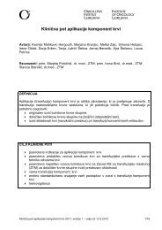
mammography (craniocaudal and lateral<br />
view), which showed a mass in the upper<br />
quadrant of the right <strong>breast</strong>, with mild skin<br />
retraction, with malignant characteristics<br />
(Figure 1). Breast ultrasound showed a welldefined<br />
nodular lesion with heterogeneous<br />
echo pattern posterior to acoustic enhancement.<br />
The lesion considered being malignant,<br />
and no fine needle aspiration cytology received.<br />
The resection of the tumour and auxiliary<br />
lymph nodes dissection decided to be<br />
performed therapeutically.<br />
At operation tumour was excised in<br />
healthy tissue and sent to cryobiopsy, which<br />
showed no malignant cell, but tyroid necrosis<br />
Figure 1. Mammography of primary <strong>breast</strong> <strong>tuberculosis</strong><br />
mimics <strong>breast</strong> cancer. The findings were obscure<br />
and the diagnosis set by cold biopsy.<br />
of the tissue, compatible with the inflammatory<br />
disease. No lymph nodes were removed.<br />
The pathological examination of the specimen<br />
showed that the mass was tuberculous.<br />
Mantoux test was positive. The full examination<br />
(x-ray, CT, etc) showed that <strong>tuberculosis</strong><br />
was nowhere else; that means that <strong>breast</strong> <strong>tuberculosis</strong><br />
was primary. The patient received<br />
anti-<strong>tuberculosis</strong> therapy (3 drugs combined<br />
therapy) for 9 months. There has not been recurrence<br />
for 4 years of the follow-up.<br />
Discussion<br />
Breast <strong>tuberculosis</strong> identified as primary and<br />
secondary. In the primary, <strong>breast</strong> is the only<br />
site of the disease in patients with no history<br />
of <strong>tuberculosis</strong>. In the secondary, mainly<br />
haematogenous spreading or direct extension<br />
infects <strong>breast</strong> after a contact with an infected<br />
material. The mycobacterium can infect<br />
<strong>breast</strong> haematogenous from auxilla, lungs,<br />
ribs and articular lesions, or can be infected<br />
by a direct contact through nipple, abrasions<br />
of the skin or lactiferous duct. 1,4,5,7<br />
Three types of <strong>breast</strong> <strong>tuberculosis</strong> have<br />
been described. The most common type is<br />
nodular disease, which is growing slowly and<br />
masquerades carcinoma on mammography.<br />
The second type, which also mimics carcinoma,<br />
is the diffuse type, which presents multiple<br />
foci. The third type is the sclerosing,<br />
which is painful and more common in the<br />
elderly. 3,4,8,9<br />
The differential diagnosis is quite difficult,<br />
and includes cancer, mastitis, sarcoma, actinomycosis,<br />
granulomatous mastitis, etc., although<br />
it’s not uncommon that more than<br />
one pathologies in the same <strong>breast</strong> coexist. 9,10<br />
The most common symptoms are a palpable<br />
<strong>breast</strong> with or with no auxiliary lymph<br />
nodes, usually painful with sometimes nipple<br />
discharge. 5,7<br />
Mammographic findings are not always<br />
specific for <strong>breast</strong> <strong>tuberculosis</strong>, which can be<br />
Radiol Oncol 2003; 37(1): 1-3.
Filippou DC et al. / <strong>Primary</strong> <strong>breast</strong> <strong>tuberculosis</strong> 3<br />
misdiagnosed as fibroadenoma or adenocarcinoma<br />
(inflammatory or scirrous). The two<br />
mammographic findings that are specific for<br />
<strong>breast</strong> <strong>tuberculosis</strong> are »skin bulge« and the<br />
»sinus tract site«. Ultrasonography may resemble<br />
cystic lesion, or indicates a hypoechoic<br />
heterogeneous mass with irregular borders.<br />
CT is useful in the diagnosis, particularly<br />
between primary and secondary <strong>tuberculosis</strong><br />
as can indicate lesions in other sites. 4,6,8-10<br />
More accurate information can be<br />
achieved by fine needle aspiration biopsy,<br />
which can demonstrate a granulomatous inflammatory<br />
lesion with central cessation. 7<br />
Many <strong>case</strong>s can be misdiagnosed and the<br />
diagnosis achieved after the surgical removal<br />
of the mass and histological examination of<br />
the specimen. In these <strong>case</strong>s a <strong>tuberculosis</strong><br />
infection history is negative, the mammographic<br />
and radiological findings obscure and<br />
the mass misdiagnosed as carcinoma. 3,7,9<br />
In primary <strong>breast</strong> <strong>tuberculosis</strong> the indicated<br />
treatment consists of the surgical removal<br />
of the mass and the anti-<strong>tuberculosis</strong> therapy<br />
with isoniazide, pyrazimanide, ethamboutole<br />
and rifampikin for the period from 9 months<br />
to 2 years. 2,10<br />
The increasing <strong>tuberculosis</strong> incidence in<br />
Western countries may also increase the incidence<br />
of <strong>breast</strong> <strong>tuberculosis</strong>.<br />
In conclusion, primary <strong>breast</strong> <strong>tuberculosis</strong><br />
is an uncommon <strong>breast</strong> pathology, which can<br />
mimic adenoma or carcinoma and can be misdiagnosed<br />
especially in patients with no previous<br />
history of the disease. The fine needle<br />
aspiration biopsy can lead to a correct diagnosis,<br />
which is finally achieved with the histological<br />
examination of the specimen.<br />
Patients with <strong>breast</strong> <strong>tuberculosis</strong> should undergo<br />
a surgical removal of the tumour and a<br />
long time anti-<strong>tuberculosis</strong> therapy.<br />
References<br />
1. Gupta R, Gupta AS, Duggal N. Tubercular mastitis,<br />
Int Surg 1982; 67: 422-4.<br />
2. Zandrino F, Monetti F, Gandolo N. <strong>Primary</strong> <strong>tuberculosis</strong><br />
of the <strong>breast</strong>. A <strong>case</strong> <strong>report</strong>. Acta Radiol<br />
2000; 41: 61-3.<br />
3. D’Orsi CJ, Feldhaus L, Sonnenfeld M. Unusual lesions<br />
of the <strong>breast</strong>. Radiol Clin North Am 1983; 21:<br />
67-80.<br />
4. McKeown KC, Wilkinson KA. Tuberculosis of the<br />
<strong>breast</strong>. J Surg 1962; 103: 424-7.<br />
5. Raven RW. Tuberculosis of the <strong>breast</strong>. Br Med J<br />
1949; 2: 734-7.<br />
6. Gilbert AI, McCough EC, Farell JJ. Tuberculosis of<br />
the <strong>breast</strong>. Am J Surg 1962; 103: 424-7.<br />
7. Wilson TS, MacGregor JW. The diagnosis and<br />
treatment of <strong>tuberculosis</strong> of the <strong>breast</strong>. Can Med<br />
Assoc J 1963; 89: 1118-24.<br />
8. Estrin J, Bernstein M. Tuberculous mastitis. South<br />
Med J 1994; 87: 1151-2.<br />
9. Alagaratnam TT, Ong GB. Tubereculosis of the<br />
<strong>breast</strong>. Br J Surg 1980; 67: 125-6.<br />
10. Wilson JP, Chapman SW. Tuberculous mastitis.<br />
Chest 1990; 98: 1505-9.<br />
Radiol Oncol 2003; 37(1): 1-3.