An Unusual Cause of Hemoptysis in a Young Female - OMJ
An Unusual Cause of Hemoptysis in a Young Female - OMJ
An Unusual Cause of Hemoptysis in a Young Female - OMJ

Create successful ePaper yourself
Turn your PDF publications into a flip-book with our unique Google optimized e-Paper software.
Oman Medical Journal (2011) Vol. 26, No. 6:457-458<br />
DOI 10. 5001/omj.2011.117<br />
Cl<strong>in</strong>ical Quiz<br />
<strong>An</strong> <strong>Unusual</strong> <strong>Cause</strong> <strong>of</strong> <strong>Hemoptysis</strong> <strong>in</strong> a <strong>Young</strong> <strong>Female</strong><br />
Pankaj Gupta, Raju Sharma, Surendra K. Sharma<br />
Received: 07 Jun 2011/ Accepted: 12 Aug 2011<br />
© OMSB, 2011<br />
Introduction<br />
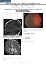
A 28-year-old female presented with recurrent epistaxis<br />
and mild hemoptysis for two years. There was no fever, dyspnea,<br />
palpitations, obstructed breath<strong>in</strong>g or hematuria. There was no<br />
history <strong>of</strong> past tuberculosis or drug <strong>in</strong>take. Both her parents died<br />
when she was 5-years <strong>of</strong> age from suspected tuberculosis. On<br />
exam<strong>in</strong>ation, she had pallor and nasal telangiectasia.<br />
Blood pressure and pulse rate was normal. There was no<br />
clubb<strong>in</strong>g, cyanosis or pedal edema. Cardiovascular system<br />
exam<strong>in</strong>ation was normal. Laboratory <strong>in</strong>vestigations revealed<br />
normal hematological pr<strong>of</strong>ile with a hemoglob<strong>in</strong> <strong>of</strong> 12.5 g, platelet<br />
count <strong>of</strong> 250,000/µl, and prothromb<strong>in</strong> time <strong>of</strong> 12 seconds. Chest<br />
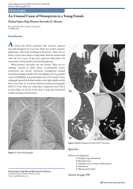
radiograph showed ill-def<strong>in</strong>ed nodules <strong>in</strong> the right middle and left<br />
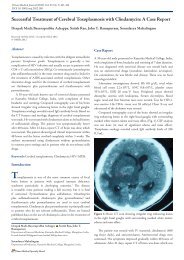
lower zones (Fig. 1). A contrast enhanced computed tomography<br />
(CECT) <strong>of</strong> the chest was undertaken. Sequential axial CECT<br />
sections (Figs. 2a and b) <strong>of</strong> the chest <strong>in</strong> lung and mediast<strong>in</strong>al<br />
w<strong>in</strong>dow sett<strong>in</strong>gs are shown below.<br />
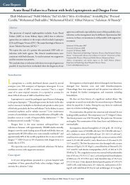
2a<br />
2b<br />
Figure 2a & b: Sequential axial CECT sections.<br />
Figure 1: Chest Radiograph.<br />
Pankaj Gupta, Raju Sharma , Surendra K Sharma<br />
All India Institute <strong>of</strong> Medical Sciences, India<br />
E-mail: raju152@yahoo.com<br />
Question<br />
What is your diagnosis?<br />
A. Wegener’s granulomatosis<br />
B. Bronchiectasis<br />
C. Multiple arterio-venous malformations<br />
D. Metastases<br />
E. Rheumatoid nodule<br />
<strong>An</strong>swer on page 458<br />
Oman Medical Specialty Board
Oman Medical Journal (2011) Vol. 26, No. 6:457-458<br />
<strong>An</strong>swer<br />
C. Multiple arterio-venous malformations<br />
Discussion<br />
Pulmonary arteriovenous malformations (PAVMs) are rare.<br />
Most are congenital associated with hereditary haemorrhagic<br />
telangiectasia (HHT). Acquired causes <strong>in</strong>clude: chest trauma,<br />
cardiovascular <strong>in</strong>terventional procedures, chronic cirrhosis,<br />
mitral stenosis, and schistosomiasis. HHT is responsible for 80%<br />
<strong>of</strong> PAVMs and PAVMs are present <strong>in</strong> 15-45% <strong>of</strong> patients with<br />
HHT. 1 HHT, or Rendu-Osler-Weber disease, is an autosomal<br />
dom<strong>in</strong>ant multiorgan fibrovascular dysplasia with a reported<br />
prevalence <strong>of</strong> 1 <strong>in</strong> 10,000 to 1 <strong>in</strong> 5000. 2 The anatomic structure<br />
<strong>of</strong> the PAVMs <strong>in</strong> HHT is classified <strong>in</strong>to two types, 3 simple, with a<br />
s<strong>in</strong>gle feed<strong>in</strong>g artery with a s<strong>in</strong>gle dra<strong>in</strong><strong>in</strong>g ve<strong>in</strong> and complex, with<br />
two or more arterial branches, and two or more dra<strong>in</strong><strong>in</strong>g ve<strong>in</strong>s.<br />
Mutations associated with HHT <strong>in</strong>clude: endogl<strong>in</strong> mutation,<br />
activ<strong>in</strong> receptor-like k<strong>in</strong>ase (ALK)-1 mutation and SMAD4<br />
mutation. 4<br />
Epistaxis secondary to nasal mucosa telangiectasia is the<br />
most common manifestation. Cl<strong>in</strong>ical manifestations related to<br />
pulmonary <strong>in</strong>volvement <strong>in</strong>clude dyspnea, cyanosis, clubb<strong>in</strong>g and<br />
hemoptysis. Complications associated with untreated PAVMs<br />
are life threaten<strong>in</strong>g hemorrhage and neurological complications.<br />
Neurological complications <strong>in</strong>clude transient ischaemic attack<br />
and stroke; results from the paradoxical embolization <strong>of</strong> leg<br />
ve<strong>in</strong> thrombus to the left-sided arterial circulation through the<br />
PAVMs. Rarely, the thrombus can orig<strong>in</strong>ate <strong>in</strong> situ <strong>in</strong> PAVMs.<br />
<strong>An</strong>other common neurological complication is cerebral abscess<br />
secondary to paradoxical embolization <strong>of</strong> bacterial bi<strong>of</strong>ilms.<br />
Diagnosis is based on the Curaçao Criteria established<br />
by the Scientific Division <strong>of</strong> the Hereditary Hemorrhagic<br />
Telangiectasia International Foundation. 5 Diagnosis is made<br />
<strong>in</strong> the presence <strong>of</strong> three <strong>of</strong> the follow<strong>in</strong>g: recurrent spontaneous<br />
epistaxis, mucocutaneous telangiectasis, an autosomal dom<strong>in</strong>ant<br />
familial distribution, and visceral AVMs. Screen<strong>in</strong>g for PAVMs<br />
is recommended <strong>in</strong> all HHT patients due to the serious<br />
complications associated with them and the availability <strong>of</strong> safe<br />
and effective <strong>in</strong>terventions. 6 The screen<strong>in</strong>g method <strong>of</strong> choice is<br />
Contrast echocardiography with a sensitivity <strong>of</strong> 90%. 1 Unenhanced<br />
Computed tomography (CT) thorax is used to confirm diagnosis<br />
and measure the size <strong>of</strong> PAVMs <strong>in</strong> patients with positive screen<strong>in</strong>g<br />
echocardiography. With the availability <strong>of</strong> multidetector CT<br />
(MDCT), contrast enhancement is not required for detection<br />
<strong>of</strong> PAVMs. 7 On CT, the f<strong>in</strong>d<strong>in</strong>gs <strong>in</strong>clude a serpig<strong>in</strong>ous mass or<br />
nodule with one or more enlarged feed<strong>in</strong>g arteries and one or more<br />
dra<strong>in</strong><strong>in</strong>g ve<strong>in</strong>s (depend<strong>in</strong>g on whether the malformation is simple<br />
or complex). Embolization <strong>of</strong> PAVM with coils is the treatment <strong>of</strong><br />
choice. 2 A feed<strong>in</strong>g artery calibre <strong>of</strong> at least 3 mm is considered the<br />
threshold for treatment <strong>of</strong> PAVMs. 5 Recurrence <strong>of</strong> PAVMs occurs<br />
<strong>in</strong> around 19% <strong>of</strong> treated patients. Therefore, follow-up CTs are<br />
recommended at 1 year and every 3-5 years thereafter.<br />
Other visceral manifestations <strong>in</strong>clude central nervous<br />
system manifestations: cerebral vascular malformations, sp<strong>in</strong>al<br />
cord vascular malformation, encephalopathy, bra<strong>in</strong> abscess and<br />
stroke. 8 Liver is <strong>in</strong>volved <strong>in</strong> 30% to 73% <strong>of</strong> patients with HHT.<br />
Hepatic fibrovascular dysplasia ranges from small telangiectases<br />
to large vascular masses. 9 Recurrent gastro<strong>in</strong>test<strong>in</strong>al bleed<strong>in</strong>g from<br />
mucosal fibrovascular dysplasia occurs <strong>in</strong> about 20% <strong>of</strong> patients<br />
with HHT.<br />
Based on the patient’s presentation with epistaxis and<br />
hemoptysis; cl<strong>in</strong>ical differential diagnoses <strong>of</strong> pulmonary<br />
tuberculosis, Wegener’s granulomatosis, bleed<strong>in</strong>g disorders,<br />
drug <strong>in</strong>take (drugs affect<strong>in</strong>g the coagulation pr<strong>of</strong>ile), and collagen<br />
vascular disorders were considered. Radiological differential<br />
diagnoses <strong>of</strong> multiple lung nodules on chest radiograph <strong>in</strong>clude<br />
neoplastic lesions (metastases, synchronous lung cancers), multiple<br />
pulmonary arterio-venous malformations, <strong>in</strong>fections (multiple<br />
abscesses, fungal <strong>in</strong>fections, hydatid disease) and immunological<br />
conditions (wegener’s granulomatosis, rheumatoid nodules,<br />
sarcoidosis, amyloidosis, organis<strong>in</strong>g pneumonia). 10<br />
Acknowledgements<br />
The authors reported no conflict <strong>of</strong> <strong>in</strong>terest and no fund<strong>in</strong>g was<br />
receive on this work.<br />
References<br />
1. Cott<strong>in</strong> V, Plauchu H, Bayle JY, Barthelet M, Revel D, Cordier JF. Pulmonary<br />
arteriovenous malformations <strong>in</strong> patients with hereditary hemorrhagic<br />
telangiectasia. Am J Respir Crit Care Med 2004 May;169(9):994-1000.<br />
2. Cott<strong>in</strong> V, Ch<strong>in</strong>et T, Lavolé A, Corre R, Marchand E, Reynaud-Gaubert<br />
M, et al; Groupe d’Etudes et de Recherche sur les Maladies “Orphel<strong>in</strong>es”<br />
Pulmonaires (GERM”O”P). Pulmonary arteriovenous malformations <strong>in</strong><br />
hereditary hemorrhagic telangiectasia: a series <strong>of</strong> 126 patients. Medic<strong>in</strong>e<br />
(Baltimore) 2007 Jan;86(1):1-17.<br />
3. White RI Jr, Lynch-Nyhan A, Terry P, Buescher PC, Farmlett EJ, Charnas<br />
L, et al. Pulmonary arteriovenous malformations: techniques and long-term<br />
outcome <strong>of</strong> embolotherapy. Radiology 1988 Dec;169(3):663-669.<br />
4. Letteboer TG, Mager JJ, Snijder RJ, Koeleman BP, L<strong>in</strong>dhout D, Ploos<br />
van Amstel JK, et al. Genotype-phenotype relationship <strong>in</strong> hereditary<br />
haemorrhagic telangiectasia. J Med Genet 2006 Apr;43(4):371-377.<br />
5. Shovl<strong>in</strong> CL, Guttmacher AE, Buscar<strong>in</strong>i E, Faughnan ME, Hyland RH,<br />
Westermann CJ, et al. Diagnostic criteria for hereditary hemorrhagic<br />
telangiectasia (Rendu-Osler-Weber syndrome). Am J Med Genet 2000<br />
Mar;91(1):66-67.<br />
6. Gossage JR. Primary pulmonary hypertension or portopulmonary<br />
hypertension? Chest 1998 Oct;114(4):1224-1225.<br />
7. Pollak JS, Saluja S, Thabet A, Henderson KJ, Denbow N, White RI<br />
Jr. Cl<strong>in</strong>ical and anatomic outcomes after embolotherapy <strong>of</strong> pulmonary<br />
arteriovenous malformations. J Vasc Interv Radiol 2006 Jan;17(1):35-44,<br />
quiz 45.<br />
8. Haitjema T, Westermann CJ, Overtoom TT, Timmer R, Disch F, Mauser H,<br />
et al. Hereditary hemorrhagic telangiectasia (Osler-Weber-Rendu disease):<br />
new <strong>in</strong>sights <strong>in</strong> pathogenesis, complications, and treatment. Arch Intern Med<br />
1996 Apr;156(7):714-719.<br />
9. Siddiki H, Doherty MG, Fletcher JG, Stanson AW, Vrtiska TJ, Hough DM,<br />
et al. Abdom<strong>in</strong>al f<strong>in</strong>d<strong>in</strong>gs <strong>in</strong> hereditary hemorrhagic telangiectasia: pictorial<br />
essay on 2D and 3D f<strong>in</strong>d<strong>in</strong>gs with isotropic multiphase CT. Radiographics<br />
2008 Jan-Feb;28(1):171-184.<br />
Oman Medical Specialty Board