Download - Oemus Media AG
Download - Oemus Media AG
Download - Oemus Media AG

You also want an ePaper? Increase the reach of your titles
YUMPU automatically turns print PDFs into web optimized ePapers that Google loves.
DeNtal tribuNe | april-June, 2010 Clinical 9<br />
Treating a peri-radicular abscess<br />
Dentist Nicolai Orsteen presents a clinical case study looking at the treatment of a maxillary left lateral front tooth<br />
The patient is a 24-year old white<br />
Northern European male. His<br />
chief complaint was pain from<br />
the maxillary left lateral front<br />
tooth, with periodic swelling of<br />
the left anterior palatal.<br />
The patient’s dental history<br />
indicated previous problems in<br />
this region, documenting an<br />
emergency appointment in<br />
March 2007 due to pain and<br />
swelling from tooth 22. He was<br />
prescribed a seven-day course of<br />
Penicillin V tablets (660mg<br />
qds*4) for acute apical periodontitis<br />
tooth 22. Following this<br />
appointment, the patient was<br />
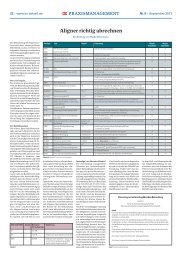
Table 1: Clinical findings<br />
referred for examination and<br />
treatment of tooth 22.<br />
Diagnosis<br />
The extra-oral examination on<br />
30 January 2008 was within<br />
normal limits, shown in Figures<br />
2 and 3.<br />
However, as is visible in Table<br />
one, the intra-oral examination<br />
revealed gingival bleeding on<br />
prodding, no sinus tract and<br />
fluctuant swelling of the palate<br />
mucosa in the area of teeth 21,<br />
22 and 23. The periodontal<br />
pockets however, were within<br />
normal limits.<br />
21 22 23<br />
Sensitivity to Cold Yes No Yes<br />
Percussion No Yes No<br />
Palpation No Yes No<br />
Mobility No No No<br />
Probing Depth (mm) 2 2 2<br />
Restoration NoneComposite (Pal) None<br />
Further radiographic investigation in April 2008 revealed that the patient was suffering from a<br />
discontinuation of the lamina dura on tooth 22, as well as a large circumscribed apical radiolucency<br />
(Ø 15mm). The radiographic findings in the coronal part of the root were diagnosed as<br />
dens-in-dente (see Figure 4).<br />
Following the investigations,<br />
the diagnosis showed that a periradicular<br />
abscess was related to<br />
non-vital tooth 22. The problems<br />
associated with the diagnosis<br />
were a wide root canal, and an<br />
open apex with large apical<br />
lesion.<br />
The structured treatment<br />
plan involved conventional root<br />
canal treatment, and to be assess<br />
for surgery after six months.<br />
The treatment plan<br />
Treatment commenced on 3<br />
April 2008. Following an initial<br />
clinical examination, the tooth<br />
was diagnosed with & apical<br />
abscess (no sinus present). Access<br />
was gained under a rubber<br />
dam and the canal was filled<br />
with exudate.<br />
The root canal length was<br />
determined both by apex locator<br />
(RootZX) and a periapical radiograph.<br />
The root canal disinfection<br />
was completed mechanically<br />
using Hedstroms files<br />
(size 90/20 mm/incisal edge).<br />
Particular care was taken<br />
during irrigation due to the<br />
open apex, & ultrasonics were<br />
used for the further cleaning<br />
of the canal. A formula of one<br />
per cent NaOCl, two per cent<br />
CHX and 17 per cent EDTA were<br />
used for chemical root canal<br />
disinfection. The canal was<br />
dressed with Ca(OH)2 and IRM<br />
was applied as a temporary<br />
filling.<br />
Five days after the completion<br />
of the treatment, the patient<br />
sought an emergency consultation<br />
because of severe pain and<br />
swelling from tooth 22. He was<br />
prescribed an eight-day course<br />
of clindamycin (500 mg x 3*3)<br />
to ease the discomfort.<br />
Following the surgery, on<br />
May 29, tooth 22 was asymptomatic<br />
and swill sensitive to percussion.<br />
The temporary filling<br />
was removed and the root canal<br />
disinfected again with Irrisafe,<br />
as well as a formula of one<br />
percen NaOCl, two per cent<br />
CHX and 17 per cent EDTA.<br />
A long-term intra- canal dressing<br />
with Ca(OH)2 was placed, and<br />
IRM was applied as a temporary<br />
filling.<br />
Preparing for root<br />
treatment<br />
The patient missed the following<br />
three appointments, but returned<br />
on October 14. On this<br />
date the tooth was still sensitive<br />
to percussion and palpation. As<br />
there were no real signs of improvement,<br />
it was decided that<br />
the tooth should be root filled &<br />
an appointment for apical surgery<br />
was made. To ease discomfort,<br />
the root canal was filled<br />
with an 8mm length of white<br />
MTA, & a wet cotton pellet was<br />
placed over the MTA. On top of<br />
the cotton pellet, a temporary<br />
filling with IRM was placed.<br />
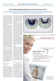
Fig. 1: Frontal view<br />
Fig. 2: Frontal view Fig. 3: Occlusal view Fig. 4: Pre-operatve periapical radiograph<br />
Fig. 5: Working lenght radiograph Fig. 6: MTA in the canal Fig. 7: MTA in the canal Fig. 8: MTA, wet cotton pellet and IRM Fig. 9: White MTA in the canal<br />
Fig. 10: Elevation of surgical flap Fig. 11: Granulation osteotomy<br />
Fig. 12: Granulation tissue removed & root-end resection performed Fig. 13: Flap sutured with 6-0 silk sutures