Download - Oemus Media AG
Download - Oemus Media AG Download - Oemus Media AG
24 Case report DeNtal tribuNe | april-June, 2010 Epulis gravidarum mimicking a neoplasm A case report by Dr Deepak Chopra, Dr Mayur Kaushik, Dr Deepak Kochar, and Dr Sidharath Malik, India Introduction aled granulation tissue with Pregnancy is a delicate condi- non-neoplastic proliferation of tion, involving complex physi- endothelial cells, suggestive of cal and physiological changes. 1 epulis gravidarum. Modification of metabolism, immunology, and high level of Case Description hormones make it possible for A 26-year-old female was refer- fetus to grow & develop, ending red with the chief complaint of an up with labor. Variations of these extensive gingival enlargement hormones cause some changes on the lower right anterior tooth on skin and oral mucosa. 2 region. The lesion was of negligi- The changes progress due to ble size when the patient first increased level of sex hormones in blood and saliva. These hor- noticed it three weeks ago, but had grown rapidly over the past Fig. 1: Pre-operative view Fig. 2: Excised lesion mones are thought to be the twenty days to attain the present reasons for occurrence of infla- size. The patient’s medical mmatory process and the epulis history revealed that she was gravidarum. 3 at five months of gestation with no systemic disease. The progesterone & estrogen receptors are situated in basal Clinical examination reve- and spinous stratum of the ep- aled an isolated exophytic, ithelium, and in the connective pedunclated lesion on the man- tissue. That is why those cells dibular right buccal side bet- are influenced by a high level of ween the interdental gingival of pregnancy hormones. 4 Proges- lateral incisor and canine. It terone dilates blood vessels, makes them more permeable, measured approximately 2.5 cm in diameter with some areas Fig. 3: Sutured with 6-0 silk sutures Fig. 4: Post-operative view and increases proliferation of capillary vessels. Estrogen regulates the proliferation, differentiation, and keratinization of the gingival tissue. These hormones increase gingival bleed- of erythema. The lesion was rough and firm in consistency on palpation that bled minimally (Fig. 1). The swelling also interfered with eating and speech. On examination, patient’s oral in that the lesion is not pus producing as “pyogenic” implies. It is however, a tumor of granulation tissue, as granuloma implies. 1 It has been called an epulis, because it is located sarcoma, kaposi’s sarcoma and non-hodgkins lymphoma. 10 Conclusion Epulis gravidarum represents an important differential diag- 7. Ababneh K, Khateeb T. Aggressive pregnancy tumor mimicking a malignant neoplasm: a case report. J Contemp Dent Pract. 2009 Nov 1; 10(6): E072-8. 8. Angelopoulos AP. Pyogenic granuloma of the oral cavity: ing, cause gingival growth, and lead to deepening of periodontal pockets as well. 5 Epulis gravidarum is a quite rare gingival disorder occurring hygiene was found good. Excisional biopsy of the swelling with a wide margin was performed (Figs. 2 & 3). The histopathological examination more frequently in the gingiva. Some other terms used are “granuloma telangiectaticum” and “pregnancy tumor”. The term “hemangiomatous granuloma” was suggested by An- nosis of oral masses and can behave in a very aggressive fashion, mimicking a malignant tumor. Excised specimens should be sent for histopathological reports to exclude malig- Statistical analysis and its clinical feature. J Oral Surg. 1971; 29, 84-9. 9. Daley TD, Nartey NO, Wysocki GP. Pregnancy tumor: an analysis. Oral Surg Oral Med Oral Pathol. 1991; 72(2): 196-99. in 1.8 to 5% of pregnant women, and it affects more commonly the anterior region of the upper jaw. 6 It is a smooth or lobulated exophytic lesion and manifests as a pink, red, or purple erythematous papule with pedunculated or sessile base. 1 It usually revealed young granulation tissue with non-neoplastic proliferation of endothelial cells and the enlargement of blood capillaries. Infiltration of acute and chronic inflammatory cells in a collagenous matrix was also present. Surface of the lesion gelopoulos to accurately reflect the characteristic histopathologic picture (hemangioma-like) and the inflammatory nature (granuloma) of the lesion. 8 Clinically it presents as a lesion that is pedunculated or nancy. It is acceptable practice to excise aggressive variants of this lesion during pregnancy to avoid distressing side effects. References 1. Jafarzadeh H, Sanatkhani M, Mohtasham N. Oral pyogenic granuloma: a review. J Oral 10. Czerninski R et al. Comparison of clinical and histological diagnosis in lesions of oral mucosa. Oral Surg Oral Med Oral Pathol Oral Radiol Endond. 2007; 103 (4): e20. DT arises in the 2 nd trimester, grows gradually over a few months time, and it also tends to bleed. After delivery of the child, it may regress and disappear entirely. 7 The purpose of this article is to describe a gingival swelling in a five months pregnant 26- year-old woman, which grew very rapidly unlike for this kind of tumor mimicking a malignant neoplasm. The lesion was not painful and grew very rapidly over a three week period. The histopathological examination reve- showed hyperplastic parakeratinised stratified squamous epithelium with areas of atrophy and ulcer. These findings were consistent with a histopathological diagnosis of epulis gravidarum. After three weeks of postoperative followup, clinical appearance of normal gingiva was present at the site of the lesion (Fig. 4). Discussion Epulis gravidarum is also known as “Pyogenic granuloma”. The term is somewhat a misnomer broad based, highly vascularized, smooth, edematous, hemorrhagic, soft, red with glossy surface and hardened when it had been longstanding. It could be a single or multiple well localized outgrowth, painless or with dull pain. It usually is not bigger than 2 cm in the diameter. 9 Differential diagnosis includes peripheral giant cell granuloma, epulis, peripheral ossifying fibroma, metastatic cancer, hemangioma, conventional granulation tissue, hyperplastic gingival inflammation, angio- Sci. 2006; 48: 167-75. 2. Erickson CV, Matus NR. Skin disorders of pregnancy. Am Fam Physic. 1994; 3: 602-10. 3. Laine MA. Effect of pregnancy on periodontal and dental health. Acta Odontol Scand. 2002; 60: 257-64. 4. Zeeman GG, Veth O, Dennison D. Focus on primary care on periodontal disease. Implications on women’s care. Obst Gynecol Survey. 2001; 56: 43-9. 5. Henry F, et al. Blood vessel changes during pregnancy: a review. Am J Clin Dermatol. 2006; 7: 65-9. 6. Paradowska A, Slawecki K, Chojak EG. Pregnancy tumor: review of literature. Dent Med Probl. 2008; 45(1): 51-4. About the authors Dr Deepak Chopra is a reader in the department of periodontology at Inderprastha Dental College at Ghaziabad, India. He can be contacted at deepakchopra2010 @gmail.com. Dr Mayur Kaushik is a reader in the department of periodontology at Subharati Dental College at Meerut, India. Dr Deepak Kochar is an assistant professor in the department of periodontology at Inderprastha Dental College at Ghaziabad, India. Dr Sidharath Malik is an assistant professor in the department of periodontology at Inderprastha Dental College at Ghaziabad, India.
DeNtal tribuNe | april-June, 2010 trends & applications 25 Miniscrews—a focal point in practice Six-part series by Dr Björn Ludwig, Dr Bettina Glasl, Dr Thomas Lietz, & Prof. Jörg A. Lisson—Part IV Figs. 1a–c: Figs. 1a–d: The uprighting of a second molar with simultaneous reshaping of the dental arch. The problem is clearly visible in the X-ray. The uprighting spring is fixed to a miniscrew (a, b). Status after five months without reactivation of the arch section (c, d). Clinical examples (2) Repositioning individual teeth The uprighting of molars The straightening of mesially tipped (2 nd ) molars in a full dentition represents a therapeutic challenge. The treatment is further complicated if the tooth is not only tipped but also partly impacted. The presence of a nonerupted third molar does not simplify the process (Fig. 1a). When planning the required appliance, it is important to consider whether it is necessary, for example, to reshape the entire dental arch (Figs. 1a–d) or just upright the tipped tooth. If miniscrews with bracket heads are used, it is possible to employ a special NiTi uprighting spring (such as the Memory Titanol spring, FORESTADENT). A standard multi-bracket appliance can be used to reshape the dental arch. At the same time, a second force element can be applied with the aid of a miniscrew and an uprighting spring (Figs. 1b–d). This avoids the loss of anchorage that inevitably occurs when only an uprighting spring is fixed to the multibracket appliance (Fig. 2). The straightening of an individual tooth may become necessary for periodontological, prosthetic or orthodontic reasons. This is a very simple procedure if a miniscrew and uprighting spring are used, and the appliance remains invisible to the observer. The tooth need only be fitted with an appropriate attachment system that makes it possible to fix this to the uprighting spring. Depending on how the spring is Figs. 3a–c: The alignment of a displaced canine using a miniscrew. After the canines have been exposed, they are attached to a bracket by means of a miniscrew (a). After removal of the screw, the dental arch can be reshaped using a conventional technique (b, c). Fig. 2: The uprighting spring fixed to the main arch not only affects the molars, but also causes displacement of the premolars (loss of anchorage). (Photo: Prof. Dominguez, São Paulo, Brasil). nents tend to move towards each other. In the worst-case scenario, only the group providing anchorage is displaced from its original position. This can occur if there is ankylosis of the retinated tooth, something that is difficult to evaluate during initial examination. If an attempt is made to move an ankylosed canine towards insufficient dental anchorage, the result will be Figs. 4a–e: Obtaining additional transverse space by means of ‘hybrid RPE’. The initial diagnosis is an asymmetrical narrow jaw with insufficient space for tooth 13 (a). After fixture of the brackets, two mini screws (OrthoEasy) were inserted during the same session (b). The hybrid RPE appliance was attached to the miniscrews and molar bands using laboratory abutments (FORESTADENT; c). The diastema shows the effect of the appliance after ten days’ use (d). Status after transverse expansion and concurrent reshaping of the dental arch (e). Fig. 5: The hybrid RPE appliance with adjuvant anterior hooks for the attachment of a Delaire mask. set, it is even possible to achieve intrusion/extrusion of the tooth. This form of treatment is in expensive for the patient and the orthodontist will find it highly effective. Alignment of retinated teeth The alignment of retained or displaced teeth, particularly in the case of canines, is one of the most common forms of surgical intervention in the field of orthodontic techniques. Numerous appliances are available— rubber bands, springs, orthodontic chains—that are effective to a greater or lesser extent. All these mechanisms have the same underlying problem: the neighbouring teeth must be used—directly or indirectly—to provide an anchorage, so that the required traction forces can be applied. Ideally, the neighbouring teeth will offer the greater resistance so that only the retained tooth moves. Realistically, however, both compo- the worst-case scenario. This can lead to an open bite in the region of the anterior teeth and premolars. Miniscrews provide the definitive form of anchorage for the alignment of displaced teeth (Figs. 3a–c). If sufficient space is available, brackets will not be needed in the initial phase of treatment. Skeletal adjustments Palatine suture expansion Rapid palatal expansion (RPE) is one of the most effective and
- Page 1 and 2: DENTAL TRIBUNE The World’s Dental
- Page 3 and 4: DeNtal tribuNe | april-June, 2010 N
- Page 6 and 7: 6 trends & applications DeNtal trib
- Page 8 and 9: 8 Case report DeNtal tribuNe | apri
- Page 10: 10 Clinical DeNtal tribuNe | april-
- Page 14 and 15: Issue 2 March 2010 Proper Technique
- Page 17 and 18: DeNtal tribuNe | april-June, 2010 P
- Page 19 and 20: DeNtal tribuNe | april-June, 2010 C
- Page 21 and 22: FDI explores preventive dentistry a
- Page 23: DeNtal tribuNe | april-June, 2010 t
- Page 27: DeNtal tribuNe | april-June, 2010 t
24<br />
Case report DeNtal tribuNe | april-June, 2010<br />
Epulis gravidarum mimicking a neoplasm<br />
A case report by Dr Deepak Chopra, Dr Mayur Kaushik, Dr Deepak Kochar, and Dr Sidharath Malik, India<br />
Introduction<br />
aled granulation tissue with<br />
Pregnancy is a delicate condi-<br />
non-neoplastic proliferation of<br />
tion, involving complex physi-<br />
endothelial cells, suggestive of<br />
cal and physiological changes. 1<br />
epulis gravidarum.<br />
Modification of metabolism,<br />
immunology, and high level of<br />
Case Description<br />
hormones make it possible for<br />
A 26-year-old female was refer-<br />
fetus to grow & develop, ending<br />
red with the chief complaint of an<br />
up with labor. Variations of these<br />
extensive gingival enlargement<br />
hormones cause some changes<br />
on the lower right anterior tooth<br />
on skin and oral mucosa. 2<br />
region. The lesion was of negligi-<br />
The changes progress due to<br />
ble size when the patient first<br />
increased level of sex hormones<br />
in blood and saliva. These hor-<br />
noticed it three weeks ago, but<br />
had grown rapidly over the past<br />
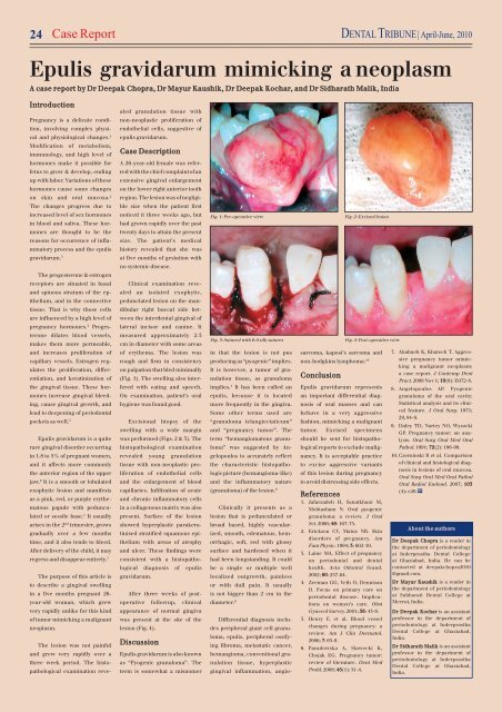
Fig. 1: Pre-operative view<br />
Fig. 2: Excised lesion<br />
mones are thought to be the<br />
twenty days to attain the present<br />
reasons for occurrence of infla-<br />
size. The patient’s medical<br />
mmatory process and the epulis<br />
history revealed that she was<br />
gravidarum. 3<br />
at five months of gestation with<br />
no systemic disease.<br />
The progesterone & estrogen<br />
receptors are situated in basal<br />
Clinical examination reve-<br />
and spinous stratum of the ep-<br />
aled an isolated exophytic,<br />
ithelium, and in the connective<br />
pedunclated lesion on the man-<br />
tissue. That is why those cells<br />
dibular right buccal side bet-<br />
are influenced by a high level of<br />
ween the interdental gingival of<br />
pregnancy hormones. 4<br />
Proges-<br />
lateral incisor and canine. It<br />
terone dilates blood vessels,<br />
makes them more permeable,<br />
measured approximately 2.5<br />
cm in diameter with some areas<br />
Fig. 3: Sutured with 6-0 silk sutures<br />
Fig. 4: Post-operative view<br />
and increases proliferation of<br />
capillary vessels. Estrogen regulates<br />
the proliferation, differentiation,<br />
and keratinization of<br />
the gingival tissue. These hormones<br />
increase gingival bleed-<br />
of erythema. The lesion was<br />
rough and firm in consistency<br />
on palpation that bled minimally<br />
(Fig. 1). The swelling also interfered<br />
with eating and speech.<br />
On examination, patient’s oral<br />
in that the lesion is not pus<br />
producing as “pyogenic” implies.<br />
It is however, a tumor of granulation<br />
tissue, as granuloma<br />
implies. 1 It has been called an<br />
epulis, because it is located<br />
sarcoma, kaposi’s sarcoma and<br />
non-hodgkins lymphoma. 10<br />
Conclusion<br />
Epulis gravidarum represents<br />
an important differential diag-<br />
7. Ababneh K, Khateeb T. Aggressive<br />
pregnancy tumor mimicking<br />
a malignant neoplasm:<br />
a case report. J Contemp Dent<br />
Pract. 2009 Nov 1; 10(6): E072-8.<br />
8. Angelopoulos AP. Pyogenic<br />
granuloma of the oral cavity:<br />
ing, cause gingival growth, and<br />
lead to deepening of periodontal<br />
pockets as well. 5<br />
Epulis gravidarum is a quite<br />
rare gingival disorder occurring<br />
hygiene was found good.<br />
Excisional biopsy of the<br />
swelling with a wide margin<br />
was performed (Figs. 2 & 3). The<br />
histopathological examination<br />
more frequently in the gingiva.<br />
Some other terms used are<br />
“granuloma telangiectaticum”<br />
and “pregnancy tumor”. The<br />
term “hemangiomatous granuloma”<br />
was suggested by An-<br />
nosis of oral masses and can<br />
behave in a very aggressive<br />
fashion, mimicking a malignant<br />
tumor. Excised specimens<br />
should be sent for histopathological<br />
reports to exclude malig-<br />
Statistical analysis and its clinical<br />
feature. J Oral Surg. 1971;<br />
29, 84-9.<br />
9. Daley TD, Nartey NO, Wysocki<br />
GP. Pregnancy tumor: an analysis.<br />
Oral Surg Oral Med Oral<br />
Pathol. 1991; 72(2): 196-99.<br />
in 1.8 to 5% of pregnant women,<br />
and it affects more commonly<br />
the anterior region of the upper<br />
jaw. 6 It is a smooth or lobulated<br />
exophytic lesion and manifests<br />
as a pink, red, or purple erythematous<br />
papule with pedunculated<br />
or sessile base. 1 It usually<br />
revealed young granulation<br />
tissue with non-neoplastic proliferation<br />
of endothelial cells<br />
and the enlargement of blood<br />
capillaries. Infiltration of acute<br />
and chronic inflammatory cells<br />
in a collagenous matrix was also<br />
present. Surface of the lesion<br />
gelopoulos to accurately reflect<br />
the characteristic histopathologic<br />
picture (hemangioma-like)<br />
and the inflammatory nature<br />
(granuloma) of the lesion. 8<br />
Clinically it presents as a<br />
lesion that is pedunculated or<br />
nancy. It is acceptable practice<br />
to excise aggressive variants<br />
of this lesion during pregnancy<br />
to avoid distressing side effects.<br />
References<br />
1. Jafarzadeh H, Sanatkhani M,<br />
Mohtasham N. Oral pyogenic<br />
granuloma: a review. J Oral<br />
10. Czerninski R et al. Comparison<br />
of clinical and histological diagnosis<br />
in lesions of oral mucosa.<br />
Oral Surg Oral Med Oral Pathol<br />
Oral Radiol Endond. 2007; 103<br />
(4): e20. DT<br />
arises in the 2 nd trimester, grows<br />
gradually over a few months<br />
time, and it also tends to bleed.<br />
After delivery of the child, it may<br />
regress and disappear entirely. 7<br />
The purpose of this article is<br />
to describe a gingival swelling<br />
in a five months pregnant 26-<br />
year-old woman, which grew<br />
very rapidly unlike for this kind<br />
of tumor mimicking a malignant<br />
neoplasm.<br />
The lesion was not painful<br />
and grew very rapidly over a<br />
three week period. The histopathological<br />
examination reve-<br />
showed hyperplastic parakeratinised<br />
stratified squamous epithelium<br />
with areas of atrophy<br />
and ulcer. These findings were<br />
consistent with a histopathological<br />
diagnosis of epulis<br />
gravidarum.<br />
After three weeks of postoperative<br />
followup, clinical<br />
appearance of normal gingiva<br />
was present at the site of the<br />
lesion (Fig. 4).<br />
Discussion<br />
Epulis gravidarum is also known<br />
as “Pyogenic granuloma”. The<br />
term is somewhat a misnomer<br />
broad based, highly vascularized,<br />
smooth, edematous, hemorrhagic,<br />
soft, red with glossy<br />
surface and hardened when it<br />
had been longstanding. It could<br />
be a single or multiple well<br />
localized outgrowth, painless<br />
or with dull pain. It usually<br />
is not bigger than 2 cm in the<br />
diameter. 9<br />
Differential diagnosis includes<br />
peripheral giant cell granuloma,<br />
epulis, peripheral ossifying<br />
fibroma, metastatic cancer,<br />
hemangioma, conventional granulation<br />
tissue, hyperplastic<br />
gingival inflammation, angio-<br />
Sci. 2006; 48: 167-75.<br />
2. Erickson CV, Matus NR. Skin<br />
disorders of pregnancy. Am<br />
Fam Physic. 1994; 3: 602-10.<br />
3. Laine MA. Effect of pregnancy<br />
on periodontal and dental<br />
health. Acta Odontol Scand.<br />
2002; 60: 257-64.<br />
4. Zeeman GG, Veth O, Dennison<br />
D. Focus on primary care on<br />
periodontal disease. Implications<br />
on women’s care. Obst<br />
Gynecol Survey. 2001; 56: 43-9.<br />
5. Henry F, et al. Blood vessel<br />
changes during pregnancy: a<br />
review. Am J Clin Dermatol.<br />
2006; 7: 65-9.<br />
6. Paradowska A, Slawecki K,<br />
Chojak EG. Pregnancy tumor:<br />
review of literature. Dent Med<br />
Probl. 2008; 45(1): 51-4.<br />
About the authors<br />
Dr Deepak Chopra is a reader in<br />
the department of periodontology<br />
at Inderprastha Dental College<br />
at Ghaziabad, India. He can be<br />
contacted at deepakchopra2010<br />
@gmail.com.<br />
Dr Mayur Kaushik is a reader in<br />
the department of periodontology<br />
at Subharati Dental College at<br />
Meerut, India.<br />
Dr Deepak Kochar is an assistant<br />
professor in the department of<br />
periodontology at Inderprastha<br />
Dental College at Ghaziabad,<br />
India.<br />
Dr Sidharath Malik is an assistant<br />
professor in the department of<br />
periodontology at Inderprastha<br />
Dental College at Ghaziabad,<br />
India.


