Radiology Clerkship Curriculum & Site Specific Contact Information
Radiology Clerkship Curriculum & Site Specific Contact Information
Radiology Clerkship Curriculum & Site Specific Contact Information

Create successful ePaper yourself
Turn your PDF publications into a flip-book with our unique Google optimized e-Paper software.
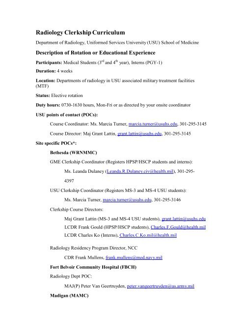
<strong>Radiology</strong> <strong>Clerkship</strong> <strong>Curriculum</strong><br />
Department of <strong>Radiology</strong>, Uniformed Services University (USU) School of Medicine<br />
Description of Rotation or Educational Experience<br />
Participants: Medical Students (3 rd and 4 th year), Interns (PGY-1)<br />
Duration: 4 weeks<br />
Location: Departments of radiology in USU associated military treatment facilities<br />
(MTF)<br />
Status: Elective rotation<br />
Duty hours: 0730-1630 hours, Mon-Fri or as directed by your onsite coordinator<br />
USU points of contact (POCs):<br />
Course Coordinator: Ms. Marcia Turner, marcia.turner@usuhs.edu, 301-295-3145<br />
Course Director: Maj Grant Lattin, grant.lattin@usuhs.edu, 301-295-3145<br />
<strong>Site</strong> specific POCs*:<br />
Bethesda (WRNMMC)<br />
GME <strong>Clerkship</strong> Coordinator (Registers HPSP/HSCP students and interns):<br />
Ms. Leanda Dulaney (Leanda.R.Dulaney.civ@health.mil), 301-295-<br />
4397<br />
USU <strong>Clerkship</strong> Coordinator (Registers MS-3 and MS-4 USU students):<br />
Ms. Marcia Turner, marcia.turner@usuhs.edu, 301-295-3146<br />
<strong>Clerkship</strong> Course Directors:<br />
Maj Grant Lattin (MS-3 and MS-4 USU students), grant.lattin@usuhs.edu<br />
LCDR Frank Gould (HPSP/HSCP students), Charles.F.Gould@health.mil<br />
LCDR Charles Ko (Interns), Charles.C.Ko.mil@health.mil<br />
<strong>Radiology</strong> Residency Program Director, NCC<br />
CDR Frank Mullens, frank.mullens@med.navy.mil<br />
Fort Belvoir Community Hospital (FBCH)<br />
<strong>Radiology</strong> Dept POC:<br />
MAJ(P) Peter Van Geertruyden, peter.vangeertruyden@us.army.mil<br />
Madigan (MAMC)
Hospital USUHS coordinator: Ms. Kathy Rogers, 253-968-<br />
5738, kathy.s.rogers.civ@mail.mil<br />
Assigned faculty:<br />
<strong>Radiology</strong> USUHS Medical Students Dr. Brian Boldt (from June 1 2013<br />
thru May 2014), brian.m.boldt.mil@mail.mil<br />
<strong>Radiology</strong> Medical Student POC (all students) Dr. Eric<br />
Roberge, eric.a.roberge.mil@mail.mil<br />
<strong>Radiology</strong> Program Coordinator SPC Blanca Ramirez 253-968-<br />
5604, blanca.d.ramirez.mil@mail.mil<br />
**<strong>Contact</strong> numbers for all doctors, please use SPC Ramirez or call 253 968-2130<br />
or 2238**<br />
<strong>Radiology</strong> Residency Program Director: LTC Neris Nieves-Robbins<br />
Email: neris.m.nievesrobbins.mil@mail.mil or nerisnieves@hotmail.com<br />
Portsmouth (NMCP)<br />
Hospital POC - <strong>Clerkship</strong> Coordinator: Paul Kinder (paul.kinder@med.navy.mil)<br />
<strong>Radiology</strong> POC - <strong>Clerkship</strong> Coordinator: CDR Rod Borgie<br />
(Roderick.borgie@med.navy.mil) 757-953-1199<br />
<strong>Radiology</strong> Residency Program Coordinator: Bridget Wakefield<br />
Tel: 757-953-7461<br />
Email: bridget.wakefield@med.navy.mil<br />
<strong>Radiology</strong> Residency Program Director: CDR Chris Kuzniewski, USN<br />
Tel: 757-953-1789<br />
Email: christopher.kuzniewski@med.navy.mil<br />
San Antonio (SAMMC, WHASC)<br />
Hospital POC - Interim GME <strong>Clerkship</strong> Coordinator: Edith Fields<br />
(edith.fields.civ@mail.mil)<br />
Commercial (210) 916-6574 / DSN 429-6574<br />
<strong>Radiology</strong> POCs - <strong>Clerkship</strong> Coordinators:
Ms. Kathy Gray, kathy.l.gray.civ@mail.mil, (210) 916-3290<br />
Ms. Susan Quintero, susan.quintero@us.af.mil, (210) 292-5290<br />
<strong>Radiology</strong> Residency Program Director, SAUSHEC: Col Paul Sherman<br />
Email: paul.sherman@us.af.mil<br />
MS-IV Course Coordinator: Ms Karen Slavick, karen.l.slavick.civ@mail.mil<br />
MS-III Course Coordinator: Ms. Laura Toscano, laura.a.toscano.civ@mail.mil<br />
Tel: 210.916.2041<br />
San Diego (NMCSD)<br />
Hospital POC – GME <strong>Clerkship</strong> Coordinator: Ms. Alexander<br />
Littleton, alexandra.littleton@med.navy.mil<br />
<strong>Radiology</strong> Residency Program Coordinator: Roberta M. Vigil<br />
Tel: 619-532-6755 DSN: 522-<br />
Email: roberta.vigil@med.navy.mil<br />
<strong>Radiology</strong> Residency Program Director: CDR Mike H. Lee, USN<br />
Tel: 619-532-6755 DSN: 522-<br />
Email: mike.lee@med.navy.mil<br />
USU Student <strong>Clerkship</strong> POC: Ms. Erin Quiko, Erin.Quiko@med.navy.mil<br />
Travis (DGMC)<br />
Hospital POC – GME office POC for clerkship scheduling: SrA Michelle Wright<br />
Email: michelle.wright.5@us.af.mil<br />
The POCs for the radiology clerkship should be Ms. Mannel and Dr. Edmonds:<br />
<strong>Radiology</strong> POCs –<br />
<strong>Radiology</strong> <strong>Clerkship</strong> and Residency Program Coordinator: Ms. Stephanie Mannel<br />
Tel: 707-423-7669<br />
Email: stephanie.mannel@us.af.mil<br />
<strong>Radiology</strong> <strong>Clerkship</strong> Director: Dr. Lance Edmonds<br />
Email: lance.edmonds@us.af.mil<br />
<strong>Radiology</strong> Residency Program Director: Lt Col Robert Jesinger, USAF<br />
Tel: 707-423-7210<br />
Email: Robert.jesinger@us.af.mil
Tripler (TAMC)<br />
Hospital POC – GME <strong>Clerkship</strong> Coordinator: Mr. Lon Pierce
Email: lon.pierce@us.army.mil<br />
<strong>Radiology</strong> POC - <strong>Clerkship</strong> Coordinator: Dr. Young W. Kim<br />
Email: young.woo.kim@us.army.mil<br />
<strong>Radiology</strong> Residency Program Coordinator: Mr. Chad Morgan<br />
Email: chad.w.morgan@us.army.mil<br />
<strong>Radiology</strong> Residency Program Director: LTC Kevin Nakamura, USA<br />
Tel: 808-433-6588<br />
Email: kevin.nakamura@us.army.mil<br />
*Note: Any MTF or other facility with a radiologist, can be a potential site for a<br />
radiology clerkship rotation. However, these rotations will need to be coordinated on a<br />
case-by-case basis between the site and the student, and will require USU Course director<br />
approval.<br />
Grading: Pass/Fail. It is the responsibility of USU medical students to ensure that Ms.<br />
Marcia Turner has received their evaluation following completion of the clerkship so that<br />
their grade can be submitted to the registrar.<br />
Pre-rotation preparation:<br />
Rotations must be scheduled through the hospital clerkship coordinator, often located<br />
within the GME office. Once your rotation has been scheduled, please follow site<br />
specific instructions regarding when and where to report on the first day of your rotation.<br />
4 th year USU medical students will need to send a Form 1304 to Ms. Marcia Turner<br />
(marcia.turner@usuhs.edu) after coordinating with the site. 3 rd year USU medical<br />
students are not required to submit a Form 1304.<br />
Brief description of rotation and rotation structure:<br />
This rotation is designed to provide trainees (medical students and interns) with<br />
experience in radiology within MTFs that have radiology departments. Daily activities<br />
will vary slightly by site but typically will consist of didactic and clinical<br />
conferences/lectures, daily medical imaging interpreted by diagnostic radiologists, tumor<br />
boards, and multidisciplinary conferences. Assessment and grading will be performed by<br />
the clerkship coordinator or designated faculty that is on site during your rotation. In an<br />
effort to standardize the rotation curriculum at the different MTFs that train students in<br />
conjunction with USU (while taking into account the variability in sites and resources),<br />
we have established a minimum level of competency in order to establish passing status<br />
for the rotation. This will be accomplished via online modules delivered via the iLearning<br />
Management System referred to as Sakai currently being used by USU. Each trainee will<br />
be required to complete and pass 4 online radiology modules (general radiology – chest<br />
and abdomen, pediatric radiology, neuroradiology, and musculoskeletal radiology). A
passing score of 70% will be required for each module but each module may be repeated<br />
as many times as required. It is anticipated that it will take approximately 10-15 hours to<br />
complete all of the modules. Please keep in mind that these modules are considered the<br />
minimum level of completion for this rotation. This will allow for increased transparency<br />
and standardization of a centrally controlled curriculum. For additional site specific<br />
requirements, please talk to your clerkship coordinator within the radiology department.<br />
It is anticipated that additional requirements may include such activities as a minimum<br />
level of participation through the different radiologic subspecialties, creation of a<br />
teaching file, oral presentations, or additional quizzes.<br />
Accessing Sakai Learning Management System:<br />
1. Go to the following<br />
site: https://cas.usuhs.edu/cas/login?service=https%3A%2F%2Flearning.usuhs.edu%2Fxs<br />
l-portal%2Flogin<br />
2. Select My <strong>Site</strong>s (right side of webpage).<br />
3. Select appropriate radiology clerkship based on year of graduation (eg. Students in the<br />
graduating class of 2015 will be enrolled in RAD03100 <strong>Radiology</strong> Selective 2015).<br />
4. Read about the modules in the “Announcements” subfolder.<br />
5. Complete each of the modules in the “Lessons” subfolder (left hand side within Course<br />
Tools).<br />
6. Pass the post-test (70% required for passing) associated with each module contained<br />
within the “Tests & Quizzes” subfolder (left hand column).<br />
7. Take the student survey contained within the “Lessons” subfolder.<br />
Teaching Methods:<br />
1. Online Sakai modules<br />
2. Didactic lectures/conferences<br />
3. Daily workload (teaching at the PACS workstation)<br />
4. Direct observation of technique or procedure<br />
5. Socratic method of questioning by faculty<br />
6. Recommended reading assignments<br />
Assessment Methods:<br />
1. Online Sakai modules –must pass each module (70%) receive a clerkship passing<br />
grade<br />
2. Intern/ student evaluation at end of rotation<br />
3. Socratic method of inquiry during rotation about assignments and cases<br />
4. Evaluations: <strong>Information</strong> will be gathered from technologists, radiology residents,<br />
fellows, patients and other individuals that the trainee may have encountered<br />
5. Quizzes (may vary by site)
6. Oral presentation by trainee (may vary by site)<br />
Level of Supervision: Direct supervision of trainees will be performed throughout the<br />
rotation, typically be a resident or staff physician. Indirect supervision will be virtually<br />
performed by the USU and on-site clerkship coordinators and directors in order to ensure<br />
trainee completion and passing of Sakai online modules. Self-directed reading and study<br />
will not be supervised.<br />
Professionalism: Unprofessional behavior will not be tolerated. Please keep in mind that<br />
unprofessional behavior within this rotation may be grounds for failure of the rotation<br />
and additional disciplinary action. Successful completion and passing of online modules<br />
will not reverse a failing grade given for unprofessional behavior.<br />
Rotation hours and leave policy: Interns and medical students do not take overnight call<br />
during this rotation. Short call or shadowing experiences may be pursued as long as<br />
Accreditation Council for Graduate Medical Education (ACGME) duty hours are not<br />
exceeded. You may take leave during this rotation with permission from your site<br />
specific clerkship coordinator or designated faculty, Program Director or Dean.<br />
ACGME duty hour policies are in effect. Any trainee who feels he/she is near or inviolation<br />
of duty hour policies should report the violation to your Program Director and/<br />
or Graduate Medical Education office. There is no reason for trainees on this rotation to<br />
exceed duty hours.<br />
Recommended reading: Raby N, Berman L, de Lacey G. Accident and Emergency<br />
<strong>Radiology</strong>, A Survival Guide, 2 nd ed.<br />
<strong>Radiology</strong> <strong>Clerkship</strong> Goals and Learning Objectives:<br />
Department of <strong>Radiology</strong>, Uniformed Services University School of Medicine<br />
Basic goals:<br />
1) Become an educated consumer of radiology consultation and services<br />
2) Learn the language of diagnostic radiology<br />
3) Develop a systematic approach to the radiologic evaluation of the acutely ill patient<br />
4) Reinforce clinical knowledge using radiographic and cross-sectional anatomy<br />
5) Understand the fundamentals of diagnostic imaging and its role in modern medicine<br />
Online module objectives:<br />
1) <strong>Radiology</strong> of the Chest<br />
a) Describe the radiographic search pattern used to interpret the adult chest<br />
radiograph<br />
b) Identify radiographic anatomy seen on the adult chest radiograph<br />
c) Correlate basic chest computed tomography (CT) landmarks to radiographic<br />
anatomy and common abnormalities
d) Apply the systematic approach to a radiographic search pattern in the setting of<br />
abnormal radiographs<br />
2) Imaging of the Abdomen<br />
a) Identify radiographic anatomy seen on the acute abdominal series<br />
b) Correlate basic abdominal CT landmarks to radiographic anatomy and common<br />
abnormalities<br />
c) Apply the systematic approach in the setting of abnormal radiographs<br />
3) Pediatric Musculoskeletal Imaging<br />
a) Identify normal vs. abnormal skeletal structures (in the younger child and<br />
adolescent)<br />
b) Identify the hallmarks of non-accidental trauma<br />
c) Identify the Salter-Harris fracture classification and fractures suspicious for child<br />
abuse<br />
d) Assess the alignment of the pediatric elbow on a radiograph<br />
e) Interpret signs of slipped capital femoral epiphysis (SCFE) and Legg-Calve-<br />
Perthes disease on radiograph and identify in which age groups these are likely to<br />
be found<br />
4) Pediatric Chest Imaging<br />
a) Identify the proper positioning of an umbilical arterial catheter and an umbilical<br />
venous catheter, and where the catheter tips should be located<br />
b) Distinguish between respiratory distress syndrome (RDS), meconium aspiration,<br />
transient tachypnea of the newborn, and neonatal pneumonia on a radiograph<br />
c) Interpret abnormal chest radiographs<br />
5) Pediatric Gastrointestinal (GI) Imaging<br />
a) Identify radiographic anatomy seen on abdominal radiographs<br />
b) Identify various radiographic findings in children, specifically: newborn<br />
gastrointestinal obstruction including midgut volvulus, Hirschsprung’s disease,<br />
intussusception, hypertrophic pyloric stenosis (HPS), and appendicitis; and,<br />
identify the best imaging technique for each condition<br />
c) Interpret abnormal GI radiographs<br />
d) Interpret a basic upper GI series, small bowel series, and barium enema<br />
6) Musculoskeletal Imaging<br />
a) Identify and diagnose musculoskeletal trauma radiology with an emphasis on<br />
deployment related injuries<br />
b) Describe a general timeline for fracture healing will be covered along with<br />
radiology pitfalls including satisfaction of search, inadequate number of<br />
projections and peripherally positioned pathology<br />
c) List stress fracture sites and identify these injuries by radiography that are<br />
prevalent in military training<br />
d) Discuss several fracture types and dislocations that are frequently missed in<br />
clinical practice<br />
e) Compare fractures vs. infection and identify findings associated with acute<br />
infection as may be seen as a result of open fracture in a combat setting<br />
7) Cervical Spine Imaging<br />
a) Identify the basic anatomy of the cervical spine<br />
b) Describe the appropriate imaging modality for cervical spine trauma
c) Diagnose the types of cervical spine injuries and their mechanisms<br />
d) Categorize which fracture types are stable versus unstable<br />
8) Traumatic Brain Injury<br />
a) Identify which patients needs brain imaging<br />
b) Select what type of brain imaging is needed<br />
c) Differentiate between extraaxial lesions and intraaxial lesions<br />
Trainee Competencies*<br />
*Adapted from Alliance of Medical Student Educators in <strong>Radiology</strong> (AMSER)<br />
Student Competencies in<br />
<strong>Radiology</strong>. http://www.aur.org/Affiliated_Societies/AMSER/amser_curriculum.cfm<br />
P A T I E N T C A R E C O M P E T E N C I E S<br />
The trainee (medical student/intern) should provide patient care that is safe,<br />
compassionate and effective in the diagnosis and management of common<br />
health problems.<br />
GOALS<br />
1. Diagnostic management skills<br />
a. Know how to order appropriate imaging tests<br />
i. Utilize the ACR (American College of <strong>Radiology</strong>)<br />
Appropriateness Criteria<br />
ii. Include patient variables into imaging selection<br />
b. Understand the importance of providing appropriate information on the<br />
radiology request form (history, physical, risk and limiting factors) so<br />
radiology can perform appropriate modality selection, protocoling, and<br />
interpretation<br />
2. <strong>Information</strong> retrieval skills<br />
a. Know how to access images and view them<br />
i. Understand the basics of a PACS workstation<br />
ii. Understand windows, levels, image linking, etc.<br />
b. Know how to access imaging reports: preliminary and final<br />
c. Perform effective, rapid clinical information search<br />
3. Visual interpretative skills<br />
a. Know basic radiological anatomy<br />
b. Understand the factors that affect image appearance and quality<br />
c. Understand the importance of using prior comparison studies
d. Recognize normal and common or critical abnormal findings on basic<br />
radiographic studies including abdominal radiographs, chest radiographs,<br />
radiographs of the bones and joints, etc.<br />
4. <strong>Information</strong> processing skills<br />
a. Synthesize history, physical exam and imaging findings to make<br />
appropriate differential diagnoses<br />
b. Correctly interpret radiology reports<br />
5. Patient safety and radiation exposure<br />
a. Understand the risks of imaging including physical, financial and<br />
emotional<br />
i. Radiation risk (ionizing) to patients and operators and methods to<br />
reduce radiation exposure<br />
ii. Contrast material risks<br />
iii. MRI safety<br />
iv. Pregnant patients and imaging<br />
v. Interventional procure risks<br />
LEARNING TOOLS<br />
• Integration and application of ACR Appropriateness criteria during small and<br />
large group didactic and case-based sessions discussing imaging for specific<br />
clinical questions.<br />
• Small group discussion of shared decision making and informed consent<br />
o Role playing of the consent process with the students alternating being the<br />
patient and the radiologist<br />
• Observe informed consent for imaging/interventional procedures<br />
• Observe discussion with pregnant patient regarding radiation and contrast risk<br />
• Didactic presentation on safety of imaging procedures and contraindications<br />
ASSESSMENT TOOLS<br />
• Pass required online module post-tests<br />
• Quizzes<br />
• Global ratings by residents, fellows and faculty<br />
• Direct observation and assessment of performance (eg. informed consent, counsel<br />
patient regarding contrast allergy or radiation risk)
M E D I C A L K N O W L E D G E C O M P E T E N C Y<br />
The trainee should demonstrate basic knowledge about normal anatomy,<br />
disease processes and radiology<br />
GOALS<br />
• Demonstrate sufficient general medical knowledge and apply this knowledge to<br />
radiologic studies<br />
• Demonstrate radiological knowledge<br />
LEARNING TOOLS<br />
• Small and large group didactic sessions<br />
• Participation in departmental and interdepartmental case conferences<br />
• Participation in the clinical activities of the radiology department<br />
• View Box (PACS) teaching<br />
• Web-based modules (Sakai Learning Management System)<br />
• Preparation of a case-based talk during the radiology rotation (may vary by site)<br />
ASSESSMENT TOOLS<br />
• Pass required online module post-tests<br />
• Quizzes<br />
• Evaluation of observed informed consent<br />
• Global rating by residents, fellows and faculty who worked with the student<br />
P R A C T I C E - B A S E D L E A R N I N G A N D I M P R O V E M E N T<br />
The trainee(s) should continually seek to improve their knowledge and skills<br />
by multiple means, be able to self-evaluate and apply new knowledge to his<br />
or her practice.<br />
GOALS<br />
1. Use of information technology and data resources<br />
a. Demonstrate awareness of key sources of data for performing evidencebased<br />
medicine<br />
i. Use established medical algorithms (Ottawa ankle rule, Ottawa<br />
knee rule, NEXUS criteria for cervical spine imaging)<br />
ii. Use National society guidelines for imaging (eg. Neurology stroke<br />
protocol, back pain, first trimester bleeding)<br />
b. Use evidence based methods for selecting imaging modalities
i. ACR Appropriateness Criteria®<br />
c. Effectively search for additional information<br />
i. Use validated sources (ie. ‘Beyond the Google’, such as<br />
Pubmed)<br />
ii. Know when additional information is needed and search<br />
spontaneously<br />
2. Perform critical assessment of the literature<br />
a. Show an awareness of current literature on common problems<br />
b. Research presentation topics appropriately using peer reviewed literature<br />
c. Appropriately interpret the results of scientific studies (eg. Validity of<br />
study)<br />
d. Be aware of some of the limitations of scientific studies (eg. Power,<br />
sample size, control subjects)<br />
3. Application of learning<br />
LEARNING TOOLS<br />
a. Effectively apply newly learned information to appropriate clinical<br />
settings<br />
i. Develop new skills<br />
ii. Apply newly acquired knowledge and skills in the appropriate<br />
clinical setting<br />
iii. Be able to propose changes in the patient care plan based on the<br />
outcomes of imaging studies<br />
b. Demonstrate improvement in existing skills and develop new skills<br />
• Journal clubs, small group or independent critical assessment of scientific<br />
literature<br />
• Didactic small or large group sessions on assessment of scientific literature<br />
• Participation in departmental conferences including mortality and morbidity as<br />
well as quality improvement conferences<br />
ASSESSMENT TOOLS<br />
• Evaluation of critical assessment of scientific literature during a journal club, case<br />
conference or while on a rotation<br />
• Pass required online module post-tests<br />
• Quizzes<br />
I N T E R P E R S O N A L C O M M U N I C A T I O N C O M P E T E N C Y
The trainee can communicate and interact effectively with patients and<br />
healthcare providers.<br />
GOALS<br />
1. Interactions with patients<br />
a. Interacts effectively with patients<br />
i. Be compassionate, friendly, professional<br />
ii. Be able to take an effective history<br />
iii. Can calm anxious patients<br />
iv. Be able to develop a potentially therapeutic relationship<br />
v. Be able to give appropriate information within their knowledge,<br />
ability and level of responsibility<br />
2. Interactions with physicians<br />
a. With radiologists (staff and residents)<br />
i. Be respectful, but not inhibited from asking questions<br />
ii. Ask appropriate insightful questions that gain knowledge<br />
iii. Not be overly intrusive, be aware of time limitations<br />
iv. Help with information technology, patient management,<br />
communication<br />
v. Understand the importance of the radiologist-clinician interaction<br />
b. With clinicians<br />
i. Can gather appropriate clinical information about patients/study<br />
requests<br />
ii. Can communicate results effectively to clinicians if asked<br />
3. Interactions with technologists/nurses<br />
a. Exhibit respectful interactions and treat them as a member of the team<br />
b. Are aware of the knowledge and training of paramedical staff<br />
4. Written communication skills<br />
a. Documents clinical data effectively when needed (e.g. electronic medical<br />
record)<br />
b. Understands need for recording of urgent findings<br />
c. Provides relevant clinical history on requisitions for medical imaging<br />
5. Presen tation skills<br />
a. Presents fluent, well-researched presentation<br />
b. Shows understanding of topic
LEARNING TOOLS<br />
c. Conveys information succinctly and memorably to audience<br />
• Collection of reading references on guidelines how to tell patients imaging results<br />
(Smith, Gunderman <strong>Radiology</strong> 2010 255:317-321)<br />
• Journal club presentations<br />
• Participation in case conferences and interdisciplinary conferences<br />
ASSESSMENT TOOLS<br />
• Global evaluation of communication skills<br />
• Evaluation of a prepared oral presentation (may vary by site)<br />
• Evaluation of journal club presentation (may vary by site)<br />
P R O F E S S I O N A L I S M C O M P E T E N C Y<br />
The trainee should demonstrate a commitment to carrying out professional<br />
responsibilities and an adherence to ethical principles.<br />
GOALS<br />
1. Demonstrate appropriate skills<br />
a. Maintains professional and medical competence by continuing to selflearn<br />
throughout career<br />
b. Seeks help and support when identifies a knowledge gap<br />
c. Continually gathers new scientific knowledge<br />
d. Strives to improve the quality of patient care by practicing at the highest<br />
level of quality<br />
2. Demonstrates appropriate behaviors<br />
a. Meets professional responsibilities by working as a member of a team<br />
b. Demonstrates honesty with patients and all members of the health care<br />
team<br />
c. Respects patient confidentiality with all information transmitted during a<br />
patient encounter<br />
d. Maintains appropriate relationships with patients to prevent boundary<br />
transgression<br />
3. Demonstrates social justice and service<br />
a. Works to improve access to care for those patients with limited resources<br />
b. Considers just distribution of finite sources when selecting imaging tests
LEARNING TOOLS<br />
c. Understands issues around conflict of interest, avoids interaction with<br />
industry for personal advantage and discloses any existing conflicts of<br />
interest<br />
• Journal clubs on ethics and professionalism in <strong>Radiology</strong><br />
• AMSER Professionalism module available on MedEdPORTAL<br />
• Small group case-based discussion on professionalism issues should include<br />
debriefing sessions (may vary by site)<br />
ASSESSMENT TOOLS<br />
• Global evaluation by technologists, nurses, residents, fellows and radiology<br />
faculty of professional behavior<br />
• AMSER Professionalism module before and after test available on<br />
MedEdPORTAL<br />
S Y S T E M S - B A S E D P R A C T I C E C O M P E T E N C Y<br />
The trainee demonstrates awareness of the complexities, interactions and<br />
considerations involved in working in the modern health care environment.<br />
(i.e. the “culture” of the workplace)<br />
GOALS<br />
1. Demonstrate awareness of the goal of cost effective imaging<br />
a. Aware of common examination charges<br />
b. Understands the basic concepts of costs and reimbursement<br />
c. Understands the financial impact on patients and society of imaging<br />
i. Understands the importance of performing appropriate imaging<br />
ii. Appreciates potential future limitations to imaging availability<br />
2. Understands the workflow patterns in radiology for effective patient management,<br />
study ordering etc<br />
3. Demonstrates effective communication between radiology and clinicians<br />
a. Appreciates the importance of the radiology-clinician interaction (verbal,<br />
written)<br />
b. Appreciates the importance of prioritizing studies based on study urgency<br />
c. Understands the importance of prompt preliminary reports<br />
d. Understands the process of dealing with discrepancies between<br />
preliminary and final reports
4. Understands the impact of medical radiation exposure on potential cancer risk for<br />
population as a whole<br />
LEARNING TOOLS<br />
a. Demonstrates knowledge of current data regarding risk<br />
b. Aware of need for reducing unnecessary imaging<br />
• Small or large group didactic session on cost effectiveness of imaging studies<br />
• Participation in departmental and multidisciplinary conferences that discuss<br />
appropriate imaging evaluation of specific diseases and cost-effectiveness<br />
• ACR/APDR videotapes on non-interpretive skills that discuss systems-based<br />
practice<br />
ASSESSMENT TOOLS<br />
• Global evaluation by technologists, nurses, residents, fellows and radiology<br />
faculty of professional behavior<br />
• Attendance at case conferences and multidisciplinary conferences<br />
• Questions from ExamWeb on cost-effectiveness and prioritization of imaging<br />
tests