Pachymetry, Specular Microscopy and Confocal Microscopy
Pachymetry, Specular Microscopy and Confocal Microscopy
Pachymetry, Specular Microscopy and Confocal Microscopy

Create successful ePaper yourself
Turn your PDF publications into a flip-book with our unique Google optimized e-Paper software.
<strong>Pachymetry</strong>, <strong>Specular</strong> <strong>Microscopy</strong><br />
<strong>and</strong> <strong>Confocal</strong> <strong>Microscopy</strong><br />
Ellison Bentley, DVM, Diplomate<br />
ACVO<br />
University of Wisconsin-Madison
<strong>Pachymetry</strong><br />
• Why measure?<br />
– Indicator of corneal health<br />
– Reflects health of endothelium<br />
– Influences tonometry measurements<br />
– Diagnosis of disease, monitoring<br />
– Can be thicker or thinner (refractive surgery)
Methods of measurement<br />
• Historically:<br />
– Calipers on fresh or fixed tissue<br />
• Poor reflection of in vivo thickness<br />
• Tissue swelling after death
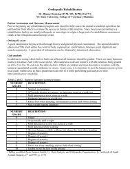
Optical pachymetry<br />
• Attachment to table<br />
mounted slit lamp<br />
• Measure oblique<br />
section of cornea with<br />
split prism<br />
• Split epithelial/<br />
endothelial images<br />
aligned by user<br />
• Various equations used<br />
to determine thickness
Optical pachymetry<br />
• Variables:<br />
– Refractive index of<br />
cornea<br />
– Anterior radius of<br />
curvature<br />
• Sources of imprecision<br />
– Equation variables<br />
– Inter-observer variability<br />
• Observer variation in<br />
alignment of images
Optical pachymetry<br />
• Automated optical<br />
pachymetry:<br />
– Significantly decreases<br />
inter-observer variability<br />
– Increases reproducibility<br />
<strong>and</strong> reliability<br />
Non-contact automated<br />
optical pachymeter
Optical pachymetry<br />
• Orbscan Anterior<br />
Segment analysis<br />
system<br />
– Series of slit beam<br />
images (9000 points)<br />
– Anterior/posterior<br />
topography<br />
– 3-D image<br />
– Thinner than with other<br />
methods<br />
http://www.augenlaser-klinik.ch/images/<br />
layout/grafiken_orbscan/<br />
orbscan2.gif&imgrefurl
Optical technique<br />
• Pentacam-rotating<br />
Schiemfplug<br />
camera-can model<br />
the anterior chamber<br />
(humans)<br />
• Scanning slit like<br />
Orbscan-increased<br />
depth of focus
Optical pachymetry<br />
• Requires clear<br />
reflections of epithelial<br />
<strong>and</strong> endothelial surface<br />
– Limited: corneal edema,<br />
scarring, deposits, etc
<strong>Specular</strong> microscopes<br />
• Another form of optical<br />
pachymetry<br />
• Use electromechanical<br />
device<br />
• Central corneal<br />
thickness; newer<br />
models mid-peripheral<br />
From Krachmer, Cornea 2005
<strong>Pachymetry</strong>-specular microscopes<br />
• Measure from posterior surface of tear film to<br />
posterior surface of Descemet’s membrane<br />
– May have error of 20-30 µm<br />
• Contact or non-contact<br />
– Corneal touch-induce some compression; thinner<br />
measurement
Ultrasound pachymetry<br />
• 1980’s<br />
• Preferred method:<br />
– Independent of patient<br />
fixation<br />
– Ease of use<br />
– Peripheral <strong>and</strong> central<br />
measurement<br />
– Improved reproducibility<br />
– Improved accuracy<br />
– Portability
Ultrasound pachymetry<br />
• Modern machines<br />
– Excellent alignment detection<br />
– Fast information retrieval <strong>and</strong> storage<br />
– Better sampling algorithms<br />
– Smaller probe tips (better for wider variety of<br />
animals, more precise measurement)
Ultrasound pachymetry<br />
• Originally-water-filled probe tips<br />
• Currently-solid tips<br />
– Measure from anterior surface of tear film to<br />
posterior surface endothelium<br />
– Automatic gain<br />
– Angle sensitivity-within 5 degrees of axis<br />
– Most 20 MHz, but up to 65 MHz
Ultrasound pachymetry<br />
• Hard plastic probes<br />
• Minimal maintenance<br />
• Easy to sterilize
Ultrasound pachymetry<br />
• Electronic pulservibrates<br />
piezoelectric<br />
crystal<br />
• Ultrasonic pulsereflects<br />
at Descemet’s;<br />
returns to crystal<br />
• Reflected waveselectric<br />
signal goes to<br />
receiver
Ultrasound pachymetry<br />
• Corneal thickness-from time it takes to pass<br />
from end of transducer, to Descemet’s <strong>and</strong><br />
back<br />
Distance = velocity x time<br />
• Velocity-speed of sound in cornea
Ultrasound pachymetry<br />
• Velocity<br />
– Human cornea-1640 m/s<br />
– Range-1550 m/s-1639 m/s-bovine, porcine, cat,<br />
human<br />
– 1639 m/s-best estimate for human but use 1640<br />
m/s-best correlate with optical pachymetry<br />
– Tang et al-velocity in canine: 1577+/- 10m/s<br />
• Since true velocity lower, most machines overestimate<br />
corneal thickness in dogs
Pach-Pen<br />
• H<strong>and</strong>-held pachymeter<br />
• 20 MHz<br />
• Speed of sound-1640<br />
m/s<br />
• Variations in species:<br />
– Feline-1590 m/s<br />
– Pach-Pen overestimates<br />
thickness
Pachymeters<br />
• Accupach <strong>and</strong> others:<br />
• LCD displays<br />
• Printing capabilities<br />
• Audible read-out<br />
• 90-999 microns<br />
• + 5 microns accuracy<br />
• Small h<strong>and</strong>held probes
High frequency ultrasound<br />
• Recently reported<br />
• Typically 50 MHz or<br />
higher<br />
• Intraobserver<br />
reproducibility-high<br />
• Interobserver<br />
reproducibility-lower<br />
• Observer perception of<br />
l<strong>and</strong>marks
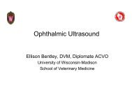
<strong>Confocal</strong> microscopy<br />
• Recently: good<br />
repeatibility<br />
• Measure corneal<br />
epithelial thickness<br />
• Bowman’s layer<br />
• Availability likely to<br />
increase-now reports in<br />
vet med<br />
www.swmed.edu/home_pages/ ophth/tscam.htm
<strong>Pachymetry</strong><br />
• Instrument comparison:<br />
– Optical pachymetry-less reproducible, less<br />
reliable than ultrasound<br />
• Both between <strong>and</strong> within observers<br />
– Optical pachymeters-Systematic left eye bias<br />
• Central left eye thicker readings
<strong>Pachymetry</strong><br />
• Studies of instrument comparison:<br />
– Automated optical pachymeter vs specular<br />
microscopy:<br />
• Identical reliability<br />
• Ultrasound pachymeters:<br />
– High intraobserver reproducibility but lower interobserver<br />
reproducibility (still higher than optical)
<strong>Pachymetry</strong><br />
• Instrument comparison:<br />
– <strong>Specular</strong> vs ultrasound pachymetry vs ultrasound<br />
biomicroscopy:<br />
• Ultrasound (both) less variability<br />
• Ultrasound pachymeter-error outputs, assess probe<br />
placement<br />
• UBM-centrality <strong>and</strong> perpendicularity can be assessed<br />
on image<br />
• Cannot assess these criteria with optical devices
Sources of error<br />
• Instrument itself<br />
• Repeated measurements<br />
• Drying of cornea<br />
• Patient positioning<br />
• Marking of cornea
Sources of error<br />
• Repeated measurements: < 1.5 % variability<br />
– Blinking between measurements decreases<br />
variability<br />
• Recumbency increases corneal thickness in<br />
humans
<strong>Pachymetry</strong> in veterinary medicine<br />
• 1985-Chan-ling-central feline corneal<br />
thickness-569 + 36 µm; diurnal variation of<br />
49; thicker after sleeping<br />
– Diurnal variation due to lid closure<br />
– Ultrasound pachymetery; Velocity=1550 m/s
<strong>Pachymetry</strong> in veterinary medicine<br />
• 1986-Carrington/Woodward-feline-755 µm<br />
– Slit lamp based optical pachymeter<br />
• 1991-Gilger-canine<br />
– Central corneal thickness-520-597 µm<br />
– Peripheral corneal thicker<br />
– Thickness increased with weight<br />
– Females thinner corneas<br />
• Ultrasonic, Velocity=1630 m/s
<strong>Pachymetry</strong> in veterinary medicine<br />
• 1993-Gilger-feline<br />
– Central <strong>and</strong> peripheral thickness similar<br />
– 578 + 64 µm<br />
– Thickness ↑ with age up to 100 months<br />
– No sex difference<br />
– Ultrasound pachymetery; Velocity=1590 m/s
<strong>Pachymetry</strong> in veterinary medicine<br />
• 1995-Schoster-feline<br />
– 13 locations<br />
– Central cornea: 546 + 48 µm<br />
– Variations in corneal thickness<br />
• Temporal <strong>and</strong> sub-periaxial areas thicker<br />
• Superio-nasal-thinnest<br />
– Ultrasound; Velocity=1640 m/s
<strong>Pachymetry</strong> in veterinary medicine<br />
• 1995-van der Woerdt-horses<br />
– Central cornea: 793 + 44 µm<br />
– Peripheral cornea significantly thicker: 831-924<br />
µm<br />
– Auriculopalpebral nerve block, xylazine, IOP,<br />
age, sex-no effect<br />
– Ultrasound; Velocity=1640 m/s
<strong>Pachymetry</strong> in veterinary medicine<br />
• 1999-Ramsey-horses<br />
– Rocky Mountain horses with cornea globosa<br />
– Thickness increases with age longer than normal<br />
horses<br />
– Ultrasound; Velocity=1640 m/s
<strong>Pachymetry</strong> in veterinary medicine<br />
• 2003-Ch<strong>and</strong>ler-dogs; post transcorneal iridal<br />
photocoagulation (diode)<br />
– No significant changes in corneal thickness<br />
– Non-contact specular microscope
<strong>Pachymetry</strong> in veterinary medicine<br />
• 2003-Plummer-Miniature horses vs full sized<br />
– Similar values between populations<br />
– Ultrasound; Velocity-1640 m/s<br />
• 2004-Gerding-dogs post intracameral<br />
lidocaine<br />
– No significant differences<br />
– Ultrasound; Velocity=1640 m/s
<strong>Specular</strong> microscopy<br />
• Typically used for endothelium exam<br />
• Light → surface: reflected, transmitted or<br />
absorbed<br />
• Usually all 3 occur<br />
• Microscope: light transmitted THROUGH<br />
substance<br />
• <strong>Specular</strong> microscope: light reflected FROM<br />
optical interface
<strong>Specular</strong> microscopy<br />
• <strong>Specular</strong> reflection = mirror like<br />
– Angle of refraction = angle of incidence<br />
– Microscope captures reflected light<br />
• Interfaces-2 media of different refractive<br />
indices<br />
– Reflection of some light<br />
– Difference in refractive indices related to amount<br />
of reflection
<strong>Specular</strong> microscopy<br />
• Optical interface between<br />
corneal endothelium <strong>and</strong><br />
aqueous humor<br />
– Also: corneal epithelium,<br />
stroma, lens<br />
• Equipment: contact or noncontact<br />
• Stationary slit, moving slit,<br />
moving spot<br />
from Krachmer, Cornea 2005
<strong>Specular</strong> microscopy<br />
• Four zones of reflection:<br />
– Zone 1-interface lens, coupling fluid, epithelium<br />
– Zone 2-light reflected from stroma<br />
– Zone 3-endothelial reflection<br />
– Zone 4-aqueous (very little reflection-dark)
<strong>Specular</strong> microscopy<br />
• Dark boundarybetween<br />
zone 3 <strong>and</strong> 4<br />
• Bright boundarybetween<br />
zone 2 <strong>and</strong> 3<br />
• Particularly noticableslit<br />
images<br />
http://www.djo.harvard.edu
<strong>Specular</strong> microscopy<br />
• Increasing angle of incidence-wider slit,<br />
larger image area<br />
– ↓ image quality-increased illumination <strong>and</strong><br />
increased scatter (stroma <strong>and</strong> epithelium)<br />
– Shortening of endothelial cells<br />
• Modern solution:<br />
– Small slits/spot-scan over tissue-↑ field of view
<strong>Specular</strong> microscopes<br />
• Difficult in awake veterinary<br />
patients<br />
• Patients typically fixate (no<br />
eye movement!)<br />
• Konan NonCon Robot-very<br />
automated<br />
– Chin rest, fixate, machine<br />
aligns by Purkinje images,<br />
focuses on endothelium,<br />
picture<br />
Cat endothelial cells-eyebank<br />
specular microscope
<strong>Specular</strong> microscopy<br />
• Analysis:<br />
– Endothelial cell density (cells/mm 2 )<br />
– Cell area (µm 2 /cell)<br />
– Pleomorphism (% of 3, 4,5, 6, 7, 8 sided cells)<br />
– Often automated counts
<strong>Specular</strong> microscopy<br />
• Density<br />
• Polymegathism<br />
Krachmer, 1997, Cornea, fig 1-10
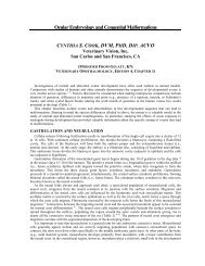
<strong>Confocal</strong> microscopy<br />
• Optical sections of living tissue<br />
– Magnification, resolution-differentiate cellular <strong>and</strong><br />
subcellular structures<br />
– Based on principle of Lukosz<br />
• Resolution improved by decreasing field of view
<strong>Confocal</strong> microscopy<br />
• 1955-Minsky<br />
– Microscope condenser-focused light w/in small<br />
area of tissue, focused objective lens on same<br />
area<br />
– ‘<strong>Confocal</strong>’-condenser, lens same focal point<br />
– Now: light source focused on small volume,<br />
confocal point detector to collect signal, scanning<br />
for full field view
<strong>Confocal</strong> microscopy<br />
• Pinhole/screen aperture for viewing-eliminates<br />
scatter from rest of tissue-very sharp image<br />
• Pinhole conjugate to focal point of lens=confocal<br />
pinhole=confocal microscope<br />
http://www.physics.emory.edu/~weeks/confocal/
• Laser-provide<br />
high intensity<br />
• Mirrors-scan<br />
across sample<br />
• Light focused on<br />
pinholemeasured<br />
by<br />
detector<br />
<strong>Confocal</strong> microscopy<br />
http://www.physics.emory.edu/~weeks/confocal
<strong>Confocal</strong> microscopy<br />
• Acquire series of thin optical sections<br />
• 3-D reconstruction<br />
• Can see all corneal layers<br />
• Better detail than specular
Anterior epithelium<br />
3 µm<br />
8 µm<br />
25µm<br />
53µm<br />
3 µm<br />
8 µm<br />
(1000x Arndt, C., doct. thesis. LMU-<br />
München, 1999)<br />
Pictures<br />
courtesy<br />
C Kafarnik<br />
25 µm<br />
53 µm
Stroma <strong>and</strong> endothelium<br />
79 µm<br />
79 µm 293 µm 487 µm<br />
293 µm<br />
487 µm<br />
517 µm<br />
535 µm<br />
539 µm<br />
517 µm 535 µm 539 µm<br />
Pictures courtesy C Kafarnik<br />
(100x; Arndt, C.,doct.<br />
thesis-LMU- München,<br />
1999)
<strong>Confocal</strong> in veterinary medicine<br />
• Kafarnik-Vet Ophth<br />
2007, 2008-dogs, cats,<br />
birds, dogs<br />
– Noted lower nerve fiber<br />
densities in<br />
brachycephalics<br />
• Ledbetter-Vet Ophth<br />
2009-horsescharacterized<br />
normal<br />
Ledbetter, VO, 2009-Eq endothelium
<strong>Specular</strong> microscopy in veterinary<br />
medicine<br />
• 1979-Stapelton-7 young dogs<br />
– 2816 + 187 cell/mm 2<br />
• 1981-Peiffer-11 cats<br />
– 2668 + 211 cell/mm 2
<strong>Specular</strong> microscopy in veterinary<br />
medicine<br />
• 1981-Befanis-endothelial repair-’young’ dogs<br />
– Froze 90%<br />
– Demonstrated re-formation of intact monolayer<br />
by 6 weeks<br />
– 2,800 cells/mm 2
<strong>Specular</strong> microscopy in veterinary<br />
medicine<br />
• 1982-Gwin-central <strong>and</strong> peripheral cornea-dog<br />
– No differences cell counts centrally vs<br />
peripherally<br />
– Cell # decreased with age<br />
• < 1 year - > 2600 cells/mm 2<br />
• 1-9 years - 2300-2500 cells/mm 2<br />
• > 10 years - 1900-2100 cells/mm 2
<strong>Specular</strong> microscopy in veterinary<br />
medicine<br />
• 1983-Gwin-21 dogs, mature cataracts, phaco<br />
<strong>and</strong> ECCE<br />
– Phaco-Cell counts ↓ 22% centrally, 13%<br />
peripherally<br />
– ECCE-Cell counts ↓ 34% centrally, 31%<br />
peripherally<br />
– Cells enlarged, pleomorphic with both surgeries
<strong>Specular</strong> microscopy in veterinary<br />
medicine<br />
• 1985-Glasser-cats, effect of BSS vs BSS plus<br />
irrigation of anterior chamber<br />
– No difference in cell density<br />
– Increased polymegathism, pleomorphism in BSS<br />
group<br />
– No change cell morphology with BSS plus
<strong>Specular</strong> microscopy in veterinary<br />
medicine<br />
• 1986-Nassise-Saline, BSS, BSS plus<br />
glutiathione-AC irrigation in dogs<br />
– 22 minute irrigation with 100mls<br />
– No changes endothelial cell density<br />
• Mild corneal thickness increase with saline group
<strong>Specular</strong> microscopy in veterinary<br />
medicine<br />
• 1990-Gerding-dogs-replace aqueous with:<br />
BSS, sodium hyaluronate, sodium<br />
chondroitin-sulfate/sodium hyaluronate,<br />
hydroxypropyl methylcellulose<br />
– No significant differences in cell density or<br />
morphology or thickness
<strong>Specular</strong> microscopy in veterinary<br />
medicine<br />
• 1992-Gerding-intracameral tPA in dogs<br />
– High dose group (50 µg/100 ml)-18% decrease in<br />
hexagonal cells<br />
– No other changes in cell density, morphology or<br />
thickness noted
<strong>Specular</strong> microscopy in veterinary<br />
• 2001-Andrew-equine<br />
– 3155 cell/mm 2<br />
medicine<br />
– Decreased cell density in ventral quadrant vs<br />
medial/temporal<br />
– Cell density dereased with age
<strong>Specular</strong> microscopy in veterinary<br />
medicine<br />
• 2002-Andrew-36 llamas, 20 alpacas<br />
– Llamas-2668 cells/mm 2<br />
– Cell density decreased with age<br />
– Alpacas-2275 cells/mm 2<br />
– Polymegathism frequent
<strong>Specular</strong> microscopy in veterinary<br />
medicine<br />
• 2003-Ch<strong>and</strong>ler-post trancorneal iridal<br />
photocoagulation in dogs<br />
– No significant difference in endothelial cell counts<br />
or morphology<br />
• 2004-Gerding-post lidocaine in AC in dogs<br />
– No significant effects on endothelial cell counts or<br />
morphology
Questions??