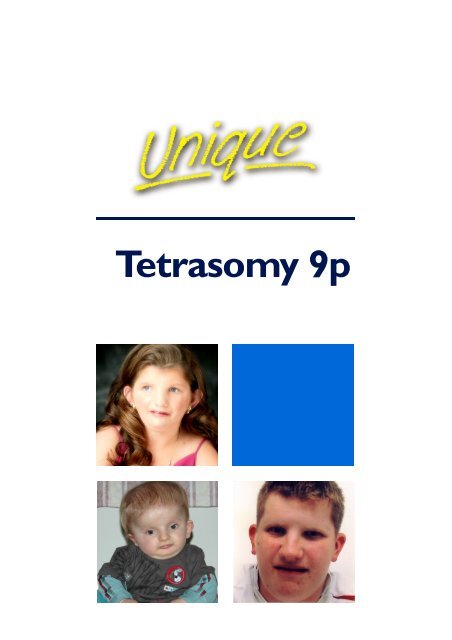
Tetrasomy 9p FTNW - Unique - The Rare Chromosome Disorder ...
Tetrasomy 9p FTNW - Unique - The Rare Chromosome Disorder ...
Tetrasomy 9p FTNW - Unique - The Rare Chromosome Disorder ...

Create successful ePaper yourself
Turn your PDF publications into a flip-book with our unique Google optimized e-Paper software.
<strong>Tetrasomy</strong> <strong>9p</strong>
Sources &<br />
references<br />
<strong>The</strong><br />
information<br />
in this leaflet<br />
is drawn<br />
partly from<br />
medical<br />
publications,<br />
where 45-50<br />
cases are<br />
reported.<br />
<strong>The</strong> firstnamed<br />
author and<br />
publication<br />
date are given<br />
so you can<br />
look for<br />
articles on<br />
the internet<br />
in PubMed.<br />
<strong>The</strong> leaflet<br />
also draws on<br />
<strong>Unique</strong>’s<br />
database.<br />
<strong>Tetrasomy</strong> <strong>9p</strong><br />
<strong>Tetrasomy</strong> <strong>9p</strong> is a rare condition in which people have too much<br />
material from one of their chromosomes. Usually they have a small<br />
extra chromosome made up of two copies of part of chromosome 9.<br />
This extra chromosome material makes it likely that people with<br />
tetrasomy <strong>9p</strong> will need support with their learning and development<br />
as well as help for some birth defects and health problems.<br />
Genes and chromosomes<br />
Our bodies are made up of billions of cells. Most cells contain a<br />
complete set of thousands of genes that act like instructions,<br />
controlling our growth, development and how our bodies work.<br />
Genes are carried on microscopically small, thread-like structures<br />
called chromosomes. We usually have 46 chromosomes, 23 inherited<br />
from our mother and 23 inherited from our father, so we have two<br />
sets of 23 chromosomes in ‘pairs’. People with tetrasomy <strong>9p</strong> usually<br />
have a small 47 th chromosome.<br />
Apart from two sex chromosomes (two Xs for a girl and an X and a Y<br />
for a boy), chromosomes are numbered 1 to 22, generally from largest<br />
to smallest. So chromosome 9 is a medium-sized chromosome. Each<br />
chromosome has a short (p) arm (at the top in the diagram below)<br />
and a long (q) arm (at the bottom). People with tetrasomy <strong>9p</strong> have<br />
two extra copies of material from the short (p) arm. <strong>The</strong>y usually have<br />
two normal chromosome 9s as well, so they have four copies of <strong>9p</strong> in<br />
all. <strong>The</strong> name ‘tetrasomy <strong>9p</strong>’ comes from ‘tetra’, the Greek word<br />
for four.<br />
<strong>9p</strong><br />
<strong>9p</strong><br />
<strong>9p</strong><br />
+ +<br />
<strong>9p</strong><br />
9q<br />
9q<br />
Two chromosomes 9 and an extra<br />
chromosome made up of two short arms<br />
<strong>Chromosome</strong> 9<br />
2<br />
Looking at the extra material from chromosome <strong>9p</strong><br />
You cannot see chromosomes with the naked eye, but if you stain<br />
them and magnify their image under a microscope, you can see that<br />
each one has a distinctive pattern of light and dark bands. <strong>The</strong> bands<br />
show that the extra 47 th chromosome in tetrasomy <strong>9p</strong> is made up of<br />
two copies of the short arm of chromosome 9. An extra chromosome<br />
made up of two copies of the same part is called an isochromosome.<br />
Sometimes the isochromosome also contains part of the long arm.
<strong>The</strong> karyotype<br />
Your genetic specialist can tell you more about how much material there is in the extra<br />
chromosome. You will almost certainly be given a karyotype, a shorthand code that<br />
usually tells you the band where chromosome 9 has broken. <strong>The</strong> chromosome usually<br />
breaks in the short arm close to the centromere (the point where the short arm meets<br />
the long arm). Sometimes it breaks in the long arm, usually close to the centromere.<br />
47,XY,+i(<strong>9p</strong>) This shows that there are 47 chromosomes, it’s a boy (XY), and the<br />
extra (+) chromosome is an isochromosome (i) made up of material from the short (p)<br />
arm of chromosome 9.<br />
47,XX,iso(9)(q12)de novo This shows that it’s a girl (XX) and that the extra<br />
chromosome is made up of the short arm and material as far as band q12 in the long<br />
arm. De novo means that the parents’ chromosomes have been examined and<br />
are normal. Iso was used by laboratories in the past but today would be (i) or, strictly<br />
speaking in this case, idic (see below).<br />
47,XY,+idic(9)(q13) This shows that the extra chromosome is made up of material<br />
from the entire short arm and the long arm as far as q13, including the centromeres<br />
where the short arms meet the long arms. An extra isochromosome that includes the<br />
centromeres is called an isodicentric chromosome, shortened to idic (eye-dick).<br />
47,XX,+mar.ish i(9)(p10)(wcp9+) dn This means that it’s a girl (XX) with a small<br />
extra chromosome that couldn’t at first be identified. A small unidentified chromosome<br />
like this is called a marker (mar). Using a technique called FISH (.ish) that allows<br />
chromosomes to be examined in greater detail, the marker was found to consist of<br />
material from chromosome 9. <strong>The</strong> chromosome has broken off at band p10, another<br />
way of describing the centromere. <strong>The</strong> specific technique used was whole chromosome<br />
painting (wcp), in which the test ‘recognises’ different parts of the extra chromosome<br />
as coming from chromosome 9. You can write de novo as dn, meaning that the parents’<br />
chromosomes have been examined and are normal.<br />
How is tetrasomy <strong>9p</strong> diagnosed?<br />
<strong>Tetrasomy</strong> <strong>9p</strong> is usually found in highest concentrations in blood, so diagnosis in a baby,<br />
child or adult means taking and analysing a blood sample. Confirmation and an<br />
estimation of the ratio of normal to tetrasomy <strong>9p</strong> cells can use a sample of skin or a<br />
scraping from inside the cheek (buccal smear) (Lloveras 2004).<br />
Diagnosis during pregnancy is more difficult as tetrasomy <strong>9p</strong> cells may not be found in<br />
fetal cells in amniotic fluid. It is therefore quite possible for a pregnancy to be affected<br />
by tetrasomy <strong>9p</strong> but for the amniotic fluid or chorionic villus sample to show only cells<br />
with normal chromosomes (Chen 2007; Eggermann 1998; Grass 1993).<br />
One possible solution in this situation is to take a sample of blood from the umbilical<br />
cord, as this blood is part of the fetal circulation (Papenhausen 1990). This procedure<br />
can be risky, however, and one couple at high risk of having a baby with tetrasomy <strong>9p</strong><br />
who lost a pregnancy after fetal blood sampling chose instead to have future<br />
pregnancies intensively monitored by amniocentesis and ultrasound with the<br />
sonographer looking for findings such as an enlarged head, growth delay and palate, lip<br />
and kidney anomalies (Henriques-Coelho 2005; McAuliffe 2005; Lloveras 2004; Dhanda<br />
2002; Xu 1998).<br />
3
Why are some people with tetrasomy <strong>9p</strong> more severely affected<br />
than others?<br />
<strong>The</strong>re are quite a few reasons. One is likely to be the size of the isochromosome,<br />
although researchers do not always agree how important it is if you have a larger<br />
isochromosome with more chromosome material. Some believe it matters and children<br />
with additional material from 9q seem more severely affected (Chen 2007; Wisniewski<br />
1978). Others are not so sure (Stumm 1999). It is also true that individual differences<br />
between children even with the same karyotype can be fairly marked.<br />
Often the karyotype from a blood or skin sample shows two different cell lines, one<br />
with tetrasomy <strong>9p</strong> and one with normal chromosomes. Where there are two different<br />
cell lines within the same tissue, it is known as mosaicism. When all the cells show<br />
tetrasomy <strong>9p</strong>, it is known as non-mosaicism. Babies found to have mosaic tetrasomy <strong>9p</strong><br />
are likely to do better than those with non-mosaic tetrasomy <strong>9p</strong>, especially in terms of<br />
survival beyond the newborn period.<br />
47,XY,+i(9)(p10)[4]/46,XY[16] This is a boy in whom 20 cells were tested. Four<br />
[4] showed tetrasomy <strong>9p</strong> while sixteen [16] showed a normal karyotype for a<br />
boy or man.<br />
<strong>The</strong> cells tested for the karyotype are most often taken from blood. In tetrasomy <strong>9p</strong>,<br />
blood cells often show non-mosaicism (that is, only tetrasomy <strong>9p</strong> cells), even where<br />
there is mosaicism or indeed cell lines with only normal cells in other parts of the body.<br />
So a baby who has only had a blood test may be thought to have non-mosaic tetrasomy<br />
<strong>9p</strong>, when in fact there is mosaicism in other tissues. For this reason, most genetics<br />
centres recommend testing samples from more than one part of the body, usually<br />
either from the skin or from inside the cheek (Lloveras 2004).<br />
Common sense might suggest that when a higher proportion of tetrasomy <strong>9p</strong> cells is<br />
found, the effects are likely to be more severe. But this is not necessarily true. One<br />
reason for this discrepancy may be varying proportions of tetrasomy <strong>9p</strong> cells in tissues<br />
of importance fro development, tissues which have not been investigated.<br />
Are there people with tetrasomy <strong>9p</strong> who are healthy, have no major birth<br />
defects and have developed normally?<br />
Yes, there are. Out of 47 reports in the medical literature, there are three people with<br />
apparently normal development and a possible fourth. One adult is an accountant and<br />
was discovered to have tetrasomy <strong>9p</strong> when infertility was investigated. Another adult<br />
was investigated for skin lesions. Another case is of a child who has developed normally<br />
to the age of five, although he showed growth delay before birth; a six-month-old baby<br />
also had no apparent abnormalities at the age of 6 months (McAuliffe 2005; Lloveras<br />
2004; Sait 2003; Nakamura 1990). Among <strong>Unique</strong>’s 13 members, two children were<br />
only diagnosed after being investigated for developmental delay in the late primary<br />
school or early secondary school years.<br />
Why is tetrasomy <strong>9p</strong> so variable?<br />
<strong>The</strong>re are four key reasons:<br />
Mosaic or not mosaic – when only tetrasomy <strong>9p</strong> cells and no normal cells are found<br />
in both blood and skin cells or amniotic fluid, the effects are likely to be more obvious<br />
and more severe. Babies with mosaic tetrasomy <strong>9p</strong> survive the newborn period better.<br />
4
Tissue-limited mosaicism – <strong>The</strong> proportions of tetrasomy <strong>9p</strong> cells are different in<br />
different body tissues. In blood there are more tetrasomy <strong>9p</strong> cells and sometimes no<br />
normal cells are found. In other tissues, especially skin and mucous membranes, the<br />
proportion of tetrasomy <strong>9p</strong> cells is usually lower and no tetrasomy <strong>9p</strong> cells may be<br />
found. Organs, such as the brain and lungs, may have different proportions again and<br />
this variability is very likely to affect the outcome (Lloveras 2004; Dhanda 2002).<br />
Size of isochromosome – that is, the amount of extra chromosome material.<br />
Common sense suggests that those with larger extra chromosomes will be more<br />
severely affected.<br />
Specific duplicated regions – a boy with three copies of the section of chromosome<br />
9 between band p13 and p22 shared some similarities with children with an<br />
isochromosome <strong>9p</strong>, but had no major anomalies apart from enlarged ventricles within<br />
the brain. This could suggest that the part of <strong>9p</strong> that he did not have extra copies of<br />
(<strong>9p</strong>23 to the tip of <strong>9p</strong>) is the part that causes important birth defects (Verheij 1999).<br />
Most likely features among those with mosaic tetrasomy <strong>9p</strong><br />
- found in at least half of all children and adults reported or described so far (de<br />
Azevedo 2003; Dhanda 2002; <strong>Unique</strong>)<br />
Developmental delay<br />
In 4/7 <strong>Unique</strong> cases, a delay in expected development was the first sign of anything<br />
wrong. Signs were first noticed between three months and early secondary school age.<br />
Central nervous system (brain and spinal cord) anomaly<br />
Abnormal features of arms or legs. <strong>The</strong>se can include dislocated joints and clubfeet<br />
Growth delay before or after birth<br />
Heart defects<br />
Abnormalities of the kidneys or urinary system. In boys, minor anomalies of the<br />
genitals or undescended testicles are common<br />
At birth, wide gaps between the bony plates of the skull. <strong>The</strong> front soft spot<br />
(fontanelle) may be large<br />
Short neck, sometimes with excess skin<br />
Typical facial features including wide set eyes, small jaw (chin), oddly formed or<br />
positioned ears and a bulbous or beaked nose<br />
Cleft lip, palate or a high-arched palate<br />
Other typical features<br />
Unusually formed nails; a single crease across the palm; incurving fingers, especially<br />
the fifth finger; short hands and feet with small toe and finger joints<br />
Widely spaced eyes; small skin folds across the inner corner of the eyes; eyes that<br />
slant somewhat downwards; a small lower jaw, set back from the upper jaw;<br />
downwards slanting mouth<br />
Low muscle tone, making the body feel floppy (hypotonia)<br />
Unusual head size – small (microcephaly) or large (macrocephaly). Enlarged fluidfilled<br />
ventricles within the brain (hydrocephaly)<br />
Strabismus (squint) or short sight (myopia)<br />
Dimple near the base of the spine<br />
Sunken eyes due to eyeball being recessed within the orbit<br />
Missing or underdeveloped bones<br />
5
What is the outlook?<br />
<strong>The</strong> outlook for babies diagnosed with tetrasomy <strong>9p</strong> is extremely variable. In some<br />
babies there is little or no effect on development or health, while in others the effects<br />
are obvious and sadly survival may not be possible. Those babies diagnosed with nonmosaic<br />
tetrasomy <strong>9p</strong> appear to be at greatest risk and typically do not survive the<br />
newborn period. Babies with a mosaic form of tetrasomy <strong>9p</strong> have a better outcome.<br />
As tetrasomy <strong>9p</strong> is most likely to appear to be non-mosaic in blood, it is important that<br />
all babies with the diagnosis made from a blood test have tissue from another part of<br />
the body (such as skin or mucous membranes) examined as well, as this is more likely<br />
to show mosaicism (Tang 2004; Moreira 2003; Dhanda 2002).<br />
<strong>The</strong>re is very little published information on the long term outlook for babies born with<br />
tetrasomy <strong>9p</strong>. Among <strong>Unique</strong> members, seven (out of eight) aged 1 to 19 were healthy<br />
and active and taking no regular medications apart from vitamins. One baby had a<br />
febrile episode after the first course of vaccinations against diphtheria, whooping cough<br />
and tetanus; another child has Raynaud’s disease, with spasm of the blood vessels in the<br />
extremities; two teenagers have hayfever (<strong>Unique</strong>).<br />
How did this happen?<br />
In the great majority of children, the extra material from <strong>9p</strong> appears as a separate, small<br />
chromosome. When the blood of parents with an affected child is examined, it has so<br />
far always revealed normal chromosomes (McAuliffe 2005). In this situation, studies<br />
have indicated that that the most common reason for the extra chromosome is the<br />
failure of the chromosomes 9 in one parent to separate in the process of preparing the<br />
eggs or sperm, leading to an extra whole chromosome 9. This extra chromosome is<br />
believed to then undergo an unusual type of division, leaving it with two top halves<br />
(short arms) while the two bottom halves (the long arms) are lost (Dutly 1998).<br />
Separation failure, termed non-disjunction, is more common in older mothers (as in<br />
Down’s syndrome) and the age of mothers having a child with tetrasomy <strong>9p</strong> is slightly<br />
higher than average. <strong>The</strong> average age of mothers of babies with the mosaic form is 33.7<br />
years (range 20 to 42 years), compared with a population average of 26-9 years, while<br />
that of fathers was 34.9 years, compared with a current UK population average of 32<br />
years.<br />
Can it happen again?<br />
So long as the parents have normal chromosomes, the extremely unusual sequence of<br />
events that led to a fetus with tetrasomy <strong>9p</strong> is very unlikely to happen again. Many<br />
couples will want the reassurance of having their next baby’s chromosomes tested<br />
during pregnancy by chorionic villus sampling or amniocentesis. Although these<br />
techniques do not necessarily show tetrasomy <strong>9p</strong>, a normal result is reassuring.<br />
Can it be passed on?<br />
As we have said, the great majority of children with tetrasomy <strong>9p</strong> have parents with<br />
normal chromosomes. But there are some people - and no-one really knows how many<br />
of them there are - who themselves have tetrasomy <strong>9p</strong> but are very mildly affected by<br />
it. <strong>The</strong>y can pass it on. <strong>The</strong>y can also have normal children. One man with tetrasomy <strong>9p</strong><br />
had two children with normal chromosomes, but his wife also had five pregnancies<br />
resulting in miscarriage (McAuliffe 2005).<br />
6
A child with tetrasomy <strong>9p</strong><br />
Two babies with tetrasomy <strong>9p</strong><br />
Facial appearance<br />
Certain facial features are found more often in children with tetrasomy <strong>9p</strong> than in<br />
other children. <strong>The</strong>se features do not matter to your child, but they may mean that you<br />
see unexpected similarities between your child and others with tetrasomy or even<br />
trisomy <strong>9p</strong>. <strong>The</strong> common features are: unusually formed or positioned ears; a small<br />
lower jaw (micrognathia) that may also be receding (retrognathia); widely spaced eyes<br />
that can be deep set or even sunken and slant upwards or downwards; a broad,<br />
bulbous or beaked nose; a large mouth with downturned corners or a short groove<br />
between the upper lip and the nose; a short neck or too much nuchal skin; and skin<br />
folds at the inner corner of the eyes. Your child’s head may have an unusual shape and<br />
may be small or enlarged. Newborn babies may have wide gaps between the bony<br />
plates of the skull and a very large soft spot (fontanelle) on top of the head that can<br />
take years to close (Henriques-Coelho 2005; Dhanda 2002; Park 1995; Schaefer 1991;<br />
Moedjono 1980; <strong>Unique</strong>).<br />
Birth weights at term: Non-mosaics 2lb 11oz /1.21kg to 5lb 15oz /2.7kg<br />
Mosaics 3lb 10oz/1.644kg to 8lb 8oz/ 3.856kg<br />
Feeding<br />
Feeding problems are common in babies and children with a chromosome disorder but<br />
in this group breastfeeding was possible for many and in one case continued to two<br />
years. Some babies were diagnosed with allergy to a milk formula and fed on a<br />
replacement milk such as soya or goat’s milk. It is not known whether a mild degree of<br />
reflux (bringing feeds back) was interpreted as a milk allergy (<strong>Unique</strong>).<br />
Only one feeding report was received for a<br />
baby with significant heart disease; not<br />
unexpectedly, this child, with significant heart<br />
problems, was fed by gastrostomy tube direct<br />
to the stomach. Quite a few babies with<br />
tetrasomy <strong>9p</strong> have a cleft palate (a split in the<br />
roof of the mouth), sometimes with a split in<br />
the upper lip as well but feeding was even<br />
problem-free in one baby with a cleft lip and<br />
palate (Orye 1975). Typically, babies with a<br />
cleft lip or palate have greater feeding<br />
difficulties until their condition is stabilised or<br />
surgically corrected.<br />
7
Growth<br />
Height and body build are variable, with some adults short (5’ 1”/1.55m in a girl of 19<br />
years) while others are of average height (5’ 9”/1.75m in a boy of 16 years; 5’ 11”/1.8m<br />
in a boy of 15). <strong>The</strong>re does not appear to be a clear link between growth delay before<br />
birth and childhood height, with examples of small-for-gestational-age babies growing<br />
into average-height children (Lloveras 2004) and others of babies of average birth<br />
weight later failing to thrive (Stumm 1999; <strong>Unique</strong>). Body build also varies from stocky<br />
to slight, with more children described as slight. It is not known whether youngsters<br />
with tetrasomy <strong>9p</strong> will continue to grow into their 20s, as those with trisomy <strong>9p</strong> do.<br />
Development: sitting, moving, walking (gross motor skills)<br />
Many babies and children are late to achieve their ‘milestones’ of sitting and walking and<br />
are helped by regular physiotherapy. <strong>The</strong>re is a wide range of eventual ability, however,<br />
with some children acquiring mobility skills around the same age as ‘typical’ children<br />
and others showing more obvious delay. Among 15 children, mostly from <strong>Unique</strong>,<br />
rolling over was achieved between four and 10 months, sitting between seven and 15<br />
months, crawling or bottom shuffling between 6 and 18 months and walking<br />
independently between 14 months and three years and five months.<br />
Mobility is affected by abnormal muscle tone and many children either have low tone<br />
(hypotonia) or high tone (hypertonia). Babies with low muscle tone at birth feel floppy<br />
to hold and have obvious head lag when held to sit. Low muscle tone generally<br />
improves with maturity and regular physiotherapy.<br />
Eventual walking style also varies between individuals. While some achieve total<br />
mobility and learn to climb stairs, to run, ride a bicycle and to swim, others retain an<br />
uneven and uncoordinated walking style and rely on wheelchair use for long distances<br />
outdoors. In general, it appears that those least affected or unaffected in early<br />
childhood are most likely to achieve normal mobility and sporting prowess as adults,<br />
while those children who have obvious mobility problems early on achieve a more<br />
limited degree of mobility.<br />
Joint abnormalities are a known feature of tetrasomy <strong>9p</strong> at birth, with extremely<br />
loose joints and dislocations (elbows, wrists, knees, hips) often observed (De Azevedo<br />
2003; Tonk 1997; Leichtman 1996; Iinuma 1994; Cavalcanti 1987; Shapiro 1985;<br />
Moedjono 1980). Children with very loose joints may need additional braces (supports,<br />
splints) before they are able to walk. In some cases, joints are unusually tight and may<br />
require surgery and tendon lengthening to extend their range of movement.<br />
8<br />
Activity in one child
A wide variety of specific abnormalities of foot position are also a common feature<br />
of tetrasomy <strong>9p</strong>. <strong>The</strong>se may include pes cavus (‘claw foot’), pes planus (flat feet), rocker<br />
bottom feet (the sole is curved without an instep, like a chair rocker), pes planovalgus<br />
(the feet are flat and stick out), pes equinovarus (club feet, with the foot turned<br />
inwards, the soles pointing towards each other), pes adductus (so-called ‘banana foot’,<br />
where the toes point inwards) and other less common positions. Babies born with feet<br />
affected in this way will receive specific physiotherapy which may avoid the need for<br />
corrective surgery and plaster casting. Treatment is tailored to the individual child and<br />
in some cases surgical correction will best enhance eventual mobility.<br />
Some children have a degree of hip dysplasia, in which the hip joints are easily<br />
dislocated. This may be apparent at birth or develop later. In either case it is treated<br />
with splinting and if necessary immobilisation in plaster and possibly surgery<br />
(Eggermann 1998; Papenhausen 1990).<br />
Some babies are born with or develop a spinal curvature, either curving sideways<br />
(scoliosis) or forwards (kyphosis). Underlying the curve may be abnormalities of muscle<br />
tone and in some cases the bones of the spine (vertebrae) may be fused together or<br />
incorrectly formed. <strong>The</strong> curvature can be treated with physiotherapy and exercises, but<br />
a support brace may be needed. If the curve becomes marked it is possible to<br />
straighten the spine using rods (Dhanda 2002; Stumm 1999; Verheij 1999; Dutly 1998;<br />
Melaragno 1992; Sjöstedt 1989; Balestrazzi 1983; Moedjono 1980; <strong>Unique</strong>).<br />
Development: hand use and coordination (fine motor skills)<br />
Development of hand use and hand-eye coordination are frequently delayed but the<br />
evidence from <strong>Unique</strong> is that adults are generally able to carry out daily personal care<br />
tasks. One family supplied a detailed timetable of their son’s fine motor development:<br />
used a spoon at 13 months; placed 3 bricks in a tower at 21 months; used a knife and<br />
fork at 27 months; bounced and caught a ball at 27 months; drew a circle at 36 months;<br />
drew a person at 42 months. As an adult, this young man is able to dress and care for<br />
himself but needs help with small buttons and shoe laces.<br />
<strong>The</strong> hands are frequently affected by tetrasomy <strong>9p</strong> with common features such as bent<br />
and shortened fingers and thumbs, occasionally overlapping each other or joined by a<br />
bridge of skin and tissue, as well as abnormal or missing fingernails. <strong>The</strong> tips of the<br />
fingers may be noticeably shortened and even missing. <strong>The</strong> <strong>Unique</strong> experience is that<br />
these features do not generally affect the way a child uses their hands and only need<br />
correction when hand use is affected.<br />
Learning<br />
<strong>The</strong> range of learning ability is very broad. At one end of the<br />
spectrum is an adult with a professional career and children<br />
who attend mainstream schools, are able to follow the normal<br />
curriculum, sometimes with help for specific learning difficulties,<br />
and achieve a range of school-leaving qualifications. At the other<br />
end are children and adults with a moderate to severe learning<br />
disability.<br />
<strong>The</strong> evidence on learning comes chiefly from <strong>Unique</strong> and shows<br />
that in general children learned to read and write between the<br />
9
ages of five and 11 years. Age of first reading does not necessarily predict eventual<br />
ability as one of the highest achieving adolescents was a relatively late reader. Specific<br />
areas of high ability and specific learning difficulties occur and a common theme appears<br />
to be a facility for visual learning.<br />
• A 19-year-old girl has a highly developed memory for people, upcoming events, lost<br />
or misplaced property and travelling directions. She is better able to learn practical<br />
tasks than academic tasks. She is extremely observant and interested in people. She<br />
can sign her name, copy most letters and write them with verbal prompts. She<br />
attended a mainstream primary and special secondary school.<br />
• A 16-year-old boy who has attended a mainstream school with 1:1 support passed<br />
the United Kingdom school-leaver examinations in mathematics, science, art and<br />
design and shows particular strengths in subjects where word use is minimised. He<br />
has a talent for abstract art and impressionism.<br />
• One 15-year-old shows an excellent - even remarkable - long term memory but has<br />
poor short term recall and working memory. He is a strong visual learner and is good<br />
at mathematics. He has dyslexia which prevents avid reading but he listens to stories<br />
such as Harry Potter. His drawing and writing are normal. He attends a private<br />
school for students with normal intelligence but learning disability.<br />
• A 13-year-old girl, attending a mainstream secondary school, is reading books for 8-<br />
10 year olds and writing at a similar standard. Another girl, aged 10, has a very good<br />
memory and is able at English. She attends a special school where her learning is<br />
most helped by her determination, by observation of people and things around her<br />
and her good memory.<br />
• A 6-year-old girl also has a very good memory and is willing to learn but lacks<br />
concentration and confidence. She reads school books, can write her name, some<br />
letters and some numbers and can draw people and butterflies. She attends a<br />
mainstream school with additional support.<br />
Speech and communication<br />
<strong>The</strong> ability to speak and converse generally reflects learning abilities, so children who<br />
need greater learning support tend to be those who start speaking later and develop<br />
less complex language. Children whose learning ability falls within the normal range may<br />
show little or no delay in initially acquiring speech and language and go on to develop<br />
complex conversational skills and a broad vocabulary.<br />
First words have generally emerged between nine months and four to five years and<br />
linked words and longer phrases by 10 years, but not everyone acquires speech. <strong>The</strong>re<br />
are wide differences between individuals in understanding, with understanding and<br />
expression on a par in some children while in others expressive skills outstrip receptive<br />
language or vice versa. Where individuals have no speech, communication has still been<br />
successful through signing and assistive technology such as a voice output<br />
communicator.<br />
Even among the fluent speakers, some unclarity has tended to persist with a small<br />
cluster of families remarking on their child’s disordered phonology and inability to<br />
discriminate between s, f, th and v sounds.<br />
10
A mild to moderate hearing loss appears to be common, affecting 3/5 <strong>Unique</strong> children.<br />
Hearing tests at birth are typically normal, with hearing loss developing due to glue ear,<br />
made worse for a few children by unusually narrow external ear canals (Tonk 1997;<br />
Melaragno 1992; Orye 1975). Glue ear is typically treated by inserting aeration tubes<br />
(grommets) into the eardrum and this surgical operation may need to be repeated.<br />
Normal hearing may not be achieved with aeration of the space behind the eardrum<br />
(middle ear) and hearing aids may help as a temporary or longer-lasting measure.<br />
As children are at risk of speech delay, parental concerns should be acted on early and<br />
home or school-based therapy provided.<br />
Behaviour<br />
<strong>The</strong> evidence from <strong>Unique</strong> shows that children and adults with tetrasomy <strong>9p</strong> are loving,<br />
caring individuals and generally speaking have an open and sociable temperament. <strong>The</strong>re<br />
is no obvious relationship between behaviour and learning ability, although those<br />
children with greater functioning difficulties will be in an environment where less is<br />
expected of them. Individuals do have difficulties in interpreting and responding suitably<br />
to social cues and this is most apparent in those who are in a mainstream environment<br />
outside their family. <strong>The</strong>y may well be popular with their peers but find it easier to<br />
relate to people older or younger than themselves.<br />
Within the family, children may experience difficulties with frustration and in<br />
accommodating their brothers and sisters.<br />
Early access to advice, input and therapy will help those families who find themselves in<br />
difficulties with their child’s behaviour. One child has a diagnosis of attention deficit<br />
hyperactivity disorder (ADHD) but methylphenidate (Ritalin) medication controls<br />
restlessness and inappropriate comments. Another child with frustration difficulties<br />
within the home has been helped by a behaviour chart and a clear reward system.<br />
A very outgoing personality who copes quite well socially. She can approach strangers<br />
but will withdraw or proceed depending on the reaction she gets. She does pick up on<br />
social cues and is very keen to help. She will initiate assistance and predict your needs<br />
- age 19<br />
A likeable personality, very affectionate. Most of the time he is lovely and continues<br />
to be well behaved outside the family environment. He can be rather obsessive at<br />
times, eg tidiness, washing clothes etc.<br />
Socially, he is popular but his immaturity<br />
makes it difficult for him to have close<br />
friendships with people of his own age<br />
- age 16<br />
A very sweet, loving child, he has an acute<br />
perception of people’s ‘true selves’ - age 15<br />
Socially, she gets on brilliantly with<br />
strangers; gets on OK with the family until<br />
she can’t do something she wants - age 10<br />
Very friendly, loves to stop and chat with<br />
people. Happy and cheerful and her happiness<br />
rubs off on everyone - age 6<br />
11
Health concerns<br />
Eyesight Vision difficulties 17/29<br />
Known difficulties include a squint (strabismus) (3), including exotropia (divergent<br />
squint), frequently intermittent, and lack of stereoscopic vision (teaming of the eyes)<br />
causing loss of 3D vision and depth perception. Strabismus may be treated with<br />
patching, glasses, exercises or surgical correction. In at least one <strong>Unique</strong> child, the<br />
squint self corrected by the age of six years.<br />
<strong>The</strong>re are six cases of marked short sight and one child is registered as partially sighted.<br />
Other children have sunken eyes; lazy eye (amblyopia); uncontrolled eye movements<br />
(nystagmus); an abnormal development of the iris; damage to the part of the back of<br />
the eye known as the chorioretinal area; a single eye (Lloveras 2004; Cazorla Calleja<br />
2003; Dutly 1998; Tonk 1997; Papenhausen 1990; Balestrazzi 1983; Cuoco 1982;<br />
Garcia-Cruz 1982; Abe 1977; Orye 1975; <strong>Unique</strong>). In at least one child the development<br />
of good vision was affected by raised pressure within the brain (Stumm 1999).<br />
Head and brain 24/37<br />
Many babies were born with a very large soft spot (fontanelle) or wide spaces between<br />
the bony plates of the skull. <strong>The</strong> front fontanelle was also often slow to close and in<br />
one child was still open at age 4 (<strong>Unique</strong>). Additionally, some babies have an unusual<br />
head shape or size (‘strawberry skull’; asymmetric head shape; microcephaly – small<br />
head; macrocephaly – large head; brachycephaly – the head is disproportionately wide<br />
ear-to-ear compared to the measurement from front to back).<br />
In some babies and children, a structural abnormality of the brain was found, much<br />
more commonly among youngsters with a non-mosaic form of tetrasomy <strong>9p</strong>. <strong>The</strong> rate<br />
among <strong>Unique</strong> members seems lower, with only 3/15 children and adolescents known<br />
to be affected. Structural anomalies such as enlarged ventricles (fluid-filled spaces)<br />
within the brain, absence or underdevelopment of the corpus callosum – the bundle of<br />
nerve fibres that links the brains two hemispheres, and Dandy Walker may be detected<br />
on prenatal ultrasound. <strong>The</strong> Dandy Walker anomaly is a cyst in the balance control part<br />
of the brain (cerebellum) that is involved with the fourth ventricle, one of the fluid-filled<br />
spaces within the brain. This may interfere with the body's ability to drain cerebrospinal<br />
fluid from the brain, resulting in hydrocephalus, a build-up of fluid within the brain<br />
(Lloveras 2004; Cazorla Calleja 2003; Stumm 1999; Andou 1994; Melaragno 1992;<br />
Balestrazzi 1983; Cuoco 1982; Garcia-Cruz 1982; Peters 1982; Ghymers 1973; <strong>Unique</strong>).<br />
Other brain anomalies include underdevelopment of the grey matter both in the<br />
cerebellum and the cerebral hemispheres; lissencephaly (smooth rather than ridged<br />
brain surface); pachygyria (where the ‘hills’ in the undulating landscape of the brain’s<br />
surface are unusually large); polymicrogyria (where the ‘hills’ are many and small)<br />
(Cazorla Calleja 2003).<br />
Biochemical tests revealed raised levels of lactate in cebrebrospinal fluid in one child,<br />
although what this finding might mean was not clear (Eggermann 1998). <strong>The</strong> most<br />
frequent problem that occurs after birth is hydrocephalus, usually requiring a shunt to<br />
drain the excess cerebrospinal fluid and relieve pressure on the growing brain. <strong>The</strong><br />
build-up of hydrocephalus may occur even when a child’s head is unusually small<br />
(microcephaly). <strong>The</strong> experience of treatment for hydrocephalus is challenging for<br />
12
families, but one <strong>Unique</strong> family whose baby son had a shunt fitted at six months<br />
reported that at 12 months old he ‘has come a long way and is developing very well,<br />
even though he is still behind’. Despite the very high rate of brain anomalies, only one<br />
child has been described as having had a seizure (not repeated) and among <strong>Unique</strong><br />
members none has reported seizures (Andou 1994).<br />
Palate Abnormalities of the roof of the mouth affected 23 babies out of 41.<br />
Abnormalities can range from those invisible to the casual onlooker (a high palate, a<br />
divided uvula, the projection of soft tissue that hangs down from the back of the<br />
mouth) to an obvious defect with a divided upper lip and a large gap in the roof of the<br />
mouth. More serious defects were much more common in those with a non-mosaic<br />
tetrasomy <strong>9p</strong>. Among the 41 babies with mosaic tetrasomy <strong>9p</strong>, nine had a cleft and 14<br />
had a high or arched palate. A cleft lip and palate is caused by an error in fusion when<br />
the fetus is forming. <strong>The</strong> lip and palate fuse from pieces that start on opposite sides of<br />
the head. <strong>The</strong> lip fuses around weeks 6-7 and the palate at around 12 weeks. A cleft<br />
occurs when the pieces come round but do not join. A cleft lip palate causes difficulties<br />
in feeding and speech production. Surgical repair eases these and may eliminate them.<br />
Teeth<br />
Children with rare chromosome disorders are at risk for dental problems. In this<br />
group, 9/15 children were affected. Both crowding (with malpositioning) and failure of<br />
milk teeth to fall out as permanent teeth came through were common and children had<br />
a high rate of dental extractions. One child lost her first milk tooth at nine years, seven<br />
months. In two children the milk teeth were late to emerge. A high standard of dental<br />
care is important to minimise damage by decay and erosion (by grinding) (Garcia-Cruz<br />
1982; Peters 1982; <strong>Unique</strong>).<br />
Heart 14/41<br />
A structural heart anomaly has been found in around one third of children and adults<br />
with mosaic tetrasomy <strong>9p</strong>. <strong>The</strong> rate among <strong>Unique</strong> members was slightly lower at 27%.<br />
<strong>The</strong>re was a wide variety of heart<br />
problems and outcomes. Among <strong>Unique</strong><br />
members, in one teenager, the heart is<br />
positioned to the right instead of the left<br />
of the chest (dextrocardia), without any<br />
effect on function or development; one<br />
youngster has insufficient mitral valves, a<br />
condition in which the valve between<br />
the upper left heart chamber and the<br />
lower left chamber does not close well<br />
enough to prevent back flow of blood<br />
when the ventricle contracts; this usually<br />
needs correction with surgery. A further<br />
youngster had mitral valve prolapse, in<br />
which the flaps of the mitral valve do not<br />
work well and allow back flow of blood<br />
from the ventricle to the atrium. This<br />
13
condition occurs in 1:20 people in the general population and often does not need<br />
treatment.<br />
Among the cases reported in the medical literature, seven babies had a persisting<br />
feature of fetal circulation known as persistent left superior vena cava. This usually<br />
causes no problems but can be associated with other heart problems. In this group, five<br />
babies had additional heart problems; unfortunately, none of these babies survived the<br />
newborn period. Other babies had complex heart problems including holes between<br />
the upper and lower heart chambers (atrial septal defect/ ASD; ventricular septal<br />
defect/ VSD), narrowed or thickened heart valves and further persisting features of<br />
fetal circulation including most prominently a patent ductus arteriosus that were not<br />
compatible with life. However, not all babies with heart problems requiring surgical<br />
correction had a gloomy outcome. One <strong>Unique</strong> baby was born with multiple heart<br />
problems which were corrected surgically at one week of age and a baby with a VSD<br />
and patent ductus arteriosus repaired at four months was doing well at the age of three<br />
(Tang 2004; Lloveras 2004; Cazorla Calleja 2003; Dutly1998; Tonk 1997; Papenhausen<br />
1990; Calvieri 1988; Melaragno 1992; Orye 1975; Ghymers 1973; <strong>Unique</strong>).<br />
Minor anomalies of genitals<br />
Among boys with a mosaic tetrasomy <strong>9p</strong>, eight (out of 19) and three girls (out of 22)<br />
were affected. Three boys were born with undescended testicles (cryptorchidism). <strong>The</strong><br />
testicles begin their descent from the abdomen during fetal life and have usually arrived<br />
in the scrotum by birth. In a significant number of boys without any chromosome<br />
abnormality, that journey is not complete by birth but is completed within the next few<br />
months. When descent does not occur, the testicles can be brought down in a surgical<br />
operation and anchored in the scrotum. Natural descent occurred during the first year<br />
of life in one boy. Two boys were born with a small penis (micropenis). One adult,<br />
otherwise normal, had no genital anomalies but low levels of sperm (oligospermia)<br />
(McAuliffe 2005; Tonk 1997; Melaragno 1992; Sjöstedt 1989; Balestrazzi 1983; Garcia-<br />
Cruz 1982; Peters 1982; Abe 1977; <strong>Unique</strong>).<br />
Among those with a non-mosaic form of tetrasomy <strong>9p</strong>, more babies were affected and<br />
generally more severely.<br />
Skeleton and bones<br />
A variety of unusual features of the skeleton has been reported, including<br />
underdeveloped shoulder blades, missing ribs in three babies; a prominent collar bone;<br />
underdevelopment of collar bones causing marked sloping of the shoulders; uneven<br />
skeletal growth with one side of the body larger than the other, a condition known as<br />
hemihypertrophy (Stumm 1999; Dutly 1998; Calvieri 1988; Balestrazzi 1983; Cuoco<br />
1982; Garcia-Cruz 1982; <strong>Unique</strong>).<br />
Kidneys 17/41<br />
<strong>The</strong> kidneys were affected in more than one baby or child out of three with mosaic<br />
tetrasomy <strong>9p</strong>. Among <strong>Unique</strong> members, only one child was affected, having repeated<br />
urinary infections as a child that needed preventive treatment with antibiotics. Among<br />
babies reported in the literature, cystic kidneys occurred once. Fluid-filled sacs form in<br />
the kidneys, usually during fetal life. A solitary cyst may not interfere with function<br />
unless it is large but multiple cysts may stop the affected kidney from working.<br />
14
A multicystic kidney may be removed if it is causing discomfort. <strong>The</strong> important thing is<br />
to ensure optimal function of the other kidney. Hydronephrosis – enlarged kidneys –<br />
occurred in three cases and horseshoe kidneys in one.<strong>The</strong> bottom points of the two<br />
usually separate kidneys are joined, creating a U (horseshoe) shape. In itself this is not<br />
harmful and around one third of children with horseshoe kidney have no symptoms and<br />
may need no treatment. However, a horseshoe kidney can increase the risk of urinary<br />
tract infections. One child had a single kidney (Tang 2004; Cazorla Calleja 2003; Dutly<br />
1998; Melaragno 1992; Balestrazzi 1983; Ghymers 1973; <strong>Unique</strong>).<br />
A <strong>Unique</strong> member with hemihypertrophy (one side of the body larger than the other)<br />
was regularly screened for Wilm’s tumours (a type of cancer) of the kidneys until the<br />
age of 5. <strong>The</strong>re is no known association between tetrasomy <strong>9p</strong> and Wilm’s tumours.<br />
Spine 3/41<br />
A sacral dimple (dimple or hole in the skin just above the crease between the<br />
buttocks) is also sometimes seen but is six times as common in babies with non-mosaic<br />
tetrasomy <strong>9p</strong> as in babies with the mosaic form. <strong>The</strong> sacral dimple may be shallow so<br />
you can see the base, but stools can collect there before your child is toilet trained, so<br />
keeping it clean and protected is important. A sacral pit may be deep and even connect<br />
to the spinal canal or the colon. If there is any concern about this, your baby’s spine will<br />
be imaged, usually with ultrasound or an MRI scan (Tonk 1997; Calvieri 1988; <strong>Unique</strong>).<br />
Other medical concerns<br />
Missing gallbladder (Dutly 1998)<br />
Umbilical hernia (Henriques-Coelho 2005; Dutly 1998; Eggermann 1998; Cavalcanti<br />
1985)<br />
Seen in non-mosaic form only Underdeveloped lungs, possibly due to diminished<br />
fetal movement, sometimes with unusual lobe pattern and bronchopulmonary dysplasia<br />
(Henriques-Coelho 2005; Deurloo 2004; Dhanda 2002; Park 1995; van Hove 1994;<br />
Shaefer 1991)<br />
Malrotation of part of the intestine (Dhanda 2002; Park 1995; van Hove 1994)<br />
Diaphragmatic hernia (Henriques-Coelho 2005; Wisniewski 1978)<br />
Underdeveloped bladder (Dhanda 2002)<br />
Biliary atresia (Henriques-Coelho 2005) Inflammation of bile duct to the liver, causing<br />
blockage of the flow of bile and jaundice. <strong>The</strong> condition is treated through an operation<br />
called Kasai-portoenterostomy in which a loop of bowel is used to form a duct to drain<br />
bile from the liver.<br />
15
Support and Information<br />
<strong>Rare</strong> <strong>Chromosome</strong> <strong>Disorder</strong><br />
Support Group,<br />
PO Box 2189,<br />
Caterham,<br />
Surrey CR3 5GN,<br />
UK<br />
Tel/Fax: +44(0)1883 330766<br />
info@rarechromo.org<br />
www.rarechromo.org<br />
This leaflet is not a substitute for personal medical advice. Families should consult a<br />
medically qualified clinician in all matters relating to genetic diagnosis, management<br />
and health. <strong>The</strong> information is believed to be the best available at the time of<br />
publication. It was compiled by <strong>Unique</strong> and reviewed by Professor Fionnuala<br />
McAuliffe, University College Dublin and National Maternity Hospital, Republic of<br />
Ireland and by Professor Maj Hulten BSc PhD MD FRCPath, Professor of Medical<br />
Genetics, University of Warwick, UK 2007.<br />
Copyright © <strong>Unique</strong> 2007<br />
<strong>Rare</strong> <strong>Chromosome</strong> <strong>Disorder</strong> Support Group Charity Number 1110661<br />
Registered in England and Wales Company Number 5460413<br />
16