Uncommon Breast Mass - OMJ
Uncommon Breast Mass - OMJ
Uncommon Breast Mass - OMJ

Create successful ePaper yourself
Turn your PDF publications into a flip-book with our unique Google optimized e-Paper software.
Oman Medical Journal (2011) Vol. 26, No. 5:374-375<br />
DOI 10. 5001/omj.2011.93<br />
Clinical Quiz<br />
<strong>Uncommon</strong> <strong>Breast</strong> <strong>Mass</strong><br />
Pankaj Gupta, 1 Manisha Jana, 2 Smriti Hari 2<br />
Received: 29 May 2011/ Accepted: 18 Jun 2011<br />
© OMSB, 2011<br />
Introduction<br />
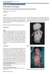
A 40-year old female presented to the <strong>Breast</strong> Clinic with a<br />
painless lump in the right breast for one month with no associated<br />
fever, nipple discharge or skin redness. The patient gave a history<br />
of a hard, slightly painful lump in the left breast for which<br />
she had undergone breast conservation surgery. Review of the<br />
records revealed that the patient had an early stage invasive ductal<br />
carcinoma. On local inspection, the right breast was normal in size<br />
with no nipple retraction and obvious swelling. On palpation; a<br />
5×4 cm soft mobile lump was felt in the upper outer quadrant. It<br />
was non-tender and the skin was freely mobile over it. A separate<br />
2×2 cm firm, non-tender, mobile lump was palpated in the upper<br />
outer quadrant, below the larger lump. There were no palpable<br />
lymph nodes in the axilla. The left breast was asymmetrically<br />
small with a large scar as a result of the previous operation. On<br />
palpation, no lump could be palpated and there was no axillary<br />
lymphadenopathy. The patient was referred to the mammography<br />
unit for bilateral mammograms. Selected mammogram (Figs. 1, 2]<br />
and ultrasound images (Fig. 3) are given as below.<br />
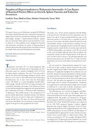
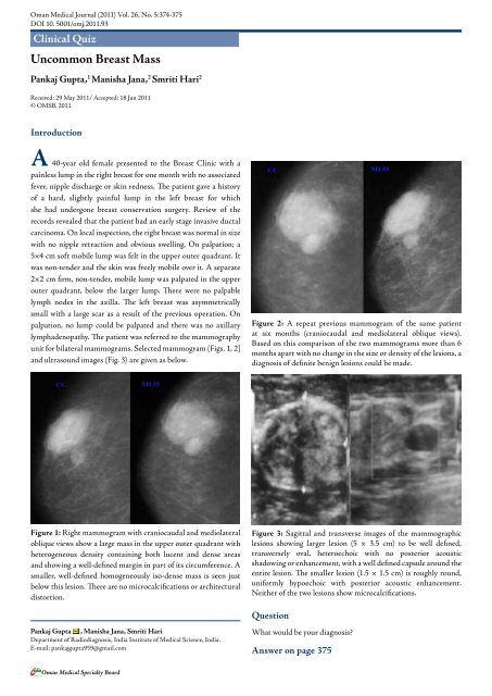
Figure 2: A repeat previous mammogram of the same patient<br />
at six months (craniocaudal and mediolateral oblique views).<br />
Based on this comparison of the two mammograms more than 6<br />
months apart with no change in the size or density of the lesions, a<br />
diagnosis of definite benign lesions could be made.<br />
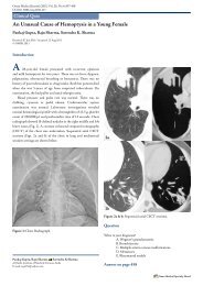
Figure 1: Right mammogram with craniocaudal and mediolateral<br />
oblique views show a large mass in the upper outer quadrant with<br />
heterogeneous density containing both lucent and dense areas<br />
and showing a well-defined margin in part of its circumference. A<br />
smaller, well-defined homogeneously iso-dense mass is seen just<br />
below this lesion. There are no microcalcifications or architectural<br />
distortion.<br />
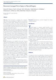
Figure 3: Sagittal and transverse images of the mammographic<br />
lesions showing larger lesion (5 × 3.5 cm) to be well defined,<br />
transversely oval, heteroechoic with no posterior acoustic<br />
shadowing or enhancement, with a well defined capsule around the<br />
entire lesion. The smaller lesion (1.5 × 1.5 cm) is roughly round,<br />
uniformly hypoechoic with posterior acoustic enhancement.<br />
Neither of the two lesions show microcalcifications.<br />
Question<br />
Pankaj Gupta , Manisha Jana, Smriti Hari<br />
Department of Radiodiagnosis, India Institute of Medical Science, India.<br />
E-mail: pankajgupta959@gmail.com<br />
What would be your diagnosis?<br />
Answer on page 375<br />
Oman Medical Specialty Board
Oman Medical Journal (2011) Vol. 26, No. 5:374-375<br />
Answer<br />
Fibroadenoma appears as a well-circumscribed mass<br />
diagnoses are fibroadenoma, lipoma, fat necrosis, and galactocele. 4 with coarse calcifications of varying morphology; however<br />
The mammographic and ultrasound appearance of the lesion<br />
suggests a diagnosis of breast hamartoma.<br />
mammographic appearances can be highly variable. Lipomas are<br />
usually not detected on mammography unless they are large, where<br />
they are seen as well-defined entirely lucent lesion. 5 Fat necrosis<br />
Discussion<br />
has a spectrum of mammographic appearances; however the<br />
pathognomonic appearance is that of a lipid cyst which is seen as<br />
Hamartoma is a rare benign tumor of the breast. It is known<br />
by many synonyms; lipofibroadenoma, fibroadenolipoma or<br />
adenolipoma, which is based on the predominant components<br />
within the mass. 1 The etiology of this disease entity is obscured.<br />
It most commonly presents in middle aged females as a painless<br />
lump. On examination, more than half of these lesions are soft<br />
and non-palpable. Gross pathological examination of a section<br />
gives the appearance of a "slice of salami" or "breast within breast." 1<br />
The tumor is composed of varying admixture of fibrous, glandular<br />
a well-defined, smooth bordered, round or oval lucent mass with a<br />
thin rim which may calcify. 6 Typical mammographic appearance of<br />
a galactocele is a solitary or multiple masses having density similar<br />
to or less than the fibro-glandular parenchyma. The presence of<br />
fat-fluid levels within a well-defined mass is pathognomonic. 7<br />
Malignancy associated with hamartoma is rare but should be kept<br />
in mind. Surgical resection of the mass is the definitive treatment.<br />
Follow up of the lesions is recommended as recurrences have been<br />
reported in several studies. 8<br />
and fatty components. The margin of the tumor is formed by<br />
compressed breast parenchyma known as the "pseudocapsule." Acknowledgements<br />
Histologically; the glandular component forms prominent lobules<br />
in matrix containing fat and fibrous stroma. This organization of<br />
glandular tissue differentiates hamartoma from fibro adenoma.<br />
The authors reported no conflict of interest and no funding was<br />
received for this work.<br />
The mammographic appearance is known as a "piece of cut<br />
sausage." 2 They are well circumscribed masses with both fat and References<br />
soft tissue densities, and are surrounded at least partly by a thin<br />
radio-opaque line which represents the pseudo capsule.<br />
1. Tse GM, Law BK, Ma TK, Chan AB, Pang L-M, Chu WC, et al. Hamartoma<br />
of the breast: a clinicopathological review. J Clin Pathol 2002 Dec;55(12):951-<br />
Based on this classic appearance, a mammographic diagnosis<br />
954.<br />
is possible, as was in our patient. The ultrasound appearance is 2. Paulus DD. Benign diseases of the breast. Radiol Clin North Am 1983<br />
highly variable and it rarely contributes primarily to a differential Mar;21(1):27-50.<br />
3. Gogas J, Markopoulos C, Gogas H, Skandalakis P, Kontzoglou K, Stavridou<br />
diagnosis. However, the typical sonographic appearance is that of a<br />
A. Hamartomas of the breast. Am Surg 1994 Jun;60(6):447-450.<br />
well circumscribed solid mass, which is predominantly hypoechoic 4. Pui MH, Movson IJ. Fatty tissue breast lesions. Clin Imaging 2003 Maybut<br />
has hyper-echogenicity in the form of lines or bands. They have<br />
a variable acoustic shadowing. MRI characteristics of hamartoma<br />
Jun;27(3):150-155.<br />
5. Lanng C, Eriksen BO, Hoffmann J. Lipoma of the breast: a diagnostic<br />
dilemma. <strong>Breast</strong> 2004 Oct;13(5):408-411.<br />
include; fat density within the mass with smooth well defined hypointense<br />
rim and heterogenous contrast enhancement. While fine J Roentgenol 1991 Aug;157(2):271-273.<br />
6. Evers K, Troupin RH. Lipid cyst: classic and atypical appearances. AJR Am<br />
needle aspiration cytology (FNAC) fails to diagnose most of these<br />
lesions, mainly due to the scantly material provided by FNAC,<br />
7. Gómez A, Mata JM, Donoso L, Rams A. Galactocele: three distinctive<br />
radiographic appearances. Radiology 1986 Jan;158(1):43-44.<br />
as well as the lack of any distinctive architectural or cytological<br />
characteristics of hamartoma. 1,3 Even a core biopsy may not prove<br />
8. Daya D, Trus T, D’Souza TJ, Minuk T, Yemen B. Hamartoma of the breast,<br />
an underrecognized breast lesion. A clinicopathologic and radiographic study<br />
of 25 cases. Am J Clin Pathol 1995 Jun;103(6):685-689.<br />
to be useful in the absence of clinical and strong radiological<br />
suspicion. The most important clinical and radiological differential<br />
Oman Medical Specialty Board