Introduction to Neutron and X-Ray Scattering - Spallation Neutron ...
Introduction to Neutron and X-Ray Scattering - Spallation Neutron ...
Introduction to Neutron and X-Ray Scattering - Spallation Neutron ...

Create successful ePaper yourself
Turn your PDF publications into a flip-book with our unique Google optimized e-Paper software.
X-<strong>Ray</strong> <strong>Scattering</strong> Studies of Thin<br />
Polymer Films<br />
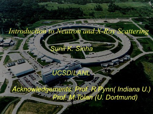
<strong>Introduction</strong> <strong>to</strong> <strong>Neutron</strong> <strong>and</strong> X-<strong>Ray</strong> <strong>Scattering</strong><br />
Sunil K. Sinha<br />
UCSD/LANL<br />
Acknowledgements: Prof. R.Pynn( Indiana U.)<br />
Prof. M.Tolan (U. Dortmund)
Wilhelm Conrad Röntgen 1845-1923<br />
1895: Discovery of<br />
X-<strong>Ray</strong>s
Nobel Prizes for Research<br />
with X-<strong>Ray</strong>s<br />
1901 W. C. Röntgen in Physics for the discovery of x-rays.<br />
1914 M. von Laue in Physics for x-ray diffraction from crystals.<br />
1915 W. H. Bragg <strong>and</strong> W. L. Bragg in Physics for crystal structure determination.<br />
1917 C. G. Barkla in Physics for characteristic radiation of elements.<br />
1924 K. M. G. Siegbahn in Physics for x-ray spectroscopy.<br />
1927 A. H. Comp<strong>to</strong>n in Physics for scattering of x-rays by electrons.<br />
1936 P. Debye in Chemistry for diffraction of x-rays <strong>and</strong> electrons in gases.<br />
1962 M. Perutz <strong>and</strong> J. Kendrew in Chemistry for the structure of hemoglobin.<br />
1962 J. Watson, M. Wilkins, <strong>and</strong> F. Crick in Medicine for the structure of DNA.<br />
1979 A. McLeod Cormack <strong>and</strong> G. Newbold Hounsfield in Medicine for computed axial<br />
<strong>to</strong>mography.<br />
1981 K. M. Siegbahn in Physics for high resolution electron spectroscopy.<br />
1985 H. Hauptman <strong>and</strong> J. Karle in Chemistry for direct methods <strong>to</strong> determine<br />
x-ray structures.<br />
1988 J. Deisenhofer, R. Huber, <strong>and</strong> H. Michel in Chemistry for the structures<br />
of proteins that are crucial <strong>to</strong> pho<strong>to</strong>synthesis.<br />
2006 R. Kornberg in Chemistry for studies of the molecular basis of eukaryotic
NSE FRET NMR<br />
0.5-50 nm length scale<br />
ps - µs time scale<br />
orientational average<br />
q~2π/length<br />
Functional domain dynamics in proteins<br />
fixed defined position<br />
> µs timescale ps - ms timescale<br />
small proteins<br />
dye<br />
dye<br />
phosphoglycerate kinase
<strong>Neutron</strong> Advantages<br />
• Penetrating, but does no damage <strong>to</strong> sample<br />
• H/D contrast matching can be used <strong>to</strong> study<br />
macromolecules in solution, polymers, etc.<br />
• Strongly interacts with magnetic moments<br />
• Energies match those of phonons, magnons,ro<strong>to</strong>ns,<br />
etc.
Nobel Prize in Physics, 1994<br />
Awarded for “pioneering contributions <strong>to</strong> the development<br />
of neutron scattering techniques for studies of condensed matter”<br />
Bertram N. Brockhouse Clifford G. Shull<br />
Development of<br />
neutron spectroscopy<br />
Development of the<br />
neutron diffraction technique
First Study of an Antiferromagnetic Structure<br />
Antiferromagnetic Structure of MnO<br />
(Shull <strong>and</strong> Wollan Phys. Rev. 83, 333 (1951)
Magnetic Structure of the Rare Earth Metals<br />
(W.C. Koehler (1965))
Science with X-<strong>Ray</strong>s<br />
• Diffraction <strong>and</strong> crystal structures<br />
• Structure Fac<strong>to</strong>rs of liquids <strong>and</strong> glasses<br />
• Structures of Thin Films<br />
• ARPES<br />
• EXAFS, XANES<br />
• Studies of Magnetism with resonant XMS<br />
• Inelastic X-ray scattering: phonons, electronic excitations<br />
• X-ray Pho<strong>to</strong>n Correlation Spectroscopy<br />
• Microscopy<br />
• Imaging/Tomography
Why<br />
Synchrotron-<br />
radiation ?<br />
Intensity !!!
Example 1: X-<strong>Ray</strong> Diffraction & structural biology<br />
• D.C. Phillips presents the<br />
3-D structure of lysozyme<br />
<strong>to</strong> the Royal Society in 1965<br />
• Linear polypeptide chain<br />
• Folded model of the same<br />
amino acid sequence<br />
• July 2009:<br />
58,588 structures in<br />
Protein Data Bank<br />
A single protein structure used <strong>to</strong> be the project of a scientific lifetime<br />
Synchrotron Radiation - 8301 structures solved in 2009
Advantages of <strong>Neutron</strong>s <strong>and</strong> X-<strong>Ray</strong>s<br />
• Penetrating/ Non Destructive N (X)<br />
• Right wavelength/energy N,X<br />
• Magnetic probe N,X<br />
• Contrast matching N<br />
• Weakly interacting-Born approxn. N,X<br />
• Global Statistical information N,X<br />
• Buried Interfaces—depth dependence N,X
<strong>Neutron</strong> <strong>and</strong> X-ray <strong>Scattering</strong>:<br />
“small” science at big<br />
facilities!
His<strong>to</strong>ric accomplishments (<strong>Neutron</strong>s)<br />
•Antiferromagnetic Structures<br />
•Rare earth spirals <strong>and</strong> other spin structures<br />
•Spin wave dispersion<br />
•Our whole underst<strong>and</strong>ing of the details of exchange<br />
interactions in solids<br />
•Magnetism <strong>and</strong> Superconductivity<br />
•Phonon dispersion curves in crystals; quantum crystals <strong>and</strong><br />
anharmonicity<br />
•Crystal fields<br />
•Excitations in normal liquids<br />
•Ro<strong>to</strong>ns in superfluid helium<br />
•Condensate fraction in helium
Recent Applications<br />
• Quantum Phase Transitions <strong>and</strong> Critical points<br />
• Magnetic order <strong>and</strong> magnetic fluctuations in the high-Tc cuprates<br />
• Gaps <strong>and</strong> low-lying excitations (including phonons) in High-Tc<br />
• Magnetic Order <strong>and</strong> spin fluctuations in highly-correlated systems<br />
• Manganites<br />
• Magnetic nanodot/antidot arrays<br />
• Exchange bias
Applications in Soft Matter <strong>and</strong><br />
Materials<br />
• Scaling Theory of polymers<br />
• Reptation in Polymers<br />
• Alpha <strong>and</strong> beta relaxation in glasses<br />
• Structures of surfactants <strong>and</strong> membranes<br />
• Structure of Ribozome<br />
• Excitations <strong>and</strong> Phase transitions in confined Systems (phase<br />
separation in Vycor glass; Ripplons in superfluid He films, etc.)<br />
• Momentum Distributions<br />
• Materials—precipitates, steels, cement, etc.
Recent Applications (contd.)<br />
• Pro<strong>to</strong>n motion in carbon nanotubes<br />
• Protein dynamics<br />
• Glass transition in polymer films<br />
• Pro<strong>to</strong>nation states in biological macromolecules from<br />
nuclear density maps<br />
• Studies of protein diffusive motion in hydrated enzymes<br />
• Boson peaks in glasses<br />
• Phase diagrams of surfactants<br />
• Lipid membranes
Applications of Surface/Interface<br />
<strong>Scattering</strong><br />
• study the morphology of surface <strong>and</strong> interface roughness<br />
• wetting films<br />
• film growth exponents<br />
• capillary waves on liquid surfaces (polymers, microemulsions, liquid<br />
metals, etc.)<br />
• isl<strong>and</strong>s on block copolymer films<br />
• pitting corrosion<br />
• magnetic roughness<br />
• study the morphology of magnetic domains in magnetic films.<br />
• Nanodot arrays<br />
• Tribology, Adhesion, Electrodeposition
X-rays <strong>and</strong> neutrons are<br />
complementary <strong>to</strong> SPM’s<br />
• Yield GLOBAL statistical properties about<br />
assemblies of particles<br />
• Can be used <strong>to</strong> study BURIED interfaces or<br />
particles<br />
• Impervious <strong>to</strong> sample environmental<br />
conditions, magnetic fields, etc.<br />
• Can also be used <strong>to</strong> study single<br />
nanoparticles ( synchrotron nanoprobe)
S.R. <strong>and</strong> neutron based research<br />
can help us <strong>to</strong> underst<strong>and</strong>:<br />
• How the constituent molecules selfassemble<br />
<strong>to</strong> form nanoparticles.<br />
• How these self-organize in<strong>to</strong> assemblies<br />
• How structure <strong>and</strong> dynamics lead <strong>to</strong><br />
function<br />
• How emergent or collective properties arise
Brightness & Fluxes for <strong>Neutron</strong> &<br />
Brightness<br />
X-<strong>Ray</strong> Sources<br />
dE/E<br />
<strong>Neutron</strong>s 2<br />
Rotating<br />
Anode<br />
Bending<br />
Magnet<br />
Undula<strong>to</strong>r<br />
(APS)<br />
10<br />
15<br />
20<br />
10<br />
0.02<br />
0.1<br />
10<br />
Divergence<br />
10�10<br />
Flux<br />
�1<br />
�2<br />
�1<br />
2<br />
�1 �2<br />
( s m ster ) (%) ( mrad ) ( s m )<br />
27<br />
10<br />
33<br />
10<br />
0. 5�10<br />
0. 1�5<br />
0. 01�<br />
0.<br />
1<br />
11<br />
10<br />
14<br />
5�10<br />
20<br />
5�10<br />
24<br />
10
Synchrotron-<br />
<strong>and</strong> <strong>Neutron</strong><br />
<strong>Scattering</strong><br />
Places
The pho<strong>to</strong>n also has wave <strong>and</strong><br />
particle properties<br />
E=h� =hc/� = hck<br />
Charge = 0 Magnetic Moment = 0<br />
Spin = 1<br />
E (keV) � (Å)<br />
0.8 15.0<br />
8.0 1.5<br />
40.0 0.3<br />
100.0 0.125
Intrinsic Cross Section: <strong>Neutron</strong>s
Intrinsic Cross Section:<br />
E i,rad (R,t) �<br />
X-<strong>Ray</strong>s<br />
e<br />
4��0c 2 R ai (t � R / c)<br />
a r (t � R / c) � � e<br />
m �(�)Eur i� R/c<br />
0e<br />
Ei,rad(R,t) � �r0�(�) eikR<br />
R cos�<br />
E in<br />
Thomson <strong>Scattering</strong> Length<br />
of the Electron<br />
(classical electron radius):<br />
r 0 �<br />
�<br />
E<br />
in<br />
�<br />
�<br />
E<br />
0<br />
� �<br />
i(<br />
k�r<br />
��t<br />
)<br />
e 2<br />
4�� 0 mc 2 � 2.82 � 10�15 m<br />
e<br />
E �<br />
rad
2<br />
2<br />
0<br />
2<br />
0<br />
2<br />
2<br />
2<br />
2<br />
2<br />
0<br />
2<br />
in<br />
rad<br />
)<br />
(<br />
)<br />
cos<br />
1<br />
(<br />
2<br />
1<br />
d<br />
d<br />
)<br />
(<br />
)<br />
(<br />
)<br />
(<br />
)<br />
,<br />
(<br />
�<br />
�<br />
�<br />
�<br />
�<br />
�<br />
�<br />
r<br />
R<br />
f<br />
P<br />
R<br />
r<br />
E<br />
t<br />
R<br />
E<br />
�<br />
�<br />
�<br />
�<br />
�<br />
�<br />
�<br />
�<br />
�<br />
�<br />
�<br />
�<br />
Intrinsic Cross<br />
Section: X-<strong>Ray</strong>s<br />
0<br />
d<br />
d ��<br />
�<br />
�<br />
�<br />
�<br />
�<br />
�<br />
r<br />
�<br />
�<br />
1 2 3 4<br />
0<br />
��<br />
�<br />
�<br />
�<br />
�<br />
�<br />
i<br />
)<br />
( 2<br />
2<br />
r<br />
2<br />
�<br />
�<br />
�<br />
Resonance<br />
<strong>Scattering</strong><br />
r<br />
�<br />
� �<br />
Thomson <strong>Scattering</strong><br />
P<br />
r 2<br />
0<br />
0<br />
r<br />
d<br />
d<br />
�<br />
�<br />
�<br />
�<br />
�<br />
�<br />
�<br />
�<br />
�<br />
��<br />
�<br />
�<br />
�<br />
4<br />
�<br />
�<br />
<strong>Ray</strong>leigh<br />
<strong>Scattering</strong>
Adding up phases at the detec<strong>to</strong>r of the<br />
wavelets scattered from all the scattering<br />
centers in the sample:
Wave vec<strong>to</strong>r transfer is defined as<br />
q = k f - k i
X-rays<br />
d� = r 0 2 �1 + Cos 2 (2�)� S(q)<br />
d� 2<br />
S(q) = �� ij exp[-iq.(r i-r j)]�<br />
{r i} == electron positions.
Now, � i exp[-iq.R i] = � N(q) Fourier Transform of nuclear density<br />
[ sometimes also referred <strong>to</strong> as F(q) ]<br />
Proof:<br />
� N(r) = � i �( r - R i)<br />
� N(q) = ∫ � N(r) exp[-iq.r] dr = ∫ � i �( r - R i) exp[-iq.r] dr<br />
Similarly,<br />
= � i exp[-iq.R i]<br />
� i exp[-iq.r i] = � el(q) Fourier Transform of electron density<br />
So, for neutrons, S(q) = � � N(q) � N * (q) �<br />
And, for x-rays, S(q) = � � el(q) � el * (q) �
H has large incoherent � ( 10.2 x 10 -24 cm 2 )<br />
but small coherent � ( 1.8 x 10 -24 cm 2 )<br />
D has larger coherent � ( 5.6 x 10 -24 cm 2 )<br />
<strong>and</strong> small incoherent � ( 2.0 x 10 -24 cm 2 )<br />
C, O have completely coherent �’s<br />
V is almost completely incoherent (� coh ~ 0.02 x10 -24<br />
cm 2 ; � incoh ~ 5.0 x10 -24 cm 2)
<strong>Neutron</strong>s<br />
I(q) = d�/d� = ∑ K,K� b K b K� S K K�(q)<br />
X-<strong>Ray</strong>s<br />
I(q) = ∑ K,K� (r 0) 2 Z K Z K� f K(q) f* K� (q) [ (1 + cos 2 (�))/2] S K K�(q)<br />
(K, K� = Different A<strong>to</strong>mic Species)<br />
S K K�(q) = �∑ l(K),m(K�)exp{-i q.[R l(K) - R m(K�)]}� ---> Partial<br />
Structure Fac<strong>to</strong>r<br />
These can be unscrambled by simultaneous measurement of<br />
d�/d� for neutrons with different iso<strong>to</strong>pes <strong>and</strong>/or X-rays.
Diffraction from Crystals
q y<br />
q x<br />
q z
(S = 6) D s = Surface fractal dimension.<br />
If D s =2, S(q) ~ 1/q 4 (Porod’s Law for<br />
smooth internal surfaces)<br />
If 2 < D s < 3, S(q) ~ 1/q n where 3< n
<strong>Scattering</strong> Geometry & Notation<br />
k i<br />
q<br />
q x<br />
q z<br />
k f<br />
Wave-Vec<strong>to</strong>r: q = k f – k i<br />
Reflectivity:<br />
q x= q y = 0<br />
q z= (4���)sin� i
Reflection of Visible Light
Perfect & Imperfect „Mirrors“
Basic Equation: X-<strong>Ray</strong>s<br />
Helmholtz-Equation & Boundary Conditions<br />
� 2 E( r ) + k 2 n 2 ( r ) E ( r ) = 0
Refractive Index: X-<strong>Ray</strong>s & <strong>Neutron</strong>s<br />
Minus!!<br />
+<br />
Dispersion<br />
magnetic<br />
part<br />
+<br />
magnetic<br />
part<br />
Absorption
Derivation of n for neutrons:<br />
Consider Schrodinger Eqn.<br />
-(ћ 2 /2m)� 2 � + (V -E)� = 0 E = (ћ 2 /2m)k 0 2<br />
can be written:<br />
� 2 � +[1 - (2m/ћ 2 k 0 2 )V] k0 2 � = 0<br />
V= (2�ћ 2 /m)b N; k 0 = 2�/�<br />
so:<br />
n 2 = (1 - (2m/ћ 2 k 0 2 )V) = 1 - (� 2 b/�) N<br />
2nd term
Refractive Index: X-<strong>Ray</strong>s<br />
E = 8 keV � = 1.54 Å<br />
Electron Density<br />
Profile !
Single Interface: Vacuum/Matter<br />
Fresnel-<br />
Formulae<br />
Reflected<br />
Amplitude<br />
Transmitted<br />
Amplitude<br />
Wave-<br />
Vec<strong>to</strong>rs
Total External Reflection<br />
cos � i � (1– �) cos � t � t=0<br />
GRAZING ANGLES !!!<br />
Critical Angle:<br />
� c � �2� ~ 0.3°
Fresnel Reflectivity: R F(� i)<br />
Total External<br />
Reflection<br />
Regime
Reformulation for Interfaces<br />
Fresnel-Reflectivity<br />
of the Substrate<br />
The „Master Formula“<br />
�<br />
Electron Density Profile
R(qz) = R 2<br />
Fexp(-qz �2 )<br />
Roughness Damps Reflectivity<br />
� j � 10 Å<br />
� � 1.54 Å
X-<strong>Ray</strong> Reflectivity:<br />
Water Surface<br />
Difference<br />
Experiment-<br />
Theory:<br />
Roughness !!<br />
Braslau et al.<br />
PRL 54, 114 (1985)<br />
Measurement<br />
Fresnel Reflectivity
Calculation of Reflectivity<br />
Slicing of Density Profile<br />
� ~ 1Å<br />
Slicing<br />
&<br />
Parratt-Iteration<br />
Reflectivity<br />
from<br />
Arbitrary<br />
Profiles !<br />
• Drawback:<br />
Numerical Effort !
Example: PS Film on Si/SiO 2<br />
X-<strong>Ray</strong> Reflectivity (NSLS)<br />
� � 1.19Å d � 109Å<br />
Data & Fit<br />
Density Profile
Grazing-Incidence-Diffraction
<strong>Scattering</strong> Geometry & Notation<br />
k i<br />
Q<br />
Q x<br />
Q z<br />
k f<br />
Wave-Vec<strong>to</strong>r: Q = k f – k i<br />
Reflectivity:<br />
Q x= Q y = 0<br />
Q z= (4���)sin� i
What do Specular <strong>and</strong> Off-<br />
specular scattering measure?<br />
• Specular reflectivity measures variations<br />
in scattering density normal <strong>to</strong> surface<br />
(averaged over x,y plane)<br />
• Off-specular scattering measures (x,y)<br />
variations of scattering density, e.g. due <strong>to</strong><br />
roughness, magnetic domains, etc.
Almost all real surfaces are<br />
rough!
Self-Affine Fractal Surfaces<br />
Let �z(r) be height fluctuation about average<br />
surface at point r in 2D plane.<br />
R.m.s. roughness � is defined by<br />
� 2 = � [�z(r)] 2 �<br />
Consider quantity<br />
G(R) = � [�z(r) - �z(r+R)] 2 �.<br />
For self-affine surfaces,<br />
G(R) = AR 2h 0
AFM/FIB Studies-Electrodeposition<br />
M.C. Lafouresse et al., PRL 98, 236101 (2007)<br />
Cu Films
<strong>Scattering</strong> from a Self-Affine<br />
S(q r 2 2<br />
2 2 �qz �<br />
) � (Ar0 / qz )e<br />
Fractal Surface<br />
��<br />
dXdYe<br />
SKS et al., Phys. Rev. B 38, 2297 (1988)<br />
2<br />
qz C(R)<br />
e �i(q x X �qyY )
Jun Wang et<br />
al.,<br />
Europhys.<br />
Lett, 42<br />
283-288<br />
(1998)<br />
Mo layers
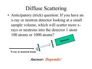
Example of Diffuse <strong>Scattering</strong> of X-<strong>Ray</strong>s<br />
from a single rough surface
Multilayers
Vec<strong>to</strong>r Diagram for Q in GISAXS<br />
Q y = (2�/�)Cos � fSin �<br />
Q x = (2�/�)(Cos � f -Cos � 1Cos �)
Measurement of GISAXS
Synchrotron<br />
Setup<br />
X-<strong>Ray</strong> Reflec<strong>to</strong>meters<br />
Labora<strong>to</strong>ry<br />
Setup<br />
HASYLAB: CEMO
Reflectivity from Liquids I<br />
Synchrotron<br />
Setup (APS)
Magnetic <strong>Neutron</strong> <strong>Scattering</strong>
spin polarized<br />
3d b<strong>and</strong>s<br />
2p 3/2<br />
2p 1/2<br />
Core level resonances<br />
e 0<br />
f = f 1 + if 2<br />
E F<br />
e f<br />
Fe<br />
Kortright et al., Phys. Rev. B 65, 12216 (2000)
NEUTRONS:<br />
R + +( Q z) - R - - (Q z) ~ M xy,�� (Q z) n (Q z)<br />
R + - (Q z) = R - + (Q z) ~ � M xy,� (Q z) � 2<br />
X-RAYS:<br />
R + ( Q z) - R - (Q z) ~ M �� (Q z) n (Q z)
� l = � 2 � D�<br />
= �(D� ��) -1<br />
� t = � R � s<br />
(� hor., � vert.)<br />
Coherence Lengths
g 2<br />
Pho<strong>to</strong>n Correlation Spectroscopy<br />
Brownian Motion of 100 particles<br />
QuickTime <strong>and</strong> a<br />
Video decompressor<br />
are needed <strong>to</strong> see this picture.<br />
Intensity-intensity au<strong>to</strong> correlation<br />
1.15<br />
1.1<br />
1.05<br />
1<br />
10 0<br />
10 1<br />
delay<br />
g ( q,<br />
� ) �<br />
2<br />
I(<br />
q,<br />
t)<br />
I(<br />
q,<br />
t ��<br />
)<br />
I(<br />
q,<br />
t)<br />
10 2<br />
2<br />
10 3<br />
Diffraction Pattern<br />
Speckles<br />
QuickTime <strong>and</strong> a<br />
Video decompressor<br />
are needed <strong>to</strong> see this picture.<br />
QuickTime <strong>and</strong> a<br />
Video decompressor<br />
are needed <strong>to</strong> see this picture.
coherent<br />
beam<br />
Pho<strong>to</strong>n Correlation Spectroscopy<br />
sample detec<strong>to</strong>r<br />
X-ray speckle pattern from a static silica aerogel
“Oversampling”:<br />
Non-crystals:<br />
pattern continuous,<br />
can do finer sampling<br />
of intensity<br />
Finer sampling;<br />
larger array;<br />
smaller transform;<br />
“finite support”<br />
(area around specimen<br />
must be clear!)<br />
6/20/2011 Miao thesis 124
Reconstruction<br />
Equations can still not be solved analytically<br />
•Positivity of<br />
electron<br />
density helps!<br />
Fienup iterative algorithm<br />
Reciprocal space Real space<br />
Impose<br />
diffraction<br />
magnitudes<br />
Impose<br />
finite<br />
support<br />
6/20/2011 Miao thesis 125
DIFFRACTION IMAGING BY J. MIAO ET AL<br />
• From Miao, Ishikawa, Johnson,<br />
Anderson, Lai, Hodgson PRL<br />
Aug 2002<br />
• SEM image of a 3-D Ni<br />
microfabricated object with two<br />
levels 1 µm apart<br />
• Diffraction pattern taken at<br />
2 Å wavelength at SPring 8<br />
• 2-D reconstruction with<br />
Fienup-type algorithm<br />
• Both levels show because<br />
the depth of focus is<br />
sufficient<br />
• Only <strong>to</strong>p level shows <strong>to</strong> useful<br />
• Resolution = 8 nm (new<br />
record)<br />
extent<br />
6/20/2011 from Howells 126
MIAO ET AL 3-D RECONSTRUCTIONS<br />
• Miao et al 3-D<br />
reconstruction of the<br />
same object pair<br />
• a <strong>and</strong> b are sections<br />
through the image<br />
• c is 3-D density<br />
• Resolution = 55 nm
Imaging of individual nanoparticles at the APS<br />
Ross Harder, University of Illinois, Champaign<br />
Coherent diffraction pattern<br />
from 170 nm Ag particle<br />
170 nm silver cubes<br />
I.K. Robinson, et al., Science 298 2177 (2003)<br />
inversion of<br />
diffraction pattern<br />
‘lensless imaging’