Sircol COLLAGEN Assay Manual - Nordic Biosite
Sircol COLLAGEN Assay Manual - Nordic Biosite
Sircol COLLAGEN Assay Manual - Nordic Biosite

You also want an ePaper? Increase the reach of your titles
YUMPU automatically turns print PDFs into web optimized ePapers that Google loves.
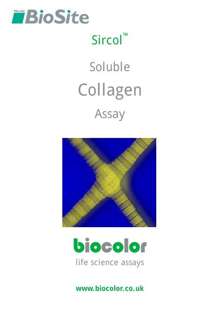
<strong>Sircol</strong> <br />
Soluble<br />
Collagen<br />
<strong>Assay</strong><br />
biocolor<br />
life science assays<br />
www.biocolor.co.uk
<strong>Assay</strong> Reference Images<br />
+30min<br />
Mix <strong>Sircol</strong> reagent and<br />
collagen reference<br />
standard<br />
+40min<br />
Centrifuge and remove<br />
supernatant carefully<br />
+50min<br />
Add alkali<br />
reagent<br />
(picture obtained<br />
using 0-50μg<br />
collagen reference<br />
standard)<br />
Biocolor Ltd.<br />
Unit 35<br />
8 Meadowbank Road<br />
BT38 8YF<br />
UK<br />
tel: +44 (0) 2893 350 258<br />
fax: +44 (0) 2893 369 716<br />
email: info@biocolor.co.uk
<strong>Sircol</strong> Soluble Collagen <strong>Assay</strong><br />
Time Req: 1 hour<br />
Detection Limit: 2.5μg*<br />
Set up assay:<br />
Label a set of 1.5 ml microcentrifuge tubes.<br />
If sufficient material is available, run duplicate<br />
samples.<br />
Prepare;<br />
[1] Reagent blanks: (100 µl of distilled water or the<br />
test sample buffer).<br />
[2] Collagen standards: (aliquots containing 5, 10,<br />
25, 50 µg).<br />
[3] Test samples, (volumes: 10 to 100 µl *).<br />
Adjust the contents of all tubes to 100 µl with<br />
distilled water or appropriate buffer.<br />
To each tube add 1 ml <strong>Sircol</strong> Dye reagent and cap<br />
all of the tubes; mix contents by inverting.<br />
Mixing:<br />
Place tubes in a mechanical shaker for 30 minutes,<br />
(or manually shake at 5 minute intervals).<br />
During this time period the <strong>Sircol</strong> Dye will bind to<br />
soluble collagens.<br />
The dye reagent is designed so that the collagendye<br />
complex will precipitate out of solution.<br />
10000<br />
Centrifuging:<br />
Transfer the tubes to a micro centrifuge and spin<br />
the tubes at >10,000 x g for a 10 minute period *.<br />
It is important to firmly pack the insoluble pellet of<br />
the collagen-dye complex at the bottom of the<br />
tubes, so as to avoid any loss during draining.
Draining:<br />
The unbound dye solution is removed by carefully<br />
inverting and draining the tubes.<br />
Any remaining droplets can be removed from the<br />
tubes by gently tapping the inverted tube on a<br />
paper tissue or a cotton wool bud can be used for<br />
removing droplets of dye from the rim of the<br />
tubes.<br />
Do not attempt to physically remove any fluid that<br />
is in close contact to the deposit.<br />
Release of bound dye:<br />
To each tube add 1 ml of the Alkali reagent.<br />
Re-cap the tubes and release the bound dye into<br />
solution. A vortex mixer is suitable.<br />
When the bound dye has been dissolved, usually<br />
within 10 minutes, the samples are ready for<br />
measurement.<br />
The colour is light stable, but should be read<br />
within 2 to 3 hours.<br />
Keep the tubes capped until ready for<br />
measurement.<br />
Measurement:<br />
(a) Spectrophotometer, set wavelength to 540<br />
nm.<br />
Use semi-micro glass, or plastic disposable<br />
cuvettes.<br />
(b) Colorimeter, set using a blue-green filter.<br />
Use semi-micro cuvettes or tubes.<br />
(c) Multiwell plate reader, set using a blue-green<br />
filter. Transfer 200 µl aliquots of samples<br />
from tubes to the wells of a 96 well,<br />
multiwell plate.<br />
Set the above instruments to zero using water.<br />
Measure absorbance of reagent blanks, collagen<br />
standards and the test samples.<br />
Subtract the reagent blank reading from the<br />
standard and test sample readings. Check<br />
duplicates are within ±10%.<br />
Plot standards on graph and use the graph to<br />
calculate the collagen content of the test samples.<br />
(see manual)
<strong>Sircol</strong><br />
Soluble Collagen<br />
<strong>Assay</strong><br />
TECHNICAL INFORMATION<br />
Test material suitable for analysis 1<br />
<strong>Assay</strong> components and storage conditions 1<br />
Mode of action of the <strong>Sircol</strong> <strong>Assay</strong> 3<br />
<strong>Assay</strong> Protocol 4<br />
Test sample preparation 8<br />
[1] salt‐soluble collagen 9<br />
[2] acid‐soluble collagen 10<br />
[3] pepsin‐soluble collagen 10<br />
[4] insoluble collagen, as gelatin 11<br />
Collagenase and gelatinase assays 12<br />
Other methods for measuring collagen 14<br />
<strong>Sircol</strong> <strong>Assay</strong> Kits and Component Units 17
The <strong>Sircol</strong> <strong>Assay</strong> has been designed<br />
for in vitro research work only<br />
Handle the<br />
<strong>Sircol</strong> <strong>Assay</strong> Kit<br />
using<br />
GOOD LABORATORY PRACTICE<br />
Read <strong>Manual</strong> Before Use<br />
5th Edition 2007<br />
<strong>Sircol</strong> <strong>Manual</strong><br />
Not to be reproduced in part or in whole, without written permission,<br />
unless required for personal, non‐commercial use.<br />
© Biocolor Ltd., 2007<br />
<strong>Sircol</strong> is a Trademark of Biocolor Ltd<br />
Published by<br />
Biocolor Ltd.<br />
8 Meadowbank Road, Carrickfergus, BT38 8YF<br />
Northern Ireland.<br />
www.biocolor.co.uk
Intended Applications:<br />
<strong>Sircol</strong> <strong>Assay</strong><br />
<strong>Manual</strong><br />
The <strong>Sircol</strong> Collagen <strong>Assay</strong> is a quantitative dye‐binding method designed for the analysis of<br />
acid‐soluble collagens extracted from mammalian tissues and collagens released into culture<br />
medium by mammalian cells during in vitro culture.<br />
Collagen forms that can be measured:<br />
[i] salt‐soluble collagens (1 M NaCl in 0.05M Tris, pH 7.5.)<br />
[ii] acid‐soluble collagens (0.01 to 1.0 M acetic acid)<br />
[iii] pepsin‐soluble collagens (0.5 M acetic acid + pepsin)<br />
[iv] total soluble collagens (composed of [i],[ii] & [iii])<br />
Collagen Types that can be measured:<br />
[1] Mammalian collagens, Types I to V, can be measured<br />
[2] Collagen Types VI to XIV can be assayed, but have not been calibrated due to<br />
insufficient purified material available for the preparation of standard curves<br />
[3] <strong>Sircol</strong> dye binding does not discriminate between collagen types<br />
[4] The dye reagent binds specifically to the [Gly‐X‐Y]n helical structure found in<br />
all collagens<br />
[5] <strong>Sircol</strong> dye binding decreases, gradually with the thermal denaturation of<br />
collagen (collagen to gelatin)<br />
[6] Non‐vertebrate collagens bind less dye, due in part to lower denaturation<br />
temperature and less hydroxyproline residues, and would require species<br />
matched purified preparations for standard calibration curves<br />
<strong>Sircol</strong> <strong>Assay</strong> Kit components:<br />
[1] The dye reagent contains Sirius Red in picric acid and has been formulated for<br />
specific binding to collagen under the conditions defined in the <strong>Sircol</strong> <strong>Assay</strong><br />
<strong>Manual</strong>.<br />
[2] Alkali reagent contains 0.5 M sodium hydroxide, for releasing <strong>Sircol</strong> Dye from<br />
the collagen‐dye complex.<br />
[3] Salt soluble collagen precipitating reagent; contains L-lysine<br />
monohydrochloride.<br />
1
[4] Collagen standard, acid soluble Type I, supplied as a sterile solution in 0.5 M<br />
acetic acid within a sealed vial. Concentration: 1 mg/ml.<br />
This is sterile bovine skin collagen that has been imported from the USA, and<br />
was obtained from disease free animals.<br />
In countries that forbid the importation of bovine derived material, a rat tail<br />
collagen standard, acid soluble Type I, is supplied as a sterile solution in 0.5 M<br />
acetic acid within a sealed vial. Concentration: 0.5 mg/ml.<br />
[5] This <strong>Sircol</strong> <strong>Assay</strong> <strong>Manual</strong>; (also available as a pdf, see www.biocolor.co.uk)<br />
Other components required, but not supplied:<br />
[a]<br />
[b]<br />
[c]<br />
[d]<br />
[e]<br />
Capped 1.5ml capacity microcentrifuge tubes<br />
Variable volume micropipettors and pipette tips<br />
A mechanical mixer for the microcentrifuge tubes. Any equipment that<br />
provides consistent shaking, rolling or rotation of the tubes is suitable<br />
A centrifuge with a 1.5ml tube rotor head and capable of at least 10,000 x g, to<br />
firmly pack the collagen‐dye pellet.<br />
A spectrophotometer, a colorimeter or a microwell plate reader with a bluegreen<br />
filter.<br />
Recommended storage conditions for components:<br />
Unopened;<br />
All components have been prepared for long term stability (at least 12 months), when stored<br />
at room temperature.<br />
Do not freeze as complete re‐solubilisation may not occur.<br />
Opened;<br />
The assay components will retain their shelf‐life, providing the glass vials of collagen<br />
standard and lysine solution are:<br />
(i) stored at +4ºC when not in use<br />
(ii) the metal seal is not removed<br />
The contents of both vials are best sampled as follows:<br />
Remove the centre metal disc only from the vial tops. Obtain aliquots from the vials, when<br />
required, using a plastic syringe fitted with a sterile hypodermic needle. The butyl rubber<br />
seal on the vial has a thin centre disc, suitable for needle insertion into the vial. Discard vial<br />
if solution contents become turbid.<br />
2
Mode of action of the <strong>Sircol</strong> dye reagent with soluble collagens:<br />
The <strong>Sircol</strong> dye reagent contains Sirius Red. The Colour Commission name is Direct Red 80.<br />
The molecular structure of the dye is shown below (Fig. 1).<br />
Mechanism by which the dye reacts with collagen:<br />
Sirius Red is an anionic dye with sulphonic acid side chain groups. These groups react with<br />
the side chain groups of the basic amino acids present in collagen.<br />
The specific affinity of the dye for collagen, under the assay conditions is due to the<br />
elongated dye molecules becoming aligned parallel to the long, rigid structure of native<br />
collagens that have intact triple helix organisation.<br />
Dye affinity is much reduced when collagen is heat denatured (>45ºC to the form of random<br />
chains of gelatin) and the triple helix unwinds.<br />
Dye binding is gradually lost when collagen (and gelatin) are exposed to bacterial<br />
collagenases.<br />
O<br />
O Na<br />
S<br />
O<br />
N<br />
N<br />
Na O<br />
S<br />
O<br />
O<br />
N<br />
N<br />
O<br />
O<br />
S<br />
O<br />
OH<br />
Na<br />
HN<br />
HN<br />
O<br />
HO<br />
O<br />
O S<br />
Na O<br />
N<br />
N<br />
O<br />
S O<br />
O Na<br />
N N<br />
S<br />
O<br />
O<br />
O<br />
Na<br />
Fig. 1<br />
The molecular structure of the <strong>Sircol</strong> Dye<br />
3
Test sample compositional requirements:<br />
As the <strong>Sircol</strong> <strong>Assay</strong> is a colorimetric procedure, samples for analysis should be free of any<br />
particulate material (cell debris and insoluble extracellular matrix).<br />
[1] The test sample can be in a salt buffer solution, acetic acid or culture medium.<br />
[2] If a surfactant has been used during tissue extraction, it is recommended that this<br />
extraction solution is <strong>Sircol</strong> tested with the Reference Collagen Standard to check that<br />
it has no adverse effects on collagen‐dye binding.<br />
[3] The presence of soluble proteins in samples, including proteoglycans, tropoelastin<br />
and other soluble ECM materials, does not interfere with the <strong>Sircol</strong> <strong>Assay</strong>.<br />
[4] Cell culture medium with foetal calf serum supplements of up to 5% does not<br />
interfere with the collagen assay. When higher serum supplements have been used,<br />
the increasing bulk of serum proteins, relative to the amount of collagen present, can<br />
cause problems.<br />
In this latter case the following options could be examined:<br />
[a] reduce the serum supplement to 5%, either after cell attachment has occurred,<br />
or reduce serum to 5% for the culture medium that will be collected and used<br />
for assay;<br />
[b] selectively remove the bulk of the serum albumin from the test sample by<br />
affinity chromatography (Blue‐Sepharose CL‐6B);<br />
[c] precipitate the collagen out of solution, adding NaCl to a concentration of 4 M<br />
for samples with neutral pH; or 2 M NaCl for samples in acetic acid.<br />
Centrifuge, drain well and re‐solubilize the collagen pellet in 0.5 M acetic acid.<br />
<strong>Assay</strong> of test solutions containing < 25 µg/ml collagen, without prior concentration:<br />
Method<br />
The <strong>Sircol</strong> Dye Reagent contains sufficient dye to permit test sample volumes to be increased<br />
up to 200 µl, before adding 1.0ml of Dye Reagent.<br />
Note 1<br />
It is not recommended that more than 200 µl of sample volumes be used with 1.0 ml<br />
of Dye Reagent. Excessive dye dilution, by increasing sample volumes above 200 µl,<br />
can result in loss of collagen dye saturation.<br />
Note 2<br />
For test samples with collagen levels less than 25 µg/ml, but with more than 5 µg/ml,<br />
it is possible to directly obtain assay results without sample concentration by<br />
increasing both sample and dye volumes, using larger volume conical centrifuge<br />
tubes.<br />
Add 1000 µl of test solution, followed by 5.00ml of Dye Reagent, contained in a 15 ml<br />
capacity conical centrifuge tube. The collagen bound dye complex, recovered after<br />
centrifugation, is then solubilised in 1.00 ml of the Alkali Reagent.<br />
No multiplication factor is required, when using 1.00 ml test samples/5.00 ml dye volumes,<br />
to express the results as µg/ml.<br />
4
Set up assay:<br />
To duplicate 1.5 ml microcentrifuge tubes, add sample volumes of between 10 and 100 µl:<br />
(a)<br />
(b)<br />
collagen standards<br />
test samples and reagent blanks (100 µl of 0.5 M acetic acid, extraction buffer or fresh<br />
unused tissue culture medium).<br />
Adjust the volume in all tubes to 100 µl.<br />
Working Standards:<br />
It is recommended that the collagen standard is initially run, in duplicate, at three<br />
concentrations; using 12.5, 25 and 50 µl aliquots of the supplied Reference Collagen<br />
Standard.<br />
The standards, with the reagent blanks, are used to produce a calibration curve with the<br />
selected spectrophotometer, colorimeter or microwell plate reader.<br />
In subsequent assay batches a minimum requirement is duplicates of a mid‐range collagen<br />
standard and reagent blanks.<br />
In repeated assays these secondary standard and reagent blanks should give absorption<br />
values, at 540 nm, to within ± 5% of that defined by the initial standard curve.<br />
Test samples:<br />
With test samples, where the approximate collagen concentrations are as yet unknown<br />
(collagen absorption at 280 nm is very low due to the limited number of aromatic amino<br />
acids and, therefore, cannot be used as a guide), 50 µl aliquots are suggested for the first run.<br />
If required use more or less, in the next batch of assays to bring all test sample readings<br />
within the concentration range covered by the standard curve.<br />
Dye reagent and mixing:<br />
Add 1.0 ml <strong>Sircol</strong> dye reagent to all tubes. Cap tubes and mix contents. First by inversion, as<br />
the density/viscosity difference between the sample and the dye reagent can differ<br />
considerably. Then gently mix tube contents at room temperature for 30 ± 5 minutes.<br />
The same mechanical mixer, at the same setting, should if possible be used for all assay<br />
batches to minimise experimental variations.<br />
Centrifuge to recover collagendye complex:<br />
Transfer tubes to a microcentrifuge and centrifuge at 10,000 x g for 10 minutes. This<br />
operation is to pack the collagen‐dye pellet at the bottom of the tubes.<br />
A minimum RCF of 10,000 x g is required; higher values, if available, can be used as this will<br />
force more unbound dye out of the pellet. This also reduces the risk of any pelleted material<br />
being lost when the tube contents are decanted.<br />
5
Removal of unbound dye:<br />
Remove tubes from centrifuge and uncap. The supernatants are drained off and discarded.<br />
The collagen bound dye remains as a pellet at the bottom of the tubes.<br />
While the tube is still inverted, use an absorbent paper tissue or a cotton bud to remove any<br />
dye solution from the top end of the tube wall. Do not remove any beads of fluid close to the<br />
collagen‐dye precipitate on the side wall of the tube.<br />
The <strong>Sircol</strong> dye reagent contains a surfactant to aid draining from the non‐wettable plastic<br />
tubes. A wash step is not recommended, but may be considered necessary for low concentration<br />
test samples. Wash with 500 µl of ethanol (99% pure; and methanol free).<br />
Do not wash with water. An ethanol wash step can reduce reagent blank readings close to<br />
zero, but increases the experimental variation between duplicate samples to above that obtainable<br />
without the wash step. This is due to the dye precipitate not packing as firmly after<br />
the ethanol wash, which causes a loss of some bound dye during the second draining step.<br />
Recovery of collagen bound dye:<br />
To the collagen‐dye pellet add 1.0 ml of the Alkali reagent and then cap the tubes. Bring the<br />
collagen bound dye back into solution; a vortex tube mixer is most convenient, but holding<br />
the top of the tube in one hand while flicking the bottom of the tube with a finger of the other<br />
hand is also effective.<br />
The dye should be in solution within 5 minutes. Pellets that are centrifuged at a high RCF<br />
may take a little longer. Gelatin samples can require several mixing operations to fully release<br />
gelatin‐bound dye.<br />
Measurement of collagen bound dye:<br />
The alkaline dye solution is stable to indoor light, but should be measured within 3 hours.<br />
Ensure tubes remain capped, to avoid loss due to evaporation, until ready for reading.<br />
Spectrophotometer;<br />
Use semi‐micro glass, or plastic disposable cuvettes. Set the instrument wavelength<br />
to 540 nm and use water to set the absorbance reading to zero. Read and record the<br />
absorbance values of assay blanks, standards and test solutions.<br />
6<br />
Colorimeter;<br />
The instrument should have a sample cell or cuvette suitable for reading test volumes<br />
within 1 ml, and a 1 cm light path length.<br />
The range of filters in colorimeters varies. A blue‐green filter, often labelled 500, 510<br />
or 550 nm, will usually be found suitable. To confirm that the filter selected is suitable,<br />
use the three concentrations of collagen standards. Ensure that these readings<br />
produce a straight line standard curve, that passes through zero.<br />
Microwell plate colorimeter;<br />
Transfer 200 µl aliquots of the alkali dye solutions from the assay tubes to the wells<br />
of a microwell plate.<br />
The selection and testing of a suitable colour filter should follow the recommendations<br />
given for the colorimeter above.
Calculation of collagen concentration in test samples:<br />
Subtract the reagent blank reading from all the standard and test readings. The reagent<br />
blank value should be between 0.15 and 0.18 (a guide range only − based on the equipment<br />
used). Higher values (>0.25) indicate that the draining and removal of unbound dye<br />
technique could be improved.<br />
When low reagent blank values are obtained consistently, it may be more convenient to set<br />
the measuring instrument to zero with the reagent blank; thus avoiding the need to subtract<br />
the background value from the test samples.<br />
Monitor the variation in absorbance readings between duplicate samples. Initially some<br />
wide variations may occur. Assuming that this is not due to a pipetting error, the most likely<br />
source of error is the draining step. A little practice with draining and drying of the top of<br />
the tubes leads to a consistent mode of practice, and the ability to bring experimental error<br />
of duplicate samples to within ± 5% of the mean.<br />
Using a computer spreadsheet with graphical output, plot the three Collagen Reference<br />
Standard absorbance means against their known collagen concentrations. On joining the<br />
points, these should produce a straight line graph which can then be extended to pass<br />
through zero (absorbance and concentration).<br />
Test sample absorbance values can now be read off the graph to determine their collagen<br />
concentration. Readings below 0.05 and higher than 1.00 are unreliable and should be reassayed<br />
− after either concentration or dilution of the test material.<br />
Where test material contains uncommonly used salts, detergents or biological molecules, it<br />
may be necessary to initially evaluate their suitability with the assay. This can be performed<br />
by adding known amounts of the Collagen Reference Standard to the test samples and<br />
checking collagen recovery, following completion of the assay.<br />
1.00<br />
0.75<br />
A 540<br />
0.50<br />
0.25<br />
0.00<br />
0 10 20 30 40 50<br />
Collagen (μg)<br />
Fig. 2 A typical straight line calibration curve for dye bound by acid soluble<br />
collagen.<br />
The above calibration graph was prepared using aliquots of the collagen standard<br />
solution; according to the assay procedure outlined on the inside cover.<br />
7
General comments<br />
TEST SAMPLE PREPARATION<br />
Collagen is ubiquitous to all animals and is found within, or surrounding, all tissues and<br />
organs. Collagen is the most abundant animal protein, accounting for about one third of the<br />
total protein of mammals.<br />
The range of collagen types and their functional roles ensures that they play a key role in all<br />
aspects of growth, maturity, pregnancy and ageing. Because of their widespread occurrence,<br />
changes to collagens occur in most chronic and some acute diseases.<br />
Given the diversity of animal species and tissue material that are used to study the above<br />
processes, there can be no single universal sample preparation procedure. Some of the more<br />
commonly used preparations and extractions are described below. A further valuable source<br />
of information is in published research papers (see coloured insert for recent research<br />
papers that have used the <strong>Sircol</strong> <strong>Assay</strong>).<br />
IN VITRO STUDIES<br />
Application for low collagen concentrations in cell culture medium:<br />
Common practice during cell culture is to employ the culture medium at a volume of ~0.25<br />
ml/cm 2 surface area of the selected culture flask, dish or plate. This culture medium ratio<br />
has been found to provide an acceptable balance between providing sufficient medium to<br />
meet the cells nutritional requirement, while avoiding excessive concentrations of cellular<br />
metabolites that accumulate during culture. The 0.25 ml/cm 2 ratio also ensures that culture<br />
medium volume, above anchorage dependent cells, does not unduly restrict exchange of CO2<br />
and O2.<br />
When monitoring the secretion of collagen the 0.25 ml/cm 2 culture medium ratio can<br />
frequently produce collagen concentrations of ~ 5 µg/100 µl (50 µg/ml) in the culture<br />
medium.<br />
The amount and rate of production of collagen can vary considerable from this 'average'<br />
value under various test conditions; collagen synthesis is often found to increase as the cell<br />
population nears confluence.<br />
Examination of the 'standard curve' in the <strong>Sircol</strong> manual reveals that for test samples with<br />
collagen concentrations of ~5 µg/100 µl the absorbance reading is too close to the reagent<br />
blank absorbance value to provide confidence in the collagen value obtained.<br />
The need to concentrate test samples, prior to the assay, would cause a substantial delay and<br />
additional work before results can be obtained. A delay that detracts from a major benefit of<br />
using the <strong>Sircol</strong> <strong>Assay</strong> − the convenience of obtaining results in one hour.<br />
For direct procedures to measure test samples with collagen concentrations below 25 µg/ml,<br />
and more than 5 µg/ml, see page 4.<br />
8
IN VIVO STUDIES<br />
Extraction of soluble collagens from tissue, cartilage and organs:<br />
Samples for analysis should be collected under aseptic conditions, where possible. Material<br />
sampled post‐mortem should be collected as soon after death as possible. Briefly wash the<br />
external surface with sterile water or saline to remove any debris and blood stains. If the<br />
sample contains attached adipose tissue this should also be trimmed off using a scalpel.<br />
If extraction is not to be carried out immediately, then the samples should be placed into<br />
labelled, sealable, plastic envelopes and frozen as quickly as possible (weigh prior to freezing).<br />
Do not store at 0 to 5ºC, even if extraction is to be performed the following day. The<br />
major risk at this early stage of preparation is proteases; these are released by dead cells and<br />
by contaminating bacterial enzymes (many active at low temperatures).<br />
Stored frozen samples are best 'thawed‐out', in the plastic envelope, within a refrigerator at<br />
5ºC. Decide if collagen content of the test samples is to be expressed as 'dry weight' or 'wet<br />
weight'. If dry weight, it will be necessary to take a representative sample, obtain its wet<br />
weight and then dry the sample in a heated, or un‐heated, drying cabinet containing drying<br />
granules. The samples are weighed daily until a constant dry weight value is obtained; most<br />
tissues and cartilages are ~ 70% water.<br />
To optimise collagen extraction the tissue sample should be 'diced' into small cubes, using a<br />
sharp scalpel. Avoid producing cubes of less than 2 to 3 mm as the 'squeezing' of these small<br />
tissue particles can result in fluid being lost from the cut surfaces. Weigh the prepared samples<br />
into sterile flasks or beakers. Use as large a weight sample as possible. You cannot have<br />
too much collagen and larger sample sizes also reduce variation, due to non‐homogeneous<br />
collagen distribution within the tissue.<br />
[1] Salt soluble collagens<br />
The salt soluble collagen fraction represents the most recent collagen secreted by the cell.<br />
Within a few hours in the extracellular matrix (or in the cell culture medium) this salt soluble<br />
collagen (tropocollagen monomers) will 'crystallise' into collagen fibrils and become salt<br />
insoluble. The salt soluble collagen fraction will be small, needing a large sample weight to<br />
produce <strong>Sircol</strong> detectable amounts of collagen (>2.5 µg). The salt soluble collagen fraction is<br />
also the most vulnerable to protease degradation.<br />
Extraction: Salt soluble collagen solvent is a 0.05 M Tris buffer, pH 7.5, containing 1.0 M<br />
sodium chloride. This solvent should also include a 'Protease Inhibitor Cocktail' (ready to<br />
use cocktail mixtures are available from Sigma‐Aldrich).<br />
Beware if preparing a DIY cocktail from dry components, as many of these agents are toxic.<br />
Extraction solvent volumes will depend on the material being extracted. A 10 volumes of<br />
solvent to wet tissue weight ratio is suggested. The sample should be stirred overnight at 0<br />
to 5ºC. To obtain a transparent solution, containing the salt soluble collagen, centrifuge at<br />
15000 x g for 60 minutes. As the <strong>Sircol</strong> <strong>Assay</strong> is a colorimetric assay, turbid or translucent<br />
extracts are not suitable for analysis. If centrifugation does not produce a transparent supernatant,<br />
consider filtering this solution through a 0.4 or 0.8 µm filter unit. Initial trials should<br />
be performed on non‐essential tissue; to determine test sample weights, weight to solvent<br />
ratio and whether a second extraction of the residue is required for quantitative extraction.<br />
9
[2] Acid soluble collagens<br />
Dilute acetic acid (0.5 M) solubilises non‐cross linked, and some cross linked forms of<br />
collagen. The pH of 0.5 M acetic acid is ~3.0, so as with salt extraction a protease inhibitor<br />
cocktail is recommended. The solvent to tissue ratio and extraction times are also similar to<br />
salt extraction.<br />
This method of extraction represents the quickest and simplest procedure for recovering the<br />
recently synthesised collagen pool from tissues. The yield of acid soluble collagen recovered<br />
will be dependent on the age of the animal (more collagen is synthesised during early<br />
growth).<br />
In adult animals most of the collagens have long term stability, usually exceeding the life<br />
span of the animal. Increases in acid soluble collagens, however, are found in various disease<br />
processes; where the extracellular matrix is being destroyed or collagen is being laid down<br />
to replace cell loss in tissues and organs.<br />
[3] Pepsin soluble collagens<br />
This extraction procedure is usually the 'method of choice' for recovering the recently<br />
synthesised collagen pool from tissues. It produces a larger yield of collagen than when 0.5<br />
M acetic acid is used alone.<br />
This is due to pepsin cleaving off part of the C‐ terminal, non‐helical region of the alphachains<br />
that make up the triple helix of tropocollagen. This C‐ terminal non‐helical region<br />
contains the initial covalent cross link between the alpha‐chains. This cross‐link aligns the<br />
three tightly wound left handed helices, permitting them to form the right handed super<br />
helix of tropocollagen. In the ECM further cross‐linking occurs between adjacent alphachains<br />
and other tropocollagen molecules that are packed into the forming collagen fibrils.<br />
The pepsin (EC 3.4.23.1) should have good activity, and is dissolved in 0.5 M acetic acid. As a<br />
general rule, use about a 1:10 ratio of pepsin: tissue wet weight. Aseptic conditions should<br />
apply during overnight extraction at room temperature. Stir vigorously during this time<br />
period.<br />
Treatment of Extracts prior to <strong>Assay</strong>:<br />
The <strong>Sircol</strong> <strong>Assay</strong> is a colorimetric assay and it is essential that test samples are transparent.<br />
Opalescence or turbidity will result in the non‐specific attachment of <strong>Sircol</strong> Dye to suspended<br />
material. Coloured transparent solutions are suitable. It is not uncommon to have reddishbrown<br />
extracts due to the presence of haemoglobin and/or myoglobin. These soluble<br />
proteins do not cause interference with the <strong>Sircol</strong> <strong>Assay</strong>.<br />
When turbidity occurs, it should be removed prior to assay. High speed centrifugation is<br />
often effective. Filtering a small representive aliquot of the test solution through a 0.4 or 0.8<br />
µm filter unit (attached to a 2 or 5 ml syringe) is also usually effective.<br />
Final Note: Beware of microbial protease activity; assay samples as soon as possible<br />
following extraction.<br />
10
Crosslinked insoluble collagen:<br />
The fraction of collagen remaining in the tissue residue after salt, acid and pepsin extraction<br />
is insoluble, covalently cross‐linked collagen. Extraction treatments that can solubilise this<br />
collagen do so by either causing peptide cleavage into fragments or by denaturation of the<br />
collagen to gelatin.<br />
Gelatin can be measured by the <strong>Sircol</strong> assay, but the degree of protein denaturation effects<br />
how much dye will be bound. If heat is used to solubilise the insoluble collagen, then providing<br />
the temperature/time are standardized, and similarly treated insoluble collagen standards<br />
are included as controls, the assay can be used to measure insoluble collagen.<br />
Extraction procedure:<br />
Insoluble collagen when suspended in water and heated at temperatures above 60ºC will be<br />
gradually converted into water soluble gelatin. The time required will depend, in part, on<br />
the nature and frequency of cross‐links and the collagen: water ratio. However, the major<br />
factor that the time will depend on is the temperature used for extraction. The higher the<br />
temperature the shorter the time required, but also the more collagen that is denatured. The<br />
more collagen that is denatured the less <strong>Sircol</strong> dye that can be bound by the gelatin.<br />
The time‐temperature effect on dye binding by gelatin is shown below. A temperature of<br />
80ºC is recommended, as this permits accurate temperature control by using a water‐bath<br />
with a thermostat and a lid to reduce evaporation loss. It is important to ensure that all of<br />
the insoluble collagen has been solubilised from the test samples. Use small samples of test<br />
material and run similar weights of insoluble collagen standards as controls. Insoluble collagens<br />
are readily available from biochemical suppliers at low cost.<br />
The heat extraction procedure, although prolonged, can after the initial calibrations be performed<br />
with limited supervision.<br />
Dye bound (as a<br />
% of unheated gelatin)<br />
100 60°C<br />
80°C<br />
75<br />
100°C<br />
120°C<br />
50<br />
25<br />
0<br />
0 50 100 150 200<br />
Time (mins)<br />
Fig. 3<br />
The effect of temperature/time on gelatin, and the subsequent dye‐binding of the <strong>Sircol</strong> Dye<br />
11
Collagenase assays:<br />
These assays aim to examine test material for the presence of collagenase activity or the<br />
presence of activators/inhibitors of collagenase. Studies to determine the amount of<br />
collagen remaining at known time intervals, or at a fixed time periods with increasing<br />
concentrations of the test agent, can usually be completed within one working day.<br />
The collagen standard supplied with the <strong>Sircol</strong> <strong>Assay</strong> Kit can be used as a collagenase<br />
substrate. If larger quantities are required, a convenient source is rat tail tendons. Some<br />
commercial sources of 'acid‐soluble' collagens may be found to be only ~40% acid‐soluble;<br />
this appears to be due to the acid‐soluble collagen 'ageing' (in the presence of a trace of<br />
moisture) during storage. Freshly prepared acid‐soluble collagen is a preferable substrate.<br />
Collagenase activity assay:<br />
To dried, formaldehyde vapour fixed, collagen and/or gelatin films on microwell plates add<br />
test solutions, buffer blanks, reference enzymes etc. Adjust all test volumes to a common<br />
value e.g. 100 µl. Seal the wells by covering the plate in 'cling film' or cover the top surface<br />
with clear plastic adhesive tape strips.<br />
Incubate at the optimum temperature for the enzyme(s) under investigation. The time<br />
required for 20‐50% digestion of the substrate(s) will need to be determined. With bacterial<br />
collagenase of ~10 U, a 3 hour period at 20ºC, aided by the use of a rocking type mixer, can<br />
digest ~ 40% of the collagen film (this data is offered as a starting guide, results can vary<br />
considerably due to differences in enzyme purity and activity). On completion of the<br />
incubation period remove the plastic film from the plate.<br />
Drain the contents from the wells into a sink. The plate is then inverted and allowed to drain<br />
dry on a paper towel. Tap the plate firmly on to the towel so as to dislodge any fluid<br />
remaining within the wells. Replace the plate, open‐well side up, and add 200 µl <strong>Sircol</strong> dye<br />
reagent to each well. Carefully cover the fluid‐full wells and leave for 30 minutes, then drain<br />
the dye from the plate.<br />
To measure the amount of bound dye present, and thus the amount of collagen remaining,<br />
add 100 µl <strong>Sircol</strong> Alkali reagent to each well. If possible place the plate on a mechanical<br />
rocker; otherwise periodically move the plate in a rotational manner, without lifting it of the<br />
bench surface. A 15 minute extraction period is sufficient to bring the bound dye into<br />
solution. Do not drain the fluid contents from the wells. The samples are now ready for<br />
inspection and/or measurement.<br />
Visual inspection:<br />
Place the microwell plate on a light‐box and examine the well staining pattern. A map of the<br />
results can be made for permanent record (a blank map is provided) or photographed.<br />
Quantitative Measurement:<br />
Place the plate into a multiwell plate colorimeter. Select a suitable colour filter (see collagen<br />
assay for advice on filter selection). Read the absorbance of each well and obtain a printout<br />
of the absorbance readings.<br />
The collagen plus water/buffer samples will have the highest absorbance readings as no<br />
collagen was removed/digested.<br />
12
The wells in column 12 will have the lowest reading as no collagen was present.<br />
The test wells values can be expressed as a percentage of the 100% standard or if a range of<br />
collagen concentrations were used the quantity (mg or moles) of collagen digested by the<br />
test samples can be calculated.<br />
collagen<br />
substrate<br />
gelatin<br />
substrate<br />
A<br />
B<br />
C<br />
D<br />
E<br />
F<br />
G<br />
H<br />
1 2 3 4 5 6 7 8 9 10 11 12<br />
Wells: A‐D / 1‐11: Collagen substrate<br />
Wells: E‐H / 1‐11: Gelatin substrate<br />
Wells: A‐H / 12 only: with no substrate (assay blanks)<br />
Map key:<br />
Strong coloured wells<br />
fill in completely<br />
Medium red coloured wells<br />
half‐filled circle<br />
Orange‐red coloured wells<br />
quarter‐filled circles<br />
Yellow‐orange coloured wells<br />
unfilled circles<br />
Fig. 4<br />
Design of a microplate assay for protease detection and quantification<br />
13
Other methods for the measurement of collagen:<br />
Where the analyst has been using other, indirect, methods for measuring collagen it is useful<br />
to run comparison duplicates with the <strong>Sircol</strong> dye‐binding assay. The results from earlier<br />
work will then be comparable with planned future work.<br />
The collagen standards used in the previous method and the collagen standard supplied in<br />
the <strong>Sircol</strong> assay kit should be measured by both methods. In case the test material contains<br />
any interfering substance(s), a selected number of test samples (with and without added<br />
collagen) should also be assayed by both methods; with as small a time difference between<br />
the two assay methods as is practical.<br />
Likewise method comparisons should be obtained, where necessary, to be able to compare<br />
collagen assayed by the <strong>Sircol</strong> dye‐binding method with earlier published work that has used<br />
amino acid analysis, hydroxyproline determinations or dpm measurements of H 3 /C 14<br />
labelled proline.<br />
The initial effort reuired in running these correlation trials will be rewarded by the<br />
substantial savings in time, labour and cost upon subsequent conversion to the <strong>Sircol</strong><br />
collagen assay.<br />
A major advantage of the <strong>Sircol</strong> assay is that it can be carried out in a 'non‐biochemical'<br />
laboratory. The correlation of the <strong>Sircol</strong> collagen dye‐binding assay with the hydroxyproline<br />
method is shown in Fig. 5.<br />
Collagen measurement by hydroxyproline determination:<br />
Many methods and modifications for the determination of hydroxyproline have been<br />
published. The procedure can be both difficult and unreliable.<br />
The assay procedure can be subdivided into four steps:<br />
[1] The collagen containing sample is placed into a glass ampoule, and sufficient<br />
concentrated HCl added to provide a final acid concentration of 6 M. The ampoules<br />
are sealed by melting the glass neck in a hot flame.<br />
The sealed ampoules, including ampoules containing known amounts of collagen and<br />
hydroxyproline standards, are placed in a heating block, or a sand bath and<br />
maintained at 110ºC for 18 hours.<br />
[2] Remove and allow cooling, before opening the ampoules. All the protein that had<br />
been present, including collagen, has been digested to a mixture of free amino acids.<br />
The excess HCl needs to be removed, either by titrating with strong NaOH (add a<br />
drop of methyl red) or by evaporation of the contents to dryness.<br />
The former method produces a high concentration of NaCl which may interfere in the<br />
subsequent steps. The drying method requires a fan extracted fume cupboard and heat/time<br />
to drive off the HCl vapour. Some of the residues formed can be difficult to redissolve.<br />
Where the test material contained much carbohydrate or glycoprotein a brown/black<br />
residue can form that interferes in subsequent steps.<br />
14
[3] Aliquots of the hydolysed protein, containing an estimated hydroxyproline concentration<br />
of between 1 and 5 mg, are added to labelled glass stoppered tubes (~15 x<br />
150mm).<br />
Standards, collagen digests, hydroxyproline standards (hydrolysed treated and untreated)<br />
and reagent blanks are similarly treated.<br />
To all tubes add 1ml of a freshly prepared solution of Chloramine‐T (0.05 M) [the<br />
Chloramine‐T should be of analytical quality and the solid sealed and stored from<br />
moisture/light]. Stopper all tubes, mix and leave for 10 minutes.<br />
To all tubes add 1 ml p‐dimethylaminobenzaldehyde (20%) cap tubes and ensure<br />
good mixing of this dense addition with the tube contents.<br />
Transfer to a 60ºC water bath and maintain at this temperature for 20 minutes, then<br />
remove and allow cooling to room temperature.<br />
Measure absorbance at 560 nm within one hour.<br />
Plot standard curve and determine the hydroxyproline content of the test samples,<br />
within the linear range of the curve. Test samples below 1 mg, or above 5 mg, will<br />
require either concentration or dilution before being re‐assayed.<br />
[4] Conversion of hydroxyproline concentration to collagen concentration:<br />
Collagens are frequently cited to contain 14% hydroxyproline by weight; this value is<br />
based on mammalian type I collagens, other collagens will contain more or less hydroxyproline.<br />
Foetal collagens can contain much less hydroxyproline, as indeed do<br />
many non‐mammalian collagens.<br />
The 14% hydroxyproline value should therefore be used as an estimate for comparison<br />
with the actual value obtained from the test standard collagen; which should be<br />
the same species and type as that present in the test samples.<br />
50<br />
1.00<br />
Collagen (μg)<br />
40<br />
30<br />
20<br />
10<br />
0<br />
0 1 2 3 4 5 6 7<br />
Hydroxyproline (μg)<br />
0.75<br />
0.50<br />
0.25<br />
0.00<br />
A 540<br />
Fig. 5<br />
<strong>Assay</strong> correlation;<br />
<strong>Sircol</strong> dye binding versus hydroxyproline. (bovine skin collagen)<br />
15
Absorbance<br />
1.5<br />
1.4<br />
1.3<br />
1.2<br />
1.0<br />
1.1<br />
0.9<br />
0.8<br />
0.7<br />
0.5<br />
0.6<br />
0.4<br />
0.3<br />
0.2<br />
0.0<br />
0.1<br />
400<br />
450 550 650<br />
500 600 700<br />
wavelength (nm)<br />
Fig. 6<br />
Absorption spectrum for the <strong>Sircol</strong> Dye in Alkali Reagent<br />
Abs max @ 555nm<br />
<strong>COLLAGEN</strong> SOURCE REFERENCES<br />
Biochemistry, biophysics; preparation, extraction & analysis<br />
Balazs, E.A., (Ed). 1970 ‘Chemistry and Molecular Biology of the Intercellular Matrix: Volume 1’<br />
Academic Press, New York<br />
Cunningham, L.W. & Frederiksen, D.W., (Eds). 1982 ‘Structural and Contractile Proteins; Part A;<br />
Extracellular Matrix’<br />
Methods in Enzymology, 82, 1‐558.<br />
Gustavson, K.H. 1956 ‘The Chemistry and Reactivity of Collagen’<br />
Academic Press, New York<br />
Hall, D.A., (Ed). 1976 ‘The Methodology of Connective Tissue Research.’<br />
Joynson‐Bruvvers, Oxford<br />
Ramachandran, G.N., (Ed). 1967 ‘Treatise on Collagen: Volume 1; Chemistry of Collagen.’<br />
Academic Press, New York<br />
16
<strong>Sircol</strong><br />
Collagen<br />
<strong>Assay</strong> Kit<br />
Options<br />
Standard <strong>Assay</strong> Kit<br />
Product Code: S1000<br />
Components of the assay kit: (120 assays)<br />
<strong>Sircol</strong> Dye Reagent<br />
120 ml<br />
Alkali Reagent<br />
120 ml<br />
Salt‐soluble collagen Precipitating Reagent 15 ml<br />
Collagen [Type I]<br />
5ml<br />
1mg/ml standard in 0.5 M acetic acid,<br />
(Bovine; produced from USA disease free animals)<br />
<strong>Sircol</strong> <strong>Assay</strong> <strong>Manual</strong><br />
24 pages<br />
(also available to download from our website )<br />
<strong>Assay</strong> Large Ecomony Pack Product Code: S5000<br />
Components of this assay kit, as S1000 (475 assays)<br />
Supplementary Unit Packs<br />
<strong>Sircol</strong> Dye Reagent only [3 x 120 ml]<br />
Product Code: S1005<br />
Collagen Standard [3 x 5 ml ampoules]<br />
Product Code: S1010<br />
Sterile bovine acid‐soluble collagen; [Type I]<br />
Concentration: 1 mg/ml in 0.5M acetic acid.<br />
Collagen Standard [3 x 5 ml ampoules]<br />
Product Code: S1011<br />
Sterile rat tail acid‐soluble collagen; [Type I]<br />
Concentration: 0.5 mg/ml in 0.5 M acetic acid.<br />
17
Other <strong>Assay</strong>s available from Biocolor<br />
APOPercentage Apoptosis <strong>Assay</strong><br />
For mammalian cells that have a conventional phospholipid composition membrane. <strong>Assay</strong><br />
is not recommended for non‐mammalian cells and is not suitable for neural cells. Requires<br />
an inverted microscope, (magn. x 100), for detection and a microplate reader for measurement.<br />
Digital microphotograph analysis option using Adobe Photoshop.<br />
<strong>Assay</strong> sensitivity; single apoptotic cell<br />
<strong>Assay</strong> run time; 1 hour<br />
Fastin Elastin <strong>Assay</strong><br />
Suitable for insitu tissue insoluble cross‐linked elastin as found in elastic cartilages and in<br />
soft tissues, such as the aorta and skin. Elastin produced during invitro cell culture. Hot oxalic<br />
acid extraction is required prior to analysis to bring the elastin into solution.<br />
<strong>Assay</strong> sensitivity; 5ug<br />
<strong>Assay</strong> run time; 4 hours<br />
Blyscan Sulfated Glycosaminoglycans <strong>Assay</strong><br />
For analysis of sulfated glycosaminoglycan components of proteoglycans including decorin,<br />
biglycan, fibromodulin, aggrecan, syndecan, betaglycan. Galactosaminoglycans: chondroitin<br />
sulfates, dermatan sulfate. Glucosaminoglycans: heparan sulfate, heparin, keratan sulfate.<br />
Test material; cell culture medium, amniotic fluid, urine, synovial fluid and tissue extracts.<br />
<strong>Assay</strong> sensitivity; 0.5ug<br />
<strong>Assay</strong> run time; 1 hour<br />
FOR FURTHER INFORMATION AND UPDATES VISIT OUR WEBSITE<br />
www.biocolor.co.uk<br />
18