Structure of the nervous system
Structure of the nervous system
Structure of the nervous system
Create successful ePaper yourself
Turn your PDF publications into a flip-book with our unique Google optimized e-Paper software.
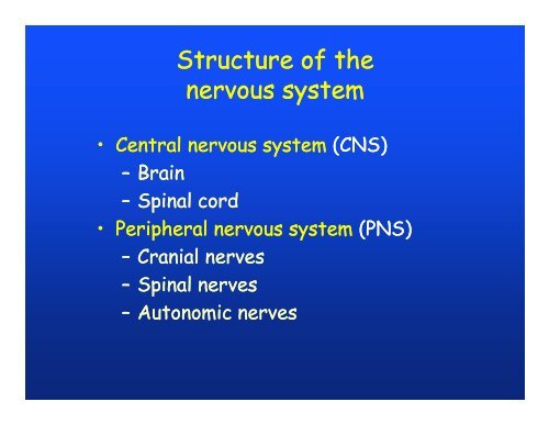
<strong>Structure</strong> <strong>of</strong> <strong>the</strong><br />
<strong>nervous</strong> <strong>system</strong><br />
• Central <strong>nervous</strong> <strong>system</strong> (CNS)<br />
– Brain<br />
– Spinal cord<br />
• Peripheral <strong>nervous</strong> <strong>system</strong> (PNS)<br />
– Cranial nerves<br />
–Spinal nerves<br />
– Autonomic nerves
Afferent<br />
Efferent<br />
Efferent
• Extend throughout CNS.<br />
Nerve fibers<br />
– Aggregated into functional units: fiber tracts.<br />
– Cluster <strong>of</strong> cell bodies: nucleus.<br />
• Bundled into fascicles and nerves in PNS.<br />
– Wrapped in connective tissue:<br />
• Endoneurium, perineurium, epineurium.<br />
• Blood vessels.<br />
– Cluster <strong>of</strong> cell bodies: ganglion.
Embryonic development<br />
<strong>of</strong> <strong>the</strong> neural tube<br />
• Hollow tube, filled with cerebrospinal fluid<br />
(CSF).<br />
• Brain:<br />
– Develops from anterior expansion <strong>of</strong> hollow tube.<br />
– Differentiated into three major ventricles.<br />
• Spinal cord:<br />
– Develops from remainder <strong>of</strong> hollow tube.<br />
– Differentiated into three embryonic layers.
Neurulation and<br />
neural crest
Neurula stage
Spinal nerves<br />
develop in<br />
conjunction<br />
with<br />
developing<br />
myotomes
Embryonic development<br />
<strong>of</strong> <strong>the</strong> spinal cord<br />
• Differentiated into three embryonic layers:<br />
– Inner germinal layer:<br />
• Active mitotic division.<br />
• CNS neuroblasts proliferate into mantle layer.<br />
– Middle mantle layer:<br />
• Neuron cell bodies congregate.<br />
• Form “gray matter”.<br />
– Outer marginal layer:<br />
• Neurons form dendrites and axons.<br />
• Myelin sheaths form “white matter”.
<strong>Structure</strong> <strong>of</strong> <strong>the</strong><br />
<strong>nervous</strong> <strong>system</strong><br />
• Central <strong>nervous</strong> <strong>system</strong> (CNS)<br />
– Brain<br />
– Spinal cord<br />
• Peripheral <strong>nervous</strong> <strong>system</strong> (PNS)<br />
– Cranial nerves<br />
– Spinal nerves<br />
– Autonomic nerves
Embryonic development<br />
<strong>of</strong> <strong>the</strong> brain<br />
• Develops embryonically as three vesicles:<br />
– Forebrain (prosencephalon):<br />
• Olfaction and vision.<br />
– Midbrain (mesencephalon):<br />
• Integration <strong>of</strong> input signals, especially visual.<br />
– Hindbrain (rhombencephalon):<br />
•Hearing<br />
• Coordination <strong>of</strong> movement, muscle tone, posture.<br />
• Integration in electromagnetic-generating and<br />
electromagnetic-detecting fishes.
<strong>Structure</strong> <strong>of</strong> <strong>the</strong> brain<br />
• 3 embryonic regions form 5 functional regions:<br />
– Prosencephalon:<br />
• Telencephalon (~cerebrum, cerebral hemispheres).<br />
• Diencephalon.<br />
--- Cephalic flexure ---<br />
– Mesencephalon.<br />
--- Pontine flexure ---<br />
–Rhombencephalon:<br />
• Metencephalon (~cerebellum).<br />
• Myelencephalon (=medulla oblongata).<br />
--- Cervical flexure ---<br />
– Spinal cord.
• Neurons:<br />
Human brain development<br />
– Develop in fetus at >250,000/min.<br />
–At birth, ~trillion (10 9 ) neurons.<br />
– Few neurons added after birth.<br />
– Thereafter, neurons die at rate <strong>of</strong><br />
thousands/day.<br />
• Glial (neuroglial) cells:<br />
– Outnumber neurons 50:1.<br />
– Regulate internal environment<br />
<strong>of</strong> brain.<br />
• Blood flow.<br />
– Help to create and destroy<br />
neural circuits.<br />
it
<strong>Structure</strong> <strong>of</strong><br />
adult vertebrate brain<br />
(5) Myelencephalon l (posterior)<br />
(4) Metencephalon<br />
(3) Mesencephalon<br />
(2) Diencephalon<br />
(1) Telencephalon (anterior)
(5) Myelencephalon (medulla oblongata)<br />
• <strong>Structure</strong> similar to spinal cord:<br />
– External white matter, internal gray matter.<br />
– Central canal expanded: 4th ventricle.<br />
– Ro<strong>of</strong> thin, highly vascularized (posterior<br />
choroid plexus).<br />
• Gas, nutrient exchange.<br />
• Secretions.<br />
• Nerve fibers grouped by function:<br />
– Somatic sensory (dorsal, afferent)<br />
– Visceral sensory<br />
– Visceral motor<br />
– Somatic motor (ventral, efferent)
Afferent<br />
Efferent
(5) Myelencephalon (medulla oblongata)<br />
• Basic function: switchboard for autonomic receptors<br />
and effectors <strong>of</strong>:<br />
–Eye.<br />
– Pharyngeal arches and derivatives:<br />
• Gills, tongue, larynx<br />
– Visceral organs:<br />
• Heart<br />
• Respiratory organs<br />
•Digestive tract<br />
• Acoustico-lateralis <strong>system</strong>
(4) Metencephalon<br />
• Ro<strong>of</strong> enlarged into cerebellum.<br />
• Functions:<br />
– Coordination <strong>of</strong> skeletal muscles.<br />
– Rhythmic locomotory actions.<br />
– Posture.<br />
• Correlation between cerebellum size and amount &<br />
intricacy <strong>of</strong> body movements:<br />
– Small in lampreys, less active fishes, amphibians,<br />
reptiles.<br />
– Large in active fishes, birds, mammals.
(4) Metencephalon<br />
• Afferent nerves (via medulla oblongata) from:<br />
– Skeletal-muscle proprioceptors.<br />
– Acoustico-lateralis <strong>system</strong>.<br />
– Eye (via mesencephalon).<br />
– Cutaneous receptors.<br />
• Efferent nerves to skeletal muscles.<br />
• Reversal <strong>of</strong> gray and white matter:<br />
– Gray cortex.<br />
– Interior white nerve fibers.
(3) Mesencephalon<br />
• Ro<strong>of</strong> enlarged into optic lobes (=optic tectum).<br />
• Function: reception and integration <strong>of</strong> visual signals.<br />
– Fishes & amphibians:<br />
• Primary visual center.<br />
– Reptiles & birds:<br />
• Visual center, along with cerebrum (forebrain).<br />
– Mammals:<br />
• Secondary visual center.<br />
• Cerebrum is primary visual center.<br />
• Cranial nerves control eye movement.<br />
• Center <strong>of</strong> learning in lower vertebrates.
(2) Diencephalon<br />
• Subdivided d d into three regions:<br />
a) Ro<strong>of</strong>: epithalamus<br />
b) Walls: thalamus<br />
c) Floor: hypothalamus
Anterior<br />
Posterior
(a) Epithalamus<br />
• Three structures formed by evagination:<br />
–Paraphysis:<br />
• Present in vertebrate embryos.<br />
• Absent in adults.<br />
• Function unknown.<br />
–Parietal(=parapineal) and pineal (=epiphysis) bodies:<br />
• Primitive function: medial eye.<br />
• Pineal operational in lampreys and hagfishes.<br />
• Parietal operational in some lizards.<br />
– Derived function n <strong>of</strong> pineal body in amniotes:<br />
• Control <strong>of</strong> internal cycles (e.g., circadian rhythms).
Pineal body<br />
• Transduces neural information into endocrine<br />
information.<br />
• Secretes <strong>the</strong> hormone melatonin on circadian cycle:<br />
– High concentrations at night.<br />
– Low during day.<br />
• Tracks secondary lunar cycles:<br />
– Mating and reproductive cycles.<br />
– Immunocompetence.<br />
–Aging.<br />
g
(b) Thalamus<br />
• Important center for relay and integration <strong>of</strong><br />
signals between <strong>the</strong> cerebrum and midbrain /<br />
hindbrain.<br />
n.<br />
• Most important in mammals due to enlarged<br />
cerebrum.
(c) Hypothalamus<br />
• Three important functional regions:<br />
– Optic chiasma: optic stalks.<br />
– Control <strong>of</strong> <strong>the</strong> autonomic (“involuntary”) <strong>nervous</strong><br />
<strong>system</strong>:<br />
• Temperature regulation, O 2 / CO 2 balance.<br />
• Heart rate, breathing rate.<br />
• Biological clock: sleep rhythms, hormone<br />
cycles.<br />
– Interacts with epithalamus.<br />
• Emotional responses.<br />
– Neurohypophysis:<br />
• At tip <strong>of</strong> infundibulum.<br />
• “Posterior” (dorsal) portion <strong>of</strong> pituitary gland.
Formation <strong>of</strong> optic stalks
Neurohypophysis<br />
• Secretes many important hormones, such as:<br />
–Vasopressin: salt-water balance.<br />
– Oxytocin: stimulates t contraction ti <strong>of</strong> smooth muscle.<br />
• Mammary glands, uterus.<br />
• Humans: related to emotional, pair-bonding and<br />
stress-related responses.<br />
• Corresponding “anterior” (ventral) pituitary gland:<br />
– Adenohypophysis.<br />
– Derived from endoderm (ro<strong>of</strong> <strong>of</strong> mouth).<br />
– Many hormones.
(1) Telencephalon<br />
• Ancestral function: primary olfactory center.<br />
– Secondary center: memory, learning, orientation.<br />
• Derived function in mammals and birds:<br />
– “Higher” brain functions.<br />
– Expanded d cerebral hemispheres.<br />
h<br />
• Wrap around posterior brain regions.<br />
• Reversal <strong>of</strong> gray and white matter.<br />
– Taken over control <strong>of</strong> many o<strong>the</strong>r functions:<br />
• Vision: color vision.<br />
• Coordination: highly integrated movement.<br />
• Memory: learning, emotional responses.
(1) Telencephalon<br />
• Subdivided into three regions:<br />
–Paleopallium: olfaction.<br />
• Dominant in fishes.<br />
• Reduced in amniotes.<br />
– Archipallium: memory and navigation.<br />
• Hippocampus in all vertebrates.<br />
• Largest in cetaceans, primates.<br />
– Neopallium: cerebral hemispheres.<br />
• Thin layer in fishes, amphibians.<br />
• Enlarged in reptiles.<br />
• Dominant in birds, mammals (neocortex).
Functional regions<br />
<strong>of</strong> <strong>the</strong> neocortex in mammals<br />
• Regions:<br />
(1) Frontal: includes speech center<br />
(Broca’s area), coordinates<br />
breathing and vocalization.<br />
(2) Parietal: includes somatosensory<br />
integration area.<br />
(3) Temporal: includes auditory region.<br />
(4) Occipital: includes visual region.<br />
• Allocations <strong>of</strong> particular functions to<br />
regions are overly simplistic.
Somatosensory cortex:
Ventral view
Limbic <strong>system</strong><br />
• = “Reptilian brain”.<br />
• Functions: fear, hunger, fighting/fleeing, memory,<br />
sensory input, and sexual behavior.<br />
• <strong>Structure</strong> is progressive in vertebrates:<br />
– Primitively, main function is processing olfactory<br />
stimuli.<br />
– Somewhat developed in fishes, amphibians.<br />
– Full development in <strong>the</strong> early reptiles.<br />
• Mammalian and avian limbic <strong>system</strong>s st not much<br />
different from crocodilian.
Limbic <strong>system</strong><br />
– Derived function in humans, primates, and many<br />
o<strong>the</strong>r mammals:<br />
• Regulation <strong>of</strong> emotional processes.<br />
– Humans: join “higher” mental functions (reasoning)<br />
with “lower” functions (fear, pleasure).<br />
• Accounts for why:<br />
– Sexual behavior and eating seem pleasurable.<br />
– Mental stress causes high blood pressure.<br />
–Etc.<br />
• Disorders in regulating mechanisms lead to craving and<br />
addiction behavioral disorders.
Evolutionary development<br />
<strong>of</strong> <strong>the</strong> brain<br />
• Cephalochordates (e.g., amphioxus) have slight dilation<br />
<strong>of</strong> neural tube in head: cerebral vesicle.<br />
• Gene expression patterns and microanatomy indicate:<br />
–Diencephalon:<br />
• Small region homologous with vertebrate lateral<br />
eyes.<br />
• Lamellar ar organ homologous ogous with vertebrate rat pineal<br />
photoreceptors.<br />
– Small midbrain.<br />
– Hindbrain.
Trends in vertebrate brains<br />
• Ancestral (“lower” vertebrates):<br />
– Posterior brain well developed.<br />
– Straight tube, large ventricles.<br />
– Cerebellum enlarged.<br />
• Derived (“higher” vertebrates):<br />
– Anterior brain well developed.<br />
– Ventricles reduced, walls thickened. Cerebral<br />
hemispheres enlarged .<br />
–Brain highly folded and elaborated.<br />
– Increased regionalization; functions shifted<br />
forward.<br />
– Enhanced limbic <strong>system</strong>.
Trends in vertebrate brains<br />
• New elements added to existing elements, ra<strong>the</strong>r<br />
than replacing <strong>the</strong>m.<br />
– Newer elements control older elements.<br />
– “Higher” <strong>nervous</strong> <strong>system</strong>:<br />
• Superimposed control circuits.<br />
• More complicated behaviors.<br />
• Repeated and parallel increases in relative brain<br />
size within major groups.<br />
– Teleost brains almost 2x size <strong>of</strong> shark brains.<br />
– “Higher” teleost brains almost 2x size <strong>of</strong> “lower”<br />
teleost brains.<br />
– Etc.
Brain size<br />
• Brain size is relatively larger in more derived<br />
vertebrate groups.<br />
• But brain size is highly hl correlated with body size<br />
(allometry).
• Increases in brain size<br />
over evolutionary time:<br />
– Average increases.<br />
– Widening size<br />
distributions.<br />
– Few replacements <strong>of</strong><br />
smaller-brained by<br />
larger-brained<br />
animals.<br />
Brain size
Brain size in hominoids<br />
Homo sapiens<br />
Homo erectus<br />
Australopi<strong>the</strong>cus afarensis<br />
olume<br />
Brain vo<br />
Body weight
Brain size in hominoids


