Ocular rosacea Crotoxin for paralysis of extraocular muscles ...
Ocular rosacea Crotoxin for paralysis of extraocular muscles ... Ocular rosacea Crotoxin for paralysis of extraocular muscles ...
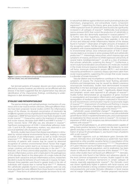
Artigo de Revisão | Review Article Ocular rosacea - a review Rosácea ocular - revisão Ana Carolina Cabreira Vieira 1 , Ana Luisa Höfling-Lima 1 , Mark J Mannis 2 ABSTRACT Rosacea is a prevalent chronic cutaneous disorder with variable presentation and severity. Although considered a skin disease, rosacea may evolve the eyes in 58-72% of the patients, causing eyelid and ocular surface inflammation. About one third of the patients develop potentially sight-threatening corneal involvement. Untreated rosacea may cause varying degrees of ocular morbidity. The importance of early diagnosis and adequate treatment cannot be overemphasized. There is not yet a diagnostic test for rosacea. The diagnosis of ocular rosacea relies on observation of clinical features, which can be challenging in up to 90% of patients in whom accompanying roseatic skin changes may be subtle or inexistent. In this review, we describe the pathophysiologic mechanisms proposed in the literature, clinical features, diagnosis and management of ocular rosacea, as well as discuss the need for a diagnostic test for the disease. Keywords: Rosacea/diagnosis; Eye manifestations; Eye diseases; Rosacea/drug therapy; Doxycycline; Visual acuity RESUMO A rosácea é uma condição cutânea crônica, que possui apresentações clínicas variáveis. Apesar de considerada uma doença dermatológica, os olhos podem ser acometidos em 58-72% dos casos, causando inflamação palpebral e da superfície ocular. Aproximadamente um terço dos pacientes desenvolve acometimento corneano, podendo causar baixa visual significativa. Diagnóstico precoce e tratamento adequado são de extrema importância, devido à significativa morbidade ocular que a doença pode causar. Não há, até o momento, um teste diagnóstico para rosácea. O diagnóstico da rosácea ocular depende da observação das manifestações clínicas, o que pode ser bastante desafiador em até 90% dos pacientes, em que os achados cutâneos são discretos ou inexistentes. Nesta revisão, descrevemos os mecanismos fisiopatológicos propostos na literatura, manifestações clínicas, diagnóstico e tratamento da rosácea ocular, assim como abordamos a necessidade de um teste diagnóstico. Descritores: Rosácea/diagnóstico; Manifestações oculares; Oftalmopatias; Rosácea/qui - mioterapia; Doxiciclina; Acuidade visual INTRODUCTION Rosacea is a widely prevalent chronic cutaneous disorder with variable presentation and severity. It primarily affects blood vessels and pilosebaceous units of the central facial skin (cheeks, chin, nose, and central forehead), causing transient or persistent erythema, telangiectasias, papules, pustules, and phymatous changes (Fi gu re 1) (1) . Although considered a skin disease, rosacea may evolve the eyes in up to 58-72% of the patients, causing eyelid and ocular surface inflammation (1,2) . About one third of the patients develop corneal involvement, which may be sight-threatening (1,3) . Rosacea, albeit common, is often overlooked by general practitioners and ophthalmologists (4) . Mild rosacea patients may not seek medical help and may not be diagnosed in clinical practice. Ocular rosacea, in particular, is frequently underdiagnosed. Its symptoms and signs can be quite non-specific, and in up to 90% of patients, accompanying roseatic skin changes may be very subtle. More importantly, in 20% of the cases, ocular signs may even precede characteristic skin involvement (3) . Patients often do not mention ocular symptoms in a dermatology clinic, unless directly asked about them. Conversely, skin manifestations are uncommonly examined during ophthalmology consults. As a result, a certain number of cases remain undetected (3) . Chronic, untreated rosacea may cause varying degrees of ocular morbidity, facial disfigurement, emotional distress, and social impairment (5,6) . The importance of early diagnosis and adequate treatment cannot be overemphasized due to the negative impact this disorder may have on the quality of life of patients and the potential sight-threatening complications of this disease. Epidemiology Rosacea affects over 16 million Americans (7) , and a Swedish survey revealed a prevalence as high as 10% (8) . A recent large observational study on the epidemiology of rosacea in the United Kingdom revealed an incidence rate of 1.65/1,000 person-years as diagnosed by general practitioners (9) . Women are more commonly diagnosed with rosacea than men, and they tend to be diagnosed earlier. A possible explanation for this is that women may seek medical care more often and earlier than men. On the other hand, men are more prone to phymatous changes (9,10) . Rhinophyma is most commonly seen in men over 40 years of age, in more advanced stages of the disease (11,12) . Ocular rosacea affects both genders equally (9) . The incidence of ocular rosacea varies among ophthalmologic and dermatologic studies, ranging from 6-72%, being more prevalent in ophthalmology clinics (2,3,13) . Rosacea may be found in early childhood as well as in the elderly, but it most commonly affects middle-aged adults (14) . Spoendlin et al., found that 80% of rosacea patients in the UK were at or above 30 years of age, with a peak in the incidence rate between the ages of 40 and 59 years (9) . Pediatric rosacea is thought to be rare, but may be underreported since the dermatologic features are often absent or difficult to identify (15-17) . Submitted for publication: August 7, 2012 Accepted for publication: August 23, 2012 Study carried out at Universidade Federal de São Paulo - UNIFESP - São Paulo (SP), Brazil. 1 Physician, Department of Ophthalmology, Universidade Federal de São Paulo - UNIFESP - São Paulo (SP), Brazil. 2 Physician, Department of Ophthalmology, University of California, Davis, CA, USA. Funding: No specific financial support was available for this study. Disclosure of potential conflicts of interest: A.C.C.Vieira, None; A.L.Höfling-Lima, employee of Federal Governement of Brazil; M.J.Mannis, None. Correspondence address: Ana Carolina Cabreira Vieira, Rua Visconde de Silva, 52/503 - Rio de Janeiro - RJ - 22271-092 - Brazil - E-mail: anacarolinavieira31@gmail.com Arq Bras Oftalmol. 2012;75(5):363-9 363
Ocular rosacea - a review Figure 1. Cutaneous manifestations of rosacea with characteristic involvement of central facial skin (cheeks, nose, and central forehead). Fair-skinned patients of European descent are more commonly affected by rosacea; however, any ethnicity can be afflicted with the disease. It has been suggested that skin pigmentation may obscure identification of the characteristic findings, contributing to underdiagnosis in dark skinned patients (18) . Etiology and pathophysiology The precise etiology and pathophysiologic mechanisms of cutaneous and ocular rosacea remain unknown, although different theories have been proposed. Several studies confirm the inflammatory nature of the disease. An elevated concentration of interleukin-1a and b and a greater activity of gelatinase B (metalloproteinase -9) and colagenase-2 (MMP-8) have been found in tear fluids of patients with ocular rosacea (19-22) . Doxycycline, used in the treatment of cutaneous and ocular rosacea, decreases both MMP-8 and MMP-9 activity (21,22) . Tumor necrosis factor (TNF-alfa) was also elevated in rosacea (23) . Significant overexpression of ICAM-1 (intercellular adhesion molecule 1) and HLA-DR, inflammatory markers, was observed in conjunctival epithelial cells of patients with rosacea (24) . Recently, studies have shown that disease exacerbation is stimulated by normal external environmental factors, leading to unique activation of pro-inflammatory systems as well as innate immune responses (23,25,26) . Molecular studies propose that the enhanced sensitivity of patients with rosacea may be caused by abnormal recognition of common environmental stimuli. Factors that trigger the innate immune system lead to an increase in the expression of certain cytokines and anti-microbial molecules in the skin (25,27) . Cathelicidin, one of these antimicrobial peptides, has both vasoactive and pro-inflammatory actions and is implicated in the pathogenesis of this disorder. Based on the observation of a single molecule that presents both vasoactive and pro-inflammatory actions, researchers decided to study the behavior of such molecule in rosacea. As hypothesized, these peptides were found in greater levels in the skin of individuals with rosacea than in normal facial skin (25,27,28) . Moreover, the cathelicidin peptides found in rosacea patients were not only more abundant but were different from those found in normal individuals (25) . These forms of cathelicidin peptides are known for its role in natural host defense against infections and in promoting leukocyte chemotaxis, angiogenesis, and extracellular matrix component expression (27) . Supporting this theory, gene array studies found that mRNA expression levels of cathelicidin were found to be significantly increased in all subtypes of rosacea (29) . Additionally, local proteases (serine protease KLK5) that control the production of cathelicidin in epidermis were also abnormally expressed in rosacea patients (25,27) . To further test their hypothesis, researchers injected abnormal cathelicidin or protease that produce these peptides in the skin of mice and observed similar inflammatory features of the disease found in humans (27) . Observation of higher amounts of receptors of the recognition system, Toll-like receptor 2 (TLR2), in the epidermis of patients with rosacea explained the overreaction of these patients to environmental stimuli, since enhanced action of TLR2 in keratinocytes leads to an increase in serine protease KLK5 and cathelicidin production (23,25) . Interestingly, tetracyclines, which improve signs and symptoms of rosacea, are known to inhibit expression and activity of several matrix metalloproteinases (21) , as well as a class of proteases that activate cathelicidin, sustaining this theory (26,27) . Furthermore, a recent study found elevated concentrations of 5 molecules involved in the innate immune response (interleukin-1b, interleukin-16, stem cell factor, monocyte chemotactic protein (MCP)-1, and monokine induced by g-interferon) in cutaneous biopsy specimens taken from ocular rosacea patients, supporting the concept that ocular rosacea is a disorder of innate immunity (30) . Vascular dilation and incompetence contribute to the signs and symptoms of rosacea. The characteristic facial flushing, persistent erythema and telangiectasia may be caused by altered cutaneous neurovascular homeostasis. Studies have demonstrated increased blood flow in the face and larger and more numerous vessels in the face than in other areas of the body (31) . Significantly dilated blood and lymphatic vessels were reported in all subtypes of rosacea (29) . Studies further demonstrated an up-regulation of genes involved in vasoregulation and neurogenic inflammation and suggested that dysregulation of mediators and receptors implicated in neurovascular and neuroimmune communication may be crucial at early stages of rosacea (26,29,32) . Improvement of erythema and flushing in rosacea by topical administration of a-adrenergic receptor agonists, oxymetazoline and xylometazoline, helps support this theory (25,33,34) . Cathelicidin causes endothelial changes (26) . Angiogenesis induced by cathelicidin is mediated by an increase in vascular endothelial growth factor (VEGF) in epidermal keratinocytes, and could represent an explanation for these vascular events in rosacea (25) . An increase in expression of VEGF and its receptors was demonstrated in the skin of patients with rosacea (35,36) . Group IIA phospholipase A 2 , an antimicrobial protein capable of killing Gram-positive bacteria, is secreted into the tears by lacrimal glands and plays a role in innate host defense. A decreased concentration of group IIA phospholipase A 2 was found in tears of patients with ocular rosacea (37) . Microbial organisms such as Helicobacter pylori, Demodex folliculorum, Demodex brevis and Staphylococcus epidermidis have been appointed as other possible causative factors in exacerbation of the disease; however this remains a controversy (23,26,31,38-43) . The prevalence of H. pylori infection was found higher in patients with rosacea when compared to the general population, and its eradication has been shown to influence the clinical outcome of this disease (31,39,44-46) . However, other studies have not found such difference (41,47-49) . Demodex, a microscopic mite found in hair follicles and sebaceous glands, is the most common ectoparasite in humans. Studies support its role in the activation of immune mechanisms in certain subtypes of rosacea, especially papulopustular rosacea (32) . The Demodex count was shown significantly higher in patients with facial rosacea, and a study demonstrated a strong correlation between positive serum immunoreactivity and ocular Demodex infestation in facial rosacea and lid margin inflammation (41,50,51) . Another proposed mechanism 364 Arq Bras Oftalmol. 2012;75(5):363-9
- Page 20 and 21: Artigo Original | Original Article
- Page 22 and 23: Santos AM, et al. Observou-se frequ
- Page 24 and 25: Espíndola RF, et al. METHODS This
- Page 26 and 27: Espíndola RF, et al. 10. Artal P,
- Page 28 and 29: Toscano DA, et al. in 1.28 seconds.
- Page 30 and 31: Toscano DA, et al. observed excelle
- Page 32 and 33: Viani GA, et al. METHODS Types of s
- Page 34 and 35: Viani GA, et al. Table 1. Character
- Page 36 and 37: Viani GA, et al. OR: odds ratio. Fi
- Page 38 and 39: Viani GA, et al. domized study whic
- Page 40 and 41: Artigo Original | Original Article
- Page 42 and 43: Magri MPF, et al. Tabela 3. Causas
- Page 44 and 45: Artigo Original | Original Article
- Page 46 and 47: Rodrigues ACL, et al. dias - 96 mes
- Page 48 and 49: Artigo Original | Original Article
- Page 50 and 51: Yaacov-Peña F, et al. recorded if
- Page 52 and 53: Guerra RLL, et al. METHODS A retros
- Page 54 and 55: Guerra RLL, et al. terial endophtha
- Page 56 and 57: Tartarella MB, et al. inactive tumo
- Page 58 and 59: Tartarella MB, et al. 15. Portellos
- Page 60 and 61: Nasser LS, et al. De acordo com a c
- Page 62 and 63: Nasser LS, et al. A heterocromia de
- Page 64 and 65: Magalhães OA, et al. Figure 1. Gel
- Page 66 and 67: Kamei RW O ultrassom mostrava um v
- Page 68 and 69: Relato de Caso | Case Report Buphth
- Page 72 and 73: Vieira ACC, et al. is that Demodex
- Page 74 and 75: Vieira ACC, et al. is often misdiag
- Page 76 and 77: Vieira ACC, et al. 51. Lazaridou E,
- Page 78: Messias K, et al. Figure 2. Potenti
- Page 81 and 82: Autoria Os critérios para autoria
- Page 83 and 84: Lista de Sítios da Internet Interf
- Page 85: TM Nova Lente Pronta Essilor Stylis
<strong>Ocular</strong> <strong>rosacea</strong> - a review<br />
Figure 1. Cutaneous manifestations <strong>of</strong> <strong>rosacea</strong> with characteristic involvement <strong>of</strong> central<br />
facial skin (cheeks, nose, and central <strong>for</strong>ehead).<br />
Fair-skinned patients <strong>of</strong> European descent are more commonly<br />
affected by <strong>rosacea</strong>; however, any ethnicity can be afflicted with the<br />
disease. It has been suggested that skin pigmentation may obscure<br />
identification <strong>of</strong> the characteristic findings, contributing to underdiagnosis<br />
in dark skinned patients (18) .<br />
Etiology and pathophysiology<br />
The precise etiology and pathophysiologic mechanisms <strong>of</strong> cutaneous<br />
and ocular <strong>rosacea</strong> remain unknown, although different theories<br />
have been proposed. Several studies confirm the inflammatory<br />
nature <strong>of</strong> the disease. An elevated concentration <strong>of</strong> interleukin-1a<br />
and b and a greater activity <strong>of</strong> gelatinase B (metalloproteinase -9) and<br />
colagenase-2 (MMP-8) have been found in tear fluids <strong>of</strong> patients with<br />
ocular <strong>rosacea</strong> (19-22) . Doxycycline, used in the treatment <strong>of</strong> cutaneous<br />
and ocular <strong>rosacea</strong>, decreases both MMP-8 and MMP-9 activity (21,22) .<br />
Tumor necrosis factor (TNF-alfa) was also elevated in <strong>rosacea</strong> (23) .<br />
Significant overexpression <strong>of</strong> ICAM-1 (intercellular adhesion molecule<br />
1) and HLA-DR, inflammatory markers, was observed in conjunctival<br />
epithelial cells <strong>of</strong> patients with <strong>rosacea</strong> (24) .<br />
Recently, studies have shown that disease exacerbation is stimulated<br />
by normal external environmental factors, leading to unique<br />
activation <strong>of</strong> pro-inflammatory systems as well as innate immune<br />
responses (23,25,26) . Molecular studies propose that the enhanced<br />
sensitivity <strong>of</strong> patients with <strong>rosacea</strong> may be caused by abnormal<br />
recognition <strong>of</strong> common environmental stimuli. Factors that trigger<br />
the innate immune system lead to an increase in the expression <strong>of</strong><br />
certain cytokines and anti-microbial molecules in the skin (25,27) . Cathelicidin,<br />
one <strong>of</strong> these antimicrobial peptides, has both vasoactive<br />
and pro-inflammatory actions and is implicated in the pathogenesis<br />
<strong>of</strong> this disorder. Based on the observation <strong>of</strong> a single molecule that<br />
presents both vasoactive and pro-inflammatory actions, researchers<br />
decided to study the behavior <strong>of</strong> such molecule in <strong>rosacea</strong>. As hypothesized,<br />
these peptides were found in greater levels in the skin <strong>of</strong><br />
individuals with <strong>rosacea</strong> than in normal facial skin (25,27,28) . Moreover,<br />
the cathelicidin peptides found in <strong>rosacea</strong> patients were not only<br />
more abundant but were different from those found in normal individuals<br />
(25) . These <strong>for</strong>ms <strong>of</strong> cathelicidin peptides are known <strong>for</strong> its role<br />
in natural host defense against infections and in promoting leukocyte<br />
chemotaxis, angiogenesis, and extracellular matrix component<br />
expression (27) . Supporting this theory, gene array studies found that<br />
mRNA expression levels <strong>of</strong> cathelicidin were found to be significantly<br />
increased in all subtypes <strong>of</strong> <strong>rosacea</strong> (29) . Additionally, local proteases<br />
(serine protease KLK5) that control the production <strong>of</strong> cathelicidin in<br />
epidermis were also abnormally expressed in <strong>rosacea</strong> patients (25,27) .<br />
To further test their hypothesis, researchers injected abnormal<br />
cathelicidin or protease that produce these peptides in the skin<br />
<strong>of</strong> mice and observed similar inflammatory features <strong>of</strong> the disease<br />
found in humans (27) . Observation <strong>of</strong> higher amounts <strong>of</strong> receptors <strong>of</strong><br />
the recognition system, Toll-like receptor 2 (TLR2), in the epidermis<br />
<strong>of</strong> patients with <strong>rosacea</strong> explained the overreaction <strong>of</strong> these patients<br />
to environmental stimuli, since enhanced action <strong>of</strong> TLR2 in keratinocytes<br />
leads to an increase in serine protease KLK5 and cathelicidin<br />
production (23,25) . Interestingly, tetracyclines, which improve signs and<br />
symptoms <strong>of</strong> <strong>rosacea</strong>, are known to inhibit expression and activity <strong>of</strong><br />
several matrix metalloproteinases (21) , as well as a class <strong>of</strong> proteases<br />
that activate cathelicidin, sustaining this theory (26,27) . Furthermore, a<br />
recent study found elevated concentrations <strong>of</strong> 5 molecules involved<br />
in the innate immune response (interleukin-1b, interleukin-16, stem<br />
cell factor, monocyte chemotactic protein (MCP)-1, and monokine<br />
induced by g-interferon) in cutaneous biopsy specimens taken from<br />
ocular <strong>rosacea</strong> patients, supporting the concept that ocular <strong>rosacea</strong><br />
is a disorder <strong>of</strong> innate immunity (30) .<br />
Vascular dilation and incompetence contribute to the signs and<br />
symptoms <strong>of</strong> <strong>rosacea</strong>. The characteristic facial flushing, persistent<br />
erythema and telangiectasia may be caused by altered cutaneous<br />
neurovascular homeostasis. Studies have demonstrated increased<br />
blood flow in the face and larger and more numerous vessels in the<br />
face than in other areas <strong>of</strong> the body (31) . Significantly dilated blood<br />
and lymphatic vessels were reported in all subtypes <strong>of</strong> <strong>rosacea</strong> (29) .<br />
Studies further demonstrated an up-regulation <strong>of</strong> genes involved<br />
in vasoregulation and neurogenic inflammation and suggested that<br />
dysregulation <strong>of</strong> mediators and receptors implicated in neurovascular<br />
and neuroimmune communication may be crucial at early stages<br />
<strong>of</strong> <strong>rosacea</strong> (26,29,32) . Improvement <strong>of</strong> erythema and flushing in <strong>rosacea</strong><br />
by topical administration <strong>of</strong> a-adrenergic receptor agonists, oxymetazoline<br />
and xylometazoline, helps support this theory (25,33,34) .<br />
Cathelicidin causes endothelial changes (26) . Angiogenesis induced<br />
by cathelicidin is mediated by an increase in vascular endothelial<br />
growth factor (VEGF) in epidermal keratinocytes, and could represent<br />
an explanation <strong>for</strong> these vascular events in <strong>rosacea</strong> (25) . An increase in<br />
expression <strong>of</strong> VEGF and its receptors was demonstrated in the skin <strong>of</strong><br />
patients with <strong>rosacea</strong> (35,36) .<br />
Group IIA phospholipase A 2<br />
, an antimicrobial protein capable <strong>of</strong><br />
killing Gram-positive bacteria, is secreted into the tears by lacrimal<br />
glands and plays a role in innate host defense. A decreased concentration<br />
<strong>of</strong> group IIA phospholipase A 2<br />
was found in tears <strong>of</strong> patients<br />
with ocular <strong>rosacea</strong> (37) .<br />
Microbial organisms such as Helicobacter pylori, Demodex folliculorum,<br />
Demodex brevis and Staphylococcus epidermidis have been<br />
appointed as other possible causative factors in exacerbation <strong>of</strong> the<br />
disease; however this remains a controversy (23,26,31,38-43) . The prevalence<br />
<strong>of</strong> H. pylori infection was found higher in patients with <strong>rosacea</strong> when<br />
compared to the general population, and its eradication has been<br />
shown to influence the clinical outcome <strong>of</strong> this disease (31,39,44-46) . However,<br />
other studies have not found such difference (41,47-49) . Demodex,<br />
a microscopic mite found in hair follicles and sebaceous glands, is<br />
the most common ectoparasite in humans. Studies support its role<br />
in the activation <strong>of</strong> immune mechanisms in certain subtypes <strong>of</strong><br />
<strong>rosacea</strong>, especially papulopustular <strong>rosacea</strong> (32) . The Demodex count<br />
was shown significantly higher in patients with facial <strong>rosacea</strong>, and<br />
a study demonstrated a strong correlation between positive serum<br />
immunoreactivity and ocular Demodex infestation in facial <strong>rosacea</strong><br />
and lid margin inflammation (41,50,51) . Another proposed mechanism<br />
364 Arq Bras Oftalmol. 2012;75(5):363-9


