Chest pain - Henry Ford Health System
Chest pain - Henry Ford Health System
Chest pain - Henry Ford Health System

Create successful ePaper yourself
Turn your PDF publications into a flip-book with our unique Google optimized e-Paper software.
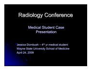
Department of Radiology<br />
<strong>Henry</strong> <strong>Ford</strong> <strong>Health</strong> <strong>System</strong><br />
Detroit, Michigan<br />
<strong>Chest</strong> <strong>pain</strong><br />
Elise van Holsbeeck MS4<br />
MSUCOM<br />
Resident: Dr. Farhaan Mir PGY 2
Chief Complaint<br />
46 year old male with <strong>pain</strong><br />
reproducible by chest palpation
History<br />
• HPI: 1 year of anterior chest wall <strong>pain</strong><br />
beginning after lifting TV. 2 weeks ago<br />
chest <strong>pain</strong> got worse usually after exertion.<br />
Pt also noticed a small bump over chest<br />
wall.<br />
• PMH/PSH: none<br />
• Social: smoker<br />
• Family: mother renal CA, brother lung CA<br />
• ROS: negative except for the <strong>pain</strong>
Radiographs<br />
No significant abnormality
Clinical Dx<br />
• Slipping Rib Syndrome<br />
• Next step: US
Ultrasound
Ultrasound<br />
• 3.0 x 3.1 cm round heterogeneous mass in<br />
the region of the xyphoid<br />
• Mostly hypoechoic with bright hyperechoic<br />
speckles suggesting chondroid matrix or<br />
residual destructed bone<br />
• Internal vascularity was demonstrated by<br />
color Doppler
<strong>Chest</strong> CT
<strong>Chest</strong> CT
CT <strong>Chest</strong>
CT <strong>Chest</strong>
<strong>Chest</strong> CT<br />
• Lytic expansile 3.6 x 3.4 x 3.2 cm lesion in<br />
the inferior body of the sternum<br />
• Soft tissue density<br />
• Small higher density areas likely<br />
fragmentation of cortex, not chondroid<br />
matrix
Differential<br />
• Plasmocytoma<br />
• Multiple Myeloma<br />
• Metastasis<br />
• Sarcoma<br />
• Giant Cell tumor<br />
• Chondrosarcoma
US guided biopsy<br />
• Diagnosis made by<br />
percutaneous Ultrasound<br />
directed biopsy of mass<br />
• Pathology showed a grade II<br />
chondrosarcoma<br />
• 24 days later, the pt’s s distal<br />
sternum was resected ‘en<br />
bloc’
Diagnosis: Chondrosarcoma<br />
• 2 nd most common primary bone<br />
malignancy (25%)<br />
• Usually pt’s s >40 y.o. (rare in children)<br />
• Cartilaginous origin therefore matrix is<br />
entirely chondroid<br />
• 5 year survival rate ranges from 90% to<br />
29% depending on grade<br />
• Tumors often recur 5-105<br />
years after<br />
surgery<br />
http://emedicine.medscape.com/article/388869-overview
Location<br />
• Flat bones<br />
• Pelvis: 25%<br />
• Ilium: 15%<br />
• Pubis and ischium: 9%<br />
• Scapula: 5%<br />
• Ribs and sternum: 12%<br />
https://my.statdx.com/STATdxMain.jsp#2
Gross pathology
Tomosynthesis
Lobular Tumor Architecture<br />
2<br />
1
Bibliography<br />
Hide, Geoff. Chondrosarcoma.<br />
http://emedicine.medscape.com/article/388869-<br />
overview. . Nov 3, 2009.<br />
Bredella, Miriam A. Dx. Chondrosarcoma.<br />
https://my.statdx.com/STATdxMain.jsp#2<br />
05-27<br />
27-08.<br />
Unni, Krishnan K. and Inwards, Carrie Y. Dahlin’s s Bone<br />
Tumors. . Lippincott Williams & Wilkins: 2009.