A theoretical study of anthrax lethal factor inhibition by a set of ... - ISP
A theoretical study of anthrax lethal factor inhibition by a set of ... - ISP
A theoretical study of anthrax lethal factor inhibition by a set of ... - ISP

Create successful ePaper yourself
Turn your PDF publications into a flip-book with our unique Google optimized e-Paper software.
Journal <strong>of</strong> Molecular Structure: THEOCHEM 821 (2007) 139–144<br />
www.elsevier.com/locate/theochem<br />
A <strong>theoretical</strong> <strong>study</strong> <strong>of</strong> <strong>anthrax</strong> <strong>lethal</strong> <strong>factor</strong> <strong>inhibition</strong><br />
<strong>by</strong> a <strong>set</strong> <strong>of</strong> novel carbamimidolyl-aryl-vinyl-carboxamidines:<br />
A possible mechanism involving zinc-ligation <strong>by</strong> amidine<br />
Tam Luong Nguyen a , Rekha G. Panchal a , Igor A. Topol b , Douglas Lane a ,<br />
Tara Kenny a , James C. Burnett a , Ann R. Hermone a , Connor McGrath a ,<br />
Stanley K. Burt b , Rick Gussio c , Sina Bavari d, *<br />
a Target Structure-Based Drug Discovery Group, SAIC-Frederick, Inc., NCI-Frederick, Frederick, MD 21702, USA<br />
b Advanced Biomedical Computing Center, SAIC-Frederick, Inc., NCI-Frederick, Frederick, MD 21702, USA<br />
c Target Structure-Based Drug Discovery Group, Information Technology Branch, Developmental Therapeutics Program,<br />
National Cancer Institute, Frederick, MD 21702, USA<br />
d US Army Medical Research Institute <strong>of</strong> Infectious Diseases, Fort Detrick, Frederick, MD 21702, USA<br />
Received 3 May 2007; received in revised form 2 July 2007; accepted 5 July 2007<br />
Available online 20 July 2007<br />
Abstract<br />
A congeneric <strong>set</strong> <strong>of</strong> carbamimidolyl-aryl-vinyl-carboxamidines from the National Cancer Institute (NCI) open chemical repository<br />
were identified as potent inhibitors <strong>of</strong> <strong>anthrax</strong> <strong>lethal</strong> <strong>factor</strong> (LF), a zinc-dependent metalloprotease that plays a critical role in potentiating<br />
Bacillus anthracis infection. Surprisingly, these compounds exhibited no differential change in activity with concentration. Docking<br />
studies revealed that the indole-attached amidine substituents <strong>of</strong> these inhibitors were positioned in close proximity to the biological zinc<br />
atom and could potentially function as transition-state mimetics. This broaches the stunning possibility that the dose independence <strong>of</strong><br />
these inhibitors is linked to zinc-ligation. Because the amidine functionality is highly basic and cationic, it is generally not considered a<br />
viable zinc-binding motif. However, quantum chemical calculations on small-molecule models predicted a marked decrease in the pK a <strong>of</strong><br />
the amidine functionality when it is in close proximity to zinc, thus allowing for the formation <strong>of</strong> a robust zinc–amidine bond.<br />
Published <strong>by</strong> Elsevier B.V.<br />
Keywords: Three-dimensional search query; Density functional theory; Docking <strong>study</strong>; Molecular dynamics; Zinc coordination<br />
1. Introduction<br />
Anthrax <strong>lethal</strong> <strong>factor</strong> (LF) is a zinc-dependent endopeptidase<br />
and a constituent <strong>of</strong> the <strong>anthrax</strong> toxin secreted <strong>by</strong><br />
Bacillus anthracis. LF cleaves near the amino terminus <strong>of</strong><br />
mitogen-activated protein kinase kinases (MAPKK),<br />
resulting in disruptions in key cellular signaling pathways<br />
and ultimately cell death [1]. A significant number <strong>of</strong><br />
small-molecule LF inhibitors have been described in the literature<br />
[2–10], and not surprisingly, most <strong>of</strong> these inhibitors<br />
are centered to zinc-binding groups such as<br />
* Corresponding author. Fax: +1 301 619 2348.<br />
E-mail address: sina.bavari@amedd.army.mil (S. Bavari).<br />
hydroxamate [2] and thiazolidine [3]. However, not every<br />
LF inhibitor has its mechanism <strong>of</strong> <strong>inhibition</strong> depicted in<br />
its chemical structure [8,9,11]. Among these are a <strong>set</strong> <strong>of</strong><br />
guanidinylated compounds [8,9], which were shown to be<br />
potent LF inhibitors but do not possess the prototypical<br />
zinc-binding groups [12].<br />
In this paper, we report that a second <strong>set</strong> <strong>of</strong> cationic<br />
compounds are potent LF inhibitors. These compounds<br />
are carbamimidolyl-aryl-vinyl-indole-carboxamidines and<br />
are chemically similar to reported LF inhibitors [11]. The<br />
biological activity <strong>of</strong> these compounds was shown to be<br />
statistically dose independent. A detailed computational<br />
investigation revealed an unexpected potential mechanism<br />
<strong>of</strong> action for these highly basic and cationic compounds.<br />
0166-1280/$ - see front matter Published <strong>by</strong> Elsevier B.V.<br />
doi:10.1016/j.theochem.2007.07.009
140 T.L. Nguyen et al. / Journal <strong>of</strong> Molecular Structure: THEOCHEM 821 (2007) 139–144<br />
2. Computational methodology<br />
The geometries <strong>of</strong> the [Zn(ac)(im) 2 (am)] model and the<br />
XIZZOV crystal structure were optimized with the Gaussian-03<br />
package (Gaussian, Inc., Wallingford, CT) using the<br />
DFT B3LYP approach. The conventional 6-31+G(d,p)<br />
basis <strong>set</strong>s on all atoms (except Zn) and the recently published<br />
6-31G(f) basis <strong>set</strong> on Zn [13] were utilized. The pK a ’s<br />
<strong>of</strong> the amidine/guanidine NH’s were calculated using a previously-described<br />
protocol [14] but in this instance, the<br />
hydration energy <strong>of</strong> the proton (DG solv (H + )) was taken<br />
as 264.0 kcal/mol, which is the most recently obtained<br />
value [15]. The resulting pK a values were corrected <strong>by</strong> the<br />
addition <strong>of</strong> 0.18 pK a units based on the result that the calculated<br />
pK a <strong>of</strong> the arginine–guanidine NH using this<br />
method was 12.32 whereas the textbook experimental pK a<br />
value is 12.5.<br />
Docking studies were performed using the InsightII and<br />
Cerius2 programs on a Silicon Graphics Octane 2 workstation.<br />
The X-ray structure <strong>of</strong> LF in complex with FSPA<br />
(FSPA = (2R)-2-[(4-fluoro-3-methylphenyl)sulfonylamino]-<br />
N-hydroxy-2-(tetrahydro-2H-pyran-4-yl)acetamide) (PDB<br />
code 1YQY) [2] was selected as the template and optimized<br />
using a published tethered-minimization protocol [16]. The<br />
ligands were initially docked into the catalytic site using the<br />
Ligand Docking module in Cerius2 and their conformations<br />
were refined using iterative cycles <strong>of</strong> hydropathic<br />
analysis, manual adjustment, and molecular mechanics<br />
simulations [16].<br />
3. Results and discussion<br />
3.1. Empirical basis for LF <strong>inhibition</strong> <strong>by</strong> diamidines<br />
We had previously developed a pharmacophore for LF<br />
<strong>inhibition</strong> [11]. Here we virtually screened the NCI chemical<br />
repository for compounds that fit our pharmacophore<br />
for LF <strong>inhibition</strong> [11] but that also contained functionality<br />
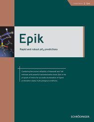
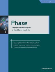
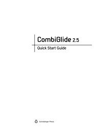
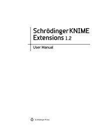
isosteric to guanidine. These three-dimensional search queries<br />
identified four candidate compounds in the carbamimidoyl-aryl-vinyl-indole-carboxamidines<br />
1–4 (Fig. 1).<br />
A HPLC-based enzymatic assay [11] was used to determine<br />
the activity <strong>of</strong> 1–4 against LF. From triplicate results,<br />
compounds 1–4 had average %LF <strong>inhibition</strong>s <strong>of</strong> 89–92% at<br />
Fig. 1. Chemical structures <strong>of</strong> compounds 1–4 with their NSC numbering<br />
given in parentheses.<br />
Table 1<br />
Activity <strong>of</strong> compounds 1–4 in an HPLC-based assay <strong>of</strong> LF <strong>inhibition</strong><br />
Compound K i (lM) %LF i (20 lM) %LF i (10 lM) %LF i (5 lM)<br />
1 2.5 ± 0.07 92 ± 5.6 84 ± 3.5 69 ± 1.4<br />
2 1.9 ± 0.07 92 ± 2.8 88 ± 1.4 67 ± 1.4<br />
3 12 ± 0.78 89 ± 4.6 59 ± 4.2 21 ± 4.2<br />
4 3.5 ± 0.78 90 ± 6.5 70 ± 0.7 49 ± 2.8<br />
The %LF <strong>inhibition</strong> (%LF i ) data <strong>of</strong> 1–4 are the averages ± standard<br />
deviation for four replicates at a compound concentration <strong>of</strong> 20 lM, and<br />
three replicates at 5 and 10 lM. Additionally, the HPLC-based assay was<br />
used for enzyme kinetic studies. The K i values <strong>of</strong> 1–4 were determined<br />
using at least six different concentrations <strong>of</strong> the inhibitor ranging from 3 to<br />
30 lM, and the standard deviations were determined from at least<br />
duplicate results.<br />
20 lM, <strong>by</strong> 59–88% at 10 lM, and <strong>by</strong> 21–69% at 5 lM<br />
(Table 1). At first glance, it appeared that the activity <strong>of</strong><br />
these compounds was dose independent. This is exemplified<br />
<strong>by</strong> compound 1, which maintained 75% <strong>of</strong> its activity,<br />
despite a fourfold decrease in concentration. Subsequent<br />
statistical analyses confirmed the dose independence <strong>of</strong><br />
compounds 1–4 (see Supplementary Information). This<br />
atypical pattern <strong>of</strong> activity warranted additional <strong>study</strong> <strong>of</strong><br />
compounds 1–4.<br />
The apparent K i ’s <strong>of</strong> 1–4 were determined (Table 1).<br />
Compound 1 had a K i <strong>of</strong> 2.5 ± 0.07 lM, compound 2 a<br />
K i <strong>of</strong> 1.9 ± 0.07 lM, compound 3 a K i <strong>of</strong> 12 ± 0.78 lM,<br />
and compound 4 a K i <strong>of</strong> 3.5 ± 0.78 lM. These K i values<br />
are in line with the potencies <strong>of</strong> inhibitors containing<br />
zinc-binding groups. For instance, acetohydroxamic acid,<br />
which has a simple molecular structure and contains a<br />
strong zinc-binding motif [12], is characterized <strong>by</strong> IC 50 values<br />
<strong>of</strong> 15–40 mM [17] for different matrix metalloproteases.<br />
Dixon plot analysis [18] <strong>of</strong> the kinetic data <strong>of</strong> 1–4 revealed<br />
linear relationships between the inhibitor concentrations<br />
and the inverse <strong>of</strong> the maximum velocity, V (Supplementary<br />
Information). This is consistent with a model <strong>of</strong> compounds<br />
1–4 binding at the LF active site.<br />
3.2. Potential zinc-binding motif in the amidine functionality<br />
The experimental results presented here suggest that<br />
compounds 1–4 bind at the LF active and are dose independent<br />
in their activities. This is in contrast to most drugs,<br />
which are typically reversible inhibitors that exhibit a dose<br />
dependence in their biological effect. The direct relationship<br />
between dose and the biological response for these<br />
drugs is due to the principle <strong>of</strong> mass action, that is, the<br />
greater the number <strong>of</strong> inhibitor molecules that are present<br />
at the active site <strong>of</strong> a receptor, the greater is the biological<br />
effect. In contrast, since the inhibitory activities <strong>of</strong> 1–4 do<br />
not directly correlate with their concentrations in solution,<br />
it would appear that compounds 1–4 do not necessarily follow<br />
the principle <strong>of</strong> mass action. This indicates that compounds<br />
1–4 may not be reversible inhibitors. The<br />
irreversible binding <strong>of</strong> 1–4 at the LF active site would<br />
explain their dose independence, since a single molecule
T.L. Nguyen et al. / Journal <strong>of</strong> Molecular Structure: THEOCHEM 821 (2007) 139–144 141<br />
would definitely inactive the enzyme and abrogate the<br />
direct relationship between inhibitor concentration and<br />
biological response. However, while this hypothesis is reasonable,<br />
compounds 1–4 do not possess identifiable zincbinding<br />
groups [12].<br />
Instead compounds 1–4 are characterized <strong>by</strong> a common<br />
indole-attached amidine moiety. While both the indole and<br />
amidine functionalities are not considered viable zinc-binding<br />
groups, this moiety is likely responsible for the potent<br />
activity <strong>of</strong> these compounds. There are several potential<br />
mechanisms for rationalizing the dose independence <strong>of</strong><br />
1–4. The indole-attached amidine group may be chemically<br />
labile and in solution, the amidine functionality is in equilibrium<br />
with a product that is a stronger Lewis base.<br />
Another possibility is that the amidine functionality itself<br />
may possess unrealized zinc-binding potential. To the<br />
authors’ knowledge, the Zn–N (amidine) coordinate bond<br />
has not been experimentally determined. However, there<br />
are examples in the literature <strong>of</strong> guanidine–metal ion bonding.<br />
There are 150 structures in the Cambridge Structural<br />
Database (CSD) that depict the guanidine–metal ion interaction<br />
[19]. One hundred and forty-six <strong>of</strong> these CSD structures<br />
involve a diguanidine moiety engaged in a metal<br />
chelation interaction via its anti-oriented sp 2 Ng lone pair<br />
electrons. The four exceptions are the CSD structures RIP-<br />
DID [20], RIPDOJ [20], ALOSUP [21], and XIZZOV [22],<br />
which have an individual guanidine functionality coordinated<br />
to metal ions via its syn-oriented sp 2 Ng lone pair<br />
electrons. These four structures may <strong>of</strong>fer a paradigm for<br />
the arginine–metal ion interaction in biological systems.<br />
In fact, three protein crystal structures have been determined<br />
that depict the L-arginine–guanidine–metal ion coordinate<br />
interaction (PDB codes 1R30 [23], 1JV0 [24], and<br />
3CEV [25]). In the 1R30, 1JV0, and 3CEV structures, the<br />
L-arginine–guanidine moieties are coordinated to Fe 2+ ,<br />
Zn 2+ ,andMn 2+ , respectively. Since guanidine is characterized<br />
<strong>by</strong> a higher pK a than amidine, the guanidine–zinc<br />
interactions in these small-molecule complexes and biological<br />
systems provide a sound structural and chemical basis<br />
for amidine–zinc bonding.<br />
3.3. Zn–guanidine bonding as the archetype<br />
An archetype for the Zn–amidine bond is the small-molecule<br />
model composed <strong>of</strong> Zn(II) bound to Cy * (Cy * = (2-<br />
guanidinyl)ethyl-cyclen)) investigated <strong>by</strong> Aoki et al. (CSD<br />
code XIZZOV) [22]. In a comprehensive biochemical and<br />
crystallographic <strong>study</strong>, Aoki et al. [22] showed that the<br />
pendant guanidine group ligated Zn to form a robust<br />
Zn–N(guanidine) bond in a neutral pH aqueous solution.<br />
The zinc complex was crystallized at pH 7.5 and its crystal<br />
structure was determined to an R-<strong>factor</strong> <strong>of</strong> 2.94 [22]. The<br />
crystal structure revealed a five-coordinate zinc atom that<br />
was bonded to the N g1(eta) atom <strong>of</strong> guanidine with a<br />
bond distance <strong>of</strong> 1.95 Å. Shown in Fig. 2a, a molecular<br />
model was created from the XIZZOV (Zn(II)–Cy * ) crystal<br />
structure, and its geometric parameters were optimized<br />
Fig. 2. DFT-optimized models <strong>of</strong> the XIZZOV crystal structure and the<br />
[Zn(ac)(im) 2 (am)] complex. Zn is rendered in CPK, and the ligands are<br />
drawn in stick. The nitrogen atoms are colored black, oxygen atoms dark<br />
grey, and the carbon atoms are colored light grey. (a) DFT-optimized<br />
model <strong>of</strong> the X-ray structure <strong>of</strong> Zn-(2-guanidinyl)ethyl-cyclen) (CSD code<br />
XIZZOV). (b) DFT-optimized structure <strong>of</strong> [Zn(ac)(im) 2 (am)] which<br />
consists <strong>of</strong> Zn(II) bound to 1H-Indole-6-carboxamidine, acetic acid, and<br />
two imidazoles.<br />
using the DFT B3LYP approach. As evident <strong>by</strong> an r.m.s.<br />
deviation <strong>of</strong> only 0.07 Å, the DFT-refined XIZZOV model<br />
and the starting crystal structure are structurally similar.<br />
Notably, the Zn–N(guanidine) bond distance in the<br />
DFT-optimized structure is 1.98 Å, which is only 0.03 Å<br />
longer than that in the XIZZOV crystal structure. This<br />
incremental change in the Zn–N(guanidine) bond distance<br />
during the DFT simulations is consistent with the robustness<br />
<strong>of</strong> the Zn–N(guanidine) bonding experimentally demonstrated<br />
<strong>by</strong> Aoki et al. [22].<br />
Concomitantly, a molecular model <strong>of</strong> the Zn–amidine<br />
bond was constructed. The XIZZOV crystal structure<br />
was used as a template for the Zn–amidine model. Since<br />
an indole-attached amidine moiety is common to<br />
compounds 1–4, this small-molecule model system<br />
consisted <strong>of</strong> Zn(II) coordinated to acetic acid, two imidazoles,<br />
and 1H-Indole-6-carboxamidine. This model is<br />
termed [Zn(ac)(im) 2 (am)]. Similarly, the geometry <strong>of</strong> the<br />
[Zn(ac)(im) 2 (am)] model was refined using DFT simulations<br />
at the B3LYP level (Fig. 2b). The resulting [Zn(ac)<br />
(im) 2 (am)] structure exhibited a strong structural correlation<br />
to the XIZZOV DFT model (Fig. 2). In the two<br />
structures, the amidine and guanidine groups have similar<br />
Zn–N–C bond angles <strong>of</strong> 130.6° and 138.8°, respectively.<br />
These bond angles are indicative <strong>of</strong> strong Zn–N(amidine<br />
or guanidine) interactions. Additionally, the amidine and<br />
guanidine have characteristic short bond distances to the<br />
zinc atom, which is again consistent with strong<br />
Zn–N(amidine or guanidine) interactions. The Zn–N(amidine)<br />
and Zn–N(guanidine) bond distances are 2.02 and<br />
1.98 Å, respectively.
142 T.L. Nguyen et al. / Journal <strong>of</strong> Molecular Structure: THEOCHEM 821 (2007) 139–144<br />
3.4. Novel chemical property <strong>of</strong> amidine in the presence <strong>of</strong><br />
zinc<br />
While these quantum chemical calculations predicted a<br />
robust Zn–N(amidine) bond, the question remains as to<br />
how a cationic and highly basic group such as amidine<br />
could ligate zinc? Amidines exist predominantly in the protonated<br />
amidinium form and therefore are poor Lewis<br />
bases. The answer may be provided <strong>by</strong> experiments performed<br />
on the Zn–Cy * model system [22]. Aoki et al. [22]<br />
had observed that the pK a <strong>of</strong> unbound guanidine decreased<br />
from 12.4 to 5.9 when it was zinc-bound. This pointed to a<br />
facile deprotonation <strong>of</strong> the guanidinium ion in the presence<br />
<strong>of</strong> zinc and allowed for zinc coordination [22].<br />
Postulating that zinc-ligation <strong>by</strong> the amidine functionality<br />
may occur <strong>by</strong> a similar mechanism, we calculated the<br />
amidine pK a values in two atomic models. The first model<br />
was [Zn(ac)(im) 2 (am)] and the second was the 1H-indole-6-<br />
carboxamidine constituent <strong>by</strong> itself. The calculated amidine<br />
pK a ’s <strong>of</strong> the unligated 1H-indole-6-carboxamidine<br />
constituent and <strong>of</strong> the [Zn(ac)(im) 2 (am)] model were<br />
12.47 and 8.37, respectively. These calculations indicate a<br />
decrease <strong>of</strong> 4.1 pK a units when the amidine group is zincbound.<br />
Additionally, since it has been established that<br />
the hydrophobic environment <strong>of</strong> an enzyme can contribute<br />
to a pK a decrease <strong>of</strong> 2–3 units [26], the amidine pK a ’s <strong>of</strong> 1–4<br />
may have effectively been decreased <strong>by</strong> 6.1–7.1 pK a units in<br />
the LF active. This marked decrease in pK a units indicates<br />
a facile deprotonation <strong>of</strong> the amidinium ion in the LF<br />
active site, thus establishing a mechanistic basis for zincligation<br />
<strong>by</strong> amidine.<br />
3.5. Structural basis for dose independence<br />
While quantum chemical methods are powerful analytical<br />
tools, in this instance, they can hardly be used to delineate<br />
the structural basis for zinc coordination <strong>by</strong> the<br />
amidine groups in the full model system. As a consequence,<br />
docking studies and molecular dynamics simulations were<br />
performed on compounds 1–4 to elucidate in atomic detail<br />
their mechanism <strong>of</strong> <strong>inhibition</strong>. To establish a template for<br />
these docking studies, a molecular model <strong>of</strong> LF complexed<br />
with the KPVLPA sequence, which represents the P5–P1’<br />
site <strong>of</strong> the native substrate MEK2 [27], was constructed.<br />
The MEK2 sequence was modeled in a transition state<br />
[28] with the Pro10-Ala11 amide cleavage site [27] positioned<br />
for nucleophilic attack <strong>by</strong> the zinc-bound water<br />
(Fig. 3a). In this conformation, the Pro10 carbonyl is zinc<br />
polarized and forms a hydrogen bond to the Tyr728 phenol.<br />
Additionally, hydrogen bonds are formed between:<br />
(1) the backbones <strong>of</strong> LF Gly657 and MEK2 Ala11, (2)<br />
the backbones <strong>of</strong> LF Tyr659 and MEK2 Leu9, and (3)<br />
the side chains <strong>of</strong> LF Glu662 and MEK2 Lys6.<br />
Compounds 1–4 were docked in the LF active in their<br />
protonated amidinium forms. Fig. 3b shows the binding<br />
model <strong>of</strong> 3, which is representative <strong>of</strong> the <strong>set</strong>. The docked<br />
poses <strong>of</strong> 1–4 exhibit a strong structural correlation with<br />
Fig. 3. Molecular models <strong>of</strong> LF complexed with the MEK2 segment and<br />
compound 3 which is representative <strong>of</strong> the inhibitors in Fig. 1. LF, MEK2<br />
and 3 are rendered in stick with the catalytic water molecule shown in<br />
CPK. Zinc is colored purple. Nitrogen and oxygen atoms are colored blue<br />
and red, respectively, while the carbon atoms are colored grey for LF and<br />
orange for 3 and MEK2. Hydrogen bonds are denoted <strong>by</strong> dashed yellow<br />
lines. (For interpretation <strong>of</strong> color mentioned in this figure legend the<br />
reader is referred to the web version <strong>of</strong> the article.)<br />
the proposed binding mode <strong>of</strong> MEK2. In each instance,<br />
the indole-attached amidinium ion is a bioisostere <strong>of</strong> the<br />
Pro10-Ala11 amide cleavage site, the indole aromatic ring<br />
itself is a bioisostere <strong>of</strong> the Pro10 side chain, the second<br />
aromatic ring is a bioisostere <strong>of</strong> the Pro7 side chain, and<br />
the second amidinium group is a bioisostere <strong>of</strong> the Lys6<br />
side chain amino. The chemical structures <strong>of</strong> 1–4 are characterized<br />
<strong>by</strong> a distance range between the two amidinium<br />
groups <strong>of</strong> 14.0–15.7 Å, and as revealed <strong>by</strong> the binding models,<br />
this metric could be mapped onto two electronegative<br />
regions on the LF surface, specifically the separate cationic<br />
sinks formed <strong>by</strong> the zinc-bound hydroxide and the carboxylate<br />
<strong>of</strong> Glu662.<br />
To delineate the chemical feasibility <strong>of</strong> zinc-ligation <strong>by</strong><br />
1–4 in the LF active site, zinc-coordination models <strong>of</strong> 1–4<br />
were generated from the nonbonded binding models. During<br />
molecular dynamics simulations, the atoms <strong>of</strong> LF were<br />
held fixed in Cartesian space, and the distances between the
T.L. Nguyen et al. / Journal <strong>of</strong> Molecular Structure: THEOCHEM 821 (2007) 139–144 143<br />
H 690<br />
H 686<br />
E 735<br />
Zn<br />
H<br />
O H<br />
O E 687<br />
O<br />
E 735<br />
H 690<br />
Zn<br />
HO E 687<br />
H 686<br />
O H O<br />
Y 728<br />
H<br />
O<br />
H<br />
H<br />
N<br />
H N<br />
H<br />
I<br />
H<br />
N<br />
Y 728<br />
H<br />
O<br />
H<br />
N H<br />
H N<br />
H<br />
II<br />
H<br />
N<br />
Y 728<br />
H 690<br />
E 735<br />
Zn<br />
E 687<br />
H 686 O H O<br />
H<br />
H<br />
H<br />
H<br />
O<br />
N N<br />
H N<br />
H<br />
Y 728<br />
H 690<br />
H 686<br />
H<br />
O<br />
E 735<br />
Zn<br />
H N<br />
H N<br />
H<br />
O H<br />
H<br />
HO E 687<br />
O<br />
H<br />
N<br />
III<br />
IV<br />
Scheme 1.<br />
inhibitor amidine nitrogen atom and the zinc atom were<br />
incrementally decreased using distance constraints to<br />
2.0 Å, which is the coordinate covalent bond distance<br />
observed in the B3LYP level models. Fig. 3c shows the<br />
coordination model <strong>of</strong> compound 3, which is representative<br />
<strong>of</strong> the <strong>set</strong>. The hydropathic quality <strong>of</strong> the initial nonbonded<br />
binding model and the coordinate Zn–amidine model were<br />
compared. Since the ratios <strong>of</strong> favorable to unfavorable<br />
intermolecular interactions in the two models were similar,<br />
this suggests that the stereoelectronic features <strong>of</strong> the LF<br />
active site are complementary to zinc-ligation <strong>by</strong> the amidine<br />
groups.<br />
3.6. Putative molecular mechanism <strong>of</strong> <strong>inhibition</strong><br />
Based on these atomic models, we can propose a molecular<br />
mechanism <strong>of</strong> <strong>inhibition</strong> involving the amidinium ion<br />
(Scheme 1). Binding <strong>of</strong> 1–4 to LF results in the formation<br />
<strong>of</strong> a hydrogen bonding network between the ligands and<br />
the catalytic engine. The indole-attached amidinium ion<br />
is hydrogen bonded to the phenol <strong>of</strong> Tyr728, and the<br />
zinc-bound water molecule, which itself is hydrogen<br />
bonded to the carboxylate <strong>of</strong> Glu687 (Scheme 1, panel I).<br />
The zinc-bound water molecule is deprotonated <strong>by</strong><br />
Glu687 to give the reactive hydroxide ion. In lieu <strong>of</strong> the<br />
nucleophilic attack <strong>of</strong> the substrate, the hydroxide anion<br />
encounters and deprotonates the amidinium ion (Scheme<br />
1, panel II). The resulting neutral amidine functionality is<br />
a markedly better Lewis base than the protonated amidinium<br />
ion, and is capable <strong>of</strong> ligating the zinc atom. In a potentially<br />
concerted mechanism, the neutral amidine<br />
functionality displaces the catalytic water molecule <strong>by</strong> mass<br />
action and coordinates the zinc atom (Scheme 1, panel III).<br />
The end result is a stable Zn–amidine complex with the<br />
indole-attached amidinium ion hydrogen bonded to the<br />
phenol <strong>of</strong> Tyr728 (Scheme 1, panel IV).<br />
4. Conclusion<br />
Four new potent LF inhibitors have been identified<br />
from the NCI chemical repository. The LF activities <strong>of</strong><br />
these carbamimidolyl-aryl-vinyl-indole-carboxamidines<br />
were shown to be statistically dose independent. Because<br />
this dose independence may be a manifestation <strong>of</strong> zinc-ligation<br />
<strong>by</strong> these inhibitors, we employed a detailed computational<br />
<strong>study</strong> to elucidate their molecular mechanism <strong>of</strong><br />
<strong>inhibition</strong>. The Zn–guanidine bond determined <strong>by</strong> Aoki<br />
et al. [22] was selected as the archetype <strong>of</strong> the Zn–amidine<br />
bond. Quantum chemical calculations predicted a stable<br />
Zn–N(amidine) bond with an atom–atom distance <strong>of</strong><br />
2.02 Å and Zn–N–C bond angle <strong>of</strong> 138.8°. Additionally,<br />
a calculated pK a decrease <strong>of</strong> 4.1–5.1 units for the amidine<br />
functionality in the presence <strong>of</strong> zinc established the mechanistic<br />
basis for zinc-ligation <strong>by</strong> a highly basic group such<br />
amidine that would otherwise to considered inert to zinc.<br />
Concomitantly, docking studies and molecular dynamics<br />
simulations delineated in atomic detail the structural basis<br />
for zinc-ligation <strong>by</strong> the amidines <strong>of</strong> 1–4 in the full LF active<br />
site.<br />
Acknowledgments<br />
The research described herein was sponsored <strong>by</strong> the US<br />
Army Medical Research and Material Command Research<br />
Plan #02-4-3U-057 and IAA #Y3-CM-100505 (MRMC<br />
and NCI). We acknowledge the National Cancer Institute
144 T.L. Nguyen et al. / Journal <strong>of</strong> Molecular Structure: THEOCHEM 821 (2007) 139–144<br />
for providing the compounds and for the allocation <strong>of</strong><br />
computing time and staff support at the Advanced Biomedical<br />
Computing Center. This project has been funded in<br />
whole or in part with federal funds from the National Cancer<br />
Institute, National Institutes <strong>of</strong> Health, under Contract<br />
N01-CO-12400. The content <strong>of</strong> this publication does not<br />
necessarily reflect the views or policies <strong>of</strong> the Department<br />
<strong>of</strong> Health and Human Services, nor does mention <strong>of</strong> trade<br />
names, commercial products, or organizations imply<br />
endorsement <strong>by</strong> the US Government. This research was<br />
supported in part <strong>by</strong> the Developmental Therapeutics Program<br />
in the Division <strong>of</strong> Cancer Treatment and Diagnosis<br />
<strong>of</strong> the National Cancer Institute.<br />
Appendix A. Supplementary data<br />
Statistical analysis and Dixon plots <strong>of</strong> assay data. Coordinates<br />
<strong>of</strong> the binding models <strong>of</strong> LF complexed with<br />
MEK2 and 3 NSC294200 before and after zinc coordination<br />
as well as coordinates for the small molecule models<br />
<strong>of</strong> XIZZOV and [Zn(ac)(im)2(am)].<br />
Supplementary data associated with this article can be<br />
found, in the online version, at doi:10.1016/j.theochem.<br />
2007.07.009.<br />
References<br />
[1] N.S. Duesbery, J. Resau, C.P. Webb, S. Koochekpour, H.M. Koo,<br />
S.H. Leppla, G.F. Vande Woude, Proc. Natl. Acad. Sci. USA 98<br />
(2001) 4089.<br />
[2] W.L. Shoop, Y. Xiong, J. Wiltsie, A. Woods, J. Guo, J.V. Pivnichny,<br />
T. Felcetto, B.F. Michael, A. Bansal, R.T. Cummings, B.R. Cunningham,<br />
A.M. Friedlander, C.M. Douglas, S.B. Patel, D. Wisniewski,<br />
G. Scapin, S.P. Salowe, D.M. Zaller, K.T. Chapman, E.M. Scolnick,<br />
D.M. Schmatz, K. Bartizal, M. MacCoss, J.D. Hermes, Proc. Natl.<br />
Acad. Sci. USA 102 (2005) 7958.<br />
[3] M. Forino, S. Johnson, T.Y. Wong, D.V. Rozanov, A.Y. Savinov, W.<br />
Li, R. Fattorusso, B. Becattini, A.J. Orry, D. Jung, R.A. Abagyan,<br />
J.W. Smith, K. Alibek, R.C. Liddington, A.Y. Strongin, M. Pellecchia,<br />
Proc. Natl. Acad. Sci. USA 102 (2005) 9499.<br />
[4] S.L. Johnson, D. Jung, M. Forino, Y. Chen, A. Satterthwait, D.V.<br />
Rozanov, A.Y. Strongin, M. Pellecchia, J. Med. Chem. 49 (2006) 27.<br />
[5] I.A. Schepetkin, A.I. Khlebnikov, L.N. Kirpotina, M.T. Quinn, J.<br />
Med. Chem. 49 (2006) 5232.<br />
[6] M. Fridman, V. Belakhov, L.V. Lee, F.S. Liang, C.H. Wong, T.<br />
Baasov, Angew. Chem. Int. Ed. Engl. 44 (2005) 447.<br />
[7] L.V. Lee, K.E. Bower, F.S. Liang, J. Shi, D. Wu, S.J. Sucheck, P.K.<br />
Vogt, C.H. Wong, J. Am. Chem. Soc. 126 (2004) 4774.<br />
[8] G.S. Jiao, L. Cregar, M.E. Goldman, S.Z. Millis, C. Tang, Bioorg.<br />
Med. Chem. Lett. 16 (2006) 1527.<br />
[9] G.S. Jiao, O. Simo, M. Nagata, S. O’Malley, T. Hemscheidt, L.<br />
Cregar, S.Z. Millis, M.E. Goldman, C. Tang, Bioorg. Med. Chem.<br />
Lett. 16 (2006) 5183.<br />
[10] S.L. Johnson, L.H. Chen, M. Pellecchia, Bioorg. Chem. 35 (2007) 306.<br />
[11] R.G. Panchal, A.R. Hermone, T.L. Nguyen, T.Y. Wong, R.<br />
Schwarzenbacher, J. Schmidt, D. Lane, C. McGrath, B.E. Turk, J.<br />
Burnett, M.J. Aman, S. Little, E.A. Sausville, D.W. Zaharevitz, L.C.<br />
Cantley, R.C. Liddington, R. Gussio, S. Bavari, Nat. Struct. Mol.<br />
Biol. 11 (2004) 67.<br />
[12] D.T. Puerta, S.M. Cohen, Curr. Top. Med. Chem. 4 (2004) 1551.<br />
[13] V.A. Rassolov, J.A. Pople, M.A. Ratner, T.L. Windus, J. Chem.<br />
Phys. 109 (1998) 1223.<br />
[14] I.A. Topol, A.V. Neuukhin, S.K. Burt, Mol. Phys. 100 (2002) 791.<br />
[15] M.W. Palascak, G.C. Shields, J. Phys. Chem. A 108 (2004) 3692.<br />
[16] T.L. Nguyen, C. McGrath, A.R. Hermone, J.C. Burnett, D.W.<br />
Zaharevitz, B.W. Day, P. Wipf, E. Hamel, R. Gussio, J. Med. Chem.<br />
48 (2005) 6107.<br />
[17] D.T. Puerta, M.O. Griffin, J.A. Lewis, D. Romero-Perez, R. Garcia,<br />
F.J. Villarreal, S.M. Cohen, J. Biol. Inorg. Chem. 11 (2006) 131.<br />
[18] M. Dixon, Biochem. J. 55 (1953) 170.<br />
[19] L. Di Costanzo, L.V. Flores Jr., D.W. Christianson, Proteins 65<br />
(2006) 637.<br />
[20] D.P. Fairlie, W.G. Jackson, B.W. Skelton, H. Wen, A.H. White,<br />
W.A. Wickramasinghe, T.C. Woon, H. Taube, Inorg. Chem. 36<br />
(1997) 1020.<br />
[21] M.K. Ammar, F.B. Amor, T. Jouini, A. Driss, J. Chem. Crystallogr.<br />
32 (2002) 87.<br />
[22] S. Aoki, K. Iwaida, N. Hanamoto, M. Shiro, E. Kimura, J. Am.<br />
Chem. Soc. 124 (2002) 5256.<br />
[23] F. Berkovitch, Y. Nicolet, J.T. Wan, J.T. Jarrett, C.L. Drennan,<br />
Science 303 (2004) 76.<br />
[24] M. Ferraroni, S. Tilli, F. Briganti, W.R. Chegwidden, C.T. Supuran,<br />
K.E. Wiebauer, R.E. Tashian, A. Scozzafava, Biochemistry 41 (2002)<br />
6237.<br />
[25] M.C. Bewley, P.D. Jeffrey, M.L. Patchett, Z.F. Kanyo, E.N. Baker,<br />
Structure 7 (1999) 435.<br />
[26] H. Vahrenkamp, Acc. Chem. Res. 32 (1999) 589.<br />
[27] G. Vitale, L. Bernardi, G. Napolitani, M. Mock, C. Montecucco,<br />
Biochem. J. 352 (Pt 3) (2000) 739.<br />
[28] W.N. Lipscomb, N. Strater, Chem. Rev. 96 (1996) 2375.