scapula fracture classification system - Kreiskrankenhaus Mechernich
scapula fracture classification system - Kreiskrankenhaus Mechernich
scapula fracture classification system - Kreiskrankenhaus Mechernich

Create successful ePaper yourself
Turn your PDF publications into a flip-book with our unique Google optimized e-Paper software.
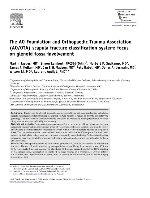
J Shoulder Elbow Surg (2013) 22, 512-520<br />
www.elsevier.com/locate/ymse<br />
The AO Foundation and Orthopaedic Trauma Association<br />
(AO/OTA) <strong>scapula</strong> <strong>fracture</strong> <strong>classification</strong> <strong>system</strong>: focus<br />
on glenoid fossa involvement<br />
Martin Jaeger, MD a , Simon Lambert, FRCSEd(Orth) b , Norbert P. S€udkamp, MD a ,<br />
James F. Kellam, MD c , Jan Erik Madsen, MD d , Reto Babst, MD e , Jonas Andermahr, MD f ,<br />
Wilson Li, MD g , Laurent Audige, PhD h, *<br />
a Department of Orthop€adie und Traumatologie, Universit€atsklinikum Freiburg, Albert-Ludwigs-Universit€at, Freiburg,<br />
Germany<br />
b Shoulder and Elbow Service, The Royal National Orthopaedic Hospital, Stanmore, UK<br />
c Department of Orthopaedic Surgery, Carolinas Medical Center, Charlotte, NC, USA<br />
d Orthopaedic Department, Oslo University Hospital, Ullevaal, Norway<br />
e Klinik f€ur Unfallchirurgie, Luzerner Kantonsspital, Luzern, Switzerland<br />
f Department for Orthopedic and Trauma Surgery, Hospital of the University of Bonn, <strong>Mechernich</strong>, Germany<br />
g Department of Orthopaedics & Traumatology, Queen Elizabeth Hospital, Kowloon, Hong Kong<br />
h AO Clinical Investigation and Documentation, D€ubendorf, Switzerland<br />
Background: Fractures of the glenoid frequently require surgical treatment. A comprehensive and reliable<br />
<strong>scapula</strong> <strong>classification</strong> <strong>system</strong> involving the glenoid <strong>fracture</strong> patterns is needed to describe the underlying<br />
pathology. The AO Scapula Classification Group introduces an appropriate novel <strong>system</strong> that is presented<br />
along with its inter-rater reliability and accuracy.<br />
Materials and methods: An iterative consensus process (involving a series of face-to-face meetings and<br />
agreement studies) with an international group of 7 experienced shoulder surgeons was used to specify<br />
and evaluate a <strong>scapula</strong>r <strong>fracture</strong> <strong>classification</strong> <strong>system</strong> with a focus on <strong>fracture</strong> patterns of the glenoid<br />
fossa. The last evaluation was conducted on a consecutive collection of 120 <strong>scapula</strong>r <strong>fracture</strong>s documented<br />
by both plain radiographs and computed tomography scans including 3-dimensional surface<br />
rendering. Inter-rater reliability was analyzed with k statistics, and accuracy was estimated by latent<br />
class modeling.<br />
Results: Of 120 <strong>scapula</strong>r <strong>fracture</strong>s, 46 involved the glenoid (38%), with 38 classified as F1 articular rim<br />
<strong>fracture</strong>s. The overall median sensitivity and specificity in identifying these <strong>fracture</strong>s were 95% and<br />
93%, respectively. Surgeons’ accuracy in classifying F1 <strong>fracture</strong>s ranged from 86% to 100% (median,<br />
94%). Subsequently, <strong>classification</strong> of simple F1 <strong>fracture</strong>s resulted in a proportion of 36% of anterior<br />
rim <strong>fracture</strong>s, 19% of posterior rim <strong>fracture</strong>s, and 45% of short oblique <strong>fracture</strong>s, with accuracies ranging<br />
from 85% to 98%.<br />
Institutional review board/ethics committee approval: not required.<br />
*Reprint requests: Laurent Audige, PhD, AO Clinical Investigation and<br />
Documentation, Stettbachstrasse 6, CH-8600 D€ubendorf, Switzerland.<br />
E-mail address: laurent.audige@aofoundation.org (L. Audige).<br />
1058-2746/$ - see front matter Ó 2013 Journal of Shoulder and Elbow Surgery Board of Trustees.<br />
http://dx.doi.org/10.1016/j.jse.2012.08.003
Classification of glenoid <strong>fracture</strong>s 513<br />
Conclusion: This new <strong>system</strong> for <strong>scapula</strong>r glenoid <strong>fracture</strong>s has proved to be sufficiently reliable and<br />
accurate when applied by experienced shoulder surgeons. Further validation of the most detailed <strong>system</strong>,<br />
as well as involvement of surgeons with different levels of training in the framework of clinical routine<br />
and research, however, should be considered.<br />
Level of evidence: Level II, Development of Diagnostic Criteria, Diagnostic Study.<br />
Ó 2013 Journal of Shoulder and Elbow Surgery Board of Trustees.<br />
Keywords: Scapula <strong>fracture</strong>; glenoid fossa; <strong>fracture</strong> <strong>classification</strong>; diagnostic; reliability; accuracy<br />
Scapular <strong>fracture</strong>s are rare and account for 1% of all <strong>fracture</strong>s<br />
and 3% to 5% of all shoulder girdle <strong>fracture</strong>s. 7 In the<br />
Swedish population, the annual incidence was estimated at 10<br />
per 10,000 inhabitants. 18 AccordingtoGoss, 13 the <strong>scapula</strong>r<br />
body is involved in up to 45% of all <strong>scapula</strong> <strong>fracture</strong>s, with the<br />
remainder involving the glenoid (approximately 10%), the<br />
<strong>scapula</strong>r neck (25%), the processes (approximately 15%), and<br />
the spine (5%). Treatment modalities depend on the <strong>fracture</strong><br />
location, pattern, and displacement on the one hand and the<br />
patient’s age, demands, and comorbidity on the other. In<br />
contrast to <strong>fracture</strong>s of the <strong>scapula</strong>r body, which are mostly<br />
treated conservatively, surgery is more often indicated for 80%<br />
of <strong>fracture</strong>s involving the glenoid. 24 This is explained by the<br />
specific shape and function of the glenoid. Glenoid rim <strong>fracture</strong>s<br />
can result in joint instability, where surface incongruity may<br />
increase the risk of post-traumatic arthrosis accompanied by<br />
pain and limited function. The surgical approach is influenced<br />
by the main pathology. Not all glenoid <strong>fracture</strong>s can be sufficiently<br />
reduced and stabilized by an anterior approach. Posterior<br />
<strong>fracture</strong> patterns have to be managed through a posterior<br />
approach that is more complex and less frequently practiced.<br />
In the clinical environment, diagnosis of <strong>scapula</strong>r <strong>fracture</strong>s<br />
is based mainly on radiographic examination of the<br />
shoulder. Three plains comprising an anteroposterior view,<br />
outlet view, and axillary view are very helpful in gathering<br />
essential information. If the glenoid is involved, it is<br />
strongly recommended to obtain an additional computed<br />
tomography (CT) scan to precisely evaluate the glenoid<br />
surface. It is also crucial to analyze the <strong>fracture</strong> <strong>system</strong>atically<br />
with the help of a clinically relevant, reliable, and valid<br />
<strong>fracture</strong> <strong>classification</strong> <strong>system</strong>. 5,9,11,19 Until now, a number of<br />
glenoid <strong>fracture</strong> <strong>classification</strong>s have been in use 1,10,14-16,20 ;<br />
however, none of these are comprehensive and validated.<br />
A comprehensive <strong>scapula</strong> <strong>classification</strong> <strong>system</strong> has been<br />
developed as a collaborative effort between the AO Foundation<br />
and the Orthopaedic Trauma Association. 17 The<br />
purpose of this study was to create and validate a focused<br />
<strong>classification</strong> of the glenoid as an extension of this <strong>system</strong>.<br />
Materials and methods<br />
Scapula <strong>classification</strong> group and phase I consensus<br />
development<br />
The international Scapula Classification Group (SCCG)<br />
comprising 7 experienced shoulder specialist surgeons was<br />
established to propose a focused <strong>system</strong> for <strong>fracture</strong>s of the glenoid<br />
fossa, along with definitions and illustrations. This <strong>system</strong> was to<br />
be descriptive, clinically relevant, sufficiently comprehensive, and<br />
intuitive for both inexperienced and experienced surgeons. Under<br />
coordination by a methodologist statistician, a phase 1 consensus<br />
development process was implemented in an iterative manner with<br />
a series of 3 agreement studies (<strong>classification</strong> sessions), followed<br />
by face-to-face review meetings to discuss disagreements and<br />
improve the <strong>system</strong>. Consensus was reached when the SCCG<br />
agreed that the <strong>system</strong> was well defined, clinically relevant, and<br />
reliable for its purpose. 4 This development process was the first of<br />
3 research phases that is required to be completed before the<br />
<strong>classification</strong> could be considered as validated.<br />
Fracture <strong>classification</strong> <strong>system</strong><br />
The <strong>classification</strong> <strong>system</strong> for bony lesions of the <strong>scapula</strong> consists<br />
of separate codes, that is, one each for the involvement of the<br />
articular segment (glenoid fossa), the body, and any of the<br />
processes. In addition, each code comprises 2 levels, where level 1<br />
represents a basic <strong>system</strong> for rapid simplified documentation 17 and<br />
level 2 provides a focused <strong>system</strong> for detailed documentation.<br />
Classification of the <strong>scapula</strong> is divided into 3 regions: the<br />
articular segment, the processes, and the body. The articular<br />
segment includes the area involving the glenoid fossa and the<br />
articular rim, which is limited by a line joining the superior articular<br />
rim to the lateral border of the supra<strong>scapula</strong>r notch and a line<br />
positioned medially and parallel to the plane of the glenoid fossa<br />
that starts cranially at the lateral border of the supra<strong>scapula</strong>r notch.<br />
In the basic <strong>system</strong>, <strong>fracture</strong>s of the articular segment (denoted<br />
F) are classified in 1 of 3 groups (Fig. 1): F0, <strong>fracture</strong> of the<br />
articular segment, not through the glenoid fossa, but resulting in<br />
the fossa being detached from any part of the <strong>scapula</strong> body; F1,<br />
<strong>fracture</strong> involving the glenoid fossa with a simple rim, transverse,<br />
or oblique <strong>fracture</strong> pattern; and F2, multifragmentary joint <strong>fracture</strong><br />
involving the glenoid fossa with 3 or more articular fragments.<br />
The focused <strong>system</strong> is an extended <strong>classification</strong> of F1 or F2<br />
<strong>fracture</strong>s according to their location in 4 quadrants within<br />
the glenoid fossa (Fig. 2). These quadrants are defined by the<br />
equatorial line and the intertubercular line running from the<br />
supraglenoid tubercle to the infraglenoid tubercle. There are 5<br />
subgroups, consisting of 3 for F1 <strong>fracture</strong>s and 2 for F2 <strong>fracture</strong>s:<br />
F1(1), simple anterior articular rim or oblique <strong>fracture</strong>; F1(2),<br />
simple posterior articular rim or oblique <strong>fracture</strong>; F1(3), simple<br />
transverse or short oblique <strong>fracture</strong>; F2(4), multifragmentary<br />
<strong>fracture</strong> with more than 1 <strong>fracture</strong> line exit point; and F2(5),<br />
central <strong>fracture</strong>-dislocation, with no exit line through the rim.<br />
Simple anterior (1) and posterior (2) articular rim or oblique<br />
<strong>fracture</strong>s are further divided into 3 categories (Fig. 3): 1a/2a,
514 M. Jaeger et al.<br />
Figure 1<br />
Fractures of the articular segment. Ó 2012, Jaeger et al.<br />
Figure 2<br />
Four areas defined within the glenoid fossa. Ó 2012, Jaeger et al.<br />
infra-equatorial <strong>fracture</strong> located in 1 quadrant (same side as the<br />
maximum glenoid meridian); 1b/2b, rim <strong>fracture</strong> anterior/posterior<br />
to the maximum glenoid meridian with exits superior/inferior to<br />
the equatorial line; and 1c/2c, <strong>fracture</strong> oblique line exiting on<br />
the opposite side (posterior/anterior) to the maximum glenoid<br />
meridian. (Initial definitions used for the last evaluation session are<br />
presented in Appendix I, available on the journal’s website at www.<br />
jshoulderelbow.org. A revision and final proposal are presented in<br />
Fig. 3).<br />
Simple transverse or short oblique <strong>fracture</strong>s (3) are also further<br />
divided into 3 categories (Fig. 4): 3a, infra-equatorial; 3b, equatorial;<br />
and 3c, supra-equatorial.<br />
For multifragmentary <strong>fracture</strong>s with a combination of 2<br />
‘‘simple’’ <strong>fracture</strong> lines, the focused codes describing this<br />
combination may be added as an optional specification (Fig. 5);<br />
for example, in the case of a transverse and rim <strong>fracture</strong>, the<br />
<strong>classification</strong> would be F2(4/1c3c).<br />
Consecutive series and <strong>classification</strong> sessions<br />
The <strong>classification</strong> <strong>system</strong> was evaluated for reliability and accuracy<br />
using a consecutive series of <strong>scapula</strong> <strong>fracture</strong>s documented<br />
with CT scans and conventional radiographs obtained from<br />
2 centers in Europe and North America. Cases were included
Classification of glenoid <strong>fracture</strong>s 515<br />
Figure 3<br />
Focused <strong>classification</strong> <strong>system</strong> of glenoid fossa rim <strong>fracture</strong>s (F1). Ó 2012, Jaeger et al.<br />
Figure 4<br />
Focused <strong>classification</strong> <strong>system</strong> of simple transverse or short oblique <strong>fracture</strong>s of the glenoid fossa (F1). Ó 2012, Jaeger et al.<br />
when diagnosed with a <strong>scapula</strong> <strong>fracture</strong> within 15 days after the<br />
injury in adult patients (mature skeleton). Whereas the first session<br />
used the 2-dimensional CT series visualized with a standard<br />
DICOM (Digital Imaging and Communications in Medicine)<br />
viewer, the last 2 sessions considered 3-dimensional (3D) reconstructions<br />
for all cases prepared by the primary author (M.J.) on<br />
video sequences (Fig. 6). All diagnostic images were collected in<br />
DICOM (Digital Imaging and Communications in Medicine)
516 M. Jaeger et al.<br />
Figure 5<br />
Focused <strong>classification</strong> <strong>system</strong> of multifragmentary joint <strong>fracture</strong>s (F2). Ó 2012, Jaeger et al.<br />
Figure 6<br />
Example of a 3D CT reconstruction video used for the last <strong>classification</strong> session. Ó 2012, Jaeger et al.<br />
format, anonymized, and distributed on DVD format to SCCG<br />
members, who classified <strong>fracture</strong>s independently ‘‘at home’’<br />
within a 3-month period. Fracture codes were collected electronically<br />
using specifically designed Excel sheets (Microsoft, Redmond,<br />
WA, USA), which were centralized for the analyses. The<br />
results including reliability and accuracy data were reviewed<br />
during face-to-face meetings to identify the reasons for coding<br />
disagreements and improving the <strong>system</strong>.<br />
The final session included 120 cases documented with the<br />
2-dimensional CT series and 3D CT reconstruction videos, as well<br />
as additional radiographs available from 105 cases. Data were<br />
managed and analyzed by use of Intercooled Stata, version 11<br />
(StataCorp LP, College Station, TX, USA). The basic <strong>system</strong> was<br />
first analyzed to assess the likely distribution of articular segment<br />
(F0/F1/F2) <strong>fracture</strong>s in the sample and identify them. The focused<br />
<strong>system</strong> was then applied to this <strong>fracture</strong> subset and separately<br />
analyzed for simple <strong>fracture</strong> patterns (F1) and multifragmentary<br />
joint <strong>fracture</strong>s (F2). Interobserver reliability was evaluated by<br />
means of k coefficients. The k coefficient is commonly used as<br />
a chance-corrected measure of agreement, which ranges from þ1<br />
(complete agreement) to 0 (agreement by chance alone) to less<br />
than 0 (less agreement than expected by chance). The k coefficient<br />
is a useful indicator of reliability, and a value of 0.70 is considered<br />
an adequate sign of reliability. Classification accuracy was estimated<br />
by latent class modeling 22,23 using Latent GOLD software,<br />
version 3.0.1 (Statistical Innovations, Belmont, MA, USA). This<br />
technique aims at identifying the most likely ‘‘true’’ distribution of<br />
<strong>fracture</strong> classes in the population of <strong>scapula</strong> <strong>fracture</strong>s based on the<br />
evaluated sample and the agreement data collected among the<br />
participating surgeons; for each class, the degree of <strong>classification</strong><br />
accuracy for each surgeon is also estimated. 6<br />
Results<br />
When identifying cases using the basic <strong>system</strong> to classify<br />
a <strong>fracture</strong> of the articular segment (denoted F), the<br />
7 shoulder specialists were in agreement for 73% of the 120
Classification of glenoid <strong>fracture</strong>s 517<br />
cases. The overall k coefficient was 0.79, and the surgeons’<br />
pair-wise k (21 pairs) ranged from 0.66 to 0.93. The sample<br />
identifying the most likely ‘‘true’’ distribution included<br />
46 cases (38%) with an F <strong>fracture</strong>, where the accuracy in<br />
identifying these <strong>fracture</strong>s (diagnosis sensitivity) ranged<br />
from 91% to 99% (median, 95%). The surgeons’ accuracy<br />
in identifying the remaining 74 cases as non–articular<br />
segment <strong>fracture</strong>s ranged from 86% to 98% (median, 93%).<br />
The 46 F <strong>fracture</strong>s were further divided as follows: 1 F0<br />
<strong>fracture</strong>, 38 F1 <strong>fracture</strong>s, and 7 F2 <strong>fracture</strong>s. Surgeons<br />
agreed on this sub<strong>classification</strong> in 72% of these cases (33),<br />
with an overall k coefficient of 0.64. The surgeons’<br />
pair-wise k (21 pairs) ranged from 0.43 to 0.92 (median,<br />
0.64). Surgeons were 86% to 100% accurate (median, 94%)<br />
in terms of classifying F1 <strong>fracture</strong>s. The number of F2<br />
<strong>fracture</strong>s was considered too small for estimation of the<br />
surgeons’ <strong>classification</strong> accuracy with an adequate degree<br />
of precision; nevertheless, 4 surgeons correctly classified<br />
6 of the 7 multifragmentary <strong>fracture</strong>s, with a minimum of<br />
4 of these <strong>fracture</strong>s correctly classified among the other<br />
3 surgeons. The only F0 <strong>fracture</strong> in the sample was<br />
correctly classified by 5 surgeons (Fig. 7), whereas the<br />
other 2 surgeons classified it as a simple fossa <strong>fracture</strong> (F1)<br />
or simple body <strong>fracture</strong> (B1).<br />
Surgeons attained full agreement in the <strong>classification</strong> of<br />
half of the 38 simple glenoid fossa (F1) <strong>fracture</strong>s according<br />
to the higher level of the focused <strong>system</strong> (Figs. 3 and 4;<br />
Appendix I, available on the journal’s website at www.<br />
jshoulderelbow.org). The overall k coefficient was 0.66,<br />
with the surgeons’ pair-wise k ranging from 0.35 to 0.82<br />
(median, 0.67) (Table I). The estimated distribution of cases<br />
within the sample included 14 F1(1) <strong>fracture</strong>s, 7 F1(2)<br />
<strong>fracture</strong>s, and 17 F1(3) <strong>fracture</strong>s with category-specific<br />
k coefficients of 0.70, 0.65, and 0.62, respectively. There<br />
was no central <strong>fracture</strong>-dislocation among the 7 multifragmentary<br />
joint <strong>fracture</strong>s (Fig. 6), and all were classified<br />
as having more than 2 <strong>fracture</strong> line exit points [code F2(4)]<br />
with full agreement among the surgeons.<br />
Classification session data supported the distinction of<br />
simple fossa <strong>fracture</strong>s into 3 classes with the following<br />
proportions: 36%, 19%, and 45% (Table II). The largest<br />
group, representing transverse or short oblique <strong>fracture</strong>s<br />
(class 3), was classified with 87% to 93% accuracy by<br />
5 surgeons and 70% accuracy (median, 91%) for the other<br />
2. The second largest <strong>fracture</strong> group represented anterior<br />
rim or oblique <strong>fracture</strong>s (class 1), showing the highest<br />
<strong>classification</strong> accuracies from 85% to 98% among the<br />
surgeons (median, 91%). The posterior rim or oblique<br />
<strong>fracture</strong>s (class 2) were classified with 80% to 96% accuracy<br />
by 4 surgeons and 64% to 69% accuracy by the other<br />
3 (median, 80%). Incorrect <strong>classification</strong> of rim and oblique<br />
<strong>fracture</strong>s as transverse and short oblique <strong>fracture</strong> types<br />
occurred more frequently, showing the higher ability to<br />
discern between anterior and posterior rim <strong>fracture</strong>s.<br />
Further <strong>classification</strong> into the 3 subgroups of simple glenoid<br />
fossa <strong>fracture</strong>s (ie, a, b, and c) showed a lower degree of<br />
interobserver reliability, with overall k coefficients of 0.46<br />
and 0.39 based on the combined group of anterior and<br />
posterior <strong>fracture</strong>s (Fig. 3; Appendix I, available on the<br />
journal’s website at www.jshoulderelbow.org) and the group<br />
of transverse <strong>fracture</strong>s, respectively (Fig. 4, Table I).<br />
Surgeons’ pair-wise k coefficients ranged from 0.07 to 0.89<br />
(median, 0.53) (anterior and posterior <strong>fracture</strong>s) and from<br />
0.03 to 0.88 (median, 0.36) (transverse <strong>fracture</strong>s). After<br />
reviewing these results, members of the SCCG unanimously<br />
agreed that a revision of categories 1c and 2c was required<br />
and a final proposal was made, as presented in Figure 3.<br />
Discussion<br />
The SCCG was established to develop and validate a clinically<br />
useful comprehensive <strong>scapula</strong> <strong>classification</strong> <strong>system</strong>. In<br />
a first phase, special interest was targeted at testing its<br />
reliability to allow its integration in a clinical setting. This<br />
study focuses on glenoid <strong>fracture</strong>s.<br />
In our series of <strong>scapula</strong> <strong>fracture</strong>s, the involvement of<br />
articular segment <strong>fracture</strong>s was estimated for 38% of the 120<br />
cases. This incidence is much higher compared with data<br />
reported by Goss 15 suggesting that <strong>fracture</strong>s of the glenoid<br />
cavity account for up to 10% of all <strong>scapula</strong> <strong>fracture</strong>s. Ideberg<br />
et al 18 also reported involvement of the glenoid cavity in<br />
about 30% of <strong>scapula</strong> <strong>fracture</strong>s, whichdsimilar to our own<br />
studydmight be due to the case selection process. Our<br />
<strong>scapula</strong>r <strong>fracture</strong> cases were consecutively collected from 2<br />
major level I trauma centers in the United States and central<br />
Europe regardless of their treatments. To achieve a precise<br />
diagnosis, both plain radiographs and CT scans were obtained.<br />
Obtaining the latter, especially in combination with<br />
3D volume rendering, is believed to increase the accuracy in<br />
detecting glenoid <strong>fracture</strong>s and, therefore, might also<br />
explain the higher proportion of glenoid <strong>fracture</strong>s detected<br />
in our study compared with those from 3 more recent<br />
publications. 2,8,21 In an earlier evaluation session using only<br />
radiographs for 33 cases, 8 cases (24%) were diagnosed with<br />
glenoid involvement. The misdiagnosis of glenoid <strong>fracture</strong>s<br />
may lead to either joint instability or joint incongruence,<br />
with the potential consequence of secondary arthrosis, so it<br />
is mandatory to identify these <strong>fracture</strong>s precisely. We<br />
strongly recommend performing a CT scan routinely in all<br />
cases of <strong>scapula</strong>r <strong>fracture</strong>s.<br />
Using the entire <strong>scapula</strong> <strong>classification</strong> <strong>system</strong> provided<br />
by the SCCG, the involved shoulder surgeons were<br />
extremely accurate (>90%) in identifying all <strong>fracture</strong>s<br />
involving the articular segment (code F). According to the<br />
focused <strong>classification</strong> <strong>system</strong>, the incidence of anterior rim<br />
<strong>fracture</strong>s was 2 times higher compared with posterior<br />
<strong>fracture</strong>s in our series. Single transverse or short oblique<br />
<strong>fracture</strong>s were slightly more frequent compared with anterior<br />
rim <strong>fracture</strong>s. Multifragmentary <strong>fracture</strong>s with central<br />
<strong>fracture</strong>-dislocation were rare; this type of <strong>fracture</strong> pattern<br />
was not found in any of the 120 cases. The use of the
518 M. Jaeger et al.<br />
Figure 7<br />
Fracture of the articular segment not involving the glenoid fossa (F0). Ó 2012, Jaeger et al.<br />
Table I Coding agreement and reliability for glenoid fossa <strong>fracture</strong>s according to focused <strong>system</strong><br />
Fracture subsets Categories) No. of cases y Full agreement k Coefficient<br />
Overall Pair-wise (21 pairs)<br />
Minimum Median Maximum<br />
Simple fossa (F1) 50% 0.66 0.35 0.67 0.82<br />
1 14 0.70<br />
2 7 0.65<br />
3 17 0.62<br />
Multifragmentary fossa (F2) 4/5 7/0 100%<br />
F1(1) and F1(2) combined a/b/c 4/9/8 24% 0.46 0.07 0.53 0.89<br />
F1(3) a/b/c 3/8/6 29% 0.39 0.03 0.36 0.88<br />
) The categories of 1 to 5 represent the following: 1, anterior simple rim or oblique <strong>fracture</strong>; 2, posterior simple rim or oblique <strong>fracture</strong>; 3, simple<br />
transverse or short oblique <strong>fracture</strong>; 4, multifragmentary fossa with more than 2 <strong>fracture</strong> line exit points; and 5, multifragmentary fossa with central<br />
<strong>fracture</strong>-dislocation (no exit line through the rim).<br />
y Estimated by latent class analysis and the distribution of the surgeons’ codes.<br />
detailed sub<strong>classification</strong> seems to be reproducible with<br />
high accuracy (>80%) in all categories of F1 (1, 2, and 3).<br />
A more detailed description of rim <strong>fracture</strong>s with respect to<br />
their location to the equator becomes less reliable and is<br />
most pronounced when differentiating short oblique <strong>fracture</strong>s<br />
[F1(3)]. This cannot be done without a CT scan and<br />
was still not highly reliable with 3D volume rendering. One<br />
reason for this outcome might be the method used in this<br />
study. To provide the same data for all participating<br />
surgeons, 1 shoulder surgeon performed the volume<br />
rendering of all CT scans. The result was presented to the<br />
surgeons as a movie sequence in a 360 horizontal and 360 <br />
vertical rotation; this setting was chosen based on common<br />
practice (usually of the radiologist) in presenting these<br />
types of data for further evaluation. Subsequently, the other<br />
shoulder surgeons did not perform their own 3D analysis,<br />
that is, they were unable to rotate or focus the 3D scans<br />
according to their own needs, and as such, their accuracy<br />
may have been limited. On the other hand, these results<br />
highlight the difficulty in distinguishing <strong>fracture</strong>s bordering<br />
between 2 categories. Because the distribution of all glenoid<br />
<strong>fracture</strong>s can be considered a continuum of possible<br />
patterns, any category is inherently associated with some<br />
level of imprecision for adjacent <strong>fracture</strong> pathologies.<br />
Former <strong>scapula</strong> <strong>classification</strong>s, especially of the glenoid,<br />
were conducted based on the assessment of plain radiographs.<br />
5,15,24 All of them were published without further<br />
reliability assessment. To our knowledge, this is the first<br />
study providing an evaluation of <strong>classification</strong> reliability<br />
and accuracy. Armitage et al 3 recently provided a frequency<br />
map of surgically treated <strong>scapula</strong> <strong>fracture</strong>s. To our knowledge,<br />
this was the first article until now describing the use<br />
of 3D CT volume rendering to enhance the mapping of<br />
<strong>scapula</strong> <strong>fracture</strong>s. In contrast to our study, Armitage et al
Classification of glenoid <strong>fracture</strong>s 519<br />
Table II Classification accuracy for patterns of simple <strong>fracture</strong>s<br />
of glenoid fossa<br />
Surgeons and <strong>fracture</strong> categories Fracture classes<br />
1 2 3<br />
Class size 36% 19% 45%<br />
Surgeon 1<br />
1: Anterior rim or oblique 85%) 34% 29%<br />
2: Posterior rim or oblique 0% 64%) 0%<br />
3: Transverse or short oblique 15% 2% 71%)<br />
Surgeon 2<br />
1: Anterior rim or oblique 88%) 1% 1%<br />
2: Posterior rim or oblique 1% 80%) 13%<br />
3: Transverse or short oblique 11% 18% 87%)<br />
Surgeon 3<br />
1: Anterior rim or oblique 91%) 2% 1%<br />
2: Posterior rim or oblique 0% 96%) 6%<br />
3: Transverse or short oblique 9% 3% 93%)<br />
Surgeon 4<br />
1: Anterior rim or oblique 98%) 2% 9%<br />
2: Posterior rim or oblique 0% 69%) 0%<br />
3: Transverse or short oblique 2% 29% 91%)<br />
Surgeon 5<br />
1: Anterior rim or oblique 98%) 18% 1%<br />
2: Posterior rim or oblique 0% 64%) 6%<br />
3: Transverse or short oblique 1% 18% 93%)<br />
Surgeon 6<br />
1: Anterior rim or oblique 98%) 2% 19%<br />
2: Posterior rim or oblique 1% 80%) 13%<br />
3: Transverse or short oblique 1% 18% 68%)<br />
Surgeon 7<br />
1: Anterior rim or oblique 91%) 2% 6%<br />
2: Posterior rim or oblique 8% 96%) 0%<br />
3: Transverse or short oblique 1% 2% 93%)<br />
Classification accuracies<br />
Minimum 85% 64% 68%<br />
Median 91% 80% 91%<br />
Maximum 98% 96% 93%<br />
) Percentages indicate surgeon <strong>classification</strong> accuracies (percentages<br />
of correct <strong>classification</strong>).<br />
mainly focused on <strong>scapula</strong> body and neck <strong>fracture</strong>s.<br />
Although they did not propose a <strong>classification</strong> <strong>system</strong> for<br />
glenoid <strong>fracture</strong>s, they documented involvement of the<br />
glenoid in 17% of all <strong>scapula</strong> <strong>fracture</strong>s. This incidence is<br />
surprisingly low considering the fact that these authors<br />
included <strong>fracture</strong>s undergoing operative treatment.<br />
According to the current treatment decision process, 24 the<br />
<strong>fracture</strong> population of Armitage et al should have a bias<br />
toward including proportionally more glenoid <strong>fracture</strong>s. On<br />
the other hand, Ada and Miller 1 reported glenoid <strong>fracture</strong>s<br />
in 10% of their series based on assessments using plain<br />
radiographs. Such heterogeneity in the proportion of glenoid<br />
<strong>fracture</strong>s may be a reflection that the distribution of<br />
<strong>fracture</strong> patterns is likely to differ between settings and, as<br />
such, a comparison between <strong>scapula</strong> <strong>fracture</strong> studies<br />
requires adequate <strong>classification</strong> and documentation.<br />
The combination of a glenoid <strong>fracture</strong> and <strong>scapula</strong> body<br />
<strong>fracture</strong> is frequent, so it is also important to have<br />
a combined <strong>classification</strong> considering both the glenoid and<br />
the rest of the <strong>scapula</strong>. This <strong>classification</strong> <strong>system</strong> should be<br />
descriptive and comprehensive, as well as aid in the<br />
decision-making process for individual therapy, especially<br />
if minimally invasive approaches are intended. 12 To our<br />
knowledge, the current <strong>scapula</strong> <strong>classification</strong> <strong>system</strong> is<br />
most comprehensive as well as highly reliable and accurate.<br />
Other existing <strong>scapula</strong> <strong>fracture</strong> <strong>classification</strong> <strong>system</strong>s lack<br />
a more focused description of glenoid <strong>fracture</strong>s. The<br />
commonly used <strong>classification</strong> for glenoid <strong>fracture</strong>s<br />
according to Ideberg et al 18 and Goss 15 does not consider<br />
the rest of the <strong>scapula</strong>; simple glenoid <strong>fracture</strong> lines<br />
between anterior and posterior rim <strong>fracture</strong>s (types Ia and<br />
Ib) and transverse <strong>fracture</strong>s (types II-V) are distinguished,<br />
and type VI <strong>fracture</strong>s represent multifragmentary <strong>fracture</strong>s.<br />
The <strong>classification</strong> of <strong>scapula</strong>r <strong>fracture</strong>s by Ada and Miller 1<br />
distinguishes among 4 types of <strong>fracture</strong>s including type III<br />
glenoid <strong>fracture</strong>s; however, further sub<strong>classification</strong>s of<br />
glenoid <strong>fracture</strong>s do not exist.<br />
This evaluation is limited by its sample size, although it is<br />
one of the largest ever used in such a project. There is often<br />
a tradeoff between increasing the case collection and making<br />
the evaluation feasible when busy clinicians are involved.<br />
Results should be considered with caution when the number<br />
of cases in any category is low, such as in the more detailed a,<br />
b, and c category <strong>system</strong>. In addition, nothing can be said<br />
about the reliability of coding multifragmentary glenoid<br />
fossa <strong>fracture</strong>s with central <strong>fracture</strong>-dislocation. Although<br />
<strong>classification</strong> of F1 simple fossa <strong>fracture</strong>s according to<br />
anterior rim, posterior rim, or transverse patterns appears<br />
reasonably reliable and accurate, location of these <strong>fracture</strong>s<br />
with regard to the 4 quadrants remains open for further<br />
clarification and evaluation. The wide range of surgeons’<br />
pair-wise k coefficients shows that different applications of<br />
the definitions occurred among surgeons; thus, further<br />
clarification and specification of the <strong>system</strong> are required.<br />
Transverse or short oblique <strong>fracture</strong>s typically separate<br />
a process (ie, coracoid 3c) or involve the body (3a and 3b).<br />
The 3a and 3b <strong>fracture</strong>s are typically located near the<br />
equator. In contrast, 1c and 2c rim <strong>fracture</strong>s are much smaller<br />
because they are located near the glenoid edge.<br />
The <strong>classification</strong> process and evaluation sessions were<br />
performed by experienced surgeons only, which may not<br />
reflect the validity of the <strong>system</strong> in general practice. As<br />
advocated by Audige et al, 4 a further study is required to<br />
assess the reliability and accuracy of this <strong>classification</strong><br />
<strong>system</strong> when used by surgeons of varying education and<br />
experience. Such evaluation would also consider the final<br />
change made to the rim <strong>fracture</strong> categories. This study could<br />
also be influenced by the preparation of the 3D volume<br />
rendering datasets by only the main author. Furthermore, this<br />
report is not intended to make recommendations regarding<br />
any treatment modalities based on this <strong>classification</strong><br />
<strong>system</strong> at this early stage; rather, our intent is to provide
520 M. Jaeger et al.<br />
a comprehensive, reliable, and accurate <strong>classification</strong> <strong>system</strong><br />
as a base for further clinical examinations.<br />
Conclusion<br />
In summary, it was possible to create a comprehensive<br />
<strong>scapula</strong> <strong>classification</strong> <strong>system</strong> focusing also on <strong>fracture</strong>s of<br />
the articular segment. This <strong>classification</strong> <strong>system</strong> is believed<br />
to be clinically relevant and appears reasonably reliable<br />
and accurate with respect to glenoid <strong>fracture</strong>s at both the<br />
basic and more detailed levels. Further validation of the<br />
most detailed <strong>system</strong> in a larger case sample with surgeons<br />
of different training backgrounds should be considered.<br />
Acknowledgments<br />
The authors thank the following persons for their participation<br />
and support in the development of the basic <strong>scapula</strong><br />
<strong>classification</strong> <strong>system</strong>: Edward Harvey (Montreal General<br />
Hospital, Montreal, Quebec, Canada), Dolfi Herscovici<br />
(Florida Orthopaedic Institute, Temple Terrace, FL, USA),<br />
and Julie Agel and Sean Nork (Harborview Medical Center,<br />
Seattle, WA, USA). The authors also thank M. Wilhelmi,<br />
PhD (AO Clinical Investigation and Documentation), for<br />
the preparation and copy editing of this manuscript.<br />
Disclaimer<br />
The authors, their immediate families, and any research<br />
foundations with which they are affiliated have not<br />
received any financial payments or other benefits from<br />
any commercial entity related to the subject of this article.<br />
The surgeons who participated in the successive faceto-face<br />
meetings were supported with regard to their travel<br />
expenses and received a small per-diem payment. Support<br />
for this study was provided by the AO Foundation.<br />
Supplementary data<br />
Supplementary data related to this article can be found<br />
online at http://dx.doi.org/10.1016/j.jse.2012.08.003.<br />
References<br />
1. Ada JR, Miller ME. Scapular <strong>fracture</strong>s. Analysis of 113 cases. Clin<br />
Orthop Relat Res 1991:174-80.<br />
2. Anavian J, Conflitti JM, Khanna G, Guthrie ST, Cole PA. A reliable<br />
radiographic measurement technique for extra-articular <strong>scapula</strong>r<br />
<strong>fracture</strong>s. Clin Orthop Relat Res 2011;469:3371-8. http://dx.doi.org/<br />
10.1007/s11999-011-1820-3<br />
3. Armitage BM, Wijdicks CA, Tarkin IS, Schroder LK, Marek DJ,<br />
Zlowodzki M, et al. Mapping of <strong>scapula</strong>r <strong>fracture</strong>s with<br />
three-dimensional computed tomography. J Bone Joint Surg Am 2009;<br />
91:2222-8. http://dx.doi.org/10.2106/JBJS.H.00881<br />
4. Audige L, Bhandari M, Hanson B, Kellam J. A concept for the validation<br />
of <strong>fracture</strong> <strong>classification</strong>s. J Orthop Trauma 2005;19:404-9.<br />
http://dx.doi.org/10.1097/01.bot.0000155310.04886.37<br />
5. Audige L, Bhandari M, Kellam J. How reliable are reliability studies<br />
of <strong>fracture</strong> <strong>classification</strong>s? A <strong>system</strong>atic review of their methodologies.<br />
Acta Orthop Scand 2004;75:184-94. http://dx.doi.org/10.1080/<br />
00016470412331294445<br />
6. Audige L, Hunter J, Weinberg A, Magidson J, Slongo T. Development<br />
and evaluation process of a paediatric long-bone <strong>fracture</strong> <strong>classification</strong><br />
proposal. Eur J Trauma 2004;30:248-54. http://dx.doi.org/10.1007/<br />
s00068-004-1428-3<br />
7. Bahk MS, Kuhn JE, Galatz LM, Connor PM, Williams GR Jr. Acromioclavicular<br />
and sternoclavicular injuries and clavicular, glenoid, and<br />
<strong>scapula</strong>r <strong>fracture</strong>s. J Bone Joint Surg Am 2009;91:2492-510.<br />
8. Bartonicek J, Tucek M, Fric V. Radiographic evaluation of <strong>scapula</strong><br />
<strong>fracture</strong>s [in Czech]. Rozhl Chir 2009;88:84-8.<br />
9. Burstein AH. Fracture <strong>classification</strong> <strong>system</strong>s: do they work and are<br />
they useful? J Bone Joint Surg Am 1993;75:1743-4.<br />
10. Euler E, Habermeyer P, Kohler W, Schweiberer L. Scapula<br />
<strong>fracture</strong>sd<strong>classification</strong> and differential therapy [in German]. Orthopade<br />
1992;21:158-62.<br />
11. Garbuz DS, Masri BA, Esdaile J, Duncan CP. Classification <strong>system</strong>s in<br />
orthopaedics. J Am Acad Orthop Surg 2002;10:290-7.<br />
12. Gauger EM, Cole PA. Surgical technique: a minimally invasive<br />
approach to <strong>scapula</strong> neck and body <strong>fracture</strong>s. Clin Orthop Relat Res<br />
2011;469:3390-9. http://dx.doi.org/10.1007/s11999-011-1970-3<br />
13. Goss TP. Frequency of <strong>scapula</strong>r <strong>fracture</strong>s. In: Rockwood CA,<br />
Matsen FA, Wirth MA, Lippitt SB, editors. Fractures of the <strong>scapula</strong>.<br />
3rd ed. Philadelphia: Saunders; 2004. p. 413-54.<br />
14. Goss TP. Scapular <strong>fracture</strong>s and dislocations: diagnosis and treatment.<br />
J Am Acad Orthop Surg 1995;3:22-33.<br />
15. Goss TP. Fractures of the glenoid cavity. J Bone Joint Surg Am 1992;<br />
74:299-305.<br />
16. Hardegger FH, Simpson LA, Weber BG. The operative treatment of<br />
<strong>scapula</strong>r <strong>fracture</strong>s. J Bone Joint Surg Br 1984;66:725-31.<br />
17. Harvey E, Audige L, Herscovici D, Agel J, Madsen JE, Babst R, et al.<br />
Development and validation of the new international <strong>classification</strong> for<br />
<strong>scapula</strong> <strong>fracture</strong>s. J Orthop Trauma 2012;26:364-9. http://dx.doi.org/<br />
10.1097/BOT.0b013e3182382625<br />
18. Ideberg R, Grevsten S, Larsson S. Epidemiology of <strong>scapula</strong>r <strong>fracture</strong>s.<br />
Incidence and <strong>classification</strong> of 338 <strong>fracture</strong>s. Acta Orthop Scand 1995;<br />
66:395-7.<br />
19. Martin JS, Marsh JL. Current <strong>classification</strong> of <strong>fracture</strong>s. Rationale and<br />
utility. Radiol Clin North Am 1997;35:491-506.<br />
20. Mayo KA, Benirschke SK, Mast JW. Displaced <strong>fracture</strong>s of the glenoid<br />
fossa. Results of open reduction and internal fixation. Clin Orthop<br />
Relat Res 1998:122-30.<br />
21. Tadros AM, Lunsjo K, Czechowski J, Corr P, Abu-Zidan FM.<br />
Usefulness of different imaging modalities in the assessment of<br />
<strong>scapula</strong>r <strong>fracture</strong>s caused by blunt trauma. Acta Radiol 2007;48:71-5.<br />
http://dx.doi.org/10.1080/02841850601026435<br />
22. Uebersax JS, Grove WM. Latent class analysis of diagnostic agreement.<br />
Stat Med 1990;9:559-72.<br />
23. Vermunt J, Magidson J. Latent class cluster analysis. In:<br />
Hagenaars JA, McCutcheon AL, editors. Applied latent class analysis.<br />
Cambridge: Cambridge University Press; 2002. p. 89-106.<br />
24. Zlowodzki M, Bhandari M, Zelle BA, Kregor PJ, Cole PA. Treatment<br />
of <strong>scapula</strong> <strong>fracture</strong>s: <strong>system</strong>atic review of 520 <strong>fracture</strong>s in 22 case<br />
series. J Orthop Trauma 2006;20:230-3. http://dx.doi.org/10.1097/<br />
00005131-200603000-00013