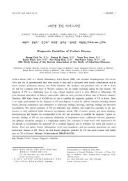
궤양성 대장염 진단 가이드라인 - 대한내과학회
궤양성 대장염 진단 가이드라인 - 대한내과학회
궤양성 대장염 진단 가이드라인 - 대한내과학회

Create successful ePaper yourself
Turn your PDF publications into a flip-book with our unique Google optimized e-Paper software.
대한소화기학회지 2009;53:145-160<br />
□ REVIEW □<br />
<strong>궤양성</strong> <strong>대장염</strong> <strong>진단</strong> <strong>가이드라인</strong><br />
중앙대학교 의과대학 내과학교실*, 이화여자대학교 의과대학 내과학교실 † , 가톨릭대학교 의과대학 내과학교실 ‡ ,<br />
서울대학교 의과대학 내과학교실 § , 한양대학교 의과대학 내과학교실 ∥<br />
최창환*ㆍ정성애 † ㆍ이보인 ‡ ㆍ이강문 ‡ ㆍ김주성 § ㆍ한동수 ∥ ㆍ대한장연구학회 IBD 연구회<br />
Diagnostic Guideline of Ulcerative Colitis<br />
Chang Hwan Choi, M.D.*, Sung Ae Jung, M.D. † , Bo In Lee, M.D. ‡ ,<br />
Kang Moon Lee, M.D. ‡ , Joo Sung Kim, M.D. § , Dong Soo Han, M.D. ∥ , and<br />
IBD Study Group of the Korean Association of the Study of Intestinal Diseases<br />
Department of Internal Medicine, Chung-Ang University College of Medicine*, Ewha Womans University<br />
College of Medicine † , The Catholic University of Korea College of Medicine ‡ , Seoul National University<br />
College of Medicine § , Hanyang University College of Medicine ∥ , Seoul, Korea<br />
Ulcerative colitis is a chronic inflammatory disorder causing mucosal inflammation of the colorectum with crypt<br />
abnormality on biopsy. It affects the rectum and a variable extent of the colon in continuity. Ulcerative colitis is<br />
characterized by a relapsing and remitting course. It arises from an interaction between genetic and environmental<br />
factors, but the precise etiology is unknown. The incidence and prevalence in Korea are still low compared with<br />
those of Western countries, but have increased in recent years. There are many challenging issues on the diagnosis<br />
of ulcerative colitis, and sometimes these lead to differences in practice between clinicians. Therefore, IBD<br />
Study Group of KASID set out the Korean diagnostic guideline of ulcerative colitis. The diagnosis is based on<br />
clinical, endoscopic, radiologic, and histologic criteria. The symptoms are dependent upon the extent and severity<br />
of disease and most commonly include bloody diarrhea, rectal bleeding, and/or urgency. The systemic symptoms<br />
of malaise, tachycardia, fever, or weight loss are features of a severe attack. The laboratory findings may reveal<br />
leucocytosis, thrombocytosis, iron deficiency anemia, hypoalbuminemia, and elevated erythrocyte sedimentation rate<br />
and C-reactive protein indicating severe disease activity or chronicity. For the elimination of infectious causes, microbial<br />
investigation with stool specimens should be performed for common enteric pathogens including assays for<br />
Clostridium difficile toxin, and sometimes for amoeba or other parasites. The most typical endoscopic features are<br />
continuous, confluent, and concentric colonic involvement proximal to the anal verge. Endoscopic severity may be<br />
best well reflected by the presence of mucosal friability, spontaneous bleeding, and deep ulcerations. Typical<br />
pathologic findings are composed of widespread crypt architectural distortion (cryptitis, crypt abscess, and crypt<br />
atrophy), heavy, diffuse lamina propria cell infiltration, and basal plasmacytosis. (Korean J Gastroenterol<br />
2009;53:145-160)<br />
Key Words: Ulcerative colitis; Diagnosis, Guideline<br />
연락처: 한동수, 471-701, 경기도 구리시 교문동 249-1<br />
한양대학교 구리병원 소화기내과<br />
Tel: (031) 560-2226, Fax: (031) 555-2998<br />
E-mail: hands@hanyang.ac.kr<br />
* 본 연구는 보건복지부 보건의료기술진흥사업의 지원에<br />
의하여 이루어진 것임(과제고유번호: A080588).<br />
Correspondence to: Dong Soo Han, M.D.<br />
Department of Internal Medicine, Hanyang University College<br />
of Medicine, 249-1, Gyomun-dong, Guri 471-701, Korea<br />
Tel: +82-31-560-2226, Fax: +82-31-555-2998<br />
E-mail: hands@hanyang.ac.kr
146 대한소화기학회지: 제53권 제3호, 2009<br />
서 론<br />
<strong>궤양성</strong> <strong>대장염</strong>은 대장 점막 또는 점막하층에 국한된 염증을<br />
특징으로 하는 원인 불명의 만성 염증성 장질환으로, 호전과<br />
악화가 반복되는 혈성 설사와 대변절박증(urgency) 및 복통 등<br />
이 주증상이다. 1,2 <strong>궤양성</strong> <strong>대장염</strong>은 유전, 환경 요인이 복합적<br />
으로 작용하여 발생하며, 전 세계적으로 분포하지만 북미와<br />
북유럽에서 가장 호발한다. 인종별로는 유태인과 코카시안에<br />
서 발생이 많고 동양인에서는 상대적으로 드물다. 2,3 하지만<br />
최근에는 남유럽과 우리나라를 포함하는 아시아 국가, 그리고<br />
다른 개발도상국에서도 발병률이 증가하고 있다. 3-5<br />
<strong>궤양성</strong> <strong>대장염</strong>으로 인해 많은 환자들이 고통을 받고 있지<br />
만 이 질병의 명확한 원인이나 치료 방법은 제시되지 않았<br />
으며, 많은 임상 연구에도 불구하고 전문가의 판단이나 의<br />
견으로밖에 답변이 어려운 문제들이 많다. 따라서 환자 진<br />
료나 치료 방법에 차이가 있을 수 있으며, 이러한 차이를 줄<br />
이기 위해서는 <strong>궤양성</strong> <strong>대장염</strong>의 <strong>진단</strong>과 치료에 대한 가이드<br />
라인이 필요하다. 하지만, 현재 국내에는 <strong>궤양성</strong> <strong>대장염</strong> 진<br />
단을 위한 <strong>가이드라인</strong>이 없어 서양과 일본의 <strong>가이드라인</strong>에<br />
따라 <strong>진단</strong>을 하고 있는 실정이다. 따라서 우리나라 실정에<br />
맞는 <strong>궤양성</strong> <strong>대장염</strong> <strong>진단</strong> <strong>가이드라인</strong> 설정이 필요하며 이를<br />
통해 잘못된 치료, 혹은 치료 지연으로 인한 문제를 예방할<br />
수 있을 것이다. 또한 질병에 관한 통일된 용어를 사용함으<br />
로써 환자를 치료하는 임상의사와 연구자들 서로 간에 의사<br />
소통의 혼란을 줄일 수 있을 것으로 기대한다.<br />
이번 <strong>궤양성</strong> <strong>대장염</strong> <strong>진단</strong> <strong>가이드라인</strong>은 대한장연구학회<br />
IBD 연구회에서 제정하였으며, 이는 <strong>진단</strong> 기준을 제시하는<br />
것이 아니고, 진료와 연구를 담당하는 의사가 <strong>궤양성</strong> 대장<br />
염을 <strong>진단</strong>하고, 질병 상태를 정의 및 분류하는 데 방향을 제<br />
시하기 위한 것이다. 이를 위하여 현재까지 국내외에서 발<br />
표된 다양한 연구 자료들을 검토하였고, 국내에서 <strong>궤양성</strong><br />
<strong>대장염</strong>을 <strong>진단</strong>하는 데 고려하여야 할 점들도 반영하였다.<br />
사안에 따라 근거 자료가 불충분하여 명확한 결론을 내기<br />
어려운 경우에는 IBD 연구회 전문가들의 토론을 통해 권고<br />
안을 제시하였으며, 이는 새로이 발표되는 연구 결과들에<br />
따라 지속적으로 수정 및 보완될 것이다. 본문의 권고 사항<br />
의 의학 근거 등급은 유럽<strong>가이드라인</strong> 6 의 형식을 따라 표기<br />
하였다(Table 1).<br />
본 론<br />
1. 정의<br />
<strong>궤양성</strong> <strong>대장염</strong>은 연속적으로 직장에서 근위부로 침범하<br />
Table 1. Levels of Evidence and Grades of Recommendation Based on the Oxford Center for Evidence Based Medicine<br />
Evidence<br />
levels (EL)<br />
Diagnostic study<br />
Therapeutic study<br />
1a<br />
1b<br />
1c<br />
2a<br />
2b<br />
2c<br />
3a<br />
3b<br />
4<br />
5<br />
Systematic review (SR) with homogeneity of level 1<br />
diagnostic studies<br />
Validating cohort study with good reference standards<br />
Specificity is so high that a positive result rules in the<br />
diagnosis or sensitivity is so high that a negative<br />
result rules out the diagnosis<br />
SR with homogeneity of level >2 diagnostic studies<br />
Exploratory cohort study with good reference standards<br />
SR with homogeneity of 3b and better studies<br />
Non-consecutive study; or without consistently applied<br />
reference standards<br />
Case-control study, poor or non-independent reference<br />
standard<br />
Expert opinion without explicit critical appraisal, or based<br />
on physiology, bench research or “first principles”<br />
Recommendation grades (RG)<br />
A<br />
B<br />
C<br />
D<br />
Consistent level 1 studies<br />
Consistent level 2 or 3 studies or extrapolations from level 1 studies<br />
Level 4 studies or extrapolations from level 2 or 3 studies<br />
Level 5 evidence or troublingly inconsistent or inconclusive studies of any level<br />
Systematic review (SR) with homogeneity of randomized<br />
controlled trials (RCTs)<br />
Individual RCT (with narrow confidence interval)<br />
All or none<br />
SR (with homogeneity) of cohort studies<br />
Individual cohort study (including low quality RCT; e.g.,<br />
최창환 외 5인. <strong>궤양성</strong> <strong>대장염</strong> <strong>진단</strong> <strong>가이드라인</strong> 147<br />
는 점막 또는 점막하층의 염증을 특징으로 하는 원인 불명<br />
의 만성 염증성 장질환으로, 호전과 악화가 반복되는 혈성<br />
설사와 대변절박증 및 복통 등이 주증상이다. 1,6,7<br />
2. 분류<br />
1) 병변의 범위 분류<br />
<strong>궤양성</strong> <strong>대장염</strong>의 병변 범위는 경구 제제와 국소치료 제<br />
제 선택을 포함한 치료 방법의 선택에 영향을 미친다[EL<br />
1b, RG B]. 또한 병변 범위는 대장암 발생에 대한 감시<br />
대장내시경검사의 시작 시점과 검사 간격(빈도)에 영향을<br />
미친다[EL 2, RG B]. 따라서 병변 범위에 따른 분류가 필<br />
요하다[EL 5, RG D]. 대장내시경검사 소견에서 육안으로<br />
염증이 있는 대장 구역에 따라 직장염(염증이 항문연에서<br />
15 cm까지만 침범), 좌측<strong>대장염</strong>(직장에서 비장만곡부위까<br />
지 침범), 그리고 광범위 <strong>대장염</strong>(비장만곡 이상의 부위까<br />
지 침범)으로 분류한다[EL 5, RG D].<br />
병변의 범위 분류는 대장내시경검사 소견에 의한 몬트리<br />
올 분류 방법을 따라 직장염, 좌측<strong>대장염</strong>, 그리고 광범위 대<br />
장염으로 분류한다(Fig. 1). 8,9 국내 <strong>궤양성</strong> <strong>대장염</strong> 환자 187<br />
명을 대상으로 한 연구에서 병변 범위에 따른 빈도는 직장<br />
염 24.1%, 좌측<strong>대장염</strong> 48.1%, 그리고 광범위 <strong>대장염</strong> 27.8%<br />
였다. 10 국내 환자 304명을 대상으로 한 다른 연구에서는 직<br />
장염 44.1%, 좌측<strong>대장염</strong> 22.7%, 그리고 광범위 <strong>대장염</strong><br />
33.2%였다. 11<br />
<strong>궤양성</strong> <strong>대장염</strong>에서 병변 범위는 치료 방법의 선택에 영향<br />
을 미친다. 예를 들어, 국소 치료 제제인 좌약은 직장염에<br />
서, 그리고 관장제는 좌측<strong>대장염</strong>에서 일차적으로 사용하며,<br />
경구 제제는 국소 제제와 동시에, 혹은 단독으로 광범위 대<br />
장염에서 사용한다[EL 1b, RG B]. <strong>진단</strong> 당시 병변 범위는<br />
대장암 발생에 대한 감시(surveillance) 대장내시경검사의 시<br />
작 시점과 검사 간격(빈도)에 영향을 미친다. 일반 인구집단<br />
을 대상으로 장기적으로 추적 관찰한 스웨덴 연구에 의하<br />
면, 병변 범위는 3,117명의 <strong>궤양성</strong> <strong>대장염</strong> 환자에서 대장암<br />
발생의 유의한 위험인자였다. 직장염에서는 대장암 발생의<br />
상대 위험도가 증가하지 않았으나, 좌측<strong>대장염</strong>에서는 2.8배,<br />
그리고 광범위 <strong>대장염</strong>에서는 14.8배로 증가하였다. 12 따라서<br />
좌측<strong>대장염</strong>, 혹은 광범위 <strong>대장염</strong> 환자에서는 증상 발생 시<br />
점의 8-10년 후부터 감시 대장내시경검사가 권장되나, 직장<br />
염 환자에서는 권장되지 않는다. 최근 유럽에서 보고된 다<br />
른 연구에서도 <strong>진단</strong> 당시 병변 범위는 대장암 발생의 유의<br />
한 위험인자였다. 13<br />
2) 임상 중증도의 분류<br />
<strong>궤양성</strong> <strong>대장염</strong> 질병 중증도에 따른 분류는 임상에서<br />
매우 유용하며, 이에 따라 환자의 치료 방법(약제의 종<br />
류, 경구제제, 주사용제 사용 및 수술 치료)이 결정된다<br />
[EL 1b, RG B]. 질병의 중증도 지표는 아직 충분히 검증<br />
되지 않았다. 하지만, 임상 양상, 혈액검사, 내시경검사,<br />
영상의학검사 및 조직검사 소견에 따른 중증도 분류는<br />
환자 치료에 도움을 준다[EL 2, RG B]. 임상 중증도는 관<br />
해, 경도, 중등도, 중증, 그리고 전격성으로 분류한다. 관<br />
해의 정의가 충분히 검증되지는 않았으나, 증상이 완전<br />
히 호전되고(혈변이 없고, 배변 횟수가 하루 3회 이하),<br />
내시경검사에서 점막이 정상, 혹은 치유된 경우로 정의<br />
한다[EL 5, RG D]. 중증 <strong>궤양성</strong> <strong>대장염</strong> 기준을 만족하는<br />
경우에는 입원이 필요하며 집중적인 내과 치료, 혹은 수<br />
술 치료가 필요하다[EL 4, RG D].<br />
<strong>궤양성</strong> <strong>대장염</strong>의 임상 및 내시경 중증도 지표로서 몇 가<br />
지가 제시되었으나 아직 충분히 검증된 지표는 없다. 대부<br />
분 중증도 지표는 임상 연구 목적으로만 사용되고 있으며,<br />
실제 임상에서는 Truelove and Witts 14 에 의해 1955년에 제안<br />
된 중증 <strong>궤양성</strong> <strong>대장염</strong> 분류 기준만이 적용하기 쉬워 현재<br />
까지 사용되고 있다(Table 2). 15<br />
우리나라에서도 진료실에서 사용의 편의를 위해 임상 중<br />
Fig. 1. <strong>궤양성</strong> <strong>대장염</strong> 병변의<br />
범위에 따른 분류.
148 The Korean Journal of Gastroenterology: Vol. 53, No. 3, 2009<br />
증도를 Truelove and Witts' Score 14 에 따라 관해(remission),<br />
경도(mild), 중등도(moderate), 중증(severe), 그리고 전격성<br />
(fulminant)으로 분류한다. 관해는 증상이 완전히 호전되고<br />
(혈변이 없고, 배변 횟수가 하루 3회 이하), 내시경검사에서<br />
점막이 정상, 혹은 치유된 경우로 정의한다. 16-18 하지만 실제<br />
임상에서 관해는 배변 횟수가 하루에 3회 이하이고, 혈변이<br />
없고, 대변절박증이 없는 경우로 정의할 수 있다. 환자에 의<br />
한 주관적인 관해 평가는 혈변과 점막의 유약성이 없는 상<br />
태로 정의된 의학적 관해에 대해 86%의 민감도(sensitivity)<br />
와 76%의 특이도(specificity)로 일치한다. 따라서 진료를 하<br />
Table 2. Truelove and Witts' Score for Clinical Severity of<br />
Ulcerative Colitis 14<br />
경도* 중등도 중증 †<br />
배변 회수 4회 이하 중증과 경증 사이 6회 이상<br />
혈변 - or + +++<br />
발열 ‡ 없음 37.5 o C 이상<br />
빈맥 § 없음 90회/분 이상<br />
빈혈 없음 헤모글로빈<br />
75% 이하<br />
ESR 정상 30 mm/h 이상<br />
* 경도는 위의 6가지 조건을 모두 만족.<br />
† 중증은 6회/일 이상의 설사와 육안적 혈변을 항상 동반.<br />
‡ 발열: 저녁시간의 평균 체온이 37.5 o C 이상이거나 4일 중<br />
에 2일 이상 체온이 37.8 o C 이상.<br />
§ 빈맥: 평균 맥박수가 90회/분 이상.<br />
면서 점막 치유를 확인하기 위한 내시경 검사는 대부분 필<br />
요하지 않다. 19 경도는 하루 배변 횟수가 4회 이하이고 혈변<br />
이 간헐적으로 소량 있으며, 전신 증상(발열, 빈맥, 빈혈,<br />
ESR 상승)이 모두 없는 경우로 한다. 중등도는 하루 4회 이<br />
상의 배변과 혈변, 그리고 경미한 전신 증상이 있는 경우로<br />
한다. 중증은 하루 6회 이상의 설사와 육안적인 혈변을 항<br />
상 동반하며, 4가지 전신 증상 중 발열과 빈맥 중 하나를 반<br />
드시 포함하여 2개 이상을 만족하는 경우로 한다. 전격성은<br />
중증 기준을 만족하고, 하루 15회 이상의 설사, 지속적인 출<br />
혈, 수혈이 필요한 정도의 빈혈, 38.0 o C 이상의 고열, 백혈구<br />
증다증, 심한 복통과 복부 압통을 동반한 경우로 한다. 국내<br />
<strong>궤양성</strong> <strong>대장염</strong> 환자 187명을 대상으로 한 연구에서<br />
Truelove and Witts' Score 14 에 따른 임상 중증도의 빈도는 경<br />
도 26.2%, 중등도 43.9%, 그리고 중증 29.9%였다. 10<br />
<strong>궤양성</strong> <strong>대장염</strong> 환자를 대상으로 한 임상 연구에서는 질병<br />
의 중증도 지표로 Mayo Score가 가장 많이 이용된다(Table<br />
3). 16,20 Mayo score 20 에 따라 분류한 국내 연구에서 의하면 경<br />
도 49.0%, 중등도 41.0%, 그리고 중증 10.0%였다. 11<br />
객관적인 임상 소견 중에 혈변 횟수, 체온, 그리고 맥박수는<br />
중요한 예후인자이다. <strong>궤양성</strong> <strong>대장염</strong>에서 직장염을 제외하면<br />
C-reactive protein (CRP)가 질병 중증도와 잘 일치하나, 21-23 크<br />
론병에 비해서는 유용성이 다소 떨어진다. 24-26 혈액 검사 지표<br />
로 erythrocyte sedimentation rate (ESR), serum procalcitonin,<br />
albumin 등이 연구되었으나 어느 것도 다른 것에 비해 유용성<br />
이 더 크지 않다. 27,28 최근에는 대변 내 만성 염증의 지표로<br />
Table 3. Mayo Score; Ulcerative Colitis Disease Activity Index (Range 0-12) 20<br />
Variables/Score Criteria<br />
Stool frequency 0 Normal number of stool<br />
1 1-2 stools more than normal<br />
2 3-4 stools more than normal<br />
3 ≥5 stools more than normal<br />
Rectal bleeding 0 No blood seen<br />
1 Streaks of blood with stool less than half the time<br />
2 Obvious blood with stool most of the time<br />
3 Blood alone passed<br />
Findings of proctosigmoidoscopy 0 Normal or inactive disease<br />
1 Mild disease (erythema, decreased vascular pattern, mild friability)<br />
2 Moderate disease (marked erythema, absent vascular pattern, friability, erosions)<br />
3 Severe disease (spontaneous bleeding, ulceration)<br />
Physician's global assessment 0 Normal<br />
1 Mild<br />
2 Moderate<br />
3 Severe<br />
1 경증 <strong>궤양성</strong> <strong>대장염</strong>: UCDAI 3-5<br />
2 중등도 <strong>궤양성</strong> <strong>대장염</strong>: UCDAI 6-10<br />
3 중증 <strong>궤양성</strong> <strong>대장염</strong>: UCDAI 11-12
Choi CH, et al. Diagnostic Guideline of Ulcerative Colitis 149<br />
calprotectin, lactoferrin, elastase, 그리고 S100A12 등이 보고되<br />
었으나 이는 단지 대장의 만성 염증만을 나타낼 뿐이며 궤양<br />
성 <strong>대장염</strong>에 특이적인 지표는 아니다. 29-33<br />
3) 내시경 중증도의 분류<br />
<strong>궤양성</strong> <strong>대장염</strong>의 내시경 중증도는 Mayo Score에 따라<br />
관해, 경도, 중등도, 그리고 중증으로 분류한다. 내시경<br />
중증도는 점막의 유약성, 자연 출혈, 그리고 깊은 궤양<br />
소견으로 간편하게 평가할 수 있다[EL 2b, RG B].<br />
<strong>궤양성</strong> <strong>대장염</strong>의 내시경 중증도 지표로서 몇 가지가 제시<br />
되었으나 아직 충분히 검증된 지표는 없다. 따라서, 임상 연<br />
구에서 가장 자주 이용되는 Mayo Score 20 에 따라 내시경 중<br />
증도를 분류한다(Table 3). 경도에서는 혈관 투견상의 감소,<br />
발적, 경도의 과립상(granularity) 점막변화 및 점막 유약성<br />
(mucosal friability) 등이 관찰되며, 중등도에서는 혈관 투견<br />
상의 소실, 심한 발적, 중등도의 유약성, 미란, 작은 궤양, 접<br />
촉 출혈, 점액 및 혈변 부착 등의 소견이 관찰된다. 중증에<br />
서는 광범위한 궤양, 혹은 뚜렷한 자연출혈이 관찰된다. 16<br />
4) 기타 분류<br />
<strong>궤양성</strong> <strong>대장염</strong> <strong>진단</strong> 당시 연령에 따른 분류는 환자의 치<br />
료에 영향을 미치지 않으므로 유용하지 않다[EL 2, RG C].<br />
현재 <strong>궤양성</strong> <strong>대장염</strong>의 치료는 환자의 연령에 따라 효과가<br />
다르지 않으며, 대장암의 발생 위험도 <strong>진단</strong> 당시의 연령이<br />
아니라 질병의 유병기간과 연관있다.<br />
<strong>궤양성</strong> <strong>대장염</strong>과 원발 경화 담관염(primary sclerosing cholangitis)의<br />
동반 유무는 환자의 치료와 예후에 관련되므로<br />
중요하다[EL 2, RG C]. 원발 경화 담관염이 동반된 경우 대<br />
장암 발생률이 높아지므로 감시(surveillance) 대장내시경검<br />
사 시행에 영향을 미친다. 12,34<br />
유전, 혹은 혈청 지표를 이용한 <strong>궤양성</strong> <strong>대장염</strong>의 분류는<br />
유용성이 없으므로 권장되지 않는다[EL 2, RG C]. <strong>궤양성</strong><br />
<strong>대장염</strong> 환자의 40-60%에서 perinuclear anti-neutrophil cytoplasmatic<br />
antibodies (pANCAs)가 양성이다. 35,36 pANCA는 크<br />
론병과의 감별에 유용할 수 있으나, 민감도가 높지 않기 때<br />
문에 <strong>궤양성</strong> <strong>대장염</strong>의 <strong>진단</strong>을 위해서는 권장되지 않는<br />
다. 37-39 염증성 장질환과 관련된 유전자 이상은 대개 크론병<br />
과 연관된 것이나 <strong>궤양성</strong> <strong>대장염</strong>에서도 일부 유전자 이상이<br />
알려져 있다. HLA 부위 40 와 염색체 1번에 위치한 인터루킨<br />
23 수용체(interleukin-23 receptor) 유전자, 41 염색체 10번에 위<br />
치한 DLG5 유전자, 42 다약제 저항성 유전자-1 (multidrug<br />
resistance gene-1), tolllike receptor 유전자 등이 <strong>궤양성</strong> 대장<br />
염과 연관된 것으로 알려져 있다. 43-51 하지만 <strong>궤양성</strong> <strong>대장염</strong><br />
은 다양한 요인이 복합적으로 작용하여 발생하며, 유전자<br />
이상만으로 <strong>진단</strong>할 수 없고, 또한 유전자 이상이 없어도 발<br />
생할 수 있다. 따라서 실제 임상에서 <strong>진단</strong>을 위한 유전자 변<br />
이 검사는 권장되지 않는다.<br />
3. 역학<br />
국내 <strong>궤양성</strong> <strong>대장염</strong>의 발병률과 유병률은 아직 서구에<br />
비해서는 낮으나, 지속적으로 증가하고 있다[EL 2b, RG<br />
B]. 크론병이나 <strong>궤양성</strong> <strong>대장염</strong> 가족력은 <strong>궤양성</strong> <strong>대장염</strong><br />
의 발생 위험을 높인다[EL 2b, RG B]. 흡연은 <strong>궤양성</strong> 대<br />
장염 발병에 예방 효과가 있으며 발병이 되더라도 경과<br />
가 더 경미하다[EL 2b, RG B]. 충수염으로 인해 충수절<br />
제술을 시행 받은 경우에는 이후 <strong>궤양성</strong> <strong>대장염</strong>의 발생<br />
이나 중증도가 감소한다[EL 2b, RG B]. 비선택 비스테로<br />
이드 항염제 사용은 <strong>궤양성</strong> <strong>대장염</strong>을 악화시킬 가능성이<br />
있으나[EL 2b, RG B] 선택 COX-2 억제제의 단기간 사용<br />
은 대체로 안전하다[EL 1b, RG B]. 한국인의 <strong>궤양성</strong> 대<br />
장염 연관 대장암의 발생빈도는 서구에 비해 낮으나 향<br />
후 증가할 가능성이 있다[EL 5, RG D].<br />
우리나라에서는 1970년대부터 <strong>궤양성</strong> <strong>대장염</strong> 증례가 보<br />
고되기 시작하여 52 1980년대 이후로 이 질환의 임상상을 정<br />
리한 논문이 발표되었으며 53-56 발병률과 유병률이 서구에<br />
비해서는 높지 않으나 최근 빠르게 증가하고 있다. 57,58 국내<br />
<strong>궤양성</strong> <strong>대장염</strong> 유병률은 인구 100,000명당 30.87명 정도로<br />
추정된다. 59 발병 연령은 서양 연구와 같이 젊은 나이에 흔<br />
하며, 20-40대의 연령에서 가장 빈도가 높다. 10,11,57,58,60 남녀<br />
비는 1:1.1-1.3으로 여성이 약간 더 많다. 57,61 국내 <strong>궤양성</strong><br />
<strong>대장염</strong> 환자의 1.6-2.0%는 <strong>궤양성</strong> <strong>대장염</strong>의 가족력이 있으<br />
며 62,63 이는 서구에 비해 낮지만 <strong>궤양성</strong> <strong>대장염</strong> 환자의 일차<br />
친족(부모, 형제, 자식)에서 <strong>궤양성</strong> <strong>대장염</strong> 발병 위험도는<br />
일반인에 비해 14.2배로 서구에 필적한다. 63<br />
흡연을 유지하는 동안에는 <strong>궤양성</strong> <strong>대장염</strong>의 발생이나 중<br />
증도가 감소하며 64,65 국내 연구에서도 <strong>궤양성</strong> <strong>대장염</strong> 환자<br />
중 흡연자의 비율이 일반인에 비해 유의하게 낮았다. 66 충수<br />
염으로 인해 충수절제술을 받는 경우 <strong>궤양성</strong> <strong>대장염</strong>의 발생<br />
이나 중증도가 감소하고 67,68 한국인에서도 <strong>궤양성</strong> <strong>대장염</strong><br />
환자의 충수절제술 빈도는 대조군에 비해 유의하게 낮다. 69<br />
비선택 비스테로이드 항염제는 <strong>궤양성</strong> <strong>대장염</strong>을 악화시키<br />
며 70,71 선택 COX-2 억제제를 단기간 사용하는 것이 더 안전<br />
하다. 72,73<br />
한국인 <strong>궤양성</strong> <strong>대장염</strong> 연관 대장암의 추정 누적 발생비는<br />
0.5%로 서구 발생빈도에 비해 매우 낮으나 이는 아직 우리<br />
나라에서 <strong>궤양성</strong> <strong>대장염</strong>의 발병률이 정점에 이르지 않았고,<br />
또 <strong>궤양성</strong> <strong>대장염</strong> 환자에서 대장암 <strong>진단</strong>이 지연되기 때문일<br />
가능성이 있다. 74
150 대한소화기학회지: 제53권 제3호, 2009<br />
4. <strong>진단</strong><br />
<strong>궤양성</strong> <strong>대장염</strong>의 표준화된 <strong>진단</strong> 방법은 없으며, 병력<br />
과 임상 양상, 전형적인 내시경검사 및 조직검사 소견을<br />
종합하여 <strong>진단</strong>한다. 감염 <strong>대장염</strong>을 배제해야 하며, 궤양<br />
성 <strong>대장염</strong> <strong>진단</strong>이 불확실한 경우에는 간격을 두고 내시<br />
경검사 및 조직검사를 다시 시행하여 확인한다[EL 5, RG<br />
D].<br />
<strong>궤양성</strong> <strong>대장염</strong>의 자연 경과는 대개 관해와 재발이 반복되<br />
나, 약 5%에서는 관해가 되지 않고 만성 지속 형태를 보이<br />
며, 또한 약 5%에서는 처음 증상 발현 이후 지속적으로 관<br />
해를 유지한다. 75 <strong>궤양성</strong> <strong>대장염</strong>에 특이적인 <strong>진단</strong> 방법은 없<br />
으며, 감염 <strong>대장염</strong>을 배제하고, 병력과 임상 양상, 전형적인<br />
내시경검사 및 조직검사 소견을 종합하여 <strong>진단</strong>한다. 1,2,6,7 확<br />
진을 위해 내시경검사 및 조직검사는 꼭 필요하며, 조직병<br />
리 소견만으로 <strong>진단</strong>하는 것은 적절하지 않으나, 조직검사<br />
소견이 정상인 경우에는 거의 활동기 <strong>궤양성</strong> <strong>대장염</strong>을 배제<br />
할 수 있다. <strong>궤양성</strong> <strong>대장염</strong>으로 <strong>진단</strong>된 환자 중 약 10%는 5<br />
년 이내에 크론병으로 <strong>진단</strong>이 바뀌거나 <strong>진단</strong>이 불확실해질<br />
수도 있으며, 76 <strong>대장염</strong> 환자 중 일부는 처음부터 염증성 장<br />
질환 중 어느 것이라 <strong>진단</strong>하여 분류하기 어려운 경우도 있<br />
다. 8 따라서, <strong>궤양성</strong> <strong>대장염</strong> <strong>진단</strong>이 불확실한 경우에는 간격<br />
을 두고 내시경검사 및 조직검사를 다시 시행하여 확인하는<br />
것이 필요하다.<br />
<strong>궤양성</strong> <strong>대장염</strong> <strong>진단</strong>을 위한 기본 검사항목[EL 5, RG D]<br />
1 임상 증상, 병력<br />
2 신체검진<br />
3 혈액검사: 일반혈액검사(CBC), ESR, CRP, 혈청생화<br />
학검사<br />
4 대변 검사: 대변 세균배양, 기생충 및 충란 검사,<br />
Clostridium difficile 배양 및 독소(toxin) 검사<br />
5 에스자결장경/대장내시경검사<br />
6 대장조직검사<br />
1) 임상 양상<br />
(1) 임상 증상<br />
<strong>궤양성</strong> <strong>대장염</strong>의 임상 증상은 질병의 범위와 중증도에<br />
따라 차이가 있으나 주증상으로 혈변, 설사, 대변절박증<br />
을 호소하며, 야간설사도 종종 호소한다. 병원을 방문하<br />
기 전 대개 수 주일에서 수 개월 동안 증상을 호소한다.<br />
중증에선 동반 증상으로 전신쇠약감, 식욕 부진, 발열 등<br />
이 발생할 수 있다[EL 5, RG D].<br />
<strong>궤양성</strong> <strong>대장염</strong>의 가장 흔한 증상은 혈변이며, 90% 이상<br />
의 환자가 호소한다. 2,6 증상은 대장 점막의 염증 정도(내시<br />
경 중증도)와 질병의 범위에 따라 다양하다. 77-85 무른변이 6<br />
주 이상 지속되는 경우에는 감염 설사보다는 <strong>궤양성</strong> <strong>대장염</strong><br />
을 시사한다. 86 광범위 <strong>대장염</strong> 혹은 좌측<strong>대장염</strong>은 대부분 혈<br />
변을 동반한 만성 설사를 호소하며, 그 외 대변절박증, 뒤무<br />
직(tenesmus), 점액변, 야간설사, 경련 복통, 배변 전의 좌하<br />
복부 통증 및 불쾌감 등을 호소한다. 그 외 중증의 경우에는<br />
동반 증상으로 식욕부진, 오심, 구역, 구토, 발열, 부종, 전신<br />
쇠약감 등이 발생할 수 있다. 하지만 직장염은 대개 직장 출<br />
혈, 대변절박증, 뒤무직 등의 증상만을 호소하며, 가끔 변비<br />
증상을 호소하기도 한다. 81,83 국내 <strong>궤양성</strong> <strong>대장염</strong> 환자 304<br />
명을 대상으로 한 연구에서 임상 증상의 빈도는 혈변<br />
90.8%, 대변절박증 70.7%, 뒤무직 70.7%, 설사 64.1%, 점액<br />
변 55.3%, 복통 53.9%, 직장통 21.4%, 그리고 체중감소<br />
14.5%였다. 11 <strong>궤양성</strong> <strong>대장염</strong>에서도 심한 설사를 호소하는<br />
경우에 단순 누공을 포함하는 항문주위 병변이 동반할 수<br />
있으나, 반복하거나 혹은 복합 항문주위 누공이 발생하면<br />
크론병을 시사한다. <strong>궤양성</strong> <strong>대장염</strong>은 점진적으로 발병하며,<br />
병원을 방문하기 전 대개 수 주일에서 수 개월 동안 증상을<br />
호소한다. 간헐적인 증상으로 병원을 방문하기도 하고, 체<br />
중감소, 발열, 빈맥, 구역, 구토 등의 전신 증상을 동반한 중<br />
증의 증상을 호소하며(약 15%) 내원하기도 한다. 87<br />
<strong>궤양성</strong> <strong>대장염</strong>은 장외 증상으로 피부질환은 결절 홍반<br />
(erythema nodosum, 약 4% 이내), 2,88 괴저 농피염(pyoderma<br />
gangrenosum, 약 1-2%), 89 구강 궤양(약 10% 이상) 등 90 이 있<br />
다. 안질환은 약 5-8%의 드문 장외 증상으로 포도막염<br />
(uveitis), 홍채염(iritis), 공막염(scleritis), 상공막염(episcleritis),<br />
홍체모양체염(iridocyclitis) 등이 발생할 수 있다. 91,92 관절질<br />
환(관절통, 관절염)은 비교적 흔히 관찰되는 장외 증상으로<br />
말초관절염(peripheral arthritis, 약 5-20%), 93 천장골염(sacroiliitis,<br />
약 10-15%), 94,95 강직 척추염(seronegative ankylosing<br />
spondylitis, 약 1-2%)이 동반될 수 있다. 94-96 간담관 질환으로<br />
는 지방간을 포함하는 경미한 간질환이 50-90% 정도로 흔히<br />
동반되며 담석증이 동반될 수 있다. 97 원발 경화 담관염<br />
(primary sclerosing cholangitis)은 약 2-8%에서 발생하며, 98,99<br />
간암과 대장암의 발생률이 높고, pANCA가 88%에서 양성으<br />
로 나타난다. 100 혈전색전증(thromboembolism)은 일반인에 비<br />
해 <strong>궤양성</strong> <strong>대장염</strong>에서 더 흔하며 질병의 중증도와 병변의<br />
범위(광범위 <strong>대장염</strong>)와 관련이 있다. 101
최창환 외 5인. <strong>궤양성</strong> <strong>대장염</strong> <strong>진단</strong> <strong>가이드라인</strong> 151<br />
(2) 병력<br />
증상의 발생 시점, 반복되는 혈변과 설사, 대변절박증,<br />
뒤무직, 복통, 변실금, 야간 설사, 그리고 장외 증상을 포<br />
함하는 충분한 병력을 조사한다. 최근 여행력, 음식 불내<br />
성(food intolerance), 감염 장염의 병력, 항생제와 비스테<br />
로이드 항염제를 포함하는 약물 복용력, 흡연력, 성생활,<br />
염증성 장질환의 가족력, 이전에 충수돌기 제거 여부 등<br />
을 조사한다[EL 5, RG D].<br />
임상 증상만으로도 <strong>궤양성</strong> <strong>대장염</strong>을 의심할 수 있으며,<br />
감염, 혹은 약인 <strong>대장염</strong>의 감별에 유의한다.<br />
(3) 신체 검진<br />
<strong>궤양성</strong> <strong>대장염</strong> 환자에서 신체 검진를 통해 전신상태,<br />
맥박수, 체온, 혈압, 키, 체중, 복부 검진(팽만, 압통), 직<br />
장 수지 검사, 외음부 시진, 구강 내부 시진, 눈, 피부, 관<br />
절 침범 등을 조사한다. 경도 혹은 일부 중등도 환자에서<br />
는 특이 소견이 없을 수 있다[EL 5, RG D].<br />
<strong>궤양성</strong> <strong>대장염</strong> 환자의 신체 검사 소견은 질병 범위와 중<br />
증도에 따라 다르다. 질병 활동도가 경도-중등도인 경우에<br />
직장 수지검사에서 관찰되는 혈변 이외에는 대개 다른 특이<br />
소견이 없다. 중증에서는 복부 팽만, 전반적인 압통과 반발<br />
압통, 장음감소, 빈맥, 발열, 기립 저혈압, 체중감소 등이 동<br />
반될 수 있다.<br />
2) 검사실 소견<br />
(1) 혈액 및 대변 검사<br />
혈액검사는 일반혈액검사(CBC), ESR, CRP, 혈청생화<br />
학검사, 혈청철분검사를 포함하여 시행한다[EL 5, RG<br />
D]. ESR과 CRP는 심한 <strong>대장염</strong>의 치료 반응을 평가하는<br />
데 유용하다[EL 2b, RG B]. 감염 설사의 감별을 위해<br />
Clostridium difficile 독소 검사를 포함하는 미생물 검사가<br />
권장된다[EL 2b, RG B]. 최근에 여행한 경력(특히, 외국)<br />
이 있는 경우에는 기생충 등에 대한 추가 대변검사가 필<br />
요하다[EL 5, RG D].<br />
모든 활동기 <strong>궤양성</strong> <strong>대장염</strong> 환자에서 일반혈액검사(CBC),<br />
ESR, CRP, 혈청생화학검사, 혈청철분검사, 그리고 감염 대<br />
장염 배제를 위한 대변 검사를 시행한다. 다른 염증성 질환<br />
과 <strong>궤양성</strong> <strong>대장염</strong>을 감별할 수 있는 질병 특이 검사는 없으<br />
나 질병의 중증도를 간접적으로 평가할 수 있으며, 백혈구<br />
증가, 철분 결핍 빈혈, 혈소판 증가, ESR 상승, CRP 상승,<br />
저알부민 혈증, pANCA 양성 등이 관찰된다. 질병 중증도가<br />
경도-중등도인 좌측<strong>대장염</strong>과 직장염에서는 혈액검사 소견<br />
이 정상일 수 있다. 지방간염, 담석증, 원발 경화 담관염 등<br />
이 동반된 경우에는 간기능 검사에서 이상 소견이 관찰될<br />
수 있다. 국내 <strong>궤양성</strong> <strong>대장염</strong> 환자 304명을 대상으로 한 연<br />
구에서 혈액검사 이상의 빈도는 빈혈 48.4%, 백혈구증다증<br />
21.7%, 혈소판증다증 22.0%, 저알부민 혈증 11.9%, ESR 상<br />
승 23.6%, CRP 상승 28.8%, 그리고 pANCA 양성 49.2%였<br />
다. 11 직장염을 제외한 <strong>궤양성</strong> <strong>대장염</strong>에서는 CRP가 질병의<br />
중증도와 잘 일치한다. 21-23 중증 환자에서는 대개 CRP 상승<br />
과 함께 ESR의 상승, 빈혈, 저알부민 혈증이 관찰되며, 이들<br />
은 대장절제 수술 시행의 예측인자로 이용될 수 있<br />
다. 26,102,103<br />
<strong>궤양성</strong> <strong>대장염</strong>을 <strong>진단</strong>할 때는 항상 감염 <strong>대장염</strong>을 배제해<br />
야 하며, Clostridium difficile toxin A와 B, Campylobacter,<br />
Escherichia coli 0157:H7 등을 포함하는 일반적인 병원균에<br />
대한 배양 검사를 시행한다. 환자에 따라 특이 병력이 있는<br />
경우에는 추가로 대변에서 기생충 및 충란 검사를 시행하<br />
며, 아메바 장염을 배제하기 위해 대변 내 원충, 혹은 대장<br />
조직 내 영양형(trophozoite)을 관찰한다.<br />
<strong>궤양성</strong> <strong>대장염</strong> 증상이 재발하였을 때 항상 미생물 검사가<br />
권장되지는 않으나, 104-106 중증이거나 치료에 반응이 없는 경<br />
우에는 동반된 감염을 배제하기 위한 검사가 필요하며, 특<br />
히 최근 3개월 이내에 항생제를 투여 받은 병력이 있는 경<br />
우 C. difficile 감염에 대한 검사를 반드시 시행한다. 107,108 위<br />
막 <strong>대장염</strong>(pseudomembranous colitis)이 의심되는 환자에서는<br />
에스자결장경검사가 대변 내 C. difficile 독소검사보다 더 유<br />
용하며, 따라서 대변 내 독소 검사가 음성인 경우에도 에스<br />
자결장경검사가 필요하다. 109 <strong>궤양성</strong> <strong>대장염</strong> 환자(특히, 중증<br />
면역억제 환자)에서 거대세포바이러스(Cytomegalovirus) 감<br />
염의 재활성화가 흔히 관찰된다. 110-112 이에 대한 임상 의의<br />
는 뚜렷하지 않으나, 거대세포바이러스 감염은 치료에 반응<br />
이 없는 심한 질병의 악화를 유발할 수 있다. 따라서 치료-<br />
불응, 혹은 중증 <strong>대장염</strong> 환자에서는 거대세포바이러스 감염<br />
을 고려해야 한다. <strong>궤양성</strong> <strong>대장염</strong>에서 임상적으로 의미 있<br />
는 거대세포바이러스 감염을 발견하기 위한 검사 방법은 아<br />
직 정립되어 있지 않다. 대장 조직검사에서 거대세포바이러<br />
스 감염에 의한 핵내 봉입체(intranuclear inclusion bodies)가<br />
관찰될 수 있는데, 핵내 봉입체가 적은 수로 드물게 존재하<br />
는 경우에는 항상 임상적으로 유의한 감염을 의미하지 않으<br />
며, 다발성으로 관찰되는 경우에는 유의한 감염을 의미한<br />
다. 113,114<br />
(2) 생물학적 표지 인자<br />
<strong>궤양성</strong> <strong>대장염</strong> <strong>진단</strong>에 있어서 혈청 및 대변 내 염증<br />
표지인자 검사는 일반적으로 권장되지 않는다. 하지만,<br />
중성구 유도 단백 중에 하나인 calprotectin은 추가 연구<br />
결과를 기대할 수 있다[EL 2b, RG B].
152 The Korean Journal of Gastroenterology: Vol. 53, No. 3, 2009<br />
염증성 장질환을 대상으로 가장 많이 연구된 혈청학 표지<br />
인자는 pANCA와 anti-Saccharomyces cerevisiae antibodies<br />
(ASCA)이다. 대부분 연구에서 pANCA는 <strong>궤양성</strong> <strong>대장염</strong> 환<br />
자의 40-60%에서 양성이며, 크론병 환자에서는 10% 미만으<br />
로 나타난다. 하지만, 낮은 민감도로 인해 임상에서 <strong>궤양성</strong><br />
<strong>대장염</strong>의 <strong>진단</strong>과 치료 방법 선택을 위한 pANCA 검사는 일<br />
반적으로 권장되지 않는다. 115 염증성 장질환 환자에서 장의<br />
염증을 나타내는 대변 내 표지인자로 calprotectin, elastase,<br />
lysozyme, lactoferrin 등과 같은 호중구 유도 단백이 연구되<br />
었으며, 이 중 calprotectin이 가장 민감하고 비침습적인 표지<br />
인자였다. 116-119 하지만 다른 대변 내 염증 표지인자와 같이<br />
calprotectin도 염증성 장질환 감별을 위한 특이도가 낮아 궤<br />
양성 <strong>대장염</strong>의 <strong>진단</strong> 검사로서 제한점이 있다.<br />
3) 에스자결장경검사/대장내시경검사 소견<br />
(1) 내시경검사 방법의 선택<br />
<strong>궤양성</strong> <strong>대장염</strong>이 의심되는 경우에는 <strong>진단</strong>과 병변의 범<br />
위 확인을 위해 대장내시경검사와 조직검사가 우선 권장<br />
된다[EL 5, RG D]. 중증 환자에서는 복부단순촬영과 에<br />
스자결장경검사를 일차로 시행한다[EL 5, RG D].<br />
<strong>궤양성</strong> <strong>대장염</strong>이 의심되는 경우에는 에스자결장경검사<br />
보다는 대장내시경검사가 추천되며 말단 회장까지 관찰하<br />
고, 가능하면 각 분절에서 생검을 시행한다. 대부분 대장내<br />
시경검사를 통해 확진하고, 병변 범위를 확인할 수 있다. 하<br />
지만 임상 상황을 고려하여 검사 방법을 선택해야 하며, 중<br />
증 <strong>대장염</strong> 환자에서는 장정결과 대장내시경검사 중 천공<br />
등의 합병증이 발생할 수 있기 때문에 에스자결장경검사가<br />
권장된다. 120,121<br />
중증 <strong>궤양성</strong> <strong>대장염</strong>에서 에스자결장경검사<br />
로 광범위한 궤양, 깊은 궤양, 궤양 주위로 고립된 점막, 그<br />
리고 자연 출혈 등을 관찰할 수 있으며, 122,123 감염증(특히,<br />
거대세포바이러스 감염)을 배제할 수 있다. 111,112,124 상부위<br />
장관내시경검사 및 조직검사는 상부위장관 증상이 있는 환<br />
자에서 시행할 수 있다. <strong>궤양성</strong> <strong>대장염</strong> <strong>진단</strong>에 있어서 캡슐<br />
내시경검사는 아직 연구가 더 필요한 상태이나, <strong>진단</strong>이 불<br />
확실한 경우에 크론병과의 감별을 위해 시행할 수 있다. 125<br />
(2) 내시경검사 소견<br />
<strong>궤양성</strong> <strong>대장염</strong>에 특이적인 내시경검사 소견은 없다.<br />
가장 유용한 내시경검사 소견은 정상 부위와 경계를 명<br />
확하게 구분할 수 있는 연속적이고 대칭적인 염증 병변<br />
과 직장 침범이다[EL 2b, RG B].<br />
<strong>궤양성</strong> <strong>대장염</strong>은 대부분 직장을 침범하며 근위부로 진행<br />
하는 연속, 대칭, 원주형 병변이 관찰된다. 광범위 <strong>대장염</strong>이<br />
아닌 경우에는 염증이 있는 부위와 정상 부위 간에 경계가<br />
명확하다. 내시경검사 소견에서 경도의 염증이 있는 경우에<br />
는 발적, 부종, 울혈, 혈관 투견상의 감소 등이 관찰되며, 중<br />
등도의 경우에는 점막의 거친 과립상, 점막 미란, 가벼운 접<br />
촉에도 출혈이 생기는 점막의 유약성 등이 관찰된다. 중증<br />
의 경우에는 자연 출혈과 궤양이 관찰된다. 2,7,121,126 궤양은<br />
표층에 국한된 병변을 보이다가 궤양이 커지면 선상, 사행,<br />
원형 또는 난원형 등 다양한 모양으로 나타난다. 크론병과<br />
다르게 궤양의 경계가 뚜렷하지 않으며 궤양 주위 점막에<br />
부종과 발적 등 염증을 시사하는 소견이 명확하다. 깊은 궤<br />
양이 존재하면 예후가 좋지않다. 126 염증이 장기간 지속되면<br />
점막이 위축되며 대장 내강이 좁아지고, 결장주름(haustral<br />
folds)의 소실 및 점막의 과도한 증식에 의한 가성 용종<br />
(pseudopolyps)이 관찰될 수 있다.<br />
이전에 치료 받은 병력이 있는 경우에는 육안 및 조직학<br />
적으로 직장에 염증 소견이 없거나 구역성으로 염증이 관찰<br />
될 수 있다. 127-131 또한 치료 받은 병력이 없는 좌측<strong>대장염</strong><br />
환자에서도 맹장에 구역 염증이 관찰될 수 있으나, 구역성<br />
염증 유무에 따라 질병의 임상 경과가 다르지 않다. 132,133 하<br />
지만 대장에서 염증이 비연속적으로 관찰되는 경우에는 크<br />
론병과의 감별을 위해 소장 검사를 고려해야 한다[EL 5,<br />
RG D]. <strong>궤양성</strong> <strong>대장염</strong>에서 구역성으로 관찰되는 충수돌기<br />
주위 염증은 환자의 75%까지도 보고되는 흔한 소견이며,<br />
크론병과 감별을 위한 추가 검사는 필요하지 않다[EL 3a,<br />
RG C]. 충수돌기 주위 염증은 좋은 임상 경과와 연관된다는<br />
연구와 대장 절제 수술 수 회낭염(pouchitis) 발생의 위험 인<br />
자라는 연구 보고가 있으나 아직 정립된 사항은 아니<br />
다. 134-137<br />
광범위 <strong>대장염</strong> 환자에서 말단 회장까지 육안 및 조직학적<br />
인 염증이 관찰되는 경우를 역류 회장염(backwash ileitis)이라<br />
고 하며, 광범위 <strong>대장염</strong>의 20%에서 보고된다. 138-140 <strong>궤양성</strong><br />
<strong>대장염</strong>에 역류 회장염이 동반된 경우에 치료 반응이 좋지 않<br />
다. 138 육안적으로 뚜렷한 역류 회장염이 관찰되는 경우에는<br />
크론병과의 감별을 위해 소장 검사를 고려해야 한다.<br />
<strong>궤양성</strong> <strong>대장염</strong>의 <strong>진단</strong>에 있어서 소장조영술 혹은 캡슐내시<br />
경검사는 일반적으로 권장되지 않는다. 하지만 비전형적인<br />
소견(정상 직장 소견, 비전형적인 증상, 육안적인 역류성 회<br />
장염 등) 관찰되는 경우에는 소장 검사를 고려할 수 있다. 141<br />
(3) 추적 대장내시경검사<br />
관해 상태의 <strong>궤양성</strong> <strong>대장염</strong> 환자에서 정기적인 대장내<br />
시경검사는 필요하지 않으며, 대장암 감시 검사를 시작<br />
하는 시기부터 시행한다[EL 5, RG D]. <strong>궤양성</strong> <strong>대장염</strong> 증<br />
상이 재발하거나 스테로이드 의존 혹은 불응성인 경우,<br />
그리고 대장절제 수술을 고려하는 경우에는 추적 내시경<br />
검사가 필요하다[EL 5, RG D].
Choi CH, et al. Diagnostic Guideline of Ulcerative Colitis 153<br />
예후와 대장암 발생 위험도를 평가하고 치료 방법을 선택<br />
하는 데 <strong>궤양성</strong> <strong>대장염</strong> 병변 범위의 판정이 중요하지만 진<br />
단 당시 대장내시경검사 이후 추적내시경검사의 유용성에<br />
대해서는 연구된 바가 없다. 따라서, 대장암 감시 프로그램<br />
이전에 병변 범위를 재평가하기 위한 내시경검사에 대해서<br />
는 논쟁의 여지가 있다. 하지만, 전문가들의 의견에 따르면<br />
관해 상태의 <strong>궤양성</strong> <strong>대장염</strong> 환자에서 정기적인 대장내시경<br />
검사는 권장되지 않는다.<br />
내시경 생검을 통해 조직병리학적으로 평가한 병변 범위<br />
는 단순한 대장조영술, 혹은 대장내시경검사로 평가한 병<br />
변 범위와 다소 차이가 있을 수 있으며, 126,142 메틸렌 블루<br />
등을 이용한 색소내시경검사를 시행하는 경우에는 조직병<br />
리학적인 병변 범위와 더 잘 일치한다. 143 또한 약물로 유도<br />
한 임상 관해가 내시경 혹은 조직 관해와 일치하지 않을<br />
수 있다. 이러한 이유로 추적내시경검사를 고려할 수 있으<br />
나, 아직 관해기의 <strong>궤양성</strong> <strong>대장염</strong> 환자에서 내시경 재평가<br />
에 대한 임상 의의는 뚜렷하지 않으며 추가 연구가 필요한<br />
상태이다. 121<br />
4) 영상의학검사 소견<br />
영상의학검사는 내시경 기기나 시술자가 없는 경우,<br />
혹은 대장 협착 등으로 인해 내시경검사가 불가능한 경<br />
우를 제외하고는 <strong>궤양성</strong> <strong>대장염</strong> <strong>진단</strong>을 위해 권장되지<br />
않는다[EL 5, RG D]. 대장 협착이 있는 경우에는 대장조<br />
영술이나 가상대장내시경검사(CT colonography) 또는<br />
MRI 대장내시경검사와 같은 영상 방법을 사용한다[EL<br />
5, RG D].<br />
단순복부촬영은 <strong>궤양성</strong> <strong>대장염</strong> <strong>진단</strong>을 위한 검사는 아니<br />
나 중증 환자에서 일차 검사로서 의미가 있으며, 두꺼워진<br />
대장벽과 대장 내강의 확장(≥6.0 cm) 소견뿐만 아니라, 병<br />
변 범위도 평가할 수 있다. 염증 병변의 근위부 경계는 대장<br />
내 분변이 존재하는 가장 원위부 위치와 대개 일치하며, 51<br />
명의 중증 <strong>궤양성</strong> <strong>대장염</strong> 환자 중 18%에서는 과도하게 평<br />
가되었고, 8%에서는 저평가되었다. 103 주변에 궤양으로 둘러<br />
싸인 작은 원형의 비투과 병변으로 관찰되는 고립된 점막<br />
소견과, 소장 내 두 부위 이상 가스가 차 있는 소견이 관찰<br />
되면 예후가 좋지않다. 144,145<br />
대장조영술은 대장협착이 있는 경우에 더욱 유용하며, 미<br />
세한 과립상 점막, 불규칙하고 두꺼워진 점막면, 결장주름<br />
의 소실, 얕은/깊은 궤양(collar-button ulcer) 등의 소견이 관<br />
찰된다. 7<br />
가상대장내시경검사는 대장협착이 있는 경우에 도움이<br />
될 수 있으나, 현재까지 근거를 보면 <strong>궤양성</strong> <strong>대장염</strong>이 의심<br />
되거나 <strong>진단</strong>된 환자에서 병변 범위를 평가하는 데 <strong>진단</strong> 가<br />
치가 뚜렷하지 않다[EL 4, RG C].<br />
복부초음파촬영은 비침습 검사이며, 소장이나 대장의 염<br />
증을 평가하는데, 80-90%의 민감도를 가진다. 그러나, 정확<br />
도면에서 검사자의 숙련도에 의존하며, <strong>궤양성</strong> <strong>대장염</strong>과 다<br />
른 염증성 장질환의 감별에 대한 특이도가 낮다. 146-148<br />
Hydrocolonic ultrasonography (대장에 물을 넣고 시행하는 초<br />
음파)는 급성 장염을 <strong>진단</strong>하는 데 높은 민감도를 보이나, 실<br />
제 임상에서 시행하기에 다소 불편한 점이 있다. 149 상, 하장<br />
간막동맥의 도플러초음파촬영은 질병 중증도와 재발 위험<br />
률을 평가하는 데 보조 역할을 할 수 있다. 150,151<br />
백혈구신티그램촬영은 안전하고, 비침습적이며 염증의<br />
존재, 범위, 중증도를 평가할 수 있으나, 특이도가 부족하다.<br />
또한 환자가 스테로이드를 복용하는 중에 시행하는 경우에<br />
는 신뢰도가 떨어진다. 152,153<br />
5) 조직검사 소견<br />
<strong>궤양성</strong> <strong>대장염</strong>의 <strong>진단</strong>을 위해서 직장을 포함하여 2<br />
분절 이상에서 다수(2개 이상)의 조직검사가 필요하다<br />
[EL 1b, RG B]. <strong>궤양성</strong> <strong>대장염</strong>의 전형적인 조직검사 소<br />
견은 만성 염증에 의한 광범위한 점막 또는 음와의 구<br />
조 변형, 음와 기저부의 형질세포증가증, 그리고 점막<br />
고유판내 다량의 미만 세포 증가이다[EL 1a, RG A].<br />
음와(crypt) 구조 변형에는 음와의 분지(branching), 변형<br />
(distortion), 농양(abscess), 위축(atrophy), 불규칙한 점막 표면<br />
등의 소견이 포함된다. 131,154-162 염증 특징으로는 점막고유판<br />
내 세포증가, 157,160 기저 형질세포증가(basal plasmacytosis),<br />
기저 림프구응집(basal lymphoid aggregates), 156,159,160 기저 호<br />
산구증가(lamina propria eosinophils) 161,163 등의 소견이 관찰<br />
된다. 그 외 상피 이상 소견으로 Paneth세포 화생(metaplasia),<br />
164 점액 감소, 156 등이 관찰될 수 있다.<br />
(1) 질병 초기 소견: 기저 형질세포증가증은 점막고유판<br />
깊은 부위 혹은 음와 아래에 형질세포가 침윤하는 것으로<br />
염증성 장질환의 <strong>진단</strong>에 매우 예측도가 높다. 이는 초기 궤<br />
양성 <strong>대장염</strong> 환자의 성인 38-100%, 155,157 소아 58%에서 관찰<br />
된다. 이는 특히 소아에서 특징적이며, 주로 직장에서 관찰<br />
되고 대장 근위부로 갈수록 적게 관찰된다. 1 간격을 두고 반<br />
복 조직 검사를 시행하면 추가적인 조직 특징을 관찰할 수<br />
있으며 정확한 <strong>진단</strong>을 하는 데 도움이 된다.<br />
음와 이상은 관찰자 간에 일치도가 높으며(83-90%), 159,165,166<br />
미만성 음와 구조 변형, 음와 수의 감소 또는 위축이 <strong>궤양성</strong><br />
<strong>대장염</strong>을 시사하지만, 156,160 질병 초기에는 관찰되지 않을 수<br />
있다. 158 또한 음와 변형과 점막 위축은 증상이 사라진 후에<br />
정상으로 돌아올 수도 있으나 여전히 관찰될 수도 있<br />
다. 128,167
154 대한소화기학회지: 제53권 제3호, 2009<br />
(2) 전형적인 소견: <strong>궤양성</strong> <strong>대장염</strong>의 전형적인 조직검사<br />
소견은 광범위한 점막 또는 음와 구조 변형, 음와 기저부의<br />
형질세포 증가, 그리고 점막고유판내 다량의 미만 세포 증<br />
가이다. 6,7<br />
광범위한 점막 또는 음와 구조 변형, 점막 위축, 그리고<br />
융모 또는 불규칙한 점막 표면 등의 소견은 대개 4주 이상<br />
질병이 진행된 후에 나타난다. <strong>궤양성</strong> <strong>대장염</strong>에서 융모 표<br />
면은 17-63%에서 관찰되며, 크론병에서는 0-24%, 그리고 감<br />
염 장염에서는 0-7%에서 관찰된다. 156 이러한 소견은 <strong>궤양성</strong><br />
<strong>대장염</strong>으로 <strong>진단</strong> 받은 소아에서 첫 번째 조직 검사의 1/3에<br />
서 관찰되나, 162 성인에서는 증상 발생 후 16-30일이 지난 환<br />
자의 약 23%에서 관찰된다. 158<br />
기저 형질세포증가와 다량의 미만 점막고유판 세포의 증<br />
가도 <strong>궤양성</strong> <strong>대장염</strong>의 전형적인 조직검사 소견이며, 156,159 치<br />
료 받은 경력이 없는 환자에서 대장을 따라 원위부에서 근<br />
위부로 갈수록 염증 세포의 침윤 정도가 감소할 수 있다.<br />
광범위한 음와 상피 호중구 침윤(음와염 또는 음와 농양)<br />
은 <strong>궤양성</strong> <strong>대장염</strong>을 시사한다. 그러나, 이러한 소견은 감염<br />
성 <strong>대장염</strong>이나 다른 장염에서도 관찰될 수 있다. 104 점막고<br />
유판과 상피내 중성구 침윤은 질병의 관해기에는 관찰될 수<br />
없다. 기저 림프구 응집은 <strong>궤양성</strong> <strong>대장염</strong>을 시사하나, 크론<br />
병에서도 관찰될 수 있으며, 155,160 또한 질병 초기에는 종종<br />
관찰되지 않아 유용성이 떨어진다.<br />
비장만곡 원위부에서 관찰되는 Paneth 세포 화생은 특이<br />
적인 소견은 아니나 <strong>궤양성</strong> <strong>대장염</strong>을 시사하는 소견이다. 161<br />
하지만 민감도가 낮으며, 질병의 초기에는 관찰되지 않는<br />
다. 155,158,160 점액 감소는 <strong>궤양성</strong> <strong>대장염</strong>을 시사하며, 질병의<br />
중증도와 잘 일치한다. 131 활동기인데도 불구하고 이러한 점<br />
액 감소가 관찰되지 않고 잘 보존되어 있는 경우에는 궤양<br />
성 <strong>대장염</strong>보다 크론병을 시사한다. 164<br />
6) 감별 <strong>진단</strong><br />
(1) 크론병<br />
(2) 장결핵<br />
(3) 베체트 장염<br />
(4) 감염 <strong>대장염</strong><br />
(5) 허혈 <strong>대장염</strong><br />
(6) 약인 <strong>대장염</strong><br />
(7) 방사선 <strong>대장염</strong><br />
(8) 현미경 <strong>대장염</strong><br />
(9) 고립성 직장 궤양 증후군<br />
결 론<br />
<strong>궤양성</strong> <strong>대장염</strong>은 호전과 악화가 반복되는 혈성 설사, 대<br />
변절박증, 그리고 복통을 주증상으로 하는 원인 불명의 만<br />
성 염증성 장질환으로 서구에 흔한 질병이나 최근 우리나라<br />
에도 그 발병률과 유병률이 빠르게 증가하고 있다. <strong>궤양성</strong><br />
<strong>대장염</strong>의 <strong>진단</strong>은 병력과 임상 양상, 혈액 및 대변검사, 내시<br />
경검사, 그리고 조직검사 소견을 종합하여 이루어진다. 국<br />
내 <strong>궤양성</strong> <strong>대장염</strong> <strong>진단</strong> <strong>가이드라인</strong>을 통해 질병의 <strong>진단</strong> 및<br />
치료 지연으로 인한 문제를 예방하고, 임상의사와 연구자들<br />
서로 간의 원활한 의사소통에 도움이 되기를 기대한다.<br />
참고문헌<br />
1. Kornbluth A, Sachar DB. Ulcerative colitis practice guidelines<br />
in adults (update): American College of Gastroenterology,<br />
Practice Parameters Committee. Am J Gastroenterol 2004;99:<br />
1371-1385.<br />
2. Su C, Lichtenstein GR. Ulcerative colitis. In: Feldman M FL,<br />
Brandt LJ, eds. Sleisenger and Fordtran's gastrointestinal and<br />
liver disease: pathophysiology, diagnosis, management. Volume<br />
2. 8th ed. Philadelphia: Saunders, 2006:2499-2548.<br />
3. Yang SK, Loftus EV Jr, Sandborn WJ. Epidemiology of inflammatory<br />
bowel disease in Asia. Inflamm Bowel Dis 2001;<br />
7:260-267.<br />
4. Shivananda S, Lennard-Jones J, Logan R, et al. Incidence of<br />
inflammatory bowel disease across Europe: is there a difference<br />
between the north and south? Results of the European<br />
Collaborative Study on Inflammatory Bowel Disease (EC-<br />
IBD). Gut 1996;39:690-697.<br />
5. Lakatos PL. Recent trends in the epidemiology of inflammatory<br />
bowel diseases: up or down? World J Gastroenterol<br />
2006;12:6102-6108.<br />
6. Stange EF, Travis SPL, Vermeire S, et al. European evidence-based<br />
Consensus on the diagnosis and management of<br />
ulcerative colitis: Definitions and diagnosis. Journal of<br />
Crohn's and Colitis 2008;2:1-23.<br />
7. Nikolaus S, Schreiber S. Diagnostics of inflammatory bowel<br />
disease. Gastroenterology 2007;133:1670-1689.<br />
8. Silverberg MS, Satsangi J, Ahmad T, et al. Toward an integrated<br />
clinical,molecular and serological classification of inflammatory<br />
bowel disease: report of a working party of the<br />
2005 Montreal World Congress of Gastroenterology. Can J<br />
Gastroenterol 2005;19(suppl A):5-36.<br />
9. Satsangi J, Silverberg MS, Vermeire S, Colombel JF. The<br />
Montreal classification of inflammatory bowel disease: controversies,<br />
consensus and implications. Gut 2006;55:749-753.<br />
10. Choi CH, Chung HW, Lee JH, et al. Clinical course in ulcerative<br />
colitis: analysis of the factors affecting the clinical<br />
courses during the first year, and the changes of the clinical
최창환 외 5인. <strong>궤양성</strong> <strong>대장염</strong> <strong>진단</strong> <strong>가이드라인</strong> 155<br />
courses during 5 years. Korean J Gastroenterol 2001;38:<br />
169-176.<br />
11. Kim YM, Park SH, Yang SK, et al. Clinical characteristics<br />
and long-term course of ulcerative colitis in Korea. Intest Res<br />
2006;4:12-21.<br />
12. Ekbom A, Helmick C, Zack M, Adami HO. Ulcerative colitis<br />
and colorectal cancer. A population-based study. N Engl J<br />
Med 1990;323:1228-1233.<br />
13. Katsanos KH, Vermeire S, Christodoulou DK, et al. Dysplasia<br />
and cancer in inflammatory bowel disease 10 years after<br />
diagnosis: results of a population-based European collaborative<br />
follow-up study. Digestion 2007;75:113-121.<br />
14. Truelove SC, Witts LJ. Cortisone in ulcerative colitis; final<br />
report on a therapeutic trial. Br Med J 1955;2:1041-1048.<br />
15. Jakobovits SL, Travis SPL. Management of acute severe<br />
colitis. Br Med Bull 2006;75-76:131-144.<br />
16. D'Haens G, Sandborn WJ, Feagan BG, et al. A review of activity<br />
indices and efficacy end points for clinical trials of<br />
medical therapy in adults with ulcerative colitis. Gastroenterology<br />
2007;132:763-786.<br />
17. Rutgeerts P, Vermeire S, Van Assche G. Mucosal healing in<br />
inflammatory bowel disease: impossible ideal or therapeutic<br />
target? Gut 2007;56:453-455.<br />
18. Travis S, Dinesen L. Remission in trials of ulcerative colitis<br />
- what does it mean? Pract Gastroenterol 2006;30:17-30.<br />
19. Higgins PDR, Schwartz M, Mapili J, et al. Patient-defined dichotomous<br />
end points for remission and clinical improvement<br />
in ulcerative colitis. Gut 2005;54:782-788.<br />
20. Schroeder KW, Tremaine WJ, Ilstrup DM. Coated oral 5-aminosalicylic<br />
acid therapy for mildly to moderately active ulcerative<br />
colitis. A randomized study. N Engl J Med 1987;317:<br />
1625-1629.<br />
21. Prantera C, Davoli M, Lorenzetti R, et al. Clinical and laboratory<br />
indicators of extent of ulcerative colitis. Serum C-reactive<br />
protein helps the most. J Clin Gastroenterol 1988;10:<br />
41-45.<br />
22. Rodgers AD, Cummins AG. CRP correlates with clinical<br />
score in ulcerative colitis but not in Crohn's disease. Dig Dis<br />
Sci 2007;52:2063-2068.<br />
23. Vermeire S, Van Assche G, Rutgeerts P. C-reactive protein as<br />
a marker for inflammatory bowel disease. Inflamm Bowel Dis<br />
2004;10:661-665.<br />
24. Fagan EA, Dyck RF, Maton PN, et al. Serum levels of C-reactive<br />
protein in Crohn's disease and ulcerative colitis. Eur J<br />
Clin Invest 1982;12:351-359.<br />
25. Solem CA, Loftus Jr EV, Tremaine WJ, et al. Correlation of<br />
creactive protein with clinical, endoscopic, histologic, and radiographic<br />
activity in inflammatory bowel disease. Inflamm<br />
Bowel Dis 2005;11:707-712.<br />
26. Turner D, Otley AR, Mack D, et al. Development, validation,<br />
and evaluation of a pediatric ulcerative colitis activity index:<br />
a prospective multicenter study. Gastroenterology 2007;133:<br />
423-432.<br />
27. Herrlinger KR, Dittmann R, Weitz G, et al. Serum procalcitonin<br />
differentiates inflammatory bowel disease and self-limited<br />
colitis. Inflamm Bowel Dis 2004;10:229-233.<br />
28. Vermeire S, Van Assche G, Rutgeerts P. The role of C-reactive<br />
protein as an inflammatory marker in gastrointestinal<br />
diseases. Nat Clin Pract Gastroenterol Hepatol 2005;2:580-<br />
586.<br />
29. Adeyemi EO, Hodgson HJ. Faecal elastase reflects disease activity<br />
in active ulcerative colitis. Scand J Gastroenterol 1992;<br />
27:139-142.<br />
30. Angriman I, Scarpa M, D'Inca R, et al. Enzymes in feces:<br />
useful markers of chronic inflammatory bowel disease. Clin<br />
Chim Acta 2007;381:63-68.<br />
31. Costa F, Mumolo MG, Bellini M, et al. Role of faecal calprotectin<br />
as non-invasive marker of intestinal inflammation.<br />
Dig Liver Dis 2003;35:642-647.<br />
32. Kaiser T, Langhorst J, Wittkowski H, et al. Faecal S100A12<br />
as non-invasive marker distinguishing inflammatory bowel<br />
disease from irritable bowel syndrome. Gut 2007;56:1706-<br />
1713.<br />
33. Kane SV, Sandborn WJ, Rufo PA, et al. Fecal lactoferrin is<br />
a sensitive and specific marker in identifying intestinal<br />
inflammation. Am J Gastroenterol 2003;98:1309-1314.<br />
34. Soetikno RM, Lin OS, Heidenreich PA, Young HS, Blackstone<br />
MO. Increased risk of colorectal neoplasia in patients<br />
with primary sclerosing cholangitis and ulcerative colitis: a<br />
meta-analysis. Gastrointest Endosc 2002;56:48-54.<br />
35. Duerr RH, Targan SR, Landers CJ, Sutherland LR, Shanahan<br />
F. Anti-neutrophil cytoplasmic antibodies in ulcerative colitis:<br />
comparison with other colitides/diarrhoeal illnesses. Gastroenterology<br />
1992;100:1590-1596.<br />
36. Sandborn WJ. Serologic markers in inflammatory bowel disease:<br />
state of the art. Rev Gastroenterol Disord 2004;4:167-<br />
174.<br />
37. Peeters M, Joossens S, Vermeire S, et al. Diagnostic value of<br />
anti-Saccharomyces cerevisiae and antineutrophil cytoplasmic<br />
autoantibodies in inflammatory bowel disease. Am J Gastroenterol<br />
2001;96:730-734.<br />
38. Quinton JF, Sendid B, Reumaux D, et al. Anti-Saccharomyces<br />
cereviasiae mannan antibodies combined with anti-neutrophil<br />
cytoplasmic autoantibodies in inflammatory bowel dis-
156 The Korean Journal of Gastroenterology: Vol. 53, No. 3, 2009<br />
ease: prevalence and diagnostic role. Gut 1998;42:788-791.<br />
39. Ruemmele FM, Targan SR, Levy G, et al. Diagnostic accuracy<br />
of serological assays in pediatric inflammatory bowel<br />
disease. Gastroenterology 1998;115:822-829.<br />
40. Cho JH, Weaver CT. The genetics of inflammatory bowel<br />
disease. Gastroenterology 2007;133:1327-1339.<br />
41. Duerr RH, Taylor KD, Brant SR, et al. A genome-wide association<br />
study identifies IL23R as an inflammatory bowel disease<br />
gene. Science 2006;314:1461-1463.<br />
42. Stoll M, Corneliussen B, Costello CM, et al. Genetic variation<br />
in DLG5 is associated with inflammatory bowel disease.<br />
Nat Genet 2004;36:476-480.<br />
43. Brant SR, Panhuysen CI, Nicolae D, et al. MDR1 Ala893<br />
polymorphism is associated with inflammatory bowel disease.<br />
Am J Hum Genet 2003;73:1282-1292.<br />
44. Croucher PJ, Mascheretti S, Foelsch UR, Hampe J, Schreiber<br />
S. Lack of association between the C3435T MDR1 gene<br />
polymorphism and inflammatory bowel disease in two independent<br />
Northern European populations. Gastroenterology<br />
2003;125:1919-1920; author reply 1920-1921.<br />
45. Farrell RJ, Murphy A, Long A, et al. High multidrug resistance<br />
(P-glycoprotein 170) expression in inflammatory bowel<br />
disease patients who fail medical therapy. Gastroenterology<br />
2000;118:279-288.<br />
46. Franchimont D, Vermeire S, El Housni H, et al. Deficient<br />
host-bacteria interactions in inflammatory bowel disease? The<br />
toll-like receptor (TLR)-4 Asp299gly polymorphism is associated<br />
with Crohn's disease and ulcerative colitis. Gut<br />
2004;53:987-992.<br />
47. Giallourakis C, Stoll M, Miller K, et al. IBD5 is a general<br />
risk factor for inflammatory bowel disease: replication of association<br />
with Crohn disease and identification of a novel association<br />
with ulcerative colitis. Am J Hum Genet 2003;73:<br />
205-211.<br />
48. Ho GT, Nimmo ER, Tenesa A, et al. Allelic variations of the<br />
multidrug resistance gene determine susceptibility and disease<br />
behavior in ulcerative colitis. Gastroenterology 2005;128:288-<br />
296.<br />
49. Potocnik U, Ferkolj I, Glavac D, Dean M. Polymorphisms in<br />
multidrug resistance 1 (MDR1) gene are associated with refractory<br />
Crohn disease and ulcerative colitis. Genes Immun<br />
2004;5:530-539.<br />
50. Roussomoustakaki M, Satsangi J, Welsh K, et al. Genetic<br />
markers may predict disease behavior in patients with ulcerative<br />
colitis. Gastroenterology 1997;112:1845-1853.<br />
51. Torok HP, Glas J, Tonenchi L, Mussack T, Folwaczny C.<br />
Polymorphisms of the lipopolysaccharide-signaling complex in<br />
inflammatory bowel disease: association of a mutation in the<br />
Toll-like receptor 4 gene with ulcerative colitis. Clin Immunol<br />
2004;112:85-91.<br />
52. Kim SJ, Cho YK, Rhee JE, Yoon CM. Two cases of ulcerative<br />
colitis. Korean J Gastroenterol 1970;2:29-31.<br />
53. Kwon KH, Park CS, Kim BR, Kim CK. A clinical review of<br />
ulcerative colitis. Korean J Gastroenterol 1984;16:121-127.<br />
54. Rhee PL, Yoon HD, Kim NY, et al. A clinical study of colitis<br />
with chronic ulceration in Korea. Korean J Med<br />
1990;38:395-405.<br />
55. Choi JH, Hyun JH. A clinical study of 39 cases of ulcerative<br />
colitis. Korean J Gastroenterol 1992;24:493-500.<br />
56. Choi YS, Kim TI, Lee HG, et al. A clinical review of ulcerative<br />
colitis. Korean J Med 1998;54:17-23.<br />
57. Yang SK, Hong WS, Min YI, et al. Incidence and prevalence<br />
of ulcerative colitis in the Songpa-Kangdong District, Seoul,<br />
Korea, 1986-1997. J Gastroenterol Hepatol 2000;15:1037-<br />
1042.<br />
58. Yang SK, Song IS, Kim YH, et al. Epidemiology of inflammatory<br />
bowel disease in the Songpa-Kangdong district,<br />
Seoul, Korea, 1986-2001. Gastroenterology 2003;124(suppl)<br />
(abstr):210A.<br />
59. Yang SK, Yun S, Kim JH, et al. Epidemiology of inflammatory<br />
bowel disease in the Songpa-Kangdong district,<br />
Seoul, Korea, 1986-2005: a KASID study. Inflamm Bowel<br />
Dis 2008;14:542-549.<br />
60. Lee CY, Suh JH, Hahn CH, et al. Characteristics of ulcerative<br />
colitis according to the age of onset. Korean J Gastroenterol<br />
2001;37:196-202.<br />
61. Yang SH. Current status and clinical characteristics of inflammatory<br />
bowel disease in Korea. Korean J Gastroenterol<br />
2002;40:1-14.<br />
62. Choi CH, Chung HW, Lee JH, et al. Clinical course in ulcerative<br />
colitis: analysis of the factors affecting the clinical<br />
courses during the first year, and the changes of the clinical<br />
courses during 5 years. Korean J Gastroenterol 2001;38:169-<br />
176.<br />
63. Park JB, Yang SK, Byeon JS, et al. Familial occurrence of<br />
inflammatory bowel disease in Korea. Inflamm Bowel Dis<br />
2006;12:1146-1151.<br />
64. Hoie O, Wolters F, Riis L, et al. Ulcerative colitis: patient<br />
characteristics may predict 10-yr disease recurrence in a<br />
European-wide population-based cohort. Am J Gastroenterol<br />
2007;102:1692-1701.<br />
65. Mahid SS, Minor KS, Soto RE, Hornung CA, Galandiuk S.<br />
Smoking and inflammatory bowel disease: a meta-analysis.<br />
Mayo Clin Proc 2006;81:1462-1471.
Choi CH, et al. Diagnostic Guideline of Ulcerative Colitis 157<br />
66. Jang JY, Kim HJ, Jung JH, et al. The role of smoking as a<br />
risk factor in inflammatory bowel diseases: single center<br />
study in Korea. Korean J Gastroenterol 2006;47:198-204.<br />
67. Rutgeerts P, D'Haens G, Hiele M, Geboes K, Vantrappen G.<br />
Appendectomy protects against ulcerative colitis. Gastroenterology<br />
1994;106:1251-1253.<br />
68. Radford-Smith GL, Edwards JE, Purdie DM, et al. Protective<br />
role of appendicectomy on onset and severity of ulcerative<br />
colitis and Crohn's disease. Gut 2002;51:808-813.<br />
69. Nam SW, Yang SK, Jung HY, et al. Appendectomy and the<br />
risk of developing ulcerative colitis: results after control of<br />
smoking factor. Korean J Gastroenterol 1998;32:55-60.<br />
70. Forrest K, Symmons D, Foster P. Systematic review: is ingestion<br />
of paracetamol or non-steroidal anti-inflammatory<br />
drugs associated with exacerbations of inflammatory bowel<br />
disease? Aliment Pharmacol Ther 2004;20:1035-1043.<br />
71. Takeuchi K, Smale S, Premchand P, et al. Prevalence and<br />
mechanism of nonsteroidal anti-inflammatory drug-induced<br />
clinical relapse in patients with inflammatory bowel disease.<br />
Clin Gastroenterol Hepatol 2006;4:196-202.<br />
72. Reinisch W, Miehsler W, Dejaco C, et al. An open-label trial<br />
of the selective cyclo-oxygenase-2 inhibitor, rofecoxib, in inflammatory<br />
bowel disease-associated peripheral arthritis and<br />
arthralgia. Aliment Pharmacol Ther 2003;17:1371-1380.<br />
73. Sandborn WJ, Stenson WF, Brynskov J, et al. Safety of celecoxib<br />
in patients with ulcerative colitis in remission: a<br />
randomized, placebo-controlled, pilot study. Clin Gastroenterol<br />
Hepatol 2006;4:203-211.<br />
74. Chang DK, Kim YH, Byeon JS, et al. The current status of<br />
ulcerative colitis-associated colorectal cancer in Korea: a<br />
KASID study. Korean J Gastroenterol 2005;46:276-282.<br />
75. Langholz E, Munkholm P, Davidsen M, Binder V. Course of<br />
ulcerative colitis: analysis of changes in disease activity over<br />
years. Gastroenterology 1994;107:3-11.<br />
76. Henriksen M, Jahnsen J, Lygren I, et al. Change of diagnosis<br />
during the first five years after onset of inflammatory bowel<br />
disease: results of a prospective follow-up study (the IBSEN<br />
Study). Scand J Gastroenterol 2006;41:1037-1043.<br />
77. Both H, Torp-Pedersen K, Kreiner S, Hendriksen C, Binder<br />
V. Clinical appearance at diagnosis of ulcerative colitis and<br />
Crohn's disease in a regional patient group. Scand J<br />
Gastroenterol 1983;18:987-991.<br />
78. Collins P, Rhodes J. Ulcerative colitis: diagnosis and management.<br />
BMJ 2006;333:340-343.<br />
79. Drossman DA, Li Z, Leserman J, Patrick DL. Ulcerative colitis<br />
and Crohn's disease health status scales for research and<br />
clinical practice. J Clin Gastroenterol 1992;15:104-112.<br />
80. Gomes P, du Boulay C, Smith CL, Holdstock G. Relationship<br />
between disease activity indices and colonoscopic findings in<br />
patients with colonic inflammatory bowel disease. Gut 1986;<br />
27:92-95.<br />
81. Lennard-Jones JE, Shivananda S. Clinical uniformity of inflammatory<br />
bowel disease a presentation and during the first<br />
year of disease in the north and south of Europe. EC-IBD<br />
Study Group. Eur J Gastroenterol Hepatol 1997;9:353-359.<br />
82. Powell-Tuck J, Day DW, Buckell NA, Wadsworth J,<br />
Lennard-Jones JE. Correlations between defined sigmoidoscopic<br />
appearances and other measures of disease activity in<br />
ulcerative colitis. Dig Dis Sci 1982;27:533-537.<br />
83. Rao SS, Holdsworth CD, Read NW. Symptoms and stool patterns<br />
in patients with ulcerative colitis. Gut 1988;29:342-345.<br />
84. Sands BE. From symptom to diagnosis: clinical distinctions<br />
among various forms of intestinal inflammation. Gastroenterology<br />
2004;126:1518-1532.<br />
85. Seo M, Okada M, Maeda K, Oh K. Correlation between endoscopic<br />
severity and the clinical activity index in ulcerative<br />
colitis. Am J Gastroenterol 1998;93:2124-2129.<br />
86. Fine KD, Schiller LR. AGA technical review on the evaluation<br />
and management of chronic diarrhea. Gastroenterology<br />
1999;116:1464-1486.<br />
87. Baumgart DC, Sandborn WJ. Inflammatory bowel disease:<br />
clinical aspects and established and evolving therapies. Lancet<br />
2007;369:1641-1657.<br />
88. Requena L, Requena C. Erythema nodosum. Dermatol Online<br />
J 2002;8:4.<br />
89. Lebwohl M, Lebwohl O. Cutaneous manifestations of inflammatory<br />
bowel disease. Inflamm Bowel Dis 1998;4:142-148.<br />
90. Gregory B, Ho VC. Cutaneous manifestations of gastrointestinal<br />
disorders. Part II. J Am Acad Dermatol 1992;26:<br />
371-383.<br />
91. Mintz R, Feller ER, Bahr RL, Shah SA. Ocular manifestations<br />
of inflammatory bowel disease. Inflamm Bowel Dis<br />
2004;10:135-139.<br />
92. Yilmaz S, Aydemir E, Maden A, Unsal B. The prevalence of<br />
ocular involvement in patients with inflammatory bowel<br />
disease. Int J Colorectal Dis 2007;22:1027-1030.<br />
93. Orchard TR, Wordsworth BP, Jewell DP. Peripheral arthropathies<br />
in inflammatory bowel disease: their articular distribution<br />
and natural history. Gut 1998;42:387-391.<br />
94. Dekker-Saeys BJ, Meuwissen SG, Van Den Berg-Loonen<br />
EM, De Haas WH, Agenant D, Tytgat GN. Ankylosing<br />
spondylitis and inflammatory bowel disease. II. Prevalence of<br />
peripheral arthritis, sacroiliitis, and ankylosing spondylitis in<br />
patients suffering from inflammatory bowel disease. Ann
158 대한소화기학회지: 제53권 제3호, 2009<br />
Rheum Dis 1978;37:33-35.<br />
95. Palm O, Moum B, Ongre A, Gran JT. Prevalence of ankylosing<br />
spondylitis and other spondyloarthropathies among patients<br />
with inflammatory bowel disease: a population study<br />
(the IBSEN study). J Rheumatol 2002;29:511-515.<br />
96. Danese S, Semeraro S, Papa A, et al. Extraintestinal manifestations<br />
in inflammatory bowel disease. World J<br />
Gastroenterol 2005;11:7227-7236.<br />
97. Broome U, Glaumann H, Hellers G, Nilsson B, Sorstad J,<br />
Hultcrantz R. Liver disease in ulcerative colitis: an epidemiological<br />
and follow up study in the county of Stockholm.<br />
Gut 1994;35:84-89.<br />
98. Ahmad J, Slivka A. Hepatobiliary disease in inflammatory<br />
bowel disease. Gastroenterol Clin North Am 2002;31:329-<br />
345.<br />
99. Talwalkar JA, Lindor KD. Primary sclerosing cholangitis.<br />
Inflamm Bowel Dis 2005;11:62-72.<br />
100. Terjung B, Worman HJ. Anti-neutrophil antibodies in primary<br />
sclerosing cholangitis. Best Pract Res Clin Gastroenterol<br />
2001;15:629-642.<br />
101. Danese S, Papa A, Saibeni S, Repici A, Malesci A, Vecchi<br />
M. Inflammation and coagulation in inflammatory bowel disease:<br />
the clot thickens. Am J Gastroenterol 2007;102:174-<br />
186.<br />
102. Lindgren SC, Flood LM, Kilander AF, Lofberg R, Persson<br />
TB, Sjodahl RI. Early predictors of glucocorticosteroid treatment<br />
failure in severe and moderately severe attacks of ulcerative<br />
colitis. Eur J Gastroenterol Hepatol 1998;10:831-<br />
835.<br />
103. Travis SP, Farrant JM, Ricketts C, et al. Predicting outcome<br />
in severe ulcerative colitis. Gut 1996;38:905-910.<br />
104. Brown WJ, Hudson MJ, Patrick S, et al. Search for enteric<br />
microbial pathogens in patients with ulcerative colitis.<br />
Digestion 1992;53:121-128.<br />
105. Rolny P, Jarnerot G, Mollby R. Occurrence of Clostridium<br />
difficile toxin in inflammatory bowel disease. Scand J<br />
Gastroenterol 1983;18:61-64.<br />
106. Weber P, Koch M, Heizmann WR, Scheurlen M, Jenss H,<br />
Hartmann F. Microbic superinfection in relapse of inflammatory<br />
bowel disease. J Clin Gastroenterol 1992;14:302-<br />
308.<br />
107. Issa M, Vijayapal A, Graham MB, et al. Impact of Clostridium<br />
difficile on inflammatory bowel disease. Clin Gastroenterol<br />
Hepatol 2007;5:345-351.<br />
108. Rodemann JF, Dubberke ER, Reske KA, Seo da H, Stone<br />
CD. Incidence of Clostridium difficile infection in inflammatory<br />
bowel disease. Clin Gastroenterol Hepatol 2007;5:<br />
339-344.<br />
109. Johal SS, Hammond J, Solomon K, James PD, Mahida YR.<br />
Clostridium difficile associated diarrhoea in hospitalised patients:<br />
onset in the community and hospital and role of flexible<br />
sigmoidoscopy. Gut 2004;53:673-677.<br />
110. Dimitroulia E, Spanakis N, Konstantinidou AE, Legakis NJ,<br />
Tsakris A. Frequent detection of cytomegalovirus in the intestine<br />
of patients with inflammatory bowel disease. Inflamm<br />
Bowel Dis 2006;12:879-884.<br />
111. Matsuoka K, Iwao Y, Mori T, et al. Cytomegalovirus is frequently<br />
reactivated and disappears without antiviral agents in<br />
ulcerative colitis patients. Am J Gastroenterol 2007;102:<br />
331-337.<br />
112. Minami M, Ohta M, Ohkura T, et al. Cytomegalovirus infection<br />
in severe ulcerative colitis patients undergoing continuous<br />
intravenous cyclosporine treatment in Japan. World J<br />
Gastroenterol 2007;13:754-760.<br />
113. Hommes DW, Sterringa G, van Deventer SJ, Tytgat GN,<br />
Weel J. The pathogenicity of cytomegalovirus in inflammatory<br />
bowel disease: a systematic review and evidencebased<br />
recommendations for future research. Inflamm Bowel<br />
Dis 2004;10:245-250.<br />
114. Kojima T, Watanabe T, Hata K, Shinozaki M, Yokoyama T,<br />
Nagawa H. Cytomegalovirus infection in ulcerative colitis.<br />
Scand J Gastroenterol 2006;41:706-711.<br />
115. Plevy S. Do serological markers and cytokines determine the<br />
indeterminate? J Clin Gastroenterol 2004;38:S51-S56.<br />
116. Konikoff MR, Denson LA. Role of fecal calprotectin as a<br />
biomarker of intestinal inflammation in inflammatory bowel<br />
disease. Inflamm Bowel Dis 2006;12:524-534.<br />
117. Langhorst J, Elsenbruch S, Mueller T, et al. Comparison of<br />
4 neutrophil-derived proteins in feces as indicators of disease<br />
activity in ulcerative colitis. Inflamm Bowel Dis 2005;<br />
11:1085-1091.<br />
118. Poullis A, Foster R, Northfield TC, Mendall MA. Review<br />
article: faecal markers in the assessment of activity in inflammatory<br />
bowel disease. Aliment Pharmacol Ther 2002;16:<br />
675-681.<br />
119. Vermeire S, Van Assche G, Rutgeerts P. Laboratory markers<br />
in IBD: useful, magic, or unnecessary toys? Gut 2006;55:<br />
426-431.<br />
120. Deutsch DE, Olson AD. Colonoscopy or sigmoidoscopy as<br />
the initial evaluation of pediatric patients with colitis: a survey<br />
of physician behavior and a cost analysis. J Pediatr<br />
Gastroenterol Nutr 1997;25:26-31.<br />
121. Fefferman DS, Farrell RJ. Endoscopy in inflammatory bowel<br />
disease: indications, surveillance, and use in clinical practice.
최창환 외 5인. <strong>궤양성</strong> <strong>대장염</strong> <strong>진단</strong> <strong>가이드라인</strong> 159<br />
Clin Gastroenterol Hepatol 2005;3:11-24.<br />
122. Carbonnel F, Lavergne A, Lemann M, et al. Colonoscopy of<br />
acute colitis. A safe and reliable tool for assessment of<br />
severity. Dig Dis Sci 1994;39:1550-1557.<br />
123. Orlandi F, Brunelli E, Feliciangeli G, et al. Observer agreement<br />
in endoscopic assessment of ulcerative colitis. Ital J<br />
Gastroenterol Hepatol 1998;30:539-541.<br />
124. Criscuoli V, Casa A, Orlando A, et al. Severe acute colitis<br />
associated with CMV: a prevalence study. Dig Liver Dis<br />
2004;36:818-820.<br />
125. Maunoury V, Savoye G, Bourreille A, et al. Value of wireless<br />
capsule endoscopy in patients with indeterminate colitis<br />
(inflammatory bowel disease type unclassified). Inflamm<br />
Bowel Dis 2007;13:152-155.<br />
126. Pera A, Bellando P, Caldera D, et al. Colonoscopy in inflammatory<br />
bowel disease. Diagnostic accuracy and proposal<br />
of an endoscopic score. Gastroenterology 1987;92:181-185.<br />
127. Kim B, Barnett JL, Kleer CG, Appelman HD. Endoscopic<br />
and histological patchiness in treated ulcerative colitis. Am J<br />
Gastroenterol 1999;94:3258-3262.<br />
128. Odze R, Antonioli D, Peppercorn M, Goldman H. Effect of<br />
topical 5-aminosalicylic acid (5-ASA) therapy on rectal mucosal<br />
biopsy morphology in chronic ulcerative colitis. Am J<br />
Surg Pathol 1993;17:869-875.<br />
129. Rajwal SR, Puntis JW, McClean P, et al. Endoscopic rectal<br />
sparing in children with untreated ulcerative colitis. J Pediatr<br />
Gastroenterol Nutr 2004;38:66-69.<br />
130. Robert ME, Skacel M, Ullman T, Bernstein CN, Easley K,<br />
Goldblum JR. Patterns of colonic involvement at initial presentation<br />
in ulcerative colitis: a retrospective study of 46<br />
newly diagnosed cases. Am J Clin Pathol 2004;122:94-99.<br />
131. Robert ME, Tang L, Hao LM, Reyes-Mugica M. Patterns of<br />
inflammation in mucosal biopsies of ulcerative colitis: perceived<br />
differences in pediatric populations are limited to<br />
children younger than 10 years. Am J Surg Pathol 2004;28:<br />
183-189.<br />
132. D'Haens G, Geboes K, Peeters M, Baert F, Ectors N,<br />
Rutgeerts P. Patchy cecal inflammation associated with distal<br />
ulcerative colitis: a prospective endoscopic study. Am J<br />
Gastroenterol 1997;92:1275-1279.<br />
133. Mutinga ML, Odze RD, Wang HH, Hornick JL, Farraye FA.<br />
The clinical significance of right-sided colonic inflammation<br />
in patients with left-sided chronic ulcerative colitis. Inflamm<br />
Bowel Dis 2004;10:215-219.<br />
134. Byeon JS, Yang SK, Myung SJ, et al. Clinical course of<br />
distal ulcerative colitis in relation to appendiceal orifice inflammation<br />
status. Inflamm Bowel Dis 2005;11:366-371.<br />
135. Ladefoged K, Munck LK, Jorgensen F, Engel P. Skip inflammation<br />
of the appendiceal orifice: a prospective endoscopic<br />
study. Scand J Gastroenterol 2005;40:1192-1196.<br />
136. Matsumoto T, Nakamura S, Shimizu M, Iida M. Significance<br />
of appendiceal involvement in patients with ulcerative<br />
colitis. Gastrointest Endosc 2002;55:180-185.<br />
137. Yang SK, Jung HY, Kang GH, et al. Appendiceal orifice inflammation<br />
as a skip lesion in ulcerative colitis: an analysis<br />
in relation to medical therapy and disease extent. Gastrointest<br />
Endosc 1999;49:743-747.<br />
138. Abdelrazeq AS, Wilson TR, Leitch DL, Lund JN, Leveson<br />
SH. Ileitis in ulcerative colitis: is it a backwash? Dis Colon<br />
Rectum 2005;48:2038-2346.<br />
139. Goldstein N, Dulai M. Contemporary morphologic definition<br />
of backwash ileitis in ulcerative colitis and features that distinguish<br />
it from Crohn disease. Am J Clin Pathol 2006;126:<br />
365-376.<br />
140. Haskell H, Andrews CW Jr, Reddy SI, et al. Pathologic features<br />
and clinical significance of "backwash" ileitis in ulcerative<br />
colitis. Am J Surg Pathol 2005;29:1472-1481.<br />
141. Stange EF, Travis SP, Vermeire S, et al. European evidence<br />
based consensus on the diagnosis and management of<br />
Crohn's disease: definitions and diagnosis. Gut 2006;55(suppl<br />
1):i1-i15.<br />
142. Floren CH, Benoni C, Willen R. Histologic and colonoscopic<br />
assessment of disease extension in ulcerative colitis.<br />
Scand J Gastroenterol 1987;22:459-462.<br />
143. Kiesslich R, Fritsch J, Holtmann M, et al. Methylene<br />
blue-aided chromoendoscopy for the detection of intraepithelial<br />
neoplasia and colon cancer in ulcerative colitis.<br />
Gastroenterology 2003;124:880-888.<br />
144. Chew CN, Nolan DJ, Jewell DP. Small bowel gas in severe<br />
ulcerative colitis. Gut 1991;32:1535-1537.<br />
145. Lennard-Jones JE, Ritchie JK, Hilder W, Spicer CC.<br />
Assessment of severity in colitis: a preliminary study. Gut<br />
1975;16:579-584.<br />
146. Hollerbach S, Geissler A, Schiegl H, et al. The accuracy of<br />
abdominal ultrasound in the assessment of bowel disorders.<br />
Scand J Gastroenterol 1998;33:1201-1208.<br />
147. Maconi G, Ardizzone S, Parente F, Bianchi Porro G.<br />
Ultrasonography in the evaluation of extension, activity, and<br />
follow-up of ulcerative colitis. Scand J Gastroenterol<br />
1999;34:1103-1107.<br />
148. Parente F, Greco S, Molteni M, et al. Role of early ultrasound<br />
in detecting inflammatory intestinal disorders and<br />
identifying their anatomical location within the bowel.<br />
Aliment Pharmacol Ther 2003;18:1009-1016.
160 The Korean Journal of Gastroenterology: Vol. 53, No. 3, 2009<br />
149. Dixit R, Chowdhury V, Kumar N. Hydrocolonic sonography<br />
in the evaluation of colonic lesions. Abdom Imaging<br />
1999;24:497-505.<br />
150. Homann N, Klarmann U, Fellermann K, et al. Mesenteric<br />
pulsatility index analysis predicts response to azathioprine in<br />
patients with Crohn's disease. Inflamm Bowel Dis 2005;11:<br />
126-132.<br />
151. Ludwig D, Wiener S, Bruning A, et al. Mesenteric blood<br />
flow is related to disease activity and risk of relapse in ulcerative<br />
colitis: a prospective follow up study. Gut 1999;45:<br />
546-552.<br />
152. Charron M, Di Lorenzo C, Kocoshis S. CT and 99m Tc-WBC<br />
vs colonoscopy in the evaluation of inflammation and complications<br />
of inflammatory bowel diseases. J Gastroenterol<br />
2002;37:23-28.<br />
153. Koutroubakis IE, Koukouraki SI, Dimoulios PD, Velidaki<br />
AA, Karkavitsas NS, Kouroumalis EA. Active inflammatory<br />
bowel disease: evaluation with 99mTc (V) DMSA scintigraphy.<br />
Radiology 2003;229:70-74.<br />
154. Bentley E, Jenkins D, Campbell F, Warren B. How could<br />
pathologists improve the initial diagnosis of colitis?<br />
Evidence from an international workshop. J Clin Pathol<br />
2002;55:955-960.<br />
155. Dundas SA, Dutton J, Skipworth P. Reliability of rectal biopsy<br />
in distinguishing between chronic inflammatory bowel<br />
disease and acute self-limiting colitis. Histopathology 1997;<br />
31:60-66.<br />
156. Jenkins D, Balsitis M, Gallivan S, et al. Guidelines for the<br />
initial biopsy diagnosis of suspected chronic idiopathic inflammatory<br />
bowel disease. The British Society of Gastroenterology<br />
Initiative. J Clin Pathol 1997;50:93-105.<br />
157. Nostrant TT, Kumar NB, Appelman HD. Histopathology differentiates<br />
acute self-limited colitis from ulcerative colitis.<br />
Gastroenterology 1987;92:318-328.<br />
158. Schumacher G, Kollberg B, Sandstedt B. A prospective<br />
study of first attacks of inflammatory bowel disease and infectious<br />
colitis. Histologic course during the 1st year after<br />
presentation. Scand J Gastroenterol 1994;29:318-332.<br />
159. Seldenrijk CA, Morson BC, Meuwissen SG, Schipper NW,<br />
Lindeman J, Meijer CJ. Histopathological evaluation of colonic<br />
mucosal biopsy specimens in chronic inflammatory<br />
bowel disease: diagnostic implications. Gut 1991;32:1514-<br />
1520.<br />
160. Surawicz CM, Belic L. Rectal biopsy helps to distinguish<br />
acute self-limited colitis from idiopathic inflammatory bowel<br />
disease. Gastroenterology 1984;86:104-113.<br />
161. Theodossi A, Spiegelhalter DJ, Jass J, et al. Observer variation<br />
and discriminatory value of biopsy features in inflammatory<br />
bowel disease. Gut 1994;35:961-968.<br />
162. Washington K, Greenson JK, Montgomery E, et al. Histopathology<br />
of ulcerative colitis in initial rectal biopsy in<br />
children. Am J Surg Pathol 2002;26:1441-1449.<br />
163. Schmitz-Moormann P, Himmelmann GW. Does quantitative<br />
histology of rectal biopsy improve the differential diagnosis<br />
of Crohn's disease and ulcerative colitis in adults? Pathol<br />
Res Pract 1988;183:481-488.<br />
164. Tanaka M, Saito H, Kusumi T, et al. Spatial distribution and<br />
histogenesis of colorectal Paneth cell metaplasia in idiopathic<br />
inflammatory bowel disease. J Gastroenterol Hepatol<br />
2001;16:1353-1359.<br />
165. Cook MG, Dixon MF. An analysis of the reliability of detection<br />
and diagnostic value of various pathological features<br />
in Crohn's disease and ulcerative colitis. Gut 1973;14:<br />
255-262.<br />
166. Rubio CA, Johansson C, Kock Y. A quantitative method of<br />
estimating inflammation in the rectal mucosa. III. Chronic<br />
ulcerative colitis. Scand J Gastroenterol 1982;17:1083-1087.<br />
167. Kleer CG, Appelman HD. Ulcerative colitis: patterns of involvement<br />
in colorectal biopsies and changes with time. Am<br />
J Surg Pathol 1998;22:983-989.