case report - jurnalulpediatrului.ro
case report - jurnalulpediatrului.ro
case report - jurnalulpediatrului.ro

You also want an ePaper? Increase the reach of your titles
YUMPU automatically turns print PDFs into web optimized ePapers that Google loves.
JURNALUL PEDIATRULUI – Year XI, Vol. XI, Nr. 43-44, july-december 2008<br />
EDITOR IN CHIEF<br />
Eugen Sorin BOIA<br />
CO-EDITOR<br />
Liviu POP<br />
SECRETARY<br />
Radu Emil IACOB<br />
EDITORIAL BOARD<br />
O Adam<br />
Valerica Belengeanu<br />
Marioara Boia<br />
A Craciun<br />
M Gafencu<br />
Daniela Iacob<br />
A Pirvan<br />
C Popoiu<br />
Maria Puiu<br />
Maria Trailescu<br />
I Velea<br />
ADDRESS<br />
Timisoara, Romania<br />
Gospodarilor Street, nr. 42<br />
Tel: +4-0256-439441<br />
cod 300778<br />
e-mail: boiaeugen@yahoo.com<br />
www.<st<strong>ro</strong>ng>jurnalulpediatrului</st<strong>ro</strong>ng>.<strong>ro</strong><br />
ISSN 2065-4855<br />
EDITORIAL<br />
CONSULTANTS<br />
M Ardelean – Salzburg, Austria<br />
Valerica Belengeanu – Timisoara, Romania<br />
ES Boia – Timisoara, Romania<br />
Maria Bortun – Timisoara, Romania<br />
V Botiu – Timisoara, Romania<br />
V Fluture – Timisoara, Romania<br />
S Ga<strong>ro</strong>fallo – Milano, Italy<br />
DG Gotia – Iasi, Romania<br />
C Ilie – Timisoara, Romania<br />
E Lazăr – Timisoara, Romania<br />
J Mayr – Basel, Switzerland<br />
Eva Nemes – Craiova, Romania<br />
L Pop – Timisoara, Romania<br />
I Popa – Timisoara, Romania<br />
Maria Puiu – Timisoara, Romania<br />
GC Rogers – Greenville, USA<br />
J Schalamon – Graz, Austria<br />
I Simedrea – Timisoara, Romania<br />
Rodica Stackievicz – Kfar Sava, Israel<br />
H Stackievicz – Hadera, Israel<br />
C Tica – Constanta, Romania<br />
JURNALUL PEDIATRULUI – Year XI,<br />
Vol. XI, Nr. 43-44, july-december 2008
JURNALUL PEDIATRULUI – Year XI, Vol. XI, Nr. 43-44, july-december 2008<br />
CONTENTS<br />
I. RADIOLOGY<br />
1. HIRSCHSPRUNG DISEASE – THE CONTRIBUTION OF THE RADIODIAGNOSTIC<br />
CASE REPORT OF ONE RARE VARIANT WITH COMPLICATIONS<br />
M Popescu, CM Popoiu ….............................................................................................................................................3<br />
II. NEONATOLOGY<br />
2. SISTEMIC CONGENITAL SYPHILIS<br />
CLINICAL AND BIOLOGICAL STUDY<br />
Marioara Boia, Daniela Iacob, Aniko Manea, Mirela Margineanu….............................................................................7<br />
III. PEDIATRICS<br />
3. DOES THE BURNOUT SYNDROME EXIST AT THE PAEDIATRICIANS<br />
FROM ARAD? - A PILOT STUDY –<br />
Simona Dumitra, Mihaela Corciu, Sabina Morgovan, Liana Precup, D Lazar.............................................................10<br />
4. THE MARKERS FOR IMMUNO-GENETIC<br />
SUSCEPTIBILITY IN CHILDHOOD DIABETES<br />
Ionela Tămăşan, Corina Paul, I Velea, I Popa……………….......................................................................................14<br />
5. ACCIDENTAL ACUTE INTOXICATION WITH DENTOCALMIN<br />
IN CHILDREN – A SEVERE FORM CASE PRESENTATION<br />
Cristina Singer, Polixenia Stancu, Simona Coşoveanu, Luciana Rotaru, Anca Maloş, C Stoicănescu.....................19<br />
6. STAPHYLOCOCCAL IMPETIGO WITH SEVERE SEPSIS - CASE REPORT<br />
Tamara Marcovici, I. Sabau, I. Simedrea, Otilia Marginean,<br />
Camelia Daescu, Oana Belei, Marinela Lesovici, Daniela Chiru.................................................................................23<br />
IV. PEDIATRIC SURGERY<br />
7. DIAGNOSIS AND TREATMENT OF BLUNT SPLEEN TRAUMA<br />
M-A Ardelean, J Schnoell, TM Boemers……………………………………..............................................................27<br />
8. EARLY VELOPLASTY VERSUS CLASSIC URANOSTAPHILORAPHY<br />
D Apostol, SG Ap<strong>ro</strong>du.................................................................................................................................................30<br />
9. PRELIMINARY RESULTS AFTER NUSS PROCEDURE<br />
IN 5 CASES OF PECTUS EXCAVATUM<br />
A Nicodin, ES Boia, G Cozma, MC Popoiu, G Nicodin, Rodica Badeti, VL David, Maria Trailescu........................35<br />
10. TRICHOBEZOAR WITH LARGE BOWEL<br />
OBSTRUCTION IN CHILDREN – CASE REPORT<br />
ES Boia, Camelia Popescu, Maria Trailescu, A Pavel..................................................................................................42<br />
11. FAMILIAL ADENOMATOUS POLYPOSIS (FAP): WHAT MUST<br />
BE KNOWN AND WHAT SHOULD BE DONE – CASE REPORT<br />
ES Boia, R Mejdi..........................................................................................................................................................45<br />
12. PARTICULAR EVOLUTION OF GIANT COMPRESSIVE<br />
PERIRENAL HAEMATOMA - CASE REPORT<br />
A Pavel, ES Boia, Camelia Popescu, Maria Trailescu, Nicoleta Pavel, Marioara Boia, M Popescu...........................50<br />
MANUSCRIPT REQUIREMENTS............................................................................................................................54
JURNALUL PEDIATRULUI – Year XI, Vol. XI, Nr. 43-44, july-december 2008<br />
I. RADIOLOGY<br />
HIRSCHSPRUNG DISEASE – THE CONTRIBUTION<br />
OF THE RADIODIAGNOSTIC CASE REPORT OF<br />
ONE RARE VARIANT WITH COMPLICATIONS<br />
M Popescu, CM Popoiu<br />
Universitary and Emergency Hospital for Children “Louis Turcanu” f<strong>ro</strong>m Timisoara<br />
Abstract<br />
The following describes the <st<strong>ro</strong>ng>case</st<strong>ro</strong>ng> of a female<br />
suckling who,at 6 weeks of life, has been hospitalized<br />
at the<br />
Infant Surgery Clinics within “Louis Turcanu”<br />
Emergency Hospital f<strong>ro</strong>m Timisoara.<br />
Both her anamnesis and clinical aspect determined<br />
the clinician (surgeon) to demand a series of<br />
investigations such as abdominal radiological exams<br />
which showed modifications (resulted f<strong>ro</strong>m the<br />
presence of some hyd<strong>ro</strong>aeric levels).<br />
The radiological exam with contrast substance<br />
confirmed our suppositions regarding the diagnostic of<br />
Hirschsprung Disease (HD). Moreover, a tho<strong>ro</strong>ugh<br />
analysis of the patient’s radiological aspect determined<br />
the radiologist to diagnose her with a rare form of<br />
Hischsprung Disease, more precisely the ultra – short<br />
segment HD.<br />
Infectious complications as ente<strong>ro</strong>colitis and<br />
septicemia, forced the clinician (surgeon) to resort a<br />
two times surgical intervention.<br />
Key words: Hirschsprung Disease (HD), congenital<br />
aganglionosis, water-soluble contrast enema (barium<br />
enema), ultra - short segment HD.<br />
Int<strong>ro</strong>duction<br />
Ha<strong>ro</strong>ld Hirschsprung published the classic<br />
description of the congenital megacolon in 1886.<br />
Hirschsprung Disease (HD) is caracterised by the<br />
absence of the myenteric and submucosal ganglion<br />
cells in the distal digestive tract. The disease result in<br />
decreased motility in the affected bowel segment (5).<br />
This causes a blockage. Intestinal contents build up<br />
behind the blockage, causing the bowel and abdomen<br />
to become swollen (8). Hirschsprung Disease causes<br />
about 25% of all newborn intestinal obstruction (8).<br />
HD is regarded as a neu<strong>ro</strong>cristopathy because it<br />
involves a premature arrest of the craniocaudal<br />
migration of vagal neural crest cells in the hindgut at<br />
weeks 5-12 of gestation to form the enteric nervous<br />
system (5). The aganglionic, aperistaltic bowel<br />
segment effectively prevent the p<strong>ro</strong>pulsion of the fecal<br />
stream, resulting in dilatation and hypert<strong>ro</strong>phy of the<br />
normal p<strong>ro</strong>ximal colon (5).<br />
Hirschsprung Disease (congenital aganglionosis) is<br />
caused by a single gene mutation of the RET p<strong>ro</strong>tooncogene<br />
on band 10q 11.2 (3). As a congenital<br />
disorder, HD is manifested mostly in the first several<br />
weeks of life (5).<br />
HD is estimated to occur at a rate of 1 <st<strong>ro</strong>ng>case</st<strong>ro</strong>ng> per<br />
5000 live births (5). Males are affected more often than<br />
females, with a ratio of 4:1 (5).<br />
HD is sometimes associated with other inherited or<br />
congenital conditions such as Down synd<strong>ro</strong>me (2,5).<br />
Tests used to help diagnose HD may include:<br />
abdominal x-ray, anal manometry, barium enema and<br />
rectal biopsy.<br />
Anamnesis and clinical findings<br />
Female suckling with habitual constipation is<br />
hospitalized at 6 (six) weeks at Arad County Hospital<br />
between May 21, 2008 and May 23, 2008. The patient<br />
is hospitalized with the following diagnoses: hemolytic<br />
anemia (infectious?), post-transfusion reaction<br />
(hemoglobinuria, hematuria, icterus) overlapped with<br />
the initial anemia, invasive acute ente<strong>ro</strong>colitis,<br />
dynamic ileus.<br />
The patient is transferred at the Universitary and<br />
Emergency Hospital for Children f<strong>ro</strong>m Timisoara<br />
where initially is hospitalized in the Clinic no.3<br />
Pediatrics. On May 23, 2008 the clinician observes:<br />
general state p<strong>ro</strong>foundly altered, no fever, intens pale<br />
teguments with scle<strong>ro</strong>tic and tegument icterus.<br />
Between May 24, 2008 and May 28, 2008 the clinician<br />
observes and notes the state of the abdomen:<br />
distended, flat and relaxed, discret distended,<br />
distended, distended. She is then transferred at the<br />
Surgery Clinic where repeated abdominal radiographic<br />
3
JURNALUL PEDIATRULUI – Year XI, Vol. XI, Nr. 43-44, july-december 2008<br />
examinations revealing modifications, the barium<br />
enema is recommended. On June 2, 2008 this<br />
examination was performed and the radiologist has<br />
offered the diagnostic – congenital megacolon; on June<br />
11, 2008 the patient has undergone a operation when<br />
the aganglionic segment was resected and the sigmoid<br />
colostomy was performed. The patient was discharged<br />
with the pathology (congenital megacolon, acute<br />
ente<strong>ro</strong>colitis, severe sepsis with ente<strong>ro</strong>ccocus, severe<br />
secondary anemia) removed.<br />
Paraclinic findings<br />
A. Laboratory tests:<br />
a. Blood cells count:<br />
Date: Arad, Timisoara 29.05.2008, 08.06.2008<br />
Hemoglobin g% 4,6 6,2<br />
Eryt<strong>ro</strong>cytes/mm3 1.690.000 2.080.000 4.310.000<br />
Leucocytes/mm3 24.400 7.230<br />
b. Serum elect<strong>ro</strong>lytes level:<br />
Date: Timisoara 29.05.2008, 03.06.2008<br />
Na mmol/l 129 128<br />
K mmol/l 4,3 4,5<br />
Ca mmol/l 1,23 1,24<br />
Cl mmol/l 93 92<br />
c. Cultures: blood cultures<br />
Date: Timisoara 02.06.2008<br />
ente<strong>ro</strong>coccus<br />
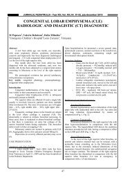
B. Radiological aspects:<br />
a. Plain abdominal radiography – Dates: 24.05.2008; 27.05.2008; 28.05.2008<br />
Marked dilatation of the bowel (small and large bowel).<br />
Air-fluid level. No gas in the pelvis (rectum) (fig. 1).<br />
b. Barium enema - Date: 02.06.2008<br />
Reduced caliber of the terminal rectum (with a tubular aspect: 43mm/length and<br />
12 – 16 mm/width) followed by a transition zone to an enlarged caliber of the p<strong>ro</strong>ximal rectum and<br />
sigmoid. Dolicosigma (fig. 2).<br />
C. Histopathological exam – Date: 16.06.2008<br />
Congenital megacolon.<br />
Fig. 1. Marked dilatation of the bowel (small and large bowel).<br />
Air-fluid level. No gas in the pelvis (rectum).<br />
4
JURNALUL PEDIATRULUI – Year XI, Vol. XI, Nr. 43-44, july-december 2008<br />
Fig. 2. Reduced caliber of the terminal<br />
rectum (with a tubular aspect: 43mm/length<br />
and 12 – 16 mm/width) followed by a<br />
transition zone to an enlarged caliber of the<br />
p<strong>ro</strong>ximal rectum and sigmoid. Dolicosigma.<br />
Discussions<br />
Normally, as a baby g<strong>ro</strong>ws in the womb, bundles<br />
of nerve cells (ganglia) begin to form between the<br />
muscle layers along the length of the colon. This<br />
p<strong>ro</strong>cess begins at the top of the colon and ends at the<br />
bottom (rectum). In children with Hirschsprung<br />
Disease, this p<strong>ro</strong>cess does not finish and the ganglia do<br />
not form along the entire length of the colon. Other<br />
times a longer portion may be affected (7).<br />
Aganglionosis begins with the anus, wich is<br />
always involved, and continues p<strong>ro</strong>ximally for a<br />
variable distance (2). The precise mechanism<br />
underlying the development of Hirschsprung disease is<br />
unknown (2).<br />
Hirschsprung Disease (HD) can be classified by the<br />
extension of the aganglionosis as follow (5):<br />
1.Classical HD (75% of <st<strong>ro</strong>ng>case</st<strong>ro</strong>ng>s): The aganglionic<br />
segment does not extend beyond the upper sigmoid.<br />
2. Long segment HD (20% of <st<strong>ro</strong>ng>case</st<strong>ro</strong>ng>s).<br />
3. Total colonic aganglionosis (3-12% of <st<strong>ro</strong>ng>case</st<strong>ro</strong>ng>s).<br />
Some rare variants include the following:<br />
4. Total intestinal aganglionosis.<br />
5. Ultra – short segment HD.<br />
Ultra – short segment HD is caracterised by a few<br />
centimeters of aganglionic bowel in the rectum,<br />
adjacent to the anus (2).<br />
Water-soluble contrast enema is the key study for<br />
diagnosis (6). Lateral views of early rectal/sigmoid<br />
colon filling are the most important images. The<br />
normal rectum should always be equal or of larger<br />
caliber than the sigmoid colon: the rectum/sigmoid<br />
ratio. In HD rectum/sigmoid ratio is reversed (6).<br />
About 10% children may present with diarrea<br />
caused by ente<strong>ro</strong>colitis, wich is thought to be related to<br />
stasis and bacterial overg<strong>ro</strong>wth. This may p<strong>ro</strong>gress to<br />
colonic perforation, causing life threatening sepsis(2).<br />
The <st<strong>ro</strong>ng>case</st<strong>ro</strong>ng> in discussion can be classifid as a rare<br />
form of HD, more specifically, the ultra – short HD,<br />
with a 4-5 cm length stenotic segment (emphasized by<br />
water soluble contrast enema), affecting not only the<br />
anal channel (1) but also the distal part of the rectum.<br />
The rectum/sigmoid ratio has been reversed.<br />
Both the ente<strong>ro</strong>colitis and septicemia, p<strong>ro</strong>ved<br />
th<strong>ro</strong>ugh positive hemoculture analyses, confirmed the<br />
fact that this <st<strong>ro</strong>ng>case</st<strong>ro</strong>ng> can be considered as belonging to the<br />
rare g<strong>ro</strong>up of 10% <st<strong>ro</strong>ng>case</st<strong>ro</strong>ng>s - of HD – diagnosed with<br />
infectious complications (2,4).<br />
Under these circumstances and taking into account<br />
the type of disease (HD associated with septicemia<br />
infection), the two times therapeutic surgical<br />
intervention was selected.<br />
Conclussions<br />
1. In Hirschsprung Disease (HD), water soluble<br />
contrast enema is the key for the diagnostic. The<br />
rectum/sigmoid ratio is reversed.<br />
2. The <st<strong>ro</strong>ng>case</st<strong>ro</strong>ng> into discussion can be classified as a<br />
rare form of HD, ultra – short HD.<br />
3. The infectious complications have determined<br />
the surgeon to choose a two times surgical<br />
intervention.<br />
5
JURNALUL PEDIATRULUI – Year XI, Vol. XI, Nr. 43-44, july-december 2008<br />
References<br />
1. And<strong>ro</strong>nescu A – Anatomia copilului; Editura<br />
Didactica si Pedagogica – Bucuresti – 1966.<br />
2. Steven L. Lee, MD and coauthors, Mar 30, 2006 –<br />
Hirschsprung Disease.<br />
3. David M. Manuel, MD and coauthors Aug 3, 2007<br />
– Hirschsprung Disease.<br />
4. Frederic N. Silverman, M.D. Caffey’s Pediatric X-<br />
ray Diagnosis, Year Book Medical Publishers,<br />
1985 – Megacolon.<br />
5. Ci<strong>ro</strong> Yoshida, Jr., MD and coauthors, Mar 31,<br />
2004 – Hirschsprung Disease.<br />
6. American Trend in Radiology, Teaching Files,<br />
2008 – Hirschsprung Disease.<br />
7. Mayo Clinic Staff, Nov. 10, 2006 –<br />
Hirschsprung’s Disease.<br />
8. Medline Plus, Medical Encyclopedia, 2008 –<br />
Hirchsprung‘s Disease.<br />
Correspondence to:<br />
Mi<strong>ro</strong>n Popescu<br />
Constantin Brancoveanu Street, No. 91a, Ap. 3,<br />
Timisoara,<br />
Romania,<br />
E-mail: <strong>ro</strong>ny.<strong>ro</strong>@mail.com<br />
6
JURNALUL PEDIATRULUI – Year XI, Vol. XI, Nr. 43-44, july-december 2008<br />
II. NEONATOLOGY<br />
SISTEMIC CONGENITAL SYPHILIS<br />
CLINICAL AND BIOLOGICAL STUDY<br />
Marioara Boia, Daniela Iacob, Aniko Manea, Mirela Margineanu<br />
„Louis Turcanu” Children’s Emergency Hospital Timisoara, Romania<br />
Abstract<br />
One of the multisystemic infection transmitted to<br />
the fetus via the placenta is the congenital, syphilis<br />
caused by Treponema pallidum. High incidence of the<br />
disease led to <strong>ro</strong>utinely screening for all pregnant<br />
women. Clinical signs in neonatal period appear in the<br />
first 5 weeks of life, but signs of the disease may occur<br />
late, after the first 2 years of life. Diagnosis based on<br />
neonatal se<strong>ro</strong>logic testing is complicated by the<br />
transplacental transfer of maternal Ig antibodies, which<br />
can cause a positive test in the absence of infection.<br />
Three significant <st<strong>ro</strong>ng>case</st<strong>ro</strong>ng>s of congenital systemic syphilis<br />
treated in the Clinic of Neonatology are presented in<br />
this paperwork.<br />
Key words: congenital syphilis, neonatal diagnosis.<br />
Int<strong>ro</strong>duction<br />
Congenital syphilis is a multisystemic infection<br />
caused by Treponema pallidum and transmitted to the<br />
fetus via the placenta.<br />
The rate of transmission is higher in women with<br />
primary and secondary syphilis than in those with<br />
tertiary syphilis. Up to 40 % of pregnant women with<br />
untreated primary syphilis have presented spontaneous<br />
abortions.<br />
High incidence of the disease led to <strong>ro</strong>utinely<br />
screening for all pregnant women.<br />
Two-thirds of the new born with syphilis are<br />
asymptomatic at birth. Clinical signs of disease can<br />
occur during fetal period, neonatal period or later in<br />
childhood with an additional perinatal mortality of<br />
25% - 30% <st<strong>ro</strong>ng>case</st<strong>ro</strong>ng>s. Without treatment, in severe forms<br />
of the disease, intrauterine death occurs in 25% of<br />
<st<strong>ro</strong>ng>case</st<strong>ro</strong>ng>s with an additional perinatal mortality of 25% -<br />
30%.<br />
Clinical signs in neonatal period appear in the first<br />
5 weeks of life and they are: ulcerative skin lesions on<br />
the palms and sole (occur in severe forms of the<br />
disease and they are highly contagious),<br />
hepatosplenomegaly, anemia, jaundice, hyd<strong>ro</strong>cephalus,<br />
lymphadenopathy, mucopurulent rhinitis, meningitis<br />
and mental retardation.<br />
In septicemic forms, X-ray examination reveals<br />
metafizar bone destruction and periosteal reaction.<br />
Signs of the disease may occur late, after the first 2<br />
years of life such as: f<strong>ro</strong>ntal bossing, mic<strong>ro</strong>gnatie, the<br />
palace pointed arch, Hutchinson’s triad, saddle nose,<br />
rhagades, optic at<strong>ro</strong>phy which leads to blindness.<br />
Because most infants born with congenital disease<br />
are free of clinical symptoms at the time of birth, final<br />
diagnosis is determined by laboratory tests. Most used<br />
are se<strong>ro</strong>logical tests and direct fluorescent antibody<br />
test.<br />
Diagnosis based on neonatal se<strong>ro</strong>logic testing is<br />
complicated by the transplacental transfer of maternal<br />
Ig antibodies, which can cause a positive test in the<br />
absence of infection. However a neonatal titer > 4<br />
times the maternal titer would not generally result f<strong>ro</strong>m<br />
passive transfer and diagnosis is considered confirmed<br />
or highly p<strong>ro</strong>bable. Therefore evaluating the new born<br />
baby must follow these steps: historical data on<br />
maternal infection, physical examination,<br />
hemoleucograma, treponemic and nontreponemic<br />
se<strong>ro</strong>logical tests, cardio-pulmonary and long bones X-<br />
ray and liver tests. T.pallidum can be identified in skin<br />
lesions, umbilical cord, placenta or autopsy.<br />
Material and method<br />
Study was conducted in the Clinic of Neonatology<br />
on three <st<strong>ro</strong>ng>case</st<strong>ro</strong>ng>s of congenital systemic syphilis which<br />
have been hospitalized in the same period.<br />
In the following we will present significant data of the<br />
three <st<strong>ro</strong>ng>case</st<strong>ro</strong>ng>s.<br />
CASE 1<br />
Patient MD, male, aged 2 weeks, delivered at term<br />
by normal vaginal <strong>ro</strong>ute with green slimy amniotic<br />
fluid, IA at 5’-8, Bw = 1980 g.(birth weigh)<br />
7
JURNALUL PEDIATRULUI – Year XI, Vol. XI, Nr. 43-44, july-december 2008<br />
The patient was admitted with extremely severe<br />
condition, intense jaundice, facial cyanosis, marked<br />
abdominal distention, hepatosplenomegaly, repeated<br />
crises of apnea and cyanosis requiring oxygen<br />
mask.On the left lower limbs he presented a<br />
erythematous macular rash with a diameter between<br />
0.5 -2 cm and with the tendency of spreading to the<br />
rest of the body.<br />
Se<strong>ro</strong>logical tests confirmed the suspicion of<br />
congenital syphilis and haematochemical<br />
investigations emphasize th<strong>ro</strong>mbocytopenia with<br />
leukocytosis and hepatocytolitic synd<strong>ro</strong>me<br />
(TGO=123U/L, TGP=112U/L). All cultures were<br />
sterile.<br />
After about a week of treatment (antibiotic +<br />
penicillin, etamsylatum, calcium, vitamins, plasma,<br />
dexamethasonum, arginine, aspatofort) CRP and<br />
aminotransferase values begin to decline slowly,<br />
jaundice gradually decreases in intensity, new born had<br />
a significant weight gain and clinical status in<br />
evolution was satisfactory(w = 3300g).<br />
CASE 2<br />
Patient K.S.- male new born baby, aged 1 day,<br />
weighing 2860 g, delivered at term by caesarean<br />
intervention, IA = 9,G1,P1.<br />
On clinical examination the patient was noted to<br />
have a extremely severe condition, jaundice,<br />
perio<strong>ro</strong>nazal (facial)cyanosis, labored respiration with<br />
retraction of the intercostal muscles, ritmic heart<br />
sounds, pulse=130 beats / min. The abdomen was soft,<br />
it p<strong>ro</strong>truded durind inspiration, the edge of the liver<br />
was palpable app<strong>ro</strong>ximately 2.5 cm below the right<br />
costal margin. Anterior fontanelle (2/3 cm) was<br />
normotensive.<br />
Biochemical investigations have confirmed the<br />
diagnosis: systemic congenital syphilis (leuKocytosis,<br />
th<strong>ro</strong>mbocytopenia, CRP positive, VDRL and TPHA<br />
positive, increased LDH, hiperbilirubinemia).<br />
Abdominal ultrasonography revealed increased<br />
liver volume, gallbladder with bold walls, normal<br />
biliary tract and normal spleen volume.<br />
Treated with penicillin-10 days, in association with<br />
other antibiotics, and with ursodeoxycholic acid,<br />
calcium, vitamins and plasma, evolution is greatly<br />
imp<strong>ro</strong>ved with the exception of transaminase that<br />
increase p<strong>ro</strong>gressively reaching a maximum at about a<br />
month of hospitalization, as follows: TGO = 535 U/l.<br />
After this time, jaundice decreases in intensity and<br />
the liver size is p<strong>ro</strong>gressively reduced.<br />
CASE 3<br />
Patient V.I. , Female, aged 1 day, delivered at term<br />
by normal vaginal <strong>ro</strong>ute, with green amniotic fluid,<br />
G2, P1, gestational age-33/34 weeks, polihidramnios,<br />
Bw = 2440 g, IA=5 at 1 minute, 6 at 5 minutes.<br />
It was admitted in the first days of life with severe<br />
condition, cyanosis, petechial elements on the legs and<br />
body, saddle nose. Balanced cardio-pulmonary, pulse<br />
132 beats / min, SaO2-99%, abdominal distension<br />
caused by gas accumulation, the liver was palpable to<br />
the right iliac tank. Eruptive pustular papules on the<br />
abdomen and thorax.<br />
Biochemical investigations (high leukocytosis,<br />
th<strong>ro</strong>mbocytopenia, hepatocitolitic synd<strong>ro</strong>me: TGO =<br />
210 U/L, TGP = 21 U/L, elevated inflammatory tests)<br />
and se<strong>ro</strong>logical tests (VDRL, TPHA-positive)<br />
confirmed the diagnosis.<br />
In evolution remain th<strong>ro</strong>mbocytopenia,<br />
leukocytosis, hepatocitolitic synd<strong>ro</strong>me and<br />
intrainfectious anemia, beginning to imp<strong>ro</strong>ve after<br />
about 2 weeks f<strong>ro</strong>m the onset.<br />
Results and discussion<br />
Although the literature does not mention an<br />
increased incidence of septicemic congenital syphilis,<br />
our clinic has faced, in a short period of time, 3 <st<strong>ro</strong>ng>case</st<strong>ro</strong>ng>s<br />
with similar clinical and biological features and also<br />
with a relatively good evolution despite data quoted in<br />
the literature that emphasizes a bad p<strong>ro</strong>gnosis : death<br />
in 40% of <st<strong>ro</strong>ng>case</st<strong>ro</strong>ng>s.<br />
Among the patognomonic features of the disease,<br />
common to our patients we mention:<br />
- Intense jaundice, with elevated bilirubinemia. At first<br />
unconjugated bilirubina and in evolution, installed<br />
colestasis led both to increased conjugated bilirubin<br />
and also elevated levels of gGT and FA.<br />
- Hepatomegaly accompanied by high levels of hepatic<br />
enzymes which after app<strong>ro</strong>ximately one month start to<br />
decline under established treatment.<br />
- Elevated values of inflammatory tests had been also<br />
present at all three patients and they slowly decreased<br />
under antibiotic therapy.<br />
Treatment was conducted in accordance with the<br />
current p<strong>ro</strong>tocols: penicillin for 10 days. It also has<br />
been done etiopatogenic treatment of concurrent<br />
infections, correction of acidobazic and<br />
hid<strong>ro</strong>elect<strong>ro</strong>litic imbalances and correction of<br />
haematological disorders.<br />
Evolution of the patient was initially serious: status<br />
toxicoseptic, needs for oxygen mask, positive<br />
se<strong>ro</strong>logy, biological values considerably raised but<br />
then slowly becoming favorable, with imp<strong>ro</strong>ving of<br />
general status, jaundice remission and normalization of<br />
transaminases. The haematological parameters have<br />
also been corrected reaching values that corresponded<br />
to the patient's age.<br />
8
JURNALUL PEDIATRULUI – Year XI, Vol. XI, Nr. 43-44, july-december 2008<br />
Conclusions<br />
1. The most commonly form of disease encountered in<br />
medical practice are asymptomatic, incidentally<br />
discovered by se<strong>ro</strong>logic test for syphilis.<br />
2. All the <st<strong>ro</strong>ng>case</st<strong>ro</strong>ng>s of manifest syphilis had a serious<br />
evolution requiring special care and treatment.<br />
3. Although the status of patients was extremely<br />
severe, joining various pathology and highly modified<br />
laboratory indices, the evolution under treatment was<br />
favorable.<br />
References<br />
1. Alford CA Jr, C<strong>ro</strong>nic congenital and perinatal<br />
infections. in Avery GB editor, Neonatology,<br />
Pathophysiology and Management of the newborn,<br />
Philadelphia, JB Lippincott, 1987<br />
2. Babcock D.S., Cranial ultrasonography of infants,<br />
Baltimore, Wiliams and Wilkins, 1981<br />
3. Bale JF, Sato Y, Eisert D, P<strong>ro</strong>gressive postnatal<br />
subependimal nec<strong>ro</strong>sis in infant with congenital<br />
cytomegalovirus infections, Pediatr Neu<strong>ro</strong>l, 1986,<br />
2, 367-370<br />
4. Bains MK, Hosseini-Ardehali M. Palatal<br />
perforations: past and present. Two <st<strong>ro</strong>ng>case</st<strong>ro</strong>ng> <st<strong>ro</strong>ng>report</st<strong>ro</strong>ng>s<br />
and a literature review. Br Dent J. 2005;199:267–<br />
269. [PubMed]<br />
5. Chawla W, Pandit PB, Nkrumach FK, Congenital<br />
syphilis in the newborn, Arch Dis Child, 1988, 63,<br />
1393-1394<br />
6. Dykes FD, Ahmann PA, Lazzara A, Cranial<br />
ultrasound in the detection of intracranial<br />
calcifications, J Pediatr, 1982, 100, 406-407<br />
7. Platou RW, Hill AJ jr, Ingraham NR jr, Early<br />
congenital syphilis. Treatement of 252 patients<br />
with penicillin, JAMA, 1947, 133, 10<br />
8. Lugo A, Sanchez S, Sanchez JL. Congenital<br />
syphilis. Paediatr Dermatol. 2006;23:121–123. doi:<br />
10.1111/j.1525-1470.2006.00194.x.<br />
9. Gurlek A, Alaybeyoglu NY, Demir CY, et al. The<br />
continuing scourge of congenital syphilis in 21 st<br />
century: a <st<strong>ro</strong>ng>case</st<strong>ro</strong>ng> <st<strong>ro</strong>ng>report</st<strong>ro</strong>ng>. Int J Paediatr<br />
Otorhinolaryngol. 2005;69:1117–1121. doi:<br />
10.1016/j.ijporl.2005.03.007.<br />
10. Tikhonova L, Salakhov E, Southwick K, for the<br />
Congenital Syphilis Investigation Team. et al.<br />
Congenital syphilis in the Russian Federation:<br />
magnitude, determinants and consequences. Sex<br />
Transm Infect. 2003;79:106–110. doi:<br />
10.1136/sti.79.2.106. [PubMed]<br />
11. Li, Y.; Gonik, B. Is congenital syphilis really<br />
congenital syphilis. Infect Dis Obstet Gynecol.<br />
2006. pp. 1–4. Article ID 81629.<br />
12. Humphrey MD, Bradford DL. Congenital syphilis:<br />
still a reality in 1996. Med J Australia.<br />
1996;165:382–385. [PubMed]<br />
Correspondence to:<br />
Marioara Boia,<br />
Gospodarilor Street, No. 42,<br />
Timisoara 300778,<br />
Romania<br />
E-mail: boiaeugen@yahoo.com<br />
9
JURNALUL PEDIATRULUI – Year XI, Vol. XI, Nr. 43-44, july-december 2008<br />
III. PEDIATRICS<br />
DOES THE BURNOUT SYNDROME EXIST AT THE<br />
PAEDIATRICIANS FROM ARAD?<br />
- A PILOT STUDY –<br />
Simona Dumitra, Mihaela Corciu, Sabina Morgovan, Liana Precup, D Lazar<br />
¹Western University “Vasile Goldis”, Arad, Romania<br />
²County Emergency Hospital, Arad, Romania<br />
Abstract<br />
Int<strong>ro</strong>duction: Burnout is a psychological term that<br />
defines the long term exhaustion and lack of interest,<br />
usually in context of work. The paediatric field needs<br />
an evaluation of the burnout as it is a medical<br />
envi<strong>ro</strong>nment that involves a lot of stress.<br />
Objectives: The aim of this study is to identify the<br />
levels of burnout of the paediatric consultants f<strong>ro</strong>m<br />
Arad, using the Maslach Burnout Inventory (MBI),<br />
created by Cristina Maslach.<br />
Methods: The study involved 13 paediatric<br />
consultants, who work in the Paediatric Emergency<br />
Unit and in the Paediatric Department of the Clinical<br />
Emergency Hospital, Arad. They were asked to submit<br />
their answers for the MBI self test. MBI considers<br />
burnout to be a multifactorial p<strong>ro</strong>blem and identifies 3<br />
subscales: emotional exhaustion, depersonalization and<br />
lack of personal accomplishment. Each subscale is<br />
measured by a different score. The higher the score on<br />
emotional exhaustion and depersonalization are, the<br />
higher the levels of burnout will be.<br />
Results: The data was int<strong>ro</strong>duced in an Excel<br />
database and analyzed statistically. A high average was<br />
obtained for the emotional exhaustion subscale (30.23)<br />
and a moderate one for depersonalization and personal<br />
accomplishment subscales (10.37 and 35.84<br />
respectively).<br />
Conclusions:<br />
1. There are high scores for emotional exhaustion, due<br />
to the large number of working hours and shifts.<br />
2. There are moderate scores for depersonalization and<br />
personal accomplishment, which can be explained<br />
th<strong>ro</strong>ugh the paediatric p<strong>ro</strong>file.<br />
3. Further studies are required to obtain a more<br />
realistic view of the p<strong>ro</strong>blem.<br />
Key words: burnout, exhaustion, depersonalization,<br />
personal accomplishment, paediatrician.<br />
Int<strong>ro</strong>duction<br />
Burnout is a psychological term for the experience<br />
of long-term exhaustion and diminished interest<br />
(depersonalization), usually in the context of work.<br />
Burnout is often construed as the result of a period of<br />
spending too much effort at work while having too<br />
little recovery.<br />
The paediatric field demands a quantification of the<br />
burnout as it is a medical envi<strong>ro</strong>nment where doctors<br />
experience a lot of stress.<br />
Objectives<br />
The aim of this study is to identify the emotional<br />
exhaustion, depersonalization and personal<br />
accomplishment scores of the paediatric consultants<br />
f<strong>ro</strong>m Arad, using the Maslach Burnout Inventory<br />
(MBI), created by Cristina Maslach.<br />
Method<br />
13 paediatric doctors submitted their answers for<br />
the MBI self test. All of them work as paediatric<br />
consultants, having f<strong>ro</strong>m 15 to 25 years of medical<br />
experience in this field, working f<strong>ro</strong>m 46 to 72 hours<br />
per week and with an average of the monthly shifts<br />
ranging f<strong>ro</strong>m 3 to 7.<br />
4 of them work in the Emergency Paediatric Unit<br />
while the others work in the Paediatric Department of<br />
the Clinical Emergency Hospital, Arad, and have, also,<br />
private cabinets.<br />
MBI identifies the most essential subscales:<br />
‣ emotional exhaustion<br />
‣ depersonalization<br />
‣ lack of personal accomplishment.<br />
Each subscale is measured by a different score.<br />
A high degree of burnout is shown by high scores<br />
of emotional exhaustion and depersonalization<br />
10
JURNALUL PEDIATRULUI – Year XI, Vol. XI, Nr. 43-44, july-december 2008<br />
subscales, and by low scores of personal<br />
accomplishment (lack of) subscale.<br />
An average degree of burnout is reflected in<br />
average scores on the three subscales.<br />
A low degree is shown by low scores of emotional<br />
exhaustion and depersonalization and by high scores of<br />
personal accomplishment subscales. (1, 2 ,3, 5).<br />
The higher the score on emotional exhaustion and<br />
depersonalization, the higher the levels of burnout.<br />
Moreover, the lack of personal accomplishment scale<br />
measures in the opposite directions, the lower the<br />
scale, the higher the level of burnout (7).<br />
The scores are analyzed according to the MBI<br />
standard, after more then 11.000 persons answered this<br />
questionnaire.<br />
MBI Low Average High<br />
Emotional<br />
exhaustion<br />
27<br />
Depersonalization 13<br />
Personal<br />
accomplishment<br />
>39 38-32
JURNALUL PEDIATRULUI – Year XI, Vol. XI, Nr. 43-44, july-december 2008<br />
Table nr.1 - The obtained scores.<br />
MBI Low Average High<br />
Emotional<br />
exhaustion<br />
Depersonalization 27<br />
30.23<br />
>13<br />
JURNALUL PEDIATRULUI – Year XI, Vol. XI, Nr. 43-44, july-december 2008<br />
Paediatricians scores/oncologists scores<br />
emotional<br />
exhaustion<br />
depersonalization<br />
personal<br />
accomplishment<br />
0 10 20 30 40<br />
oncologists<br />
paediatricians<br />
Figure 3. Comparing<br />
paediatricians scores /<br />
oncologists scores.<br />
Conclusions<br />
1. There are high scores for emotional exhaustion, due<br />
to the large number of working hours and shifts.<br />
2. There are moderate scores for depersonalization and<br />
personal accomplishment, which can be explained<br />
th<strong>ro</strong>ugh the paediatric p<strong>ro</strong>file.<br />
3. Further studies are required to obtain a more<br />
realistic view of the p<strong>ro</strong>blem.<br />
References<br />
1. Armst<strong>ro</strong>ng J, Lederberg M, Holland J. Fellows'<br />
forum: A workshop on the stresses of being an<br />
oncologist. J Cancer Educ.2004;19:88-90.<br />
2. Armst<strong>ro</strong>ng J, Holland J. Surviving the stresses of<br />
clinical oncology by imp<strong>ro</strong>ving communication.<br />
Oncology (Williston Park). 2004; 18:363-368,<br />
373-375.<br />
3. Holland JM, Neimeyer RA. Reducing the risk of<br />
burnout in end-of-life care settings: The <strong>ro</strong>le of<br />
daily spiritual experiences and training. Palliat<br />
Support Care. 2005; 3:173-181.<br />
4. Holland JC. Management of grief and loss:<br />
Medicine's obligation and challenge. J Am Med<br />
Womens Assoc. 2002; 57:95-96.<br />
5. Kash KM, Holland JC, Breitbart W, et al. Stress<br />
and burnout in oncology. Oncology (Williston<br />
Park). 2000; 14:1621-1633, 1633-1634, 1636-<br />
1637.<br />
6. Maslach C., Schaucheli W - Historical and<br />
conceptual development of burnout.<br />
7. P<strong>ro</strong>fessionnal burnout: Recent developments in<br />
theory and research. Ed. by Schaufeli W., Maslac<br />
C. Marek T. New-York, Taylor and Francis 1993.<br />
8. ***31st Congress of the Eu<strong>ro</strong>pean Society of<br />
Medical Oncology (ESMO). "Burnout is an<br />
important issue for oncology employees," lead<br />
author Senem Dubova, MD, f<strong>ro</strong>m Ege University<br />
Medical School, in Bornova-Izmir, Turkey.<br />
Correspondence to:<br />
Simona Dumitra<br />
Spitalului Street, No. 2-3,<br />
Arad,<br />
Romania<br />
Tel: +40-740013028<br />
E-mail: dumitrasimona@yahoo.com<br />
13
JURNALUL PEDIATRULUI – Year XI, Vol. XI, Nr. 43-44, july-december 2008<br />
THE MARKERS FOR IMMUNO-GENETIC<br />
SUSCEPTIBILITY IN CHILDHOOD DIABETES<br />
Ionela Tămăşan, Corina Paul, I Velea, I Popa<br />
Clinic II Pediatrics – University of Medicine and Pharmacy „V. Babeş” Timişoara<br />
Abstract<br />
Diabetes mellitus (DM) is a hete<strong>ro</strong>genous<br />
sind<strong>ro</strong>me characterized by a complex disturbance of<br />
the energetic metabolism, which affects the<br />
metabolismof both carbohydrates, lipids and p<strong>ro</strong>teins<br />
and also the other metabolisms. These disturbances<br />
result f<strong>ro</strong>m an insulin secreting defect (the decrease of<br />
β cell mass / function), associated sometimes with a<br />
degree of peripheral insulin resistance. Prediction of<br />
type 1 DM, meaning the appreciation of the risk to<br />
develop the disease, raises a great theoretical and<br />
practical interest. This is based on the acceptance of<br />
the autoimmune pathogeny in most of the <st<strong>ro</strong>ng>case</st<strong>ro</strong>ng>s (DM<br />
type1A) and the understanding of the p<strong>ro</strong>gressive,<br />
stadial evolution of the β-cell destructive p<strong>ro</strong>cess (1).<br />
The prediction strategies are using the genetic,<br />
immunologic and metabolic markers which define the<br />
risk of the patients to develop type 1 DM.<br />
Key words: susceptibility, childhood, diabetes<br />
Int<strong>ro</strong>duction<br />
F<strong>ro</strong>m a genetic point of view, diabetes is a<br />
complex, poligenic disease, involving nume<strong>ro</strong>us<br />
susceptibility genes and some p<strong>ro</strong>tective genes, all<br />
with incomplete penetrance, recip<strong>ro</strong>cally conditioning<br />
each other.<br />
Actually, is unanimously accepted that the short<br />
prediagnosis period in type 1 DM is the top of a huge<br />
iceberg, just partially explored by the modern<br />
imunogenetic studies. These studies prefigure a stadial<br />
evolution of a variable duration (2) (months, years).<br />
In this period of time the disease is ongoing<br />
th<strong>ro</strong>ugh 6 evolutive phases:<br />
genetic susceptibility (3),<br />
precipitanting event (intervention of the trigger<br />
factors),<br />
overt immunologic abnormalities<br />
(autoantibodies: GAD, ICA),<br />
p<strong>ro</strong>gressive loss of insulin release,<br />
overt diabetes,<br />
complete islet beta cell destruction.<br />
The genetic markers used in association with the<br />
family history shows that the risk of type 1 DM is (4):<br />
- 1/5.000 in <st<strong>ro</strong>ng>case</st<strong>ro</strong>ng>s without susceptibility alleles or<br />
family history<br />
- 1/4 if two risk alleles exist and a positive family<br />
history.<br />
A. Genetic markers:<br />
In the last years nume<strong>ro</strong>us genes were studied<br />
(ch<strong>ro</strong>mosomial regions); of these, two regions are<br />
mostly involved in the genetic susceptibility for type 1<br />
DM:<br />
- HLA region on short arm of ch<strong>ro</strong>mosome 6<br />
(6p21.3) noted IDDM1 and<br />
- insulin gene region on the short arm of<br />
ch<strong>ro</strong>mosome 11 (11p15), noted IDDM2. IDDM1 is<br />
responsible for almost 50% of the genetic<br />
susceptibility, while IDDM2 for 10-15% (5, 6, 7).<br />
Beside these two regions, genom –wide scan studies<br />
identified at least 18 ch<strong>ro</strong>mosomial regions (noted<br />
IDDM3, IDDM4 etc.) associated with type 1 DM. For<br />
most of these regions, the susceptibility genes have not<br />
been precisely identified yet, the mechanism of their<br />
involvement in the pathogeny of the disease still<br />
remains to be clearified (8) (table 1).<br />
The most known “diabetogenic genes” are those<br />
belonging to HLA system f<strong>ro</strong>m the MHC region of the<br />
short arm of ch<strong>ro</strong>mosome 6 (6p21.3) – with a major<br />
<strong>ro</strong>le in the immune response of the body (Fig. 1).<br />
Presently is unanimously accepted that type 1 DM in<br />
the child is associated with:<br />
- DRB1*04-DQA1*0301-DQB1*0302 allele and<br />
- DRB1*03-DQA1*0501-DQB1*0201 and the<br />
decreased frequency should explain the low<br />
incidence of DM in some countries like<br />
Romania (9).<br />
More than 90% of the diabetic patients with type 1<br />
DM have predisposing alleles type DR3-DR4 –<br />
comparatively with 40-50% in the general population.<br />
The concomitant presence of DR3-DR4 in one patient<br />
increases the risk; actually this association is<br />
encountered in 30-50% of type 1 DM patients<br />
(compared to 1-6% in the general population).<br />
14
JURNALUL PEDIATRULUI – Year XI, Vol. XI, Nr. 43-44, july-december 2008<br />
Table I. Genome screensT1DM.<br />
IDDM1 6p21 IDDM13 2q34-q35<br />
IDDM2 11p15 IDDM15 6q21<br />
IDDM3 15q26 IDDM17 10q25<br />
IDDM4 11q13 IDDM18 5q31-q35 (IL2)<br />
IDDM5 6q25-q27 1q42-qter<br />
IDDM6 18q21 8q24<br />
IDDM7 2q31 VDR, INFα 12q12-qter<br />
IDDM8 6q27-qter 16p11-p16<br />
IDDM9 3q21-q25 16q22-q24<br />
IDDM10 10p11-q11 17q24-qter<br />
IDDM11 14q24-q31 TGFβ1 19p13-q13<br />
IDDM12 2q33 (CTLA4) Xp13-p11<br />
Fig. 1 - Schematic representation of HLA,<br />
p<strong>ro</strong>jected on the short arm of ch<strong>ro</strong>mosome<br />
6 (Richard G Phelps and Andrew J Rees).<br />
Nume<strong>ro</strong>us studies, confirm that the HLA DQ<br />
molecules have a primordial <strong>ro</strong>le in the predisposition<br />
to type 1 DM. DQA1*0301-DQB1*0302 is associated<br />
with an increased susceptibility for type 1DM in most<br />
of the populational g<strong>ro</strong>up studied (10).<br />
The study of the HLA-DP alleles didn`t offer any<br />
certain p<strong>ro</strong>of concerning their involvement in the<br />
predisposition for type 1 DM.<br />
Some HLA alleles confer p<strong>ro</strong>tection for the<br />
occurence of diabetes (11); we mean especially the<br />
following HLA molecules:<br />
- DQ6 (DQB1*0602 si DQB1*0603),<br />
- DQ7 (DQB1*0301/0304),<br />
- DRB1*1401<br />
- DQA1*0201<br />
The p<strong>ro</strong>tection confered is not absolute, however,<br />
less than 1% of type1 DM patients have these alleles.<br />
These p<strong>ro</strong>tective alleles seem to have dominance upon<br />
the susceptibility alleles.<br />
The second region p<strong>ro</strong>ved to be associated with<br />
type1DM is the region for insulin gene on<br />
ch<strong>ro</strong>mosome 11 - 11p15 (IDDM2). We talk about<br />
polimorphysms f<strong>ro</strong>m a variable zone (VNTR –<br />
Variable Number of Tandem Repeats) situated in<br />
region 5' <st<strong>ro</strong>ng>report</st<strong>ro</strong>ng>ed to the insulin gene p<strong>ro</strong>motor which<br />
influences the reglatory mechanism of insulin gene<br />
transcription.<br />
At this level 3 classes of alleles may exist. Class I<br />
haplotype are associated with Type 1 DM while those<br />
f<strong>ro</strong>m class III confer p<strong>ro</strong>tection (12).<br />
The other locuses p<strong>ro</strong>oved to be involved in the<br />
predisposition for type 1 DM, include (Fig. 2):<br />
- The lymphoid-specific phosphatase (LYP) encoded<br />
by PTPN22 is involved in preventing spontaneous T-<br />
15
JURNALUL PEDIATRULUI – Year XI, Vol. XI, Nr. 43-44, july-december 2008<br />
cell activation by dephosphorylating and inactivating<br />
T-cell receptor-associated Csk kinase (13). An<br />
arginine-to-triptophan substitution at codon 620 of<br />
PTPN22 was considerently <st<strong>ro</strong>ng>report</st<strong>ro</strong>ng>ed to be associated<br />
with type 1 DM as well as other autoimmune diseases,<br />
such as rheumatoid arthritis, systemic lupus<br />
erythematosis (SLE) and Grave's disease. Genoyping<br />
of PTPN22 revealed the following alleles:<br />
- the homozygous genotype for the T allele and<br />
the hete<strong>ro</strong>zygous genotype C/T is associated<br />
with an increased risk for developing type 1<br />
diabetes<br />
- the C/C homozygous genotype is p<strong>ro</strong>tective<br />
against type 1 diabetes.<br />
- The presence of the hete<strong>ro</strong>zigous genotype C1858T<br />
in patients with type 1 DM, increases the risk to<br />
associate other autoimmune disturbances (14).<br />
- gene CTLA - 4 (Citotoxic T Lymphocite antigen) on<br />
ch<strong>ro</strong>mosome 2q33 – corresponding to IDDM12<br />
- gene for α chain of the interleukine 2 receptor<br />
(IL2RA/CD25) on ch<strong>ro</strong>mosome10p15.<br />
Possibly implicated in the predisposition for type1<br />
DM are some polymorphisms f<strong>ro</strong>m gene ICAM -1<br />
(Intercellular Cell Adhesion Molecule I) and the gene<br />
for Vitamine D Receptor (VDR - Vitamin D Receptor)<br />
(15).<br />
Fig. 2 - Summary of subset of<br />
confirmed loci f<strong>ro</strong>m whole genome<br />
screens associated with type 1A<br />
diabetes (Modified f<strong>ro</strong>m Todd et<br />
al. Nature Genetics, June 6, 2007).<br />
B. The markers for autoimmunity<br />
The autoimmune destructive p<strong>ro</strong>cess of the cells<br />
is a ch<strong>ro</strong>nic p<strong>ro</strong>cess, with variable duration and<br />
evolution velocity, individualised.<br />
Within this period, the immunologic markers<br />
might be evidentiated, in the serum, including: islet<br />
cell antibodies (ICA), insulin autoantibodies (IAA),<br />
GAD65 antibodies etc.<br />
Detection of antibodies in the serum has an<br />
important diagnostic significance, so, the high ICA is<br />
predictible for type1 DZ, before the occurence of the<br />
disease, fact that has been p<strong>ro</strong>oved in relatives of the<br />
diabetic patients. 8-10% of these, with an increased<br />
titre of these antibodies p<strong>ro</strong>gress towards DM within<br />
one year.<br />
The presence of markers in association, in the<br />
serum of some subjects, both in the general population<br />
and in some belonging to subg<strong>ro</strong>ups with increased<br />
risk for type 1 diabetes mellitus (type 1 DM), increases<br />
the p<strong>ro</strong>bability for developing this disease(16).<br />
<br />
<br />
<br />
ICA (islet cell antibodies) was first described<br />
as being associated with type 1 DM. ICA titre<br />
is expressed as JDF conventional units<br />
(Juvenile Diabetes Foundation).<br />
ICA are present in serum in 70-80% of the<br />
diabetic patients even since onset (17).<br />
Allthough technically difficult to perform, they<br />
remain the most sensitive marker for the<br />
prediction of the risk to develop DM , titres<br />
above 20 U JDF showing a p<strong>ro</strong>bability of 30-<br />
40% to develop the disease in the next 5 years.<br />
IAA (Insulin Autoantibodies) are present in<br />
serum since the onset (meaning before the<br />
initiation of insulin therapy) in 50-70% of the<br />
subjects, more frecquent in children than in<br />
adults. Their presence represents the p<strong>ro</strong>of of<br />
an ongoing β-cell destructive p<strong>ro</strong>cess and<br />
represents an important marker for the<br />
detection of the subjects at risk to develop type<br />
1 DM (18).<br />
16
JURNALUL PEDIATRULUI – Year XI, Vol. XI, Nr. 43-44, july-december 2008<br />
<br />
<br />
<br />
One of the most important autoantigenes that<br />
induces p<strong>ro</strong>duction of antibodies associated<br />
with DM is GAD (Glutamic Acid<br />
Decarboxylase) that is present in the β cells<br />
but also in CNS and the testicular tissue (19).<br />
The antibodies angainst the 65 kD peptide of<br />
GAD (GADA) are present in 70-80% of the<br />
patients type 1 DM and occur even since the<br />
prediagnostic period.<br />
Recently, a new family of β cell autoantigenes<br />
has been identified, the family of p<strong>ro</strong>teins PTP<br />
– P<strong>ro</strong>tein Ty<strong>ro</strong>sine Phosphatase. The<br />
antibodies against a 40 kDa fragment of this<br />
p<strong>ro</strong>tein also called ICA512 or IA – 2, occure<br />
in 60-70% of the subjects with type1 DM at<br />
the onset (20).<br />
There are also other citoplasmatic β cell<br />
antigenes responsible for the autoimmunity in<br />
DM. Of these, the most studied were: ICA69,<br />
Carboxipeptidase H, Gangliozide GM2-1,<br />
Imogen 38, Glima 38, Peripherina, Hsp 60<br />
(Heat Shock P<strong>ro</strong>tein 60) etc.(21).<br />
There are <st<strong>ro</strong>ng>case</st<strong>ro</strong>ng>s when healthy individuals are found<br />
with significant titres of diabetogenic antibodies that<br />
may persist years before the occurence of clinical DM<br />
or even without developing the disease (≈ 5% of the<br />
general population).<br />
For the moment, prediction (expressed as the<br />
percentage p<strong>ro</strong>bability of the risk to develop type1<br />
DM) can`t offer an absolute precision (22). The<br />
association of the immunological tests and the genetic<br />
typing, more and more accesible, even in the newborn,<br />
because of the development of rapid, automatic and<br />
cheaper techniques, increases the accuracy of<br />
prediction comparatively to isolate evaluation of the<br />
humoral immunity.<br />
In Romania there are just few studies concerning<br />
different aspects of the type 1 DM in children, but<br />
none of them p<strong>ro</strong>spective aiming for the evaluation of<br />
prediction and prevention in type 1 DM in the infantile<br />
population and the causal relationship genetic<br />
predisposition – immune status – envi<strong>ro</strong>nment factors<br />
(especially food).<br />
There are still many questions to be answered, for<br />
instance if:<br />
- the presence of the markers for cellular<br />
autoimmunity increase the risk for type 1 DM<br />
also in the general population,<br />
- all subjects with autoimmune markers will<br />
develop type 1 DM,<br />
- the detection of autoantibodies correlated with<br />
the reduction of first phase of the insulinic<br />
response increases the possibility of the<br />
disease to occure,<br />
- the associations between antibodies increase<br />
the risk.<br />
References<br />
1. Maclaren NK, Lan MS, Schatz D, Malone J,<br />
Notkins AL, Krischer J. 2003. Multiple<br />
autoantibodies as predictors of Type 1 diabetes in<br />
a general population. Diabetologia. 46:873-4.<br />
2. Atkinson MA., Eisenbarth GS – Type 1 diabetes:<br />
new perspective on the disease pathogenesis and<br />
treatment, The Lancet, 2001, 358: 221-229.<br />
3. Park Y., Eisenbarth GS – The natural history of<br />
autoimmunity in type 1 Diabetes mellitus. Disease,<br />
Prediction and Prevention, in Le Roith D., Taylor<br />
S.L., Olefsky J.M., - Diabetes mellitus – a<br />
fundamental and clinical text, 2nd Ed. Lippincot<br />
Williams and Wilkins, Philadelphia/Baltimore/<br />
New York / London, 2000.<br />
4. Pinhas-Hamiel O, Zeitler P. The global spread of<br />
type 1 diabetes mellitus in children and<br />
adolescents. J Pediatr 2005: 146: 693–700.<br />
5. Klein J, Sato A. The HLA system. First of two<br />
parts. N Engl J Med 2000; 343(10):702-709.<br />
6. Kwon OJ, Brautbar C, Weint<strong>ro</strong>b N, Sprecher E,<br />
Saphirman C, Bloch K et al. Immunogenetics of<br />
HLA class II in Israeli Ashkenazi Jewish, Israeli<br />
non-Ashkenazi Jewish, and in Israeli Arab IDDM<br />
patients. Hum Immunol 2001; 62(1):85-91.<br />
7. Undlien DE, Lie BA, Thorsby E. HLA complex<br />
genes in type 1 diabetes and other autoimmune<br />
diseases. Which genes are involved? Trends Genet<br />
2001; 17(2):93-100<br />
8. Sabbah E., Savola K., Ebeling T., Kulmala P.,<br />
Vahasalo P., IlonenJ., Salmela P.I., Knip M. –<br />
Genetic, autoimmune and clinical characteristics<br />
of childhood and adult onset type 1 diabetes.<br />
Diabetes Care 2000, 23, 1326-1332<br />
9. Ionescu Tirgoviste C., Guja C., Herr M., Cucca E.,<br />
Welsh K., Bunce M., Marshall S., Todd J.A.-<br />
Lowfrequency of HLA DRB1 03-DQB1 03 and<br />
DQB1 0302 haplotypes in Romania is consistent<br />
with the country's low incidence of Type 1<br />
diabetes. Diabetologia, 2001, 44, suppl. 3, B60-<br />
B66<br />
10. Pugliese A, Kawasaki E, Zeller M, Yu L, Babu S,<br />
Solimena M et al. Sequence analysis of the<br />
diabetes-p<strong>ro</strong>tective human leukocyte antigen-<br />
DQB1*0602 allele in unaffected, islet cell<br />
17
JURNALUL PEDIATRULUI – Year XI, Vol. XI, Nr. 43-44, july-december 2008<br />
antibody-positive first degree relatives and in rare<br />
patients with type 1 diabetes. J Clin Endocrinol<br />
Metab 1999; 84(5):1722-1728.<br />
11. Redondo MJ, Kawasaki E, Mulgrew CL, Noble<br />
JA, Erlich HA, Freed BM et al. DR and DQ<br />
associated p<strong>ro</strong>tection f<strong>ro</strong>m type 1 diabetes:<br />
comparison of DRB1*1401 and DQA1*0102-<br />
DQB1*0602. J Clin Endocrinol Metab 2000;<br />
85(10):3793-3797<br />
12. Walter M, Albert E, Conrad M, Keller E,<br />
Hummell M, Todd J. A., Bonifacio E:<br />
IDDM2/insulin VNTR modifies risk conferred by<br />
IDDM1/HLA for development of type 1 diabetes<br />
and associated autoimmunity. Diabetologia<br />
ISSN 0012-186X , 2003, vol. 46, n o 5, pp. 712-<br />
720.<br />
13. Meloni G.F., P. Lucarelli, M. Pellechia, et al.2004.<br />
A functional variant of lymphoid ty<strong>ro</strong>sine<br />
phophataze is associated with type 1 diabetes.<br />
14. Kawasaki E., T. Awata, H. Ikegami, et. Al.2006.<br />
Systematic search for single nucleotide<br />
polymorphism in a lymphoid ty<strong>ro</strong>sine phophatase<br />
(PTPN22) gene.<br />
15. Todd JA, Walker NM, Cooper JD, Smyth DJ,<br />
Downes K, Plagnol V et al. Robust associations of<br />
four new ch<strong>ro</strong>mosome regions f<strong>ro</strong>m genome-wide<br />
analyses of type 1 diabetes. Nat Genet 2007;<br />
39(7):857-864<br />
16. Park Y., Eisenbarth GS – The natural history of<br />
autoimmunity in type 1 Diabetes mellitus. Disease,<br />
Prediction and Prevention, in Le Roith D., Taylor<br />
S.L., Olefsky J.M., - Diabetes mellitus – a<br />
fundamental and clinical text, 2nd Ed. Lippincot<br />
Williams and Wilkins, Philadelphia/Baltimore/<br />
New York / London, 2000<br />
17. Vesa Eskola, Paula Vähäsalo, Hans K. Åkerblom,<br />
Mikael Knip, The Finnish ENDIT Study G<strong>ro</strong>up.<br />
Increased Frequency of Islet Cell Antibodies in<br />
Unaffected B<strong>ro</strong>thers of Children with Type 1<br />
Diabetes. Hormone research. Vol. 59. No.4.2003<br />
18. Schlosser M., Koczwara K., Kenk H., Strebelow<br />
M, Ziegler A.-G, Bonifacio E. In insulinautoantibody-positive<br />
children f<strong>ro</strong>m the general<br />
population, antibody affinity identifies those at<br />
high and low risk.<br />
Diabetologia 2005, vol. 48, no9, pp. 1830-1832<br />
19. Lindholm E, Hallengren B, Agardh CD. Gender<br />
differences in GAD antibody-positive diabetes<br />
mellitus in relation to age at onset, C-peptide and<br />
other endocrine autoimmune diseases. Diabetes<br />
Metab Res Rev. 2004 Mar-Apr;20 (2):158-64.<br />
20. Steffen G, Blanchetot C, Schepens J, Albet S,<br />
Lammers S: Multimerization of the P<strong>ro</strong>teinty<strong>ro</strong>sine<br />
Phosphatase (PTP)-like Insulin-dependent<br />
Diabetes Mellitus Autoantigens IA-2 and IA-2 β<br />
with Receptor PTPs (RPTPs). Biol. Chem., Vol.<br />
277, Issue 50, 48139-48145, December 13, 2002<br />
21. Martin S.: Islet cell autoantigen 69 antibodies in<br />
IDDM. Diabetologia 2004. pp 747.<br />
22. Samuelsson U, Sundkvist G, Borg H, Fernlund P,<br />
Ludvigsson J. 2001. Islet autoantibodies in the<br />
prediction of diabetes in school children. Diabetes<br />
Res Clin Pract. 51:51-7.<br />
Correspondence to:<br />
Dr. Ionela Tămăşan<br />
Clinic II Pediatrics<br />
Str. Evlia Celebi no 1-3<br />
300226 Timişoara, Romania<br />
Tel : 0256 – 494529<br />
E-mail: ionilop@yahoo.com<br />
18
JURNALUL PEDIATRULUI – Year XI, Vol. XI, Nr. 43-44, july-december 2008<br />
ACCIDENTAL ACUTE INTOXICATION WITH<br />
DENTOCALMIN IN CHILDREN – A SEVERE<br />
FORM CASE PRESENTATION<br />
Cristina Singer 1 , Polixenia Stancu 1 , Simona Coşoveanu 1 , Luciana Rotaru 2 , Anca Maloş 3 ,<br />
C Stoicănescu 4<br />
1 2 nd Pediatric Clinic, County Emergency Hospital Craiova; U.M.F. Craiova<br />
2 Emergency Unit, County Emergency Hospital Craiova; U.M.F. Craiova<br />
3 Intensive Care Unit, County Emergency Hospital Craiova<br />
4 2 nd Pediatric Clinic, County Emergency Hospital Craiova<br />
Abstract<br />
The authors present the <st<strong>ro</strong>ng>case</st<strong>ro</strong>ng> of a child, aged one<br />
year and seven months, who was admitted to the<br />
Emergency Unit of the County Emergency Hospital<br />
Craiova, because of an accidental ingestion of<br />
Dentocalmin, with a cardiorespiratory stop. After 25<br />
minutes of cardiorespiratory resuscitation, normal<br />
sinus rhythm is re-established; he is artificially<br />
ventilated; 48 hours after his hospitalization, he started<br />
to breath spontaneously. He is discharged after 27 days<br />
of hospitalization, with serious neu<strong>ro</strong>psychic sequels.<br />
Key words: Dentocalmin, acute intoxication, child.<br />
Int<strong>ro</strong>duction<br />
Our country annually registers about 16,000 <st<strong>ro</strong>ng>case</st<strong>ro</strong>ng>s<br />
of intoxication in children. Intoxications with drugs,<br />
caustic household p<strong>ro</strong>ducts, industrial p<strong>ro</strong>ducts<br />
(antifreeze solution, pet<strong>ro</strong>l, and gas), and alcohol are<br />
dominant.<br />
The highest frequency of the accidental acute<br />
intoxications is met in the age g<strong>ro</strong>up 0-7 years (5);<br />
among them, more than 50% are caused by drugs (2).<br />
Dentocalmin is a dental p<strong>ro</strong>duct, under the form of<br />
solution with external use. Regarding its pharmacotherapeutic<br />
action, it is a local anesthetic, an analgesic<br />
and an anti-inflammatory p<strong>ro</strong>duct. It is found under the<br />
form of bottles of 10 ml which contain: Lidocaine 2 g,<br />
Menthol 2 g and Phenol 2g (3).<br />
Case presentation<br />
The child S.G.E. (F.O. 51828/ 2007, 2 nd Pediatrics<br />
Clinic, Emergency County Hospital Craiova), male,<br />
aged 1 year and 7 months, Weight= 14 Kg is admitted<br />
in the Emergency Unit of the County Hospital Craiova,<br />
with a cardio-respiratory stop, on October, 20, 2007.<br />
The anamnesis reveals that the child’s state<br />
suddenly worsened, presenting – when in full health –<br />
a sleeping state followed by coma in app<strong>ro</strong>ximately 10<br />
minutes, after the mother administered him, by<br />
mistake, a few ml (5-6) of Dentocalmin, without using<br />
a d<strong>ro</strong>pping glass. The mother mistook the bottle of<br />
Dentocalmin for the bottle of Vigantol Oil (with a<br />
d<strong>ro</strong>pping glass), f<strong>ro</strong>m which the child used to receive a<br />
daily 2 ml dosage. He was transported by ambulance<br />
for hospitalization.<br />
Heredocolateral antecedents – young, healthy<br />
genitors; mother with higher education.<br />
Physiologic personal antecedents. Single child, on<br />
term and normal delivery, W B = 3,300 G, Apgar score<br />
10, artificial feeding when born with Milumil,<br />
Lactovit, correctly diversified when 4 months,<br />
vaccinated according to W.H.O. vaccination scheme;<br />
rickets p<strong>ro</strong>phylaxis with Vigantol Oil 2 d<strong>ro</strong>ps /day;<br />
normal physical and psychomotor development.<br />
Pathologic personal antecedents: 2<br />
hospitalizations: the first when 7 months and the<br />
second when 1 year and 6 months for acute<br />
b<strong>ro</strong>nchiolitis.<br />
Life conditions: an apartment in urban area, in a<br />
block of flats, 2 <strong>ro</strong>oms, 3 persons.<br />
When presented in the Emergency Unit, the child<br />
was in an extremely bad state, abolished conscience,<br />
marmorated teguments, cold and cyanotic extremities,<br />
absent peripheral and central pulse, absent spontaneous<br />
breath, mydriatic, non-reactive pupils.<br />
After 25 minutes of cardio-respiratory<br />
resuscitation (O.T.I. with assisted ventilation, external<br />
cardiac massage, adrenaline i.v., Na bicarbonate i.v.,<br />
E.V.P. with physiologic serum) the heart activity is reestablished.<br />
The stages E.K.G. – initial asystoly –<br />
subsequent elect<strong>ro</strong>mechanic dissociation and<br />
ventricular fibrillation; after 25 minutes, the child had<br />
a synusal rhythm, C.F.= 132 b/min [fig.1]. The state of<br />
the child remained severe, with an abolished<br />
conscience and mechanically ventilated.<br />
19
JURNALUL PEDIATRULUI – Year XI, Vol. XI, Nr. 43-44, july-december 2008<br />
E.K.G. – when admitted<br />
E.K.G. – when admitted<br />
E.K.G. – while resuscitation<br />
E.K.G. – while resuscitation<br />
E.K.G. – after resuscitation<br />
Fig. 1. E.K.G stages – initial asystoly – subsequent elect<strong>ro</strong>mechanic<br />
dissociation and ventricular fibrillation; after 25 minutes, the child had a<br />
synusal rhythm, C.F.= 132 b/min.<br />
20
JURNALUL PEDIATRULUI – Year XI, Vol. XI, Nr. 43-44, july-december 2008<br />
He is admitted to the Intensive Care Unit where<br />
the intensive treatment goes on: assisted ventilation,<br />
gastric washing, and intravenous treatment with<br />
Manitol, Fu<strong>ro</strong>semid, Dexametazone, Piracetam,<br />
vitamin B 1 , B 6 , C, Tazocin.<br />
Investigations when admitted (20 X):<br />
Hemogram: Hb= 11.8 g%, T= 240,000/mm 3 ,<br />
L= 7,500/mm 3 , N= 2%, S= 63%, E= 5%, Ly= 26%,<br />
M= 4%;<br />
Glicemy = 85 mg%;<br />
Mic<strong>ro</strong> Astrup: pO 2 159.8 mmHg; pCO 2 36.6<br />
mmHg; pH 7.24; SO 2 98.5%; BE -10.9<br />
mmol/l; BEecf -11.8 mmol/l; CHCO 3 st 15.9<br />
mmol/l; p 50 26.7 mmHg; Ct O 2 18.8%; CHCO 3<br />
15.5 mmol/l; Ct CO 2 (B) 14.3 mmol/l; SO 2 (C)<br />
98.9%;<br />
Sanguine ionogram: Na + = 143 mmol/l, K + =<br />
3.7 mmol/l, Cl - = 110 mmol/l, Ca ++ = 1.033 mmol/l.<br />
2 nd Day of hospitalization – the child breathes<br />
spontaneously and efficient (sat O 2 99%), he presents<br />
rhythmic cardiac sounds, C.F.= 127 b/min, slender<br />
abdomen, present diuresis; however, his future<br />
evolution is questionable, since the child alternates<br />
between periods of somnolence and agitation,<br />
opistotonus, horizontal nistagmus, convulsions. He is<br />
fed th<strong>ro</strong>ugh a nasogastric tube.<br />
After two weeks of hospitalization, he starts his<br />
feeding per os, but the psychomotor acquisitions are<br />
lost: he does not speak, he does not walk, he reacts<br />
only to st<strong>ro</strong>ngly painful stimuli, he presents a cerebral<br />
cry.<br />
Investigations performed while hospitalization:<br />
29 X: Hb= 8.4 g%, T= 230,000/mm 3 , L=<br />
6,800/mm 3 , N= 2%, S= 68%, E= 3%, Ly=<br />
10%, M= 7%, anisocytosis, poikilocytosis;<br />
6 XI: Hb= 11.8 g%, T= 240,000/mm 3 , L=<br />
8,500/mm 3 , N= 2%, S= 63%, E= 5%, Ly= 26%, M=<br />
4%;<br />
24 X: glicemy= 75 mg%;<br />
Sanguine ionogram 24 X: Na + = 143.4 mEq/l,<br />
K + = 4.6 mEq/l;<br />
E.g. F.O.: A.O. – normal aspect<br />
Pulmonary X-ray: no pleural-pulmonary<br />
changes, heart in normal limits.<br />
Skull C.T. (26 X): normal limits C.T. aspect,<br />
for native C.T. and postcontrast.<br />
E.g. pediatric neu<strong>ro</strong>psychiatry (30 X):<br />
vegetative status, reacting to st<strong>ro</strong>ngly painful<br />
stimuli → 15 XI: Spastic tetraparesis, psychic<br />
and motor regression.<br />
He continued to receive a treatment consisting of<br />
Diazepam, Fenobarbital, Cereb<strong>ro</strong>lizin, Piracetam,<br />
Dexametazone, physiokinetotherapy.<br />
After 27 days of hospitalization, he is discharged,<br />
balanced f<strong>ro</strong>m the cardiac and respiratory point of<br />
view, with feeding per os, a good digestive tolerance,<br />
presenting serious neu<strong>ro</strong>psychic sequels and with the<br />
following recommendations:<br />
- to carry on medical recovery -<br />
physiokinetotherapy;<br />
- to receive a drug-based treatment with<br />
Encephabol, Piracetam, and Vitamin B.<br />
A year after the child was discharged (October<br />
2008), he was admitted again in the Clinic (F.O.47113/<br />
2008) for a respiratory disease. In this period, the child<br />
followed a recovery treatment - physiokinetotherapy<br />
and drug-based treatment and p<strong>ro</strong>gress was registered:<br />
he walks if supported, utters some words, and interacts<br />
with the sur<strong>ro</strong>unding persons.<br />
Discussions<br />
We presented this <st<strong>ro</strong>ng>case</st<strong>ro</strong>ng> because of the severe<br />
intoxication p<strong>ro</strong>duced by Dentocalmin, an apparently<br />
harmless drug.<br />
The components of Dentocalmin have the<br />
following effects:<br />
Phenol (phenic acid, carbolic acid), in low<br />
concentrations (0.2 -1%), has a bacteriostatic effect,<br />
and when 3-5% it has a bactericidal action, due to the<br />
p<strong>ro</strong>tein precipitation (4).<br />
Locally applied, in concentration of 2%, the phenol is<br />
a local anesthetic, decreasing the excitability of<br />
peripheral nerves. In solutions of 5%, it is irritant and<br />
inflammable, its great power of penetration<br />
determining deep lesions, which require a long period<br />
of healing (6).<br />
Menthol (Mentholum) – is obtained f<strong>ro</strong>m the mint<br />
volatile oil (natural menthol) or th<strong>ro</strong>ugh synthesis<br />
(synthetic menthol). It is antipruriginous and<br />
a<strong>ro</strong>matizing (6).<br />
Lidocaine (Xiline) – is a local anesthetic with an<br />
amidic structure, which is active in all types of local<br />
anesthesia, including local anesthesia (under the form<br />
of solutions of 2-4% or Lidocaine ointment 5%).<br />
Intravenously administered or after absorption at the<br />
administrated place, lidocaine causes systemic effects:<br />
sedative, analgesics, anticonvulsive, antiarhythmics<br />
(3).<br />
In <st<strong>ro</strong>ng>case</st<strong>ro</strong>ng> of overdosage (the maximum admitted<br />
dose is 4 mg/Kgc/day) or fast intravenous<br />
administration, lidocaine can cause convulsions,<br />
tachycardia, lipotimy, high blood pressure, followed<br />
by coma, bradicardia, hypotension, respiratory<br />
depression. For our hospitalized child, the maximum<br />
dose of Lidocaine admitted during 24 hours was 56 mg<br />
(3). Taking into account that he received about half of<br />
the Dentocalmin bottle content, it results that he<br />
21
JURNALUL PEDIATRULUI – Year XI, Vol. XI, Nr. 43-44, july-december 2008<br />
received app<strong>ro</strong>ximately one dose of 1,000 mg of<br />
lidocaine, which represents twenty times more than the<br />
maximum admitted dose for 24 hours; hence the<br />
gravity of intoxication (the cardiac and respiratory<br />
stop, the coma).<br />
Between January, 1, 2005 and April, 1, 2008, at<br />
the Antitoxic Centre of the Emergency Clinical<br />
Hospital for Children “Grigore Alexandrescu”,<br />
Bucharest, there were admitted and <st<strong>ro</strong>ng>report</st<strong>ro</strong>ng>ed 22 <st<strong>ro</strong>ng>case</st<strong>ro</strong>ng>s<br />
of acute intoxication with Dentocalmin. The evolution<br />
was as follows: 15 <st<strong>ro</strong>ng>case</st<strong>ro</strong>ng>s with full recovery, 2 deaths,<br />
and 3 <st<strong>ro</strong>ng>case</st<strong>ro</strong>ng>s with serious neu<strong>ro</strong>psychic sequels (7).<br />
Following the requests of the Toxicology<br />
Department of the Emergency Clinical Hospital for<br />
Children “Grigore Alexandrescu”, Bucharest<br />
(P<strong>ro</strong>fessor Coriolan Ulmeanu), the Drug National<br />
Agency decided, in January 2008, an urgent<br />
withdrawal of all the Dentocalmin which was found in<br />
the communitary pharmacies. This happened because<br />
Dentocalmin had to be given according to market<br />
authorization and only within hospitals (in dental<br />
offices, respectively) (1).<br />
We have to mention that, subsequently - in<br />
September, 2008 – an infant aged 4 months was<br />
admitted in the clinic, with a Dentocalmin<br />
intoxication; he was administered by his mother, again<br />
by mistake, Dentocalmin instead of Vigantol Oil. This<br />
time, there was registered a favorable evolution.<br />
For the presented <st<strong>ro</strong>ng>case</st<strong>ro</strong>ng>, we pointed out:<br />
- the recovery potential of the heart and lungs in<br />
children – initially healthy, which after a long<br />
period of resuscitation recovered their normal<br />
activity;<br />
- the gravity of the child’s intoxication,<br />
requiring an initial treatment of cardiorespiratory<br />
resuscitation, followed by medical<br />
services in the Intensive Care unit and<br />
Pediatric Clinic;<br />
- a long period of hospitalization (27 days);<br />
- serious neu<strong>ro</strong>psychic sequels and lasting<br />
treatment for neu<strong>ro</strong>psychic recovery;<br />
- mother’s psychic impact – a feeling of guilt –<br />
the one who mistook the bottle of Vigantol Oil<br />
for the bottle of Dentocalmin<br />
References<br />
1. Agenţia Naţională a Medicamentului –<br />
Nomenclatorul Medicamentelor de uz uman, 2005,<br />
Ed. Medicală Bucureşti,<br />
2. Bates Nicola, Edwards N., Roper J., Volans S.,<br />
Paediatric Toxicology, 2006, Stockolm Press,<br />
ISBN 0-333-60951-4<br />
3. Cârlig V., Farmacologia, Ed. Aius, Craiova, 1999,<br />
Vol. I, 321-322<br />
4. Farmacopeea Română, Ed. a X-a, Ed. Medicală<br />
Bucureşti, 1998<br />
5. Goldfrank’s Toxicologic Emergencies , Ed. a 8-a ,<br />
2006<br />
6. Manolescu E., Farmacologie, Ed. Didactică şi<br />
Pedagogică Bucureşti, 1984, Ed. a II-a, p. 438<br />
7. Ninescu G.V., Petran M., Stanca S., Ulmeanu C.,<br />
Intoxicaţia acută cu Dentocalmin – intoxicaţie<br />
nouă cu un p<strong>ro</strong>dus vechi, Al IX-lea Congres<br />
Internaţional de Farmacologie, Terapeutică şi<br />
Toxicologie Clinică, 11-14 iunie 2008, Sibiu,<br />
Volum de Rezumate, Editura Alma Mater, 2008.<br />
Correspondence to:<br />
Şef lucrări Dr. Singer Cristina<br />
2 nd Pediatric Clinic, County Emergency Hospital Craiova;<br />
U.M.F. Craiova,<br />
Strada Tabaci nr.1,<br />
Craiova,<br />
judeţul Dolj,<br />
E-mail: mendelson.ltd@gmail.com;<br />
mendelson_ltd@yahoo.com<br />
22
JURNALUL PEDIATRULUI – Year XI, Vol. XI, Nr. 43-44, july-december 2008<br />
STAPHYLOCOCCAL IMPETIGO WITH<br />
SEVERE SEPSIS - CASE REPORT<br />
Tamara Marcovici 1 , I. Sabau 1 , I. Simedrea 1 , Otilia Marginean 1 , Camelia Daescu 1 , Oana Belei 1 ,<br />
Marinela Lesovici 2 , Daniela Chiru 2<br />
1 First Pediatric Clinic „Victor Babes” University of Medicine and Pharmacy Timisoara, Romania<br />
2 „Louis Turcanu” Children’s Emergency Hospital Timisoara, Romania<br />
Abstract<br />
Staphylococcus aureus is the most common cause of<br />
pyogenic infection of the skin, little infants being<br />
extremely susceptible. Staphylococci may enter in the<br />
blood with subsequent involvement of the organs. The<br />
development of staphylococcal disease is related to<br />
resistance of the host and to virulence of the organisms<br />
bacteria.We present the <st<strong>ro</strong>ng>case</st<strong>ro</strong>ng> of a five months old<br />
female infant hospitalized in our clinic for<br />
disseminated skin lesions, diarrhea with dehydration<br />
and sepsis with Staphylococcus aureus.<br />
Key words: Staphylococcus aureus, impetigo, sepsis<br />
Int<strong>ro</strong>duction<br />
Staphylococcus aureus causes a wide variety of<br />
infections. 1,2,3 It is the most common cause of pyogenic<br />
infection of the skin. 4,5,6 Little infants are extremely<br />
susceptible to staphiloccoci. A history of poor hygene<br />
and c<strong>ro</strong>wded living situations are common.<br />
Autoinfection is common and minor infection may be<br />
the source of disseminations..The intact skin and<br />
mucous membranes serve as barriers to invasion by<br />
staphylococci. 4,7 Staphylococcus aureus enters th<strong>ro</strong>ugh<br />
damaged skin and is transmitted th<strong>ro</strong>ugh direct contact<br />
6<br />
Staphylococci may also enter the blood with<br />
subsequent involvement of the organs. 1 Impetigo is<br />
most common in children. Most children are younger<br />
than 2 years. P<strong>ro</strong>gnosis may be influenced by<br />
nume<strong>ro</strong>us host factors, including nutrition,<br />
immunologic competence and the presence of other<br />
debilitating diseases. 4<br />
Case <st<strong>ro</strong>ng>report</st<strong>ro</strong>ng><br />
We present a five months old female infant admitted in<br />
the First Pediatric Clinic in „Louis Turcanu”<br />
Children’s Emergency Hospital Timisoara for high<br />
6<br />
fever, perioral , nasal, cervical, occipital and on the<br />
back crusting, diarrhoea, significant dehydration. She<br />
is the second child of an young healthy couple f<strong>ro</strong>m a<br />
town area in Timis county. The pregnancy was<br />
followed up, pathological, with imminence of<br />
miscarriege in the second trimester. The child was<br />
born in term, with a weight of 2260 g and Apgar score<br />
9. She was breastfeeded for 1 month and then with<br />
cow milk in excessive dilution with tea, with no sugar.<br />
She was precocious feeded with potato mash f<strong>ro</strong>m 3<br />
months of age. Child’s hygene was precarious.<br />
The history of illness<br />
The patient presented diaper dermatitis for 2 months.<br />
The infant had high fever 39-40ºC for five days,<br />
p<strong>ro</strong>ductive cough, diarrhoea, vomiting and<br />
erythematous rash in the perioral and cervical area,<br />
back, buttocks, perineal and thighs region..<br />
Clinical findings<br />
The infant was seriously ill, with axial hypotonia and<br />
hyperpirexia 40ºC. The skin was pale, motteled with<br />
p<strong>ro</strong>longed capillary refill time and cold extremities.<br />
There was present erythematous rash, exfoliation and<br />
crusting of perioral, cervical, perineal and flexural<br />
thighs area; purulent conjunctivitis. (fig 1, fig.2 and<br />
fig.3) She presented moderate dehydration and clinical<br />
signs of malnutrition (body weight was 4500 g).<br />
P<strong>ro</strong>ductive cough, tachypnea 50 breaths/ minute,<br />
intercostal bulging, dyspnea, alveolar and b<strong>ro</strong>nhial<br />
crackles were noticed; low blood presure (62/33<br />
mmHg) and high cardiac rate (140/’). Watery stools,<br />
oliguria and right ear pain accentuated by pressure on<br />
the tragus were present.<br />
23
JURNALUL PEDIATRULUI – Year XI, Vol. XI, Nr. 43-44, july-december 2008<br />
Fig.1. Clinical aspect: face lesions.<br />
Fig. 2. Clinical aspect: cervical and back area.<br />
Fig. 3. Clinical aspect of the child: perineal and thighs area.<br />
Consultations<br />
Chest <strong>ro</strong>entgenogram noticed bilateral lobular<br />
infiltrates in the lungs.<br />
ORL examination mentioned right ear purulent<br />
secretion.<br />
Laboratory findings<br />
Were present acute-phase reactants: leucocytosis<br />
with neut<strong>ro</strong>philia and left shift (tabel I); positive CRP,<br />
high ESR, increase serum α2 globulin fractions (tab.<br />
IV); anemia and th<strong>ro</strong>mbocytopenia (tab.lI); metabolic<br />
acidosis (pH =7.21, BE= -8) and low elect<strong>ro</strong>lytes level.<br />
(tabel III).<br />
24
JURNALUL PEDIATRULUI – Year XI, Vol. XI, Nr. 43-44, july-december 2008<br />
Tabel I. Blood cell count.<br />
Parameter<br />
Value<br />
Leucocyte/mm 3 17800<br />
Immature forms% 12<br />
Granulocytes % 78<br />
Lymphocytes % 10<br />
Hemoglobin g/dl 9,7<br />
Eryth<strong>ro</strong>cyte/mm 3 2940000<br />
Tabel II. Tests for coagulation.<br />
Parameter<br />
Value<br />
Platelet count/mm 3 87000<br />
Bleeding time<br />
3'30"<br />
Coagulation time<br />
4'20"<br />
P<strong>ro</strong>th<strong>ro</strong>mbin time 30"<br />
Tabel III. Serum elect<strong>ro</strong>lytes level.<br />
Parameter<br />
Value<br />
Na mmol/l 128<br />
K mmol/l 3,8<br />
Ca mmol/l 2,2<br />
Cl mmol/l 90<br />
Biochemical tests for liver and renal function were<br />
normal. It was present low level of seric p<strong>ro</strong>teins (45<br />
g/l) with decreased albumin level (48%).<br />
Peripherical and blood cultures were postive for<br />
Staphylococcus aureus. (tab. V) Germ’s sensitivity was<br />
good for Rocephin and Lincomicine.<br />
Tabel IV. Acute phase reactants.<br />
Parameter<br />
Value<br />
P<strong>ro</strong>tein C reactiv<br />
pozitiv<br />
ESR mm/1h 65<br />
Fibrinogen level g/l 4,99<br />
Serum α2 globuline % 22<br />
Tabel V. Bacteriological findings<br />
Specimen<br />
Result<br />
Blood<br />
Staph. aureus<br />
Occipital lesion<br />
Staph. aureus<br />
Otic secretion<br />
Staph. aureus<br />
Pharyngeal swab<br />
Staph. aureus<br />
Conjunctival secretion<br />
Staph. aureus<br />
Urine<br />
Sterile<br />
Stool<br />
Negative<br />
25
JURNALUL PEDIATRULUI – Year XI, Vol. XI, Nr. 43-44, july-december 2008<br />
Skin biopsy f<strong>ro</strong>m thigh area was performed but it was<br />
unconclusive.<br />
Medical therapy:<br />
Systemic antibiotic treatment with Rocephin<br />
0.5g/day and Gentamycin 0.02 g/day for 10<br />
days, then Lincomicyn 0.1 g/day<br />
Active immunization with antistaphylococcal<br />
vaccine, 7 doses<br />
Intravenous fluid resuscitation with isotonic<br />
sodium chloride solution; glucose 10% and<br />
elect<strong>ro</strong>lytes; correction of metabolic acidosis<br />
with NaHCO3 8,4%<br />
Oxygen and antipyretic therapy<br />
Parenteral nutrition with Aminosteril,<br />
Intralipid; administration of vitamins<br />
Diet therapy: oral glucose elect<strong>ro</strong>lyte solution<br />
(Gesol); car<strong>ro</strong>ts soup, boiled rice with 5%<br />
glucosis; low-lactose formula.<br />
Evolution<br />
Diarrhoea stopped after 5 days of treatment. Purulent<br />
otits and conjunctivitis cured in 7 days,<br />
b<strong>ro</strong>nhopneumonia in14 days and skin lesions healed in<br />
4 weeks. Weight became upward after 48 hours of<br />
therapy and total increase was 1600 g.<br />
Discussions<br />
Severe malnutrition and i<strong>ro</strong>n deficiency anemia were<br />
caused by low birth weigh and mistakes in infant’s<br />
nourishment. Sepsis with multiple metastasis in lungs,<br />
ear and conjunctiva was caused by Staphylococcus<br />
aureus. The bacteria entered th<strong>ro</strong>ugh damaged skin.<br />
The diarhhoea was secondary and dehydration drived<br />
to loss of elect<strong>ro</strong>lytes and metabolic decompensed<br />
acidosis. The app<strong>ro</strong>priate therapy lead to complete<br />
recovery without scarring.<br />
Conclusions<br />
Staphylococcal infections have a high<br />
incidence in infants<br />
Malnutrition, i<strong>ro</strong>n defficiency anemia and leak<br />
of hygene are factors that increase<br />
susceptibility for systemic staphylococcal<br />
infections<br />
Complications of skin infections are nume<strong>ro</strong>us<br />
and sepsis is a very serious one<br />
Antibiotics are the mainstay of therapy in<br />
severe staphylococcal infections<br />
References<br />
1. Forbes CD, Jackson WF.: Color Atlas and Text of<br />
Clinical Medicine, 2nd Edition, 1997: 35-36; 35,<br />
38, 95<br />
2. Berkow R, Fletcher AJ, Beers MH: The Merck<br />
Manual of Diagnosis and Therapy, Sixteenth<br />
Edition, 1992: 2416-2417<br />
3. Bell EA: New topical offers a choice in impetigo<br />
treatment, Inf Dis Child, 2007, Medline<br />
4. Behrman RE, Kliegman RM, Arvin AM: Nelson-<br />
Textbook of Pediatrics, 15th Edition, 1996: 745-8 ;<br />
1851-1852<br />
5. Davidovich CW, Wittler RR, Ruff ME, Bass JW,<br />
Brawning WC: Impetigo. Current etiology and<br />
comparison of penicillin, eryth<strong>ro</strong>mycin and<br />
cephalexin therapies, 1990, Am J Dis Child, 12<br />
(144), 1313-13115<br />
6. Rashid MR, Silverberg MA: Impetigo, 2008,<br />
eMedicine<br />
7. Johnston GA: Treatment of buloous impetigo and<br />
the Staphylococcal scalded skin synd<strong>ro</strong>me in<br />
infants, Expert Rewiew of Anti-infective Therapy,<br />
2004, 2 (3): 439-446<br />
8. King RA, de Saint Victor P: Staphylococcal<br />
scalded skin synd<strong>ro</strong>me, 2006, e Medicine<br />
Correspondence to:<br />
Marcovici Tamara,<br />
Iosif Nemoianu Street, No.2,<br />
Timisoara 300011,<br />
Romania<br />
E-mail: t_marcovici@yahoo.com<br />
26
JURNALUL PEDIATRULUI – Year XI, Vol. XI, Nr. 43-44, july-december 2008<br />
IV. PEDIATRIC SURGERY<br />
DIAGNOSIS AND TREATMENT<br />
OF BLUNT SPLEEN TRAUMA<br />
M-A Ardelean, J Schnoell, TM Boemers<br />
Clinic of Pediatric Surgery – P<strong>ro</strong>f DDr. T.M. Boemers, Salzburg, Austria<br />
Abstract<br />
The consequence of a serious accident in children<br />
is represented by a triad associated with closed cranial<br />
injuries, abdominal or thoracic lesions and long bone<br />
fractures (9). Spleen is the most involved<br />
intraperitoneal organ in closed abdominal traumas<br />
(1,8). Splenic lesions can be isolated or may be<br />
associated with other abdominal organ lesions.<br />
Sonography and CT are the most useful diagnostic<br />
methods which reveal the presence and extension of<br />
the splenic lesions.<br />
Key words: spleen trauma, sonography, nonoperative<br />
therapy<br />
Int<strong>ro</strong>duction<br />
The therapeutic concept in abdominal injuries,<br />
especially those of the parenchymatous organs, has<br />
dramatically changed in the last 25 years (2,3,9). The<br />
new therapeutic tactics are strictly conservative and<br />
has been possible due to the spectacular p<strong>ro</strong>gress in the<br />
imagistic, particularly speaking of sonography and CT.<br />
Material and method<br />
This is a ret<strong>ro</strong>spective study of patients with<br />
splenic injury treated in our service f<strong>ro</strong>m May 1990<br />
until May 2000. During this period, 27 patients were<br />
admitted with splenic lesions, with 10 of them having<br />
one or more associated major lesion: pulmonary<br />
lesions – 6 patients, fractures – 5 patients, hepatic<br />
lesions – 4 patients, cranial injury – 2 patients,<br />
pancreatic rupture – 1 patient and renal rupture – 1<br />
patient. The patient age g<strong>ro</strong>up was confined to 5-15<br />
years, with 16 male patients and 11 female patients.<br />
Based on etiology, most of the injuries took place<br />
during sport practice – 10 <st<strong>ro</strong>ng>case</st<strong>ro</strong>ng>s, followed by <strong>ro</strong>ad<br />
accidents – 8 <st<strong>ro</strong>ng>case</st<strong>ro</strong>ng>s (pedestrians - 5 <st<strong>ro</strong>ng>case</st<strong>ro</strong>ng>s), fall f<strong>ro</strong>m a<br />
height – 7 <st<strong>ro</strong>ng>case</st<strong>ro</strong>ng>s, other causes- 2. Majority of the<br />
patients (n =23) benefited of medical assistance while<br />
on their way to our hospital, 2 of whom were b<strong>ro</strong>ught<br />
intubated, while the other 4 patients were 4 patients<br />
were transported by there guardians. During admission<br />
in our clinic, 15 patients were hemodynamically stable,<br />
11 were in critical state in hypovolemic shock, while<br />
one patient was b<strong>ro</strong>ught in moribund state. Clinical<br />
examination and simultaneous re-equilibration<br />
measures were done in 11 patients who required it. The<br />
severely critical patient (with splenic and hepatic<br />
injury) was immediately transferred to the operating<br />
<strong>ro</strong>om.<br />
Emergency Sonographic Examination (ESE) was<br />
done in 22 patients (all the 15 hemo-dynamically<br />
stable patients initially had only sonographic<br />
examination), while emergency CT in 15 patients (all<br />
11 hemodynamically instable patients had emergency<br />
CT, 7 of these also had ESE). At the ESE, lesions were<br />
identified in 18 patients (81%) while at CT in 14<br />
patients (93%). Grade IV lesions (CT classification<br />
grade I-IV) was present in 3 patients, while other 7<br />
patients had grade III splenic lesions.<br />
All patients with grade III and IV lesions, all<br />
hemodynamically instable patients and those with<br />
multiple lesions were at first admitted in the intensive<br />
care unit. 8 patients required blood transfusion, in<br />
volume ranging until 30ml/kg body weight. Only 2 of<br />
the 27 patients were surgically treated: the patient<br />
b<strong>ro</strong>ught in moribund state was discovered intraoperative<br />
having both the hepatic lobes dest<strong>ro</strong>yed with<br />
lesions of the large veins of the liver as well as splenic<br />
rupture, while another patient had splenic<br />
fragmentation. In the latter patient, correction<br />
consisted in “app<strong>ro</strong>ximation” of the fragmented spleen<br />
and wrapping it in a vicryl bag.<br />
All patients, except the one who died, were<br />
followed up in evolution: clinically, the blood<br />
parameters and imagistic. Sonographic follow up was<br />
performed in all patients while CT follow up was<br />
performed only.<br />
27
JURNALUL PEDIATRULUI – Year XI, Vol. XI, Nr. 43-44, july-december 2008<br />
Results<br />
Only a single death was noted: the patient b<strong>ro</strong>ught<br />
in moribund state who died on the operating table.<br />
Evolution of all other patients was favorable, without<br />
any evidence of complications related to splenic<br />
lesions. Three of the patients with pulmonary lesions<br />
necessitated pleural drainage. One patient with<br />
epidural hematoma was bored, the hematoma<br />
evacuated and the hemostasis applied, with p<strong>ro</strong>gressive<br />
evolution without any complications. Bed rest was<br />
sustained over a variable period: 2 days in operated<br />
patients, 10-14 days in majority of patients, 14-21 days<br />
in patients with grade III and IV lesions. Discharge of<br />
patients was done after having a hemotocrite balance<br />
app<strong>ro</strong>ximately a<strong>ro</strong>und 7 days. Sports were restricted<br />
for a period of 2-3 months f<strong>ro</strong>m the accident. After the<br />
discharge, the patients were followed up clinically and<br />
imagistic in our service or other clinical units until the<br />
disappearance of lesions.<br />
Discussions<br />
The high incidence of infections after splenectomy<br />
has been described in nume<strong>ro</strong>us publications (7). The<br />
risk of sepsis after splenectomy lies between 0.5 and<br />
20%. The risk is higher in patients under 4 years and in<br />
most of the <st<strong>ro</strong>ng>case</st<strong>ro</strong>ng>s in the first 2 years after splenectomy<br />
(6). On the other hand, the current diagnosis<br />
possibilities (sonography and CT) and monitoring of<br />
patients with lesions of the parenchymatous organs<br />
have made it possible in the last many years to avoid a<br />
surgical treatment in majority of patients. More than<br />
90% of our patients were subjected to conservative<br />
treatment.<br />
Evaluation of patients with abdominal injury can<br />
be a difficult task for the physician (5). Splenic lesions<br />
should be suspected in patients with generalized<br />
abdominal pain or pain in the left hypochondria<br />
associated with rigidity in the superior abdominal<br />
quadrant (left) and sometimes with the pain radiated to<br />
the left shoulder. Shock can be present in p<strong>ro</strong>portion of<br />
over 40%. In our series, 11 patients presented with<br />
shock, while another patient was b<strong>ro</strong>ught in moribund<br />
state. Sonography is the elective imagistic tool for the<br />
paraclinical diagnosis in stable patients. Even if the<br />
lesions are not discovered in all patients at ESE, the<br />
percentage of positive results (sonographic diagnosis<br />
of the lesion) nears 100% at repeated examinations at<br />
day’s 1-2 post admission. In hemodynamically<br />
unstable patients, CT with contrast substance is the<br />
obligatory examination. This examination reveals<br />
lesions in more than 90% of the <st<strong>ro</strong>ng>case</st<strong>ro</strong>ng>s and generally<br />
p<strong>ro</strong>vides information more exact than sonography in<br />
the diagnosis of associated lesions. After ascertaining<br />
the examination mode, sonography is also indicated in<br />
these patients (easily accessible, cost effective, absence<br />
of radiations could be repeated at short intervals). Our<br />
entire grade III-IV patients – grade classification I-IV<br />
(7) – were identified at the first imagistic examination.<br />
It must be mentioned that in some patients ESE<br />
determined lesser severity of lesions than the actual<br />
grade. The final extension of the lesions was correctly<br />
identified either by CT performed immediately after<br />
sonography (unstable patients) or by repeating the<br />
sonography (in stable patients) at days 2 or 3 post<br />
admission.<br />
Effusion with peritoneal lavage (EPL) can be<br />
avoided in children when sonography and CT<br />
examination s are at hand. Rothenberg (5) published a<br />
study which showed that lapa<strong>ro</strong>tomies done for<br />
abdominal traumas based on the results of EPL were<br />
futile in p<strong>ro</strong>portion of 67.5%. EPL retains its indication<br />
in multiple trauma <st<strong>ro</strong>ng>case</st<strong>ro</strong>ng>s when the associated lesions<br />
require urgent therapy due to shortage of time for CT.<br />
Indications for surgical treatment are<br />
recommended in the following <st<strong>ro</strong>ng>case</st<strong>ro</strong>ng>s: requirement of<br />
blood transfusion exceeding amounts of more than<br />
50% of patient’s total blood volume, persistent<br />
hypotension or evidence of significant continuous<br />
hemorrhages, associated lesions that require surgery.<br />
In our series, 10 patients had grade III and IV lesions:<br />
of these only 2 were operated, while 9 required blood<br />
transfusion reaching amounts of 30ml/kg body weight.<br />
Others were stabilized by intravenous infusion of<br />
hyd<strong>ro</strong>-elect<strong>ro</strong>lyte solutions and mac<strong>ro</strong>molecule<br />
solutions. Bed rest was maintained over a variable<br />
period, the criteria being stabilizing hematocrite and<br />
hemoglobin and imp<strong>ro</strong>vement in the sonographic<br />
image and f<strong>ro</strong>m this moment another 7 days of bed<br />
rest. After this period, the patients were mobilized and<br />
were discharged the same day or the day after.<br />
American authors have suggested that hospitalization<br />
can be reduced without any negative influence on the<br />
results: patients can be discharged in 5 days after<br />
stabilizing the hematocrite (9).<br />
In conclusion, the standard treatment of isolated<br />
splenic lesions should be the conservatory treatment in<br />
90% of the <st<strong>ro</strong>ng>case</st<strong>ro</strong>ng>s. The app<strong>ro</strong>ach should be different in<br />
<st<strong>ro</strong>ng>case</st<strong>ro</strong>ng>s of the presence of associated lesions which<br />
require surgery. The paraclinical examination of<br />
choice in stable patients remains sonography in <st<strong>ro</strong>ng>case</st<strong>ro</strong>ng>s<br />
of emergency as well as for follow up in evolution<br />
while CT examination has an absolute indication in<br />
unstable patients.<br />
28
JURNALUL PEDIATRULUI – Year XI, Vol. XI, Nr. 43-44, july-december 2008<br />
References<br />
1. Almond PS, Arensman RM. Abdominal trauma in<br />
children in „Pediatric Trauma: Initial care of the<br />
Injured Child“, Raven Press, Ltd., New York,<br />
1995, pg. 77-97<br />
2. Iuchtman M, Alfici R, Sternberg E, T<strong>ro</strong>st L,<br />
Litmanovitch M. Multimodality management in<br />
severe pediatric spleen trauma. Isr Med Assoc J<br />
2000, 2: 523-5<br />
3. Lakhoo K, Bass DH, Cywes S. Blunt splenic<br />
trauma in children. S Afr J Surg 1991, 29: 108-9<br />
4. Roth<strong>ro</strong>ck SG, Green SM, Morgan R. Abdominal<br />
trauma in infants and children: p<strong>ro</strong>mpt<br />
identification and early management of serious and<br />
life-threatening injuries. Part II: Specific injuries<br />
and ED management. Pediatr Emerg Care 2000,<br />
16: 189-95<br />
5. Rothenberg S, Moore EE, Marx JA, Moore FA,<br />
McC<strong>ro</strong>skey BL. Selective management of blunt<br />
abdominal trauma in children--the triage <strong>ro</strong>le of<br />
peritoneal lavage. J Trauma 1987, 27: 1101-6<br />
6. Rowe MI, Fonkalsrud EW, O`Neill JA,Jr, Coran<br />
AG, G<strong>ro</strong>sfeld JL. Abdominal and genitourinary<br />
trauma in „Essentials of Pediatric Surgery“,<br />
Mosby, St. Louis, 1995, pg. 197 – 213<br />
7. Scorpio RJ, Wesson DE. Splenic Trauma in<br />
„Pediatric Trauma“, Mosby, St. Louis, 1993, pg.<br />
456-463<br />
8. Shoham N, Sweed Y. Abdominal trauma in<br />
childhood—the conservative app<strong>ro</strong>ach in 95 <st<strong>ro</strong>ng>case</st<strong>ro</strong>ng>s.<br />
Harefuah 1999, 2: 672-7<br />
9. Stylianos S. Cont<strong>ro</strong>versies in abdominal trauma.<br />
Seminars in Pediatric Surgery 1995, 4: 116-9<br />
Correspondence to:<br />
Dr. Mircia-Aurel Ardelean,<br />
Clinic of Pediatric Surgery,<br />
Müllner Hauptstr. 48,<br />
5020 Salzburg, Austria,<br />
E-mail: M.Ardelean@lks.at<br />
29
JURNALUL PEDIATRULUI – Year XI, Vol. XI, Nr. 43-44, july-december 2008<br />
EARLY VELOPLASTY VERSUS<br />
CLASSIC URANOSTAPHILORAPHY<br />
D Apostol, SG Ap<strong>ro</strong>du<br />
University of Medicine and Pharmacy “Gr.T.Popa” Iaşi,<br />
“Sf. Maria” Children Hospital - Pediatric Surgery and Orthopedics Clinic<br />
Abstract<br />
In the management of the cleft lip and palate the<br />
veloplasty represents an important part of the<br />
treatment. Classic, the majority of the surgeons,<br />
practices this intervention after the age of 18 months of<br />
the child, in the same time with the closure of the hard<br />
palate. Another timing to close the hard and soft palate<br />
was int<strong>ro</strong>duced by Psaume and Malek in 1983:<br />
precocious veloplasty at the age of 3 months and<br />
further, the cheiloplasty and uranoraphy in the same<br />
intervention at the age of 6-7 months. In our<br />
department we practiced a modified Malek’s p<strong>ro</strong>tocol:<br />
in the same manner, the veloplasty at the age of 3<br />
months, only the cheiloplasty of the age of 6 months<br />
and the uranoraphy, classic, after the age of 18 months.<br />
We made a comparative p<strong>ro</strong>spective study for a period<br />
of 10 years (f<strong>ro</strong>m January 1995 to December 2004)<br />
using classic app<strong>ro</strong>ach and modified Malek’s p<strong>ro</strong>tocol.<br />
F<strong>ro</strong>m a cohort of 158 patients with cleft lip and palate,<br />
125 (79,11%) were treated in the classic manner and<br />
33 (20,89%) were treated with early veloplasty. The<br />
palatal suture was b<strong>ro</strong>ken partially or totally in 10<br />
patients (30,30%), after that to these patients was made<br />
classic uranostaphiloraphy after the age of 18 months.<br />
The study also shows the advanteges and the<br />
disadvantages of the precocious veloplasty and the<br />
necessity of a long-term study to clarify the utility of<br />
this app<strong>ro</strong>ach.<br />
Key words: cleft lip and palate, veloplasty,<br />
uranoraphy, urano-staphiloraphy, cheiloplasty.<br />
Cleft lip and palate (CLP) represents a frequent,<br />
complex, malformation, which poses multiple<br />
treatment p<strong>ro</strong>blems, f<strong>ro</strong>m birth until adulthood. This<br />
malformation requires the treatment p<strong>ro</strong>vided by many<br />
specialties. Due to the complexity of the malformation<br />
and treatment, over a long period of time, there have<br />
been developed a large number of treatment schemes<br />
and nume<strong>ro</strong>us surgical methods for each treatment<br />
step. None of the treatment plans has p<strong>ro</strong>ved to be<br />
ideal. According to the <st<strong>ro</strong>ng>report</st<strong>ro</strong>ng> of the Eu<strong>ro</strong>cleft P<strong>ro</strong>ject<br />
1996 – 2000, after analyzing the <st<strong>ro</strong>ng>report</st<strong>ro</strong>ng>s received f<strong>ro</strong>m<br />
201 Eu<strong>ro</strong>pean centers that treat CLP, there have been<br />
noticed a large number of app<strong>ro</strong>aches regarding this<br />
malformation, hardly finding two resembling methods.<br />
All the surgical interventions are aimed at<br />
obtaining a result that is esthetic and morphologically<br />
normal, or as close to normal as possible. But, in the<br />
mean time, they can also be the cause of secondary<br />
bone deformation, thus leading to new ideas for<br />
various surgical techniques, and new app<strong>ro</strong>aches to<br />
this complex pathology. Surgical treatment methods<br />
can classified in two main categories:<br />
Classical Timing, presents two major,<br />
opposed, tendencies, referring to the age when<br />
the p<strong>ro</strong>cedure that closes the hard and soft<br />
palate should be performed. Classic, the<br />
treatment begins with the lip reconstruction,<br />
and, since the year 1954, it’s been applied<br />
f<strong>ro</strong>m the age of 6 months, considering that<br />
before this age the risk of retraction of the<br />
incisive region still exists ( Petit et Psaume-<br />
Ullik) (2,3). Then, after Victor Veau, the soft<br />
and hard palate it totally closed at the age of<br />
18 months, associating with this treatment<br />
method another orthodontic treatment at the<br />
age of 6 years. In the mean time,<br />
Schwekendiek was p<strong>ro</strong>moting the<br />
reconstruction of the soft palate at the age of<br />
6-8 months, and his disciples have retained the<br />
same dates as Victor Veau, but closing the<br />
hard palate at the age of 7 or 8 years,<br />
associating with this treatment an obstruction<br />
p<strong>ro</strong>sthetics device f<strong>ro</strong>m birth until later (Hotz)<br />
(2,4). Regarding these very different<br />
tendencies and treatment methods, all which<br />
cause secondary bone deformation of more or<br />
less importance, we ask ourselves if the age<br />
30
JURNALUL PEDIATRULUI – Year XI, Vol. XI, Nr. 43-44, july-december 2008<br />
when the hard palate is closed determines the<br />
secondary bone deformation, or is the cause of<br />
a different etiology (2). The necessity of<br />
associating an orthopedic treatment<br />
demonstrates that the absence of a closed hard<br />
palate is not a solution for avoiding the<br />
secondary bone deformation. It is necessary to<br />
consider the notion of scar retraction as well as<br />
the one of the abnormal muscular balance<br />
between the tongue and the upper lip (2).<br />
The Inversed Sequence ( early veloplasty ),<br />
was int<strong>ro</strong>duced by Psaume and Malek in 1983:<br />
early p<strong>ro</strong>sthetic device for restoring the <strong>ro</strong>le of<br />
the tongue. This is an obturation device with a<br />
velar extention which is used during the age of<br />
two months. This device will reduce the<br />
windening of the tongue, will help it’s tip to<br />
lower, allowing, at the same time the<br />
correction of the palatinal plates, the g<strong>ro</strong>wth of<br />
the vomer and prepares the function of the soft<br />
palate.<br />
The early staphiloraphy will lead to the correction<br />
of the pterygoid p<strong>ro</strong>cess with the nar<strong>ro</strong>wing of the<br />
cavum, and the tongue will advance forward and will<br />
lower, allowing it to play the contra-pressure <strong>ro</strong>le<br />
during the p<strong>ro</strong>cess of suction. The tongue will orient<br />
the g<strong>ro</strong>wth of the upper arcade, particularly the outer<br />
margin of the division, before the lip reconstruction.<br />
Th<strong>ro</strong>ugh this method a complete reconstruction of the<br />
malformation can be achieved, before the age of 7<br />
months. (2, 3, 5, 6, 7). This method avoids transversal<br />
scar retractions and allows the tongue to oppose the<br />
convergent unfavorable forces. The g<strong>ro</strong>wth can take<br />
place normally for the bone structure, in spite of a total<br />
surgical reconstruction, without the mandatory support<br />
of a p<strong>ro</strong>longed orthodontic treatment.<br />
Material and Method<br />
In the Pediatric Surgery and Orthopedics Clinic of<br />
the “Sf. Maria” Children Hospital, f<strong>ro</strong>m Iasi, both<br />
methods for app<strong>ro</strong>aching the CLP treatment have been<br />
used, but in the inversed sequence, the p<strong>ro</strong>cedure<br />
described by Malek in 1983 has been modified:<br />
staphiloraphy at the age of 3 months, cheiloplasty at<br />
the age of 6 months and then uranostaphiloraphy<br />
during between 18 and 24 months. Considering this<br />
new therapeutic app<strong>ro</strong>ach, we have aimed at evaluating<br />
the results and comparing them with those obtained<br />
during the classic treatment.<br />
Between January 1995 – December 2004 the study<br />
has monitored 158 patients with CLP. 125 (79,11%)<br />
have been classical treated and 33 (20,89%) with the<br />
inversed sequence. F<strong>ro</strong>m this 158 patients lot, 78<br />
(49,36%) had the lesion on the left side, 28 (17,72%)<br />
on the right side and 52 ( 32,92%) had the lesion<br />
bilateral. 99 (62,65%) were boys and 59 (37,35%)<br />
girls. For the patients who were treated using the<br />
inversed sequence, only on 2 (6,06%) the p<strong>ro</strong>tocol<br />
int<strong>ro</strong>duced by Malek was used: early veloplasty at the<br />
age of 3 months, then cheiloplasty, closing the hard<br />
palate, at the age of 6-7 months. For the other patients<br />
the modified p<strong>ro</strong>tocol mentioned above was used.<br />
Results and Discussions<br />
The study made in this period was a p<strong>ro</strong>spective<br />
study, the high difference between the number of<br />
patients treated by the two methods was due to a<br />
number of causes:<br />
The authors have practiced early veloplasty as<br />
many times it was possible, but the other<br />
surgeons used the classic timing<br />
The early veloplasty was not performed if the<br />
age of the patients exceeded 3 months and 2 weeks<br />
At the age of 3 months, some patients had<br />
respiratory diseases which counter indicated<br />
anesthesia, they were antipoliomielitic vaccine, they<br />
had an incomplete cardiac malformation or had<br />
counter indications for anesthesia during that time or<br />
had important anemia ( Hb under 8 g%)<br />
The early veloplasty was practiced using the<br />
Malek technique (fig. 1) published by the author in<br />
1983 (8, 9, 10) (fig. 2, 3).<br />
10 (30,30%) patients, f<strong>ro</strong>m the total of 33 which<br />
had a early veloplasty, had a b<strong>ro</strong>ken stitch either totally<br />
or over 50%, therefore, after the age of 18 they had<br />
classic uranostaphiloraphy (fig. 4).<br />
F<strong>ro</strong>m the 158 patients with CLP, 34 (21,51%)<br />
with classic timing had presented fistulas, and 10<br />
(6,32%) with inversed sequence, most of them, 24<br />
(70,59%) and 8 ( 80% ) the fistulas was closed after<br />
another intervention after at least one year after the<br />
primary surgical intervention. In 7 (20,59%) patients<br />
and 2 (20%) two interventions were necessary, and in<br />
3 ( 8,82%) patients ( only f<strong>ro</strong>m the first category )<br />
three interventions were needed.<br />
31
JURNALUL PEDIATRULUI – Year XI, Vol. XI, Nr. 43-44, july-december 2008<br />
Fig. 1. Early veloplasty, Malek – surgical method.<br />
A<br />
B<br />
Fig. 2. A- Patient, age of 3 months, before the early veloplasty;<br />
B – aspect of the palate at the age of 6 months, before the cheiloplasty.<br />
C<br />
D<br />
Fig. 2. C – After the chelioplasty, immediately post operator aspect;<br />
D – palate aspect at the age of 20 months, before the uranoraphy.<br />
32
JURNALUL PEDIATRULUI – Year XI, Vol. XI, Nr. 43-44, july-december 2008<br />
Fig. 3. Patient at the age of 5 months,<br />
2 months after the early veloplasty<br />
(before the chelioplasty).<br />
A<br />
B<br />
Fig. 4. A – Patient at the age of 2 years, treated with classic timing (before<br />
the uranostaphiloraphy), B – Patient at the age of 18 months, with early<br />
valeoplasty, with a partially b<strong>ro</strong>ken suture (before uranostaphiloraphy).<br />
Conclusions<br />
The authors consider that the early veloplasty<br />
represents a p<strong>ro</strong>gress in managing the children with<br />
CLP, because, after the early veloplasty, the remaining<br />
cleft palate on the hard palate, will shrink so much,<br />
that when the uranoraphy is performed, the cleft palate<br />
is between 0.5 – 1 cm wide. As a result, the<br />
uranoraphy will be performed a lot easier, with less<br />
complications, thus less fistulas in the palate, which in<br />
our study were three times less at the patients who had<br />
early veloplasty, the final closing was made easier,<br />
also reflected by the small number of interventions.<br />
The family always notice right away, post<br />
operator, imp<strong>ro</strong>vement in the feeding of breast fed<br />
children, with no reflux, with a noticeable<br />
enhancement phonation.<br />
There are however disadvantages. F<strong>ro</strong>m a surgical<br />
point of view, it is true that the cleft palate is smaller<br />
and easier to close, but in the same time, the dissection<br />
for identifying the two layers ( nasal and oral ) is much<br />
more difficult, especially in the velar layer, due to the<br />
scar tissue. Also, in <st<strong>ro</strong>ng>case</st<strong>ro</strong>ng> the veloplasty has failed and<br />
the suture was totally b<strong>ro</strong>ken, the uranoraphy is harder<br />
to practice due to the scar tissues and the cleft palate<br />
has a reduced mobility.<br />
It is true that the complications rate (30,30%) was<br />
33
JURNALUL PEDIATRULUI – Year XI, Vol. XI, Nr. 43-44, july-december 2008<br />
really high, discouraging even, but we consider that<br />
this rate can be reduced by respecting certain necessary<br />
practicing conditions for all the elective surgical<br />
interventions that we sometimes skipped, but which<br />
lead to these complications. The infants who are<br />
supposed to have surgery should have a weight<br />
correspondent to the age, and should not have anemia<br />
(Hb over 10,0 g%) and to be without respiratory<br />
diseases in that time, or less than 2 week before the<br />
operation.<br />
References<br />
1. Bill Shaw, P. Nelson, G. Semb, V. Brattst<strong>ro</strong>m, K.<br />
Molsted, B. Prahl - Anderson – The Eu<strong>ro</strong>- cleft<br />
P<strong>ro</strong>ject 1996-2000, http://books.google.com/books<br />
2. Berkowitz S. - A Multicenter Retrfospective 3D<br />
Study of Serial Complete Unilateral Cleft Lip and<br />
Palate and Complete Bilateral Cleft Lip and Palate<br />
Casts to Evaluate Treatment, Cleft Palate-<br />
Craniofacial J 1999, Vol. 36, No. 5:413-424.<br />
3. Farmand M. - Lip Repair Techniques and Their<br />
Influence on the Nose, Facial Plastic Surg 2002,<br />
Vol. 18, No. 3:155-164.<br />
4. Pellerin D. – Technique de Chirurgie Pediatrique,<br />
Ed. Masson, Paris, 1978.<br />
5. Stricker M., Raphael B. - Le périoste dans les<br />
fentes labio-palatines, Ann Chir Pediatrique 1983,<br />
Vol. 24, No. 4-5: 274-281.<br />
6. Hartridge T., Illing M.H., Sandy J.R. - The Role of<br />
Folic Acid in Oral Clefting, British Journal of<br />
Orthodontics 1999; Vol 26; No. 2: 115-120.<br />
7. Delaire J. - Intérêt de la rhinoplastie primaire.<br />
Considérations techniques, Ann Chir Pediatrique<br />
1983, Vol. 24, No. 4-5:286 - 296.<br />
8. Malek R. - Staphylorraphie précoce - P<strong>ro</strong>blèmes<br />
chirurgicaux, Ann Chir<br />
Pediatrique 1983, Vol. 24, No. 4-5: 301-304.<br />
9. Malek R.– Traitement initial des fentes labiopalatine,<br />
Ann Chir Pediatrique 1983, Vol. 24, No.<br />
4-5: 256-267.<br />
10. Malek R. - Cleft Lip and Palate: Lesions,<br />
Pathophysiology and Primary Treatment,<br />
Published by Taylor & Francis, 2001.<br />
Correspondence to:<br />
Dan Apostol<br />
Iasomiei Street, No. 4,<br />
P.O. No. 3,<br />
Iasi 700790,<br />
Romania<br />
E-mail:dan12apostol@yahoo.com<br />
34
JURNALUL PEDIATRULUI – Year XI, Vol. XI, Nr. 43-44, july-december 2008<br />
PRELIMINARY RESULTS AFTER NUSS PROCEDURE<br />
IN 5 CASES OF PECTUS EXCAVATUM<br />
A Nicodin 1 , ES Boia 2 , G Cozma 1 , MC Popoiu 2 , G Nicodin 3 , Rodica Badeti 4 , VL David 4 ,<br />
Maria Trailescu 4<br />
1 University of Medicine and Pharmacy “Victor Babes” Timisoara - Thoracic Surgery<br />
2 University of Medicine and Pharmacy “Victor Babes” Timisoara - Pediatric Surgery<br />
3 Municipal Hospital Timisoara - ATI<br />
4 Children’s Hospital “Louis Turcanu”- Department of Pediatric Surgery – Timisoara<br />
Abstract<br />
Pectus excavatum (PE) is the most frequent<br />
anterior chest deformity occurring in app<strong>ro</strong>ximately 1<br />
in 1000 live births (1). PE is a depression of the<br />
sternum and costal cartilages. PE treatment is surgical.<br />
During time more than 50 surgical p<strong>ro</strong>cedures were<br />
done. In 1998 Nuss et al int<strong>ro</strong>duced a new minimal<br />
invasive surgical p<strong>ro</strong>cedure for PE correction in which<br />
costal cartilage resection or sternal osteotomy is no<br />
longer necessary (5). We present 5 <st<strong>ro</strong>ng>case</st<strong>ro</strong>ng> of PE treated<br />
by us using Nuss technique. We present you the<br />
operative technique and the results for short and<br />
medium term. No itraoperative incidences were<br />
recorded. Immediate postoperative course was good<br />
for all patients. In the 5 th patient a left pleural effusion<br />
occurred 14 days f<strong>ro</strong>m the intervention and was<br />
immediately solved by pleural punction. Therapeutic<br />
and cosmetic results were considered good by all<br />
patients and their parents. Preliminary results indicate<br />
that Nuss p<strong>ro</strong>cedure is safe and has excellent outcomes<br />
for PE correction in children.<br />
Key words: pectus excavatum, Nuss, minim invasive,<br />
child<br />
Int<strong>ro</strong>duction<br />
Pectus excavatum (PE) is the most frequent<br />
anterior chest deformity occurring in app<strong>ro</strong>ximately 1<br />
in 1000 live births (1). PE is a depression of the<br />
sternum and costal cartilages. In most of the <st<strong>ro</strong>ng>case</st<strong>ro</strong>ng>s the<br />
deformity is asymmetric and maximum depth point is<br />
located at the inferior part of the thorax (2). In only a<br />
third of the patients the debut of the disease is during<br />
childhood (2). More often the disease becomes<br />
apparent in the prepubertal g<strong>ro</strong>wth spurt period. The<br />
most frequent associated diseases are: scoliosis (26%),<br />
cardiac malformations (1.5%) and asthma (5.2%) (3).<br />
Sometimes PE is a component of genetic disorders:<br />
Currarino-Silverman, Noonan and Marfan (4). The<br />
most frequent symptom is the limitation of the effort<br />
ability. An important aspect is the physic disturbances<br />
caused by the esthetic defect. Other symptoms like:<br />
cardiac arrhythmia or chest pain are rare.<br />
PE treatment is surgical. During time more than 50<br />
surgical p<strong>ro</strong>cedures were done. First attempt to correct<br />
PE was done by Meyer in 1911. The surgical technique<br />
int<strong>ro</strong>duced in 1949 by Ravich and modified by Adkins<br />
was in short time widely adopted and remained for<br />
almost 4 decades the main treatment method for PE.<br />
Other treatment options like: magnetic or suction<br />
elevation of sternum, sternal turnover or silicon<br />
p<strong>ro</strong>sthesis had poor outcomes and failed to impose as a<br />
treatment viable option.<br />
In 1998 Donald Nuss int<strong>ro</strong>duced a new minimal<br />
invasive operative technique. Under thoracoscopic<br />
surveillance a rigid metal bar is inserted under the<br />
sternum t<strong>ro</strong>ugh lateral thoracic incision. The bar is<br />
int<strong>ro</strong>duced with the concavity facing anterior and then<br />
turned posterior in order to correct sternal bending.<br />
Cartilage resection or sternum osteotomy is no longer<br />
necessary (5). The technique was imp<strong>ro</strong>ved in the last<br />
decade by the wildly adoption of the thoracoscopy and<br />
the int<strong>ro</strong>duction of the lateral bar stabilizers (6). Long<br />
term favorable outcomes (95%) led to its wide<br />
adoption.<br />
Material and methods<br />
Between July 2007 and September 2008 five PE<br />
patients were treated using the minimal invasive<br />
technique. The intervention represent premiere because<br />
it was performed by a team composed exclusively by<br />
Romanian surgeons. We present you the five <st<strong>ro</strong>ng>case</st<strong>ro</strong>ng>s, our<br />
operative technique and the short and medium term<br />
outcomes.<br />
The Patients<br />
1. Male 14 years old child. Severe, symmetric PE<br />
in the lower 1/3 of sternum. Effort dyspnoea<br />
was the only symptom present. EKG and<br />
cardiac echografy are normal. Haller index<br />
(HI) is 5.98. Left pleural drainage was<br />
35
JURNALUL PEDIATRULUI – Year XI, Vol. XI, Nr. 43-44, july-december 2008<br />
necessary for 2 days. He left the hospital 8<br />
days after surgery.<br />
2. Male 12 years old child. Symmetric PE.<br />
Associated disease: mitral valve p<strong>ro</strong>lapsed,<br />
myopia, isolated atrial extrasystole.<br />
Anamnestic effort dyspnoea was present for a<br />
least one year before. Sternum has a<br />
compressive effect on the right ventricle at the<br />
CT scan. HI is 3.82. After the intervention<br />
bilateral pleural drainage was necessary for 4<br />
days. He left the hospital 6 days f<strong>ro</strong>m the<br />
intervention.<br />
3. Male 18 years old patient. Symmetric PE. No<br />
symptoms are present. EKG shows a minor<br />
right bundle branch block. HI is 3.62. Right<br />
pleural drainage was maintained for 5 days. He<br />
left the hospital 8 days after surgery.<br />
4. Male 14 years old child. Left <strong>ro</strong>tated<br />
asymmetric PE. Associated disease: mitral<br />
valve p<strong>ro</strong>laps, dilated cardiophaty, scoliosis,<br />
pulmonary hypertension. The thoracic<br />
deformity increased significant and the effort<br />
dyspnoea accentuated during the past year. CT<br />
scan showed that sternum has a compressive<br />
effect on the right ventricle and the heart is<br />
displaced to the left. HI is 3.7. Bilateral pleural<br />
drainage was maintained for 4 days. He was<br />
released f<strong>ro</strong>m hospital 6 days f<strong>ro</strong>m surgery.<br />
5. Male 14 years old child. Cup shape PE slightly<br />
<strong>ro</strong>tated to the left. The deformity increased<br />
significant during the past year. Physical exam<br />
showed easy effort fatigue. HI is 4.5. The<br />
bilateral pleural drainage was removed 2 hours<br />
f<strong>ro</strong>m the intervention. (fig. 1)<br />
1 2 3<br />
Fig. 1 The patients.<br />
4 5<br />
36
JURNALUL PEDIATRULUI – Year XI, Vol. XI, Nr. 43-44, july-december 2008<br />
Pre-operative preparation<br />
Several evaluations were performed for each patient before surgery: spi<strong>ro</strong>metry, cardiologic consult, Rx, CT<br />
(fig. 2), EKG, cardiac echography, abdominal echography, genetic consult, ophthalmologic consult. Lab tests<br />
performed are: complete blood count, liver function tests, kidney function tests, inflammation tests, glycemia,<br />
blood elect<strong>ro</strong>lytes, bleeding and coagulation time.<br />
Fig. 2. CT scan with 3D reconstruction.<br />
Operative technique<br />
- Before surgery the Lorenz bar was shaped to the<br />
desired shape in order to reduce the length of the<br />
intervention.<br />
- The patient is put under general anesthesia with<br />
o<strong>ro</strong>-tracheal intubation.<br />
- The t<strong>ro</strong>car for thoracoscope is inserted in 7th<br />
right intercostal space in the mid axillary line.<br />
- Bilateral thoracic incisions are performed in the<br />
mid axillary line at the level of deepest point of the<br />
depression.<br />
- In first 4 patients the incision was transverse<br />
while in the 5 th <st<strong>ro</strong>ng>case</st<strong>ro</strong>ng> the incision was orientated<br />
vertical.<br />
- Skin tunnels are raised anterior f<strong>ro</strong>m each<br />
incision to the top of the deformity where the thoracic<br />
cavity is entered.<br />
- When the pleural cavity is opened an iat<strong>ro</strong>genic<br />
pneumothorax is made. This pneumothorax is<br />
sufficient to form the necessary work chamber and is<br />
maintained by using low ventilation pressures.<br />
- Under thoracoscopic surveillance the int<strong>ro</strong>ducer<br />
is inserted in the right pleural cavity.<br />
- Facing upwards and immediately under the<br />
sternum we slowly passed the int<strong>ro</strong>ducer th<strong>ro</strong>ugh the<br />
anterior mediastinum to the left pleural cavity.<br />
- The assistant int<strong>ro</strong>duce his finger in the left<br />
pleural cavity and elevates the sternum when the<br />
int<strong>ro</strong>ducer is passed th<strong>ro</strong>ugh the mediastinum. This<br />
maneuver increase the distance between sternum and<br />
heart.<br />
- The assistant with his finger int<strong>ro</strong>duced in the<br />
pleural cavity expect and guide out the int<strong>ro</strong>ducer. 37<br />
- The int<strong>ro</strong>ducer is than elevated and pressure<br />
applied above the sternum in order to correct the<br />
deformity.<br />
- We attached an umbilical tape to the left end of<br />
the int<strong>ro</strong>ducer and pulled th<strong>ro</strong>ugh the tunnel by<br />
withdrawing the int<strong>ro</strong>ducer f<strong>ro</strong>m the right side.<br />
- We attached the umbilical tape to the Lorenz bar<br />
and pulled the bar to the left side with the concavity<br />
facing anterior.<br />
- After is int<strong>ro</strong>duced the bar is flipped with<br />
concavity facing posterior.<br />
- Lateral stabilizer are fitted at each end and<br />
sutured to the rib cage.<br />
- The skin is closed using non-resorbable sutures.<br />
- We use bilateral pleural drainage in 3 <st<strong>ro</strong>ng>case</st<strong>ro</strong>ng>s and<br />
unilateral in 2 <st<strong>ro</strong>ng>case</st<strong>ro</strong>ng>s.<br />
- On the right side we used the thoracoscope<br />
incision for drainage.<br />
- For pain management we inserted an epidural<br />
catheter. The patient receives intravenous antibiotic, an<br />
anti-inflammatory and an analgesic drug for 5 to 6<br />
days.
JURNALUL PEDIATRULUI – Year XI, Vol. XI, Nr. 43-44, july-december 2008<br />
Fig. 3. T<strong>ro</strong>car int<strong>ro</strong>duction.<br />
Fig. 4. Transverse skin incision in mid axillary line.<br />
Fig. 5. Longitudinal skin incision in mid axillary line.<br />
Fig. 6 Tunneling.<br />
Fig. 7. The deformity is corrected by<br />
applying pressure on sternum and ribs.<br />
Fig. 8. The bar is inserted with<br />
the concavity facing anterior.<br />
38
JURNALUL PEDIATRULUI – Year XI, Vol. XI, Nr. 43-44, july-december 2008<br />
Fig. 9. The bar is flipped with<br />
the concavity facing posterior.<br />
Fig. 10. Lateral stabilizer are fitted at<br />
each end and sutured to the rib cage.<br />
Fig. 11. The thoracoscope incision<br />
is used for right pleural drainage.<br />
Fig. 12. Bilateral pleural drainage.<br />
Results<br />
No itraoperative incidences were recorded.<br />
Blood loss was minor.<br />
ime of operation was between 60 and 90 minutes.<br />
Postoperative course was good for all patients.<br />
No complication occurred in 4 of the 5 <st<strong>ro</strong>ng>case</st<strong>ro</strong>ng>s.<br />
In the 5 th <st<strong>ro</strong>ng>case</st<strong>ro</strong>ng> right pleural effusion developed 14<br />
days f<strong>ro</strong>m the intervention and was immediately<br />
solved by pleural punction with no further<br />
complication.<br />
We had no bar displacement.<br />
Postoperative pain was minor.<br />
Therapeutic and cosmetic results were considered<br />
good by all patients and their parents.<br />
Fig. 13. Postoperative aspect patient 1. Fig. 14. Postoperative aspect patient 2.<br />
39
JURNALUL PEDIATRULUI – Year XI, Vol. XI, Nr. 43-44, july-december 2008<br />
Fig. 15. Postoperative<br />
aspect patient 3.<br />
Fig. 16. Postoperative<br />
aspect patient 4.<br />
Fig. 17. Postoperative<br />
aspect patient 5.<br />
Fig.18. Postoperative Rx patient 1. Fig.19. Postoperative Rx patient 4. Fig.20. Postoperative Rx patient 5.<br />
Discussions<br />
Since its int<strong>ro</strong>duction in 1998 Nuss technique for<br />
PE correction had stimulate the interest and was<br />
adopted by a g<strong>ro</strong>wing number of surgeons all over the<br />
world. Previous studies have established that a HI<br />
greater than 3.1 surgery for PE should be considered<br />
(9). Indication for surgery was established in all our<br />
five <st<strong>ro</strong>ng>case</st<strong>ro</strong>ng> based on objective criteria (HI>3.1), clinical<br />
and psychological criteria. Age of the patient was also<br />
a key factor in the decision for surgery. The ideal age<br />
for PE correction is just before puberty, when the chest<br />
is still very malleable and the bar is in place during the<br />
pubertal g<strong>ro</strong>wth spurt, reducing so and the possibility<br />
of recurrence (6). For adult<br />
PE patient Nuss technique is still a subject of<br />
debate. Four of our patients are in pre- and puberty.<br />
One of the main advantages of Nuss technique is<br />
the absence of the anterior thoracic incision, whom in<br />
open technique, lead in many <st<strong>ro</strong>ng>case</st<strong>ro</strong>ng>s to big, unaesthetic<br />
keloids. For this reasons we modified the initial<br />
technique by performing a longitudinal instead of<br />
transversal incision. In Nuss technique costal cartilage<br />
or sternal osteotomy resection is no longer necessary<br />
and operating time is significant reduced (7).<br />
The most frequent complication cited before are:<br />
pneumothorax (6.9%), wound infection (4.5%),<br />
pericarditis (2.4%), bar displacement (1.2%) (8). None<br />
of these occurred in any of our patients. Only one<br />
patient developed a pleural effusion 14 days f<strong>ro</strong>m<br />
surgery resolved successfully by pleural punction.<br />
The initial technique used CO2 pleural insufflation<br />
for creating the necessary work chamber (5). We<br />
considered that the pneumothorax formed<br />
40
JURNALUL PEDIATRULUI – Year XI, Vol. XI, Nr. 43-44, july-december 2008<br />
spontaneously when the pleural cavity is opened offer<br />
sufficient space and positive pleural pressure is not<br />
necessary.<br />
We consider thoracoscopy necessary in order to<br />
avoid heart or lung lesions. Thoracoscopy was<br />
particularly useful for the two <st<strong>ro</strong>ng>case</st<strong>ro</strong>ng>s where the sternum<br />
was in direct contact with the hart. The assistant<br />
int<strong>ro</strong>duce his finger in the left pleural cavity to expect<br />
and guided out the int<strong>ro</strong>ducer. This adaptation of the<br />
initial technique offered a better cont<strong>ro</strong>l for int<strong>ro</strong>ducer.<br />
An additional adaptation used by us is to elevate the<br />
sternum when the int<strong>ro</strong>ducer passed th<strong>ro</strong>ugh<br />
mediastinum increasing the space between the back of<br />
the sternum and the heart. This maneuver is achieved<br />
by int<strong>ro</strong>ducing a finger inside the left pleural cavity<br />
th<strong>ro</strong>ugh the site prepared for the left side exit of the<br />
int<strong>ro</strong>ducer. In this way Rockitansky’s subxiphoid<br />
incision becomes unnecessary (10).<br />
One of the main p<strong>ro</strong>blems for Nuss technique is<br />
greater postoperative pain (7). For our patient pain<br />
level was lower than that cited before. Pain<br />
management was done mainly by intravenous drugs<br />
and for short time. The epidural catheter was necessary<br />
only in 2 <st<strong>ro</strong>ng>case</st<strong>ro</strong>ng>s and for 3 days only.<br />
We considered that intravenous antibiotics for 6<br />
days are necessary for infection p<strong>ro</strong>phylaxis.<br />
Conclusions<br />
- Preliminary results indicate that Nuss operation<br />
for PE correction is a safe surgical technique.<br />
- Postoperative outcomes are good.<br />
- Hospital stay length is short.<br />
- Blood loss is minimal.<br />
- Cosmetic outcomes are excellent, appreciated by<br />
the patients.<br />
- We are waiting for the long times results.<br />
References<br />
1. Kelly RE, Lawson ML, Paidas CN et al. Pectus<br />
excavatum in a 112-years autopsy series: anatomic<br />
findings and the effect on survival. J Pediatr Surg<br />
2005;40:1275-8<br />
2. Kelly RE jr. Pectus excavatum: historical<br />
backg<strong>ro</strong>und, clinical picture, preoperative<br />
evaluation and criteria for operation. Semin Pediatr<br />
Surg. 2008 Aug;17(3):181-93<br />
3. Popoiu MC, Popoiu A. Anterior chest wall<br />
malformations in children. Diagnostic and<br />
treatment. Romania, Timisoara: Eu<strong>ro</strong>stampa 2002<br />
4. Radulescu A, Adam O, Boia ES, Popoiu MC.<br />
Pectus excavatum. Diagnostic and treatment.<br />
Romania, Timisoara: Artpress 2005<br />
5. Nuss D, Kelly RE jr, C<strong>ro</strong>itoru DP, et al. A 10 years<br />
review of a minimal invasive technique for the<br />
correction of pectus excavatum. J Pediatr Surg<br />
1998;33:545-552<br />
6. Nuss D. Minimally invasive surgical repair of<br />
pectus excavatum. Semin Pediatr Surg. 2008<br />
Aug;17(3):209-17<br />
7. Fonkalsrud EW, Beanes S, Hebra A et al.<br />
Comparison of minimally invasive and modified<br />
Ravitch pectus excavatum repair. J Pediatr Surg.<br />
2002 Mar;37(3):413-7<br />
8. Park HJ, Lee SY, Lee CS. Complications<br />
associated with the Nuss p<strong>ro</strong>cedure: analysis of<br />
risk factors and suggested measures for prevention<br />
of complications. J Pediatr Surg. 2004<br />
Mar;39(3):391-5; discussion 391-5<br />
9. Kilda A, Basevicius A, Barauskas V et al.<br />
Radiological assessment of children with pectus<br />
excavatum. Indian J Pediatr. 2007 Feb;74(2):143-7<br />
10. Greeber MG. Chest wall and sternum resection<br />
and reconstruction. In Patterson et al, editor.<br />
Pearson’s Thoracic and Esophageal Surgery. 3d<br />
ed. Philadelphia: Churchill Livingstone Elsevier;<br />
2008. p. 1306-1328.<br />
Correspondence to:<br />
Eugen Boia,<br />
Gospodarilor Street, No. 42,<br />
Timisoara 300778,<br />
Romania,<br />
E-mail: boiaeugen@yahoo.com<br />
41
JURNALUL PEDIATRULUI – Year XI, Vol. XI, Nr. 43-44, july-december 2008<br />
TRICHOBEZOAR WITH LARGE BOWEL<br />
OBSTRUCTION IN CHILDREN – CASE REPORT<br />
ES Boia 1 , Camelia Popescu 2 , Maria Trailescu 1 , A Pavel 2<br />
¹University of Medicine and Pharmacy “Victor Babes” Timisoara<br />
²Clinical Emergency Hospital for Children ”Louis Turcanu” Timisoara - Pediatric Surgery and<br />
Orthopedics Department<br />
Abstract<br />
A trichobezoar is a mass of cumulated hair within the<br />
gast<strong>ro</strong>intestinal tract. Stomach is the common site of<br />
occurrence. Intestinal obstruction due to trichobezoar<br />
is extremely rare. In this <st<strong>ro</strong>ng>report</st<strong>ro</strong>ng>, we describe one <st<strong>ro</strong>ng>case</st<strong>ro</strong>ng><br />
of an atypical localization of a trichobezoar in an 11-<br />
year-old girl, who presented with large bowel<br />
obstruction. We performed an exploratory lapa<strong>ro</strong>tomy<br />
and we diagnosed a movable intraluminal tumor of the<br />
left angle of transverse colon. Treatment consists of<br />
milking the contents to the rectum.<br />
Key words: trichobezoar, children, bowel obstruction.<br />
Int<strong>ro</strong>duction<br />
For centuries, different types of bezoars<br />
(accumulations of foreign matter) have been known to<br />
occur in the stomach and intestines of animals and<br />
humans. Trichobezoars are classically described as<br />
consisting mostly of hair, and are often noted in<br />
psychologically impaired children who also have<br />
trichotillomania (a compulsion to pull out one's hair) [1] .<br />
Rapunzel's synd<strong>ro</strong>me, due to compulsive hair-chewing,<br />
results in the formation of a long-tailed trichobezoar, a<br />
documented variant of trichobezoars. [1]<br />
Patients with bezoars often present with abdominal<br />
pain, anorexia or vomiting, anemia, and malnutrition<br />
of different degrees. Signs and symptoms of bowel<br />
obstruction or perforation may occur. Occasionally the<br />
patient is entire asymptomatic [2] .<br />
Various imaging modalities have been recommended<br />
for detection of bezoars [3],[4] .The imaging findings are<br />
helpful in diagnosing trichobezoar. The conventional<br />
radiography shows a masse of opaque soft tissue in a<br />
swollen stomach [3],[4],[5] and can reveal bowel<br />
obstruction with dilated small bowel loops with fluid<br />
and air-filled loops p<strong>ro</strong>ximal to the site of obstruction<br />
and distal collapsed small bowel. A calcified rim may<br />
delineate the edge of the bezoar [3],[5] . The<br />
ultrasonography shows a typical curvilinear<br />
trichobezoar with bright echogenic band, this does not<br />
allow transmitting the ultrasound waves which<br />
generate a shadow over the left upper quadrant. The<br />
high echogenicity of hair and the presence of multiple<br />
acoustic interfaces created by trapped air and food<br />
limits the ultrasonography of the trichobezoars<br />
[3],[4],[5],[6] . Both, the contrast radiography and the<br />
endoscopy of the upper GI tract are the diagnostic<br />
p<strong>ro</strong>cedures of choice for establishing the<br />
diagnosis [3],[6] . The upper GI contrast radiography<br />
confirms the existence of the trichobezoar and might<br />
detect other complications such as gastric ulcers. In<br />
addition, the upper endoscopy is definitively the<br />
diagnostic support for trichobezoar; it might be used<br />
for endoscopic retrieval of p<strong>ro</strong>ximal small<br />
trichobezoars [3] . The computed tomography (CT-scan)<br />
is the most useful diagnostic tool in patients with<br />
bezoars because it reveals the localization of the bowel<br />
obstruction; it shows also a well-defined intraluminal<br />
mass of the bezoar in the transitional zone of the<br />
obstruction. A mottled gas pattern in the mass is<br />
<st<strong>ro</strong>ng>report</st<strong>ro</strong>ng>ed characterizing the bezoar, and it is supposed<br />
to be air bubbles retained within the bezoar [6],[7] .<br />
Recently, researchers have recommended magnetic<br />
resonance imaging (MRI) for the evaluation of smallbowel<br />
disease. Fast imaging techniques coupled with<br />
advantages of breath holding imp<strong>ro</strong>ved MRI<br />
visualization of bezoars. Therefore, MRI is found to be<br />
better support for determining both the site and the<br />
cause of small-bowel obstructions. MRI shows the<br />
bezoar as a mass in the small bowel containing mottled<br />
and confluent low signal intensities on both T1- and<br />
T2-weighted MR images [7] .<br />
Treatment<br />
Once the diagnosis is established, trichobezoar<br />
removal should be undertaken to avoid the<br />
complication of obstruction, hemorrhage, ulceration,<br />
perforation and peritonitis and to reestablish p<strong>ro</strong>per<br />
nutrition [2] .<br />
The upper endoscopy of GI tract might be used for<br />
endoscopic retrieval of p<strong>ro</strong>ximal small trichobezoars.<br />
Definitive treatment consists of exploratory<br />
lapa<strong>ro</strong>tomy with trichobezoar removal commonly done<br />
by gast<strong>ro</strong>tomy and/or ente<strong>ro</strong>tomy or milking of<br />
contents to the rectum. If complicated, the<br />
trichobezoars can be treated by subtotal gastrectomy<br />
42
JURNALUL PEDIATRULUI – Year XI, Vol. XI, Nr. 43-44, july-december 2008<br />
and/or intestinal resections. If trichobezoar is into the<br />
colon, then colonoscopic evacuation can be performed.<br />
Medical treatment is usually inadequate.<br />
Case <st<strong>ro</strong>ng>report</st<strong>ro</strong>ng><br />
We present the <st<strong>ro</strong>ng>case</st<strong>ro</strong>ng> of a girl R.A., 11 years’old,<br />
coming f<strong>ro</strong>m urban area, transferred in our clinic with<br />
the diagnosis: subocclusive synd<strong>ro</strong>me.<br />
History and clinical findings:<br />
She had a history of two weeks of colicky abdominal<br />
pain, nausea, vomiting, decreased oral intake, changes<br />
in bowel movements. At admission in the hospital she<br />
presented altered general state, pouched eyes, pale<br />
teguments, bilious vomiting, and constipation. The<br />
distended abdomen was diffusely sensitive to<br />
palpation, with no sign of peritoneal irritation.<br />
Abdominal auscultation revealed static intestinal<br />
sounds (borborism). Digital rectal examination<br />
revealed empty rectal ampulla, without presence of any<br />
pathological material on the hand gloves. Next day she<br />
presented signs of peritoneal irritation in the left<br />
abdomen.<br />
Paraclinical:<br />
The blood count showed a marked leucocytosis, high<br />
number of th<strong>ro</strong>mbocytes, high acute phase reactants,<br />
increased level of urea, and signs of acute dehydration<br />
with hyponatremia.<br />
Abdominal X-ray showed multiple air-fluid levels<br />
without presence of air under the diaphragm, while the<br />
ultrasound examination was negative for any<br />
abnormality. A positive diagnosis of intestinal<br />
obstruction was established based on the previous<br />
examinations.<br />
Treatment:<br />
Preoperative care consisted in gastric decompression<br />
using nose-gastric tube, parentheral nutrition,<br />
antibiotics (piperacilin tazobactam) and infusion with<br />
elect<strong>ro</strong>lytes.<br />
Surgery –consisted of the following steps:<br />
*median abdominal incision;<br />
*abdominal cavity exploration- revealed normal<br />
stomach, duodenum and ileum, inflammatory<br />
adhesions at the first jejune loops with a small blocked<br />
perforation at 5-7cm f<strong>ro</strong>m duodeno-jejunal junction.<br />
The tho<strong>ro</strong>ugh exploration of the large bowel revealed<br />
one intraluminal movable tumor of the left angle of<br />
transverse colon which could be milking to the rectum;<br />
this intraluminal tumor retained in this site compressed<br />
the first jejunal loop, affecting the vascular supply with<br />
secondary ischaemic perforation;<br />
* lysis of adhesions, jejunoraphy in double layer;<br />
*lavage of the abdominal cavity using NaCl 0.9%;<br />
*double drainage of the abdominal cavity, suture of the<br />
abdominal wall;<br />
*anal dilatation and pull-out the tumor which was a<br />
trichobezoar.<br />
The postoperative care consisted of antibiotics<br />
(piperacilin tazobactam, solution of parentheral<br />
nutrition (glucosis, aminoacids), elect<strong>ro</strong>lytes, antalgic<br />
drugs.<br />
Fig .1. The trichobezoar.<br />
Discussions<br />
Bezoars are foreign bodies in the lumen of the<br />
digestive tract. The lumen size increases in time by the<br />
accumulation of ingested nonabsorbable food or<br />
fibers [3],[4],[8],[10] .The bezoar is mostly caused by the<br />
presence of indigestible substance in the lumen. Some<br />
substances encourage stickiness and concrete<br />
formations [6],[8],[11] .Bezoar occurs mainly in female<br />
43
JURNALUL PEDIATRULUI – Year XI, Vol. XI, Nr. 43-44, july-december 2008<br />
children, who chew and swallow their hair<br />
(trichobezoar), vegetable fibers (phytobezoar),<br />
persimmon fibers (diospy<strong>ro</strong>bezoar), or semi-liquid<br />
masse of drugs (pharmacobezoar) [3],[4],[11] .<br />
Trichobezoar (hairball) is a complication of<br />
trichotillomania. It consists of recurrent hair pulling,<br />
and subsequent trichophagia or mouthing of the hair<br />
[5],[8],[9],[10] .<br />
During the time, these substances are retained by<br />
mucus and become enmeshed; this yields a mass<br />
having the shape of the stomach localization where<br />
they are usually found [8],[11] .These substances attend<br />
large size due to the ch<strong>ro</strong>nicity and delayed<br />
investigation of the affection. The age of occurrence of<br />
bezoars has been <st<strong>ro</strong>ng>report</st<strong>ro</strong>ng>ed to range between 1 and 56<br />
years old [3],[5],[8] .Although about 1 of 2000 children<br />
suffer f<strong>ro</strong>m trichotillomania, trichophagia is rarely<br />
seen, and a bezoar does not occur in all children with<br />
trichophagia [3],[10] . Reduced intestinal motility is the<br />
most quoted factor in the intestinal bezoar formation.<br />
Bezoars mostly originate at the level of the<br />
stomach [3],[5],[8],[11] , it is p<strong>ro</strong>bably related to high fat diet<br />
causing unspecific symptoms like epigastric pain,<br />
dyspepsia, and postprandial fullness. The stomach is<br />
not able to exteriorize hair and other substance out of<br />
the lumen because the friction surface is not sufficient<br />
for p<strong>ro</strong>pulsion by peristalsis. The bezoars might also<br />
References<br />
1. Andrus CH, Ponsky JL. Bezoars: classification,<br />
pathophysiology and treatment. Am J<br />
Gasetoente<strong>ro</strong>l. 1988;83:476-478.<br />
2. Stephen L.Gans and Edward Austin: Foreign<br />
bodies. Ashcraft Holders- Pediatryc surgery<br />
(second edition);86-87.<br />
3. Shadwan A, Mohammad A. Small bowel<br />
obstruction due to trichobezoar: Role of upper<br />
endoscopy in diagnosis. Gast<strong>ro</strong>intest Endoscop<br />
2000;52:784-6<br />
4. Roche C, Guye E, Coinde E, Galambrun C,<br />
Glastre C, Halabi M, et al. Trichobιzoard: À<br />
p<strong>ro</strong>pos de 5 observations. Arch Pιdiatr<br />
2005;12:1608-12<br />
5. Hoovera K, Piot<strong>ro</strong>wskib J, Pierreb K, Katzc A,<br />
Goldsteinb AM. Simultaneous gastric and small<br />
intestinal trichobezoars: A hairy p<strong>ro</strong>blem. J Pediatr<br />
Surg 2006;41:1495-7<br />
6. Billaud Y, Pilleul F, Valette PJ. Mechanical small<br />
occur with GI bleeding (6%) and intestinal obstruction,<br />
or perforation (10%) [3],[11] .The most common sites of<br />
obstruction are the gastric outlet, or duodenum.<br />
Obstructions of distal parts of the small bowel or the<br />
large bowel are extremely rare. [3] The examination of<br />
the hair content in stool would establish the diagnosis,<br />
but usually it is not done [3],[4] .It is mandatory to<br />
perform a tho<strong>ro</strong>ugh exploration of all the small<br />
intestine and the stomach searching for retained<br />
bezoars.<br />
Conclusions<br />
Trichobezoar is a rare clinical entity. Stomach is<br />
the common site of occurrence. In this <st<strong>ro</strong>ng>case</st<strong>ro</strong>ng> the large<br />
bowel obstruction occurred because of the trichobezoar<br />
itself which have migrated f<strong>ro</strong>m stomach and have<br />
stopped in the splenic angle of transvers colon. The<br />
retained trichobezoar in this site compressed the first<br />
jejunal loop, affecting the vascular supply with<br />
secondary ischaemic perforation. This <st<strong>ro</strong>ng>case</st<strong>ro</strong>ng> was<br />
diagnosed in a complicated stage(mecano-inflamatory<br />
bowel obstruction) so the pacient wiil have high risk to<br />
develop early and tardive postoperative complication<br />
(peritoneal abceses, intestinal adhesions).After surgery<br />
the pacient was treated in collaboration with a<br />
pshyatryc doctor, too.<br />
bowel obstruction due to bezoars: Correlation<br />
between CT and surgical findings. J Radiol<br />
2002;83:641-6.<br />
7. Lee JM, Jung SE, Lee KY. Small-bowel<br />
obstruction caused by phytobezoar: MR imaging<br />
findings. AJR Am J Roentgenol 2002;179:538-9.<br />
8. Wang PY, Skarsgard ED, Baker RJ. Carpet bezoar<br />
obstruction of the small intestine. J Pediatr Surg<br />
1996;31:1691-3.<br />
9. Ganpathi IS, Cheah WK. Lapa<strong>ro</strong>scopic-assisted<br />
management of small bowel obstruction due to<br />
phytobezoar. Surg Lapa<strong>ro</strong>sc Endosc Percutan Tech<br />
2005;15:30-2<br />
10. Salaam K, Carr J, Grewal H, Sholevar E, Ba<strong>ro</strong>n D.<br />
Untreated trichotillomania and trichophagia.<br />
Psychosomatics 2005;46:362-6.<br />
11. Chintamani, Durkhure R, Singh JP, Singhal V.<br />
Cotton Bezoar: A rare cause of intestinal<br />
obstruction: Case <st<strong>ro</strong>ng>report</st<strong>ro</strong>ng>. BMC Surg 2003;4:5.<br />
Correspondence to:<br />
Eugen Boia,<br />
Gospodarilor Street, No. 42,<br />
Timisoara 300778,<br />
Romania,<br />
E-mail: boiaeugen@yahoo.com<br />
44
JURNALUL PEDIATRULUI – Year XI, Vol. XI, Nr. 43-44, july-december 2008<br />
FAMILIAL ADENOMATOUS POLYPOSIS (FAP):<br />
WHAT MUST BE KNOWN AND WHAT SHOULD<br />
BE DONE – CASE REPORT<br />
ES Boia 1 , R Mejdi 1<br />
1 University of Medicine and Pharmacy “Victor Babes” Timisoara, Romania<br />
Abstract<br />
Familial adenomatous polyposis (FAP) is a<br />
neoplastic disorder of major concern to pediatric<br />
surgeons due its fatal adenoma-carcinoma sequence.<br />
This <st<strong>ro</strong>ng>case</st<strong>ro</strong>ng> <st<strong>ro</strong>ng>report</st<strong>ro</strong>ng> illustrates the attempt of surgery to<br />
alter and perhaps break such a sequence.<br />
Key words: familial adenomatous polyposis (FAP),<br />
APC gene, extracolonic cancer.<br />
Int<strong>ro</strong>duction<br />
Familial adenomatous polyposis (FAP), also<br />
known previously as adenomatous polyposis coli (the<br />
latter term is being now reserved for the relevant<br />
gene), albeit a rare condition, is the most common<br />
adenomatous polyposis synd<strong>ro</strong>me. It is classically<br />
characterized by the early onset of hundreds to<br />
thousands of polyps disseminated th<strong>ro</strong>ughout the<br />
colon. FAP is an important neoplastic disorder since it<br />
invariably evolves into carcinoma of the colon by the<br />
fifth decade of life if left untreated (adenomacarcinoma<br />
sequence.)<br />
It has an autosomal dominant pattern of<br />
inheritance due a germline mutation in the<br />
adenomatous polyposis coli (APC) tumor suppressor<br />
gene, located on band 5q21.<br />
The incidence of FAP is constant worldwide and<br />
ranges f<strong>ro</strong>m 1 in 6.000 to 1 in 12.000 births, with both<br />
sexes being equally involved.<br />
Classification and natural history<br />
Different mutations in the APC gene define a<br />
spectrum of conditions known as APC-associated<br />
polyposis which include: (1) FAP, (2) Gardner and<br />
Turcot synd<strong>ro</strong>me as overlapping variants with<br />
extracolonic manifestations, and (3) Attenuated FAP<br />
(AFAP) (or Flat adenoma synd<strong>ro</strong>me.)<br />
1. CLASSIC FAP: is diagnosed clinically in an<br />
individual with over 100 colorectal adenomatous<br />
polyps; or fewer than 100 adenomatous polyps and a<br />
relative with FAP. Up to several thousands have been<br />
described and the mean is 500 polyps at the time of<br />
diagnosis. The median ages for patients to develop<br />
polyps is 16, for bowel symptoms 29 and for colorectal<br />
carcinoma is 36 years.<br />
2.1. GARDNER SYNDROME (GS) is the<br />
association of colonic adenomatous polyposis,<br />
osteomas (mainly on the skull and mandible), dental<br />
abnormalities (Unerupted teeth, congenital absence of<br />
one or more teeth, supernumerary teeth, dentige<strong>ro</strong>us<br />
cysts, and odontomas), and soft tissue tumors<br />
(epidermoid cysts, fib<strong>ro</strong>mas, desmoid tumors). These<br />
benign extraintestinal g<strong>ro</strong>wths occur in about 20% of<br />
individuals and families with FAP.<br />
2.2. TURCOT SYNDROME (TS) is a rare<br />
combination of multicentric colonic adenomatous<br />
polyposis and CNS tumors, usually medulloblastoma.<br />
Patients develop colorectal carcinoma in young<br />
adulthood f<strong>ro</strong>m malignant transformation of the<br />
precance<strong>ro</strong>us lesions or may arise de novo in the intact<br />
intercalated epithelium.<br />
3. AFAP as the term implies is considered in an<br />
individual with fewer adenomatous polyps averaging<br />
30 and with a more p<strong>ro</strong>ximal colonic distribution when<br />
compared to classic FAP. The median ages to develop<br />
polyps is 36 and for colorectal carcinoma is 50-55<br />
years (10-15 years later than patients with classic FAP,<br />
but earlier than those with sporadic colorectal<br />
carcinoma.)<br />
Gastric polyps and adenomatous polyps of the<br />
small intestine are being increasingly detected with<br />
app<strong>ro</strong>priate upper gast<strong>ro</strong>intestinal endoscopy.<br />
No correlation could be established between the<br />
number of colonic polyps and the frequency of upper<br />
gast<strong>ro</strong>intestinal polyps. If gastric polyps are considered<br />
to have a minimal malignancy potential, the lifetime<br />
risk of small intestine malignancy is 4-12%; being the<br />
second most common malignancy in patients with<br />
FAP. The majority of small intestine carcinoma occur<br />
in the duodenum (mainly in the periampullary region,<br />
including the duodenal papilla and ampulla of Vater.)<br />
(Table 1).<br />
45
JURNALUL PEDIATRULUI – Year XI, Vol. XI, Nr. 43-44, july-december 2008<br />
Table 1 - Upper gast<strong>ro</strong>intestinal polyps in FAP.<br />
Gastric polyps<br />
hamartomatous<br />
Histology<br />
adenomatous<br />
fundic-gland<br />
Location fundus + body antrum D II + D III<br />
Polyps of the small intestine<br />
adenomatous<br />
periampullary<br />
region<br />
Frequency in FAP 50 % 10 % 50-90 % 50 %<br />
Diagnosis - tools of the trade<br />
History and physical exam<br />
80% of patients with FAP have a family history of<br />
polyps and/or colorectal cancer at age 40 years or<br />
younger.<br />
Most patients are asymptomatic until colorectal<br />
carcinoma develops. Symptoms when present include<br />
non-specific abdominal pain, a palpable abdominal<br />
mass or palpable mass on rectal examination in a<br />
young patient, change in bowel habits, a p<strong>ro</strong>gressively<br />
installed ch<strong>ro</strong>nic bloody diarrhea and unexplained<br />
rectal bleeding.<br />
Congenital hypert<strong>ro</strong>phy of the retinal pigment<br />
epithelium (CHRPE), best detected by slit-lamp<br />
examination. These are minimal flat pigmented lesions<br />
of the retina, mostly multiple and bilateral, that are<br />
highly specific for FAP.<br />
Differential diagnosis<br />
Peutz-Jeghers Synd<strong>ro</strong>me<br />
Bannayan-Riley-Ruvalcaba synd<strong>ro</strong>me<br />
Cowden disease<br />
Juvenile polyposis synd<strong>ro</strong>me<br />
C<strong>ro</strong>nkhite-Canada synd<strong>ro</strong>me<br />
Hereditary nonpolyposis colon cancer<br />
Hyperplastic polyposis<br />
Nodular lymphoid hyperplasia<br />
Lymphomatous polyposis<br />
Neu<strong>ro</strong>fib<strong>ro</strong>matosis type 1 (NF-1)<br />
Inflammatory polyposis<br />
MYH-associated polyposis<br />
Lab studies<br />
CBC - ch<strong>ro</strong>nic bloody diarrhea and rectal bleeding<br />
are often associated with anemia.<br />
P<strong>ro</strong>teinemia- late manifestations of FAP include<br />
p<strong>ro</strong>tein-losing ente<strong>ro</strong>pathy and malnutrition.<br />
Alpha-fetop<strong>ro</strong>tein (AFP).<br />
Imaging studies<br />
- Barium studies, flexible sigmoidoscopy, colonoscopy<br />
( usually reserved for AFAP because of the p<strong>ro</strong>ximal<br />
colonic distribution of the polyps), f<strong>ro</strong>nt and sideviewing<br />
esophagogast<strong>ro</strong>duodenoscopy when FAP is<br />
established ( in order to visualize gastric, duodenal,<br />
and periampullary adenomas.)<br />
- Dental panoramic and skull x-ray films to detect<br />
osteomas and dental abnormalities encountered in<br />
Gardner synd<strong>ro</strong>me.<br />
- Periodic abdominal ultrasounds and CT scans to<br />
detect intra-abdominal desmoid tumors and pancreatic<br />
cancer.<br />
Genetic studies<br />
APC gene sequencing<br />
In vit<strong>ro</strong> p<strong>ro</strong>tein synthesis assay<br />
Linkage testing<br />
Histology<br />
Tubular adenomatous polyps predominate, and<br />
later tubulovillous adenomas may be detected as they<br />
increase in size.<br />
Surgical treatment and long - term management<br />
(1) P<strong>ro</strong>ctocolectomy with permanent abdominal wall<br />
ileostomy.<br />
(2) P<strong>ro</strong>ctocolectomy with ileal pouch-anal<br />
anastomosis/IPAA (restorative p<strong>ro</strong>ctocolectomy) and<br />
temporary diverting ileostomy.<br />
(3) Subtotal colectomy and ileop<strong>ro</strong>ctostomy, with<br />
repeated postoperative p<strong>ro</strong>ctoscopy at 3-6 month<br />
intervals for fulguration of residual and subsequent<br />
rectal polyps.<br />
(4) Subtotal colectomy and rectal mucosectomy with<br />
or without a temporary diverting ileostomy and an<br />
endorectal pouch reservoir.<br />
The ideal surgery for a benign disease with an<br />
inevitable malignant transformation is p<strong>ro</strong>phylactic<br />
resection of all potentially malignant tissue. If this<br />
ideal is fulfilled by p<strong>ro</strong>ctocolectomy, aggressive pelvic<br />
dissection may leave the patient with serious sequelae<br />
(neu<strong>ro</strong>genic bladder, male impotence, female<br />
infertility.) On the other hand, leaving the rectum in<br />
place requires perseverant monitoring of residual and<br />
subsequent rectal polyps, with no permanent assurance<br />
to avoid rectal carcinoma.<br />
46
JURNALUL PEDIATRULUI – Year XI, Vol. XI, Nr. 43-44, july-december 2008<br />
Regression of residual rectal polyps, in terms of<br />
number and size, has been described after<br />
ileop<strong>ro</strong>ctostomy with or without the use of NSAIDs<br />
(Celecoxib, Sulindac or Indomethacin.)<br />
Complications and p<strong>ro</strong>gnosis<br />
If left untreated patients with FAP have a median<br />
life expectancy of 42 years. It is true that colectomy<br />
extends such a short life expectancy, but since surgery<br />
only neutralizes the risk of colorectal carcinoma at<br />
best, continuous monitoring of patients for developing<br />
extracolonic cancers cannot be overemphasized. (Table<br />
2).<br />
Desmoid tumors (diffuse mesenteric fib<strong>ro</strong>matosis),<br />
concerning about 20% of patients with FAP, typically<br />
postcolectomy.<br />
The cumulative risk for developing extracolonic<br />
cancer, mostly periampullary tumors, is 11% by age 50<br />
years and 52% by 75 years.<br />
Table 2 - Lifetime Risk of Extracolonic Cancer in FAP.<br />
Site Type of Cancer Risk of Cancer<br />
Small intestine: duodenum or<br />
4-12%<br />
periampulla<br />
Carcinoma<br />
Small intestine: distal to the<br />
Rare<br />
duodenum<br />
Stomach 0.5%<br />
Adenocarcinoma<br />
Pancreas<br />
~2%<br />
Thy<strong>ro</strong>id Papillary thy<strong>ro</strong>id carcinoma ~2%<br />
CNS Usually medulloblastoma
JURNALUL PEDIATRULUI – Year XI, Vol. XI, Nr. 43-44, july-december 2008<br />
The patient sought medical attention because of<br />
recurrent abdominal pain and diarrhea over the last 4<br />
months. The physical exam outlined a non-specific<br />
diffuse abdominal pain, both spontaneous and on<br />
palpation, with no palpable abdominal mass and no<br />
clinical sign of peritoneal involvement. Likewise, there<br />
was no palpable mass on rectal examination and no<br />
bleeding.<br />
Lab studies<br />
CBC – normal counts.<br />
P<strong>ro</strong>teinemia – normal value.<br />
Alpha-fetop<strong>ro</strong>tein (AFP) – matched value for age.<br />
Imaging studies<br />
- Barium study of the abdomen showed multiple<br />
polyps disseminated th<strong>ro</strong>ughout the colon. (Figure 2)<br />
Figure 2. Barium study - multiple polyps<br />
disseminated th<strong>ro</strong>ughout the colon.<br />
- Colonoscopy detected hundreds of sessile polyps<br />
involving the entire colon extending f<strong>ro</strong>m the rectum<br />
up to the cecum and hence establishing the diagnosis<br />
of FAP.<br />
- Upper G.I. tract endoscopy revealed no gastric or<br />
duodenal tumors.<br />
- Abdominal CT scan detected no extracolonic<br />
involvement and no desmoid tumors.<br />
Histology<br />
Bioptic polypectomy of 2 lesions was<br />
performed during colonoscopy. The first fragment was<br />
described as being a hyperplastic adenomatous tubular<br />
polyp with minimal dysplasia, and moderate fib<strong>ro</strong>sis<br />
with lymphoplasmocytic infiltrate of the chorion. The<br />
second fragment turned out to be an adenomatous<br />
tubulo-villous polyp with minimal dysplasia.<br />
Surgical treatment<br />
A p<strong>ro</strong>phylactic subtotal colectomy and<br />
ileop<strong>ro</strong>ctostomy with intraoperatory diathermy of the<br />
residual polyps seemed to be the ideal p<strong>ro</strong>cedure for<br />
this <st<strong>ro</strong>ng>case</st<strong>ro</strong>ng>. Avoiding to sacrifice the rectum was a<br />
satisfactory option because the patient had few rectal<br />
polyps and the concern about keeping a near-normal<br />
bowel movement pattern. (Figure 3-4)<br />
Figure 3 – Colon subtotally resected.<br />
48
JURNALUL PEDIATRULUI – Year XI, Vol. XI, Nr. 43-44, july-december 2008<br />
4a<br />
4b<br />
Figure 4a-4b – Resected colon was sectioned longitudinally to reveal multiple<br />
adenomatous tubulo-villous polyps involving the entire length of the colon.<br />
Long-term management<br />
1. Complete physical exam every year.<br />
2. Stool blood testing every year.<br />
3. Upper endoscopy at least every 4 years.<br />
4. Flexible sigmoidoscopy every 6 months for<br />
monitoring and diathermy of residual and subsequent<br />
rectal polyps.<br />
5. Intrarectal administration of 50 mg of indomethacin<br />
suppository once or twice daily to cont<strong>ro</strong>l the rectal<br />
remnant polyps.<br />
References<br />
1. Arensman R.M, Bambini D.A, Almond P.S,<br />
Pediatric Surgery, Landes Bioscience, 2000,<br />
44:199-200, 52:234-236.<br />
2. Ashcraft Keith W, Holder Thomas M, Pediatric<br />
Surgery, Second Edition, W.B. Saunders Co, ?<br />
458-460.<br />
3. Braun J, Dormann A, Clinical Guide Internal<br />
Medicine, 9 th Edition, Urban und Fischer Verlag,<br />
2003.<br />
4. Goldman L, Bennett J. C, Cecil Textbook of<br />
Medicine, 21 st Edition, Volume 1, W.B. Saunders<br />
Co, 2000, 139: 741-743.<br />
5. Ha<strong>ro</strong>ld E, Calne R, Watson C, General Surgery, 9 th<br />
Edition, Blackwell Science Ltd, 1998, 207.<br />
6. Keiji H, Hideaki I, Keiichi O, Regression of Rectal<br />
Polyps by Indomethacin Suppository in Familial<br />
Adenomatous Polyposis, Report of two <st<strong>ro</strong>ng>case</st<strong>ro</strong>ng>s,<br />
Journal of the Diseases of Colon and Rectum,<br />
Volume 37, Number 9, September 1994, 943-946.<br />
7. Kumar P, Clark M, Kumar & Clark Clinical<br />
Medicine, 5 th Edition, W.B.Saunders, 2002, 6:315-<br />
317.<br />
8. Passarge E, Color Atlas of Genetics, 2 nd Edition,<br />
Thieme, 2001, 326-327.<br />
9. Russel R.C.G, Williams N.S, Bulst<strong>ro</strong>de C.J.K,<br />
Bailey and Love’s Short Practice of Surgery, 23 rd<br />
Edition, Arnold International Students Edition,<br />
2000, 57: 1048-1049.<br />
10. Solomon C, Burt R.W, APC-Associated<br />
Conditions, geneclinics.org, Oct 21, 2008.<br />
11. Wehbi M, Familial Adenomatous Polyposis,<br />
emedicine, Aug 22, 2006.<br />
Correspondence to:<br />
Eugen Boia,<br />
Gospodarilor Street, No. 42,<br />
Timisoara 300778,<br />
Romania,<br />
E-mail: boiaeugen@yahoo.com<br />
49
JURNALUL PEDIATRULUI – Year XI, Vol. XI, Nr. 43-44, july-december 2008<br />
PARTICULAR EVOLUTION OF<br />
GIANT COMPRESSIVE PERIRENAL<br />
HAEMATOMA - CASE REPORT<br />
A Pavel 1 , ES Boia 2 , Camelia Popescu 1 , Maria Trailescu 2 , Nicoleta Pavel 3 ,<br />
Marioara Boia 2 , M Popescu 1<br />
1 Clinical Emergency Hospital for Children”Louis Turcanu” Timisoara - Pediatric Surgery and<br />
Orthopedics Department<br />
2 University of Medicine and Pharmacy “Victor Babes” Timisoara Pediatric Surgery Department<br />
3 General Department of Social Assistance and Child P<strong>ro</strong>tection Timis<br />
Abstract<br />
The development of a perirenal hematoma is rare<br />
and primarily the result of trauma, malignancy, or a<br />
connective tissue disease. Spontaneous perirenal<br />
haematomas are relatively rare and its diagnosis<br />
requires the absence of recent instrumentation, surgery<br />
or trauma.We <st<strong>ro</strong>ng>report</st<strong>ro</strong>ng> a particular <st<strong>ro</strong>ng>case</st<strong>ro</strong>ng> involving the<br />
development of a giant compressive perirenal<br />
haematoma in the absence of a major trauma history.<br />
Keywords: compressive perirenal hematoma,<br />
nephrectomy<br />
Int<strong>ro</strong>duction<br />
Spontaneous ret<strong>ro</strong>peritoneal hemorrhage with<br />
secondary haematoma is an uncommon condition that<br />
occurs because of bleed either f<strong>ro</strong>m the kidney or less<br />
often f<strong>ro</strong>m adjacent ret<strong>ro</strong>peritoneal structures. The<br />
most common renal causes being angiomyolipoma,<br />
and renal cell carcinoma. Vascular diseases such as<br />
polyarteritis nodosa, renal artery aneurysm, infections<br />
of kidney such as cortical abscess, pyelonephritis and<br />
renal cysts are occasional etiologic factors. Adrenal<br />
haemorrhage is seen with severe stress conditions as<br />
sepsis, burns or trauma. Patients with<br />
pheocho<strong>ro</strong>mocytoma, adrenal carcinomas, cortical<br />
adenomas also contribute as causes of ret<strong>ro</strong>peritoneal<br />
haemorrhage.<br />
Clinically, these patients have variable<br />
presentation depending on degree and duration of<br />
bleed. Nausea, vomitting, low grade fever and a<br />
decreasing haemoglobin are common findings. Mild<br />
flank and upper abdominal discomfort in <st<strong>ro</strong>ng>case</st<strong>ro</strong>ng> of<br />
minimal bleed to patients presenting in shock with<br />
oliguria in <st<strong>ro</strong>ng>case</st<strong>ro</strong>ng> of massive blood loss. Urine<br />
examination is frequently normal. [1,2] Ultrasound is a<br />
rapid non-invasive test to localize the haematoma<br />
extent and p<strong>ro</strong>bably look at primary pathology.<br />
Subscapsular haematomas do not reach as large a size<br />
as when bleeding is into the perinephric space,<br />
because of the tamponade effect of the renal capsule.<br />
This tamponade effect is not possible with distensible<br />
Ge<strong>ro</strong>ta's fascia and hence the perinephric haematoma<br />
may attain very large size. [3] CT scan in <st<strong>ro</strong>ng>case</st<strong>ro</strong>ng> of<br />
subcapsular haemorrhage will demonstrate the<br />
haematoma confined by renal capsule with<br />
parenchymal flattening. With haemorrhage in to the<br />
perinephric space, the CT scan will reveal an abnormal<br />
soft tissues density with displacement, compression or<br />
obscurity of normal ret<strong>ro</strong>peritoneal structures. As the<br />
haematoma ages, the density may decrease, while<br />
contrast enhancement of a subcapsular or perinephric<br />
haematoma does not occur unless active bleeding is<br />
taking place. [4] CT scan remains the gold standard not<br />
only to diagnose the cause of bleed but also to exactly<br />
localize the extent of haematoma.<br />
Flattened renal parenchyma compressed by<br />
haematoma is clearly seen in extracapsular<br />
haematomas. Capsular arteries remain close to the<br />
capsule in subcapsular haemorrhage. In perinephric<br />
haematoma, capsular arteries are displaced away f<strong>ro</strong>m<br />
the capsule. [5],[6],[7]<br />
A variety of pediatric perirenal/renal masses may<br />
be differentiated in the first time f<strong>ro</strong>m kidney tumors<br />
on the basis of their clinical and imaging features.<br />
Wilms tumor is distinguished by vascular invasion<br />
and displacement of structures and is bilateral in<br />
app<strong>ro</strong>ximately 10% of <st<strong>ro</strong>ng>case</st<strong>ro</strong>ng>s. Neph<strong>ro</strong>blastomatosis<br />
occurs most often in neonates and is characterized by<br />
multiple bilateral subcapsular masses, often associated<br />
with Wilms tumors.Angiomyolipoma frequently<br />
contains fat and is associated with tube<strong>ro</strong>us scle<strong>ro</strong>sis.<br />
Renal cell carcinoma is unusual in children except in<br />
association with von Hippel–Lindau synd<strong>ro</strong>me and<br />
typically occurs in the second decade. Multilocular<br />
cystic renal tumor is suggested by a large mass with<br />
multiple cysts and little solid tissue. Renal medullary<br />
carcinoma occurs in patients with sickle cell trait or<br />
hemoglobin SC disease and manifests as an infiltrative<br />
50
JURNALUL PEDIATRULUI – Year XI, Vol. XI, Nr. 43-44, july-december 2008<br />
mass with metastases. Metanephric adenoma lacks<br />
specific features but is always well defined. Renal<br />
lymphoma is characterized by multiple homogeneous<br />
masses, often with associated adenopathy.<br />
Case <st<strong>ro</strong>ng>report</st<strong>ro</strong>ng><br />
An 8 year old boy presented at the emergency<br />
department complaining of significant left-sided flank<br />
pain. Patient’s history revealed a small lumbar trauma<br />
suffered 2 weeks ago. Vital signs showed a pulse rate<br />
of 88 beats per minute, blood pressure of 110/80 mm<br />
Hg, and temperature of 36.4 0 C. Clinical examination<br />
revealed abdominal pain in the left upper and medium<br />
quadrant.<br />
Laboratory analysis of the patient’s blood was<br />
normal, except for leukocytosis (count of<br />
19,890/ml).Urine analysis revealed presence of<br />
albumin, nume<strong>ro</strong>us RBC/high-powered field and 1-3<br />
WBC/ high-powered field, high levels of vanilmandelic<br />
acid.<br />
Renal ultrasonography: the ecographic aspect<br />
suggested a mixed left renal tumor with secondary<br />
hyd<strong>ro</strong>neph<strong>ro</strong>sis (fig. 1,2).<br />
The right kidney had normal echographic aspect.<br />
Fig. 1. Echographic aspect of left kidney.<br />
Intravenous pyelography in the early excretory<br />
phase demonstrated normal excretion of contrast<br />
substance f<strong>ro</strong>m the right kidney and delayed excretion<br />
of contrast material f<strong>ro</strong>m the left kidney. Intravenous<br />
pyelography in the later excretory phase: contrast<br />
substance begins to accumulate in the dilated calyces<br />
Fig. 2. Echographic aspect of left kidney.<br />
of the left kidney, no contrast substance was seen in<br />
the left ureter on this or other images, suggesting<br />
hyd<strong>ro</strong>neph<strong>ro</strong>sis caused by urete<strong>ro</strong>-pelvic junction<br />
obstruction. The left kidney demonstrated relatively<br />
increased renal length in comparison with the right<br />
kidney.<br />
Fig. 3. Intravenous pyelography -<br />
early excretory phase.<br />
Fig. 4. Intravenous pyelography -<br />
latent excretory phase.<br />
51
JURNALUL PEDIATRULUI – Year XI, Vol. XI, Nr. 43-44, july-december 2008<br />
All these investigations suggested a mixed left<br />
renal tumor with secondary hyd<strong>ro</strong>neph<strong>ro</strong>sis, stage IV.<br />
Abdominal MRI revealed left giant perirenal<br />
hematoma with secondary hyd<strong>ro</strong>neph<strong>ro</strong>sis stage IV,<br />
reduced renal parenchyma of 3 mm. (fig.5,6)<br />
Fig. 5. Abdominal MRI.<br />
Fig. 6. Abdominal MRI (reconstruction).<br />
Treatment in this <st<strong>ro</strong>ng>case</st<strong>ro</strong>ng> consisted of great median<br />
lapa<strong>ro</strong>tomy with peritoneal organs exploration. The<br />
intraperitoneal organs showed no mac<strong>ro</strong>scopic<br />
pathologic modifications.<br />
By drifting the left colon, an entry into the<br />
ret<strong>ro</strong>peritoneal space was made. The presence of a<br />
giant tumor (with clear margins, encapsulated and<br />
mixed structures) of the left kidney was evidenced,<br />
having on its surface well defined vasculature. Based<br />
on the results of the laboratory investigation correlated<br />
with the intra-operative mac<strong>ro</strong>scopic aspect, we<br />
performed a radical left urete<strong>ro</strong>-nephrectomy within<br />
the oncological safety margins (left adrenalectomy,<br />
excision of peri-renal fatty mass and renal hilar lymph<br />
nodes). The mac<strong>ro</strong>scopic aspect of the excised tumor<br />
revealed some areas of haematoma alongside areas of<br />
“blood with no clots”<br />
Post-surgery care consisted in administration of<br />
antibiotics (cephtriaxone), solution of parenteral<br />
nutrition (glucose, aminoacids), elect<strong>ro</strong>lytes, antalgics<br />
and non-ste<strong>ro</strong>ids anti-inflammatory drugs. The<br />
evolution was favorable with swift digestive tolerance<br />
and regaining of the intestinal transit.<br />
Histopatological findings: The post operative<br />
histopathologic results confirmed the notion of the<br />
MRI findings: perirenal haematoma.<br />
Discussion<br />
The development of a perirenal haematoma is rare<br />
and primarily the result of trauma, malignancy, or a<br />
connective tissue disease.<br />
In our <st<strong>ro</strong>ng>case</st<strong>ro</strong>ng> the mechanism of p<strong>ro</strong>duction of<br />
compressive perirenal haematoma remain unidentified.<br />
The absence of major trauma, clinical findings and the<br />
echographic aspects of left kidney has raised the<br />
suspicion of a Wilm’s tumor. MRI showed left<br />
massive perirenal haematoma, left pelvicalyceal<br />
dilatation and a thinned renal cortex suggestive for a<br />
compressive perirenal haematoma with severe<br />
hyd<strong>ro</strong>neph<strong>ro</strong>sis. The ureter was collapsed. The<br />
hyd<strong>ro</strong>neph<strong>ro</strong>tics modifications could be p<strong>ro</strong>duced by<br />
the p<strong>ro</strong>gressive perirenal haematoma with compression<br />
of the upper portion of ureter. This massive perirenal<br />
haematoma was not communicating with the<br />
subcapsular parenchyma or the collecting system. The<br />
contralateral kidney was normal. In these<br />
circumstances we could not determine if perirenal<br />
haematoma was a result of a minor injury to a<br />
hyd<strong>ro</strong>neph<strong>ro</strong>tic kidney or if it was a result of a minor<br />
injury to an already existing arterio-venous renal<br />
malformation and the hyd<strong>ro</strong>neph<strong>ro</strong>sis was secondary to<br />
it.<br />
52
JURNALUL PEDIATRULUI – Year XI, Vol. XI, Nr. 43-44, july-december 2008<br />
Intraoperative we found giant perirenal haematoma<br />
which compressed the left kidney and massive<br />
distruction of kidney tissue which imposed<br />
nephrectomy. Because this patient did not show signs<br />
of acute hemmorhage, the haematoma p<strong>ro</strong>bably<br />
appeared gradually with p<strong>ro</strong>gressive compresion and<br />
disturbances of renal blood and secondary injury of<br />
neph<strong>ro</strong>scle<strong>ro</strong>sis.<br />
Histopathological examination postnephrectomy<br />
not detected tumoral changes, congenital or vascular<br />
malformations.<br />
Conclusion<br />
1. The perirenal hematomas are usually a result of a<br />
severe injury and have an obvious clinical<br />
manifestation.<br />
2. In the described <st<strong>ro</strong>ng>case</st<strong>ro</strong>ng>, the injury was insignificant<br />
and without any clinical signs.<br />
3. Preoperative differential diagnosis was suggestive<br />
for perirenal haematoma or mixed renal tumor,<br />
with the destruction of the renal parenchyma<br />
(limited to 3 mm).<br />
4. The intraoperative aspect was suggestive for a<br />
mixed renal tumor, reason for which a left<br />
nephrectomy (within oncological safety margins)<br />
was performed.<br />
5. The positive diagnosis of left giant, compressive<br />
perirenal haematoma with secondary<br />
hyd<strong>ro</strong>neph<strong>ro</strong>sis and neph<strong>ro</strong>scle<strong>ro</strong>sis was<br />
established examining the mac<strong>ro</strong> and mic<strong>ro</strong>scopic<br />
aspects of the excised mass.<br />
References<br />
1. Machuca SJ, Julve YE, Galacho BA et al.<br />
Spontaneous ret<strong>ro</strong>peritoneal haematoma - Our<br />
experience. Acias U<strong>ro</strong>logicas Espinolas<br />
1999;23:43.<br />
2. Brkovic D, Moehring K, Doersam J et al.<br />
Aetiology, diagnosis and management of<br />
spontaneous perirenal haematomas. Eu<strong>ro</strong> U<strong>ro</strong>l<br />
1996: 29: 302-307.<br />
3. Kendall AR, Senayba, Coll ME. Spontaneous<br />
subcapsular renal haematoma: Diagnosis and<br />
management. J U<strong>ro</strong>l 1988; 139: 246-250.<br />
4. Sagel SS, Siegel MJ. Stonely RJ, Jost RG.<br />
Detection of ret<strong>ro</strong>peritoneal haemorrhage by<br />
computed tomography. Am J Roentgen 1977; 129:<br />
403.<br />
5. Meyers MA, Whalen JP, Evans JA. Diagnosis of<br />
perirenal and subcapsular masses: anatomicradiologic<br />
correlation. Am J Roentgen 1974: 121:<br />
523-538.<br />
6. Pollack HM, Popley GL. Roentgenographic<br />
manifestations of spontaneous renal haemorrhage.<br />
Radiology 1974: 110: 1.<br />
7. Gupta N P, Karan S C, A<strong>ro</strong>n M, Pawar R, Ansari<br />
M S.Spontaneous perirenal haematoma: A <st<strong>ro</strong>ng>case</st<strong>ro</strong>ng><br />
<st<strong>ro</strong>ng>report</st<strong>ro</strong>ng> and review of literature. U<strong>ro</strong>l Int. 2000;<br />
64(4): 2l3-5.<br />
Correspondence to:<br />
Adrian Pavel,<br />
Iosif Nemoianu Street, No. 1-3,<br />
Timisoara,<br />
Romania,<br />
E-mail: adipavel72@yahoo.com<br />
53
JURNALUL PEDIATRULUI – Year XI, Vol. XI, Nr. 43-44, july-december 2008<br />
MANUSCRIPT<br />
REQUIREMENTS<br />
The manuscript must be in<br />
English, typed single space, one<br />
column on A4 paper, with margins:<br />
top – 3 cm, bottom – 2,26 cm, left –<br />
1,5 cm, right – 1,7cm. A 10-point<br />
font Times New Roman is required.<br />
The article should be<br />
organized in the following format:<br />
Title, Names of all authors (first<br />
name initial, surname), Names of<br />
institutions in which work was done<br />
(use the Arabic numerals,<br />
superscript), Abstract, Keywords,<br />
Text (Int<strong>ro</strong>duction, Purpose,<br />
Materials and Methods, Results,<br />
Discussions and/or Conclusions),<br />
References, and first author’s<br />
correspondence address.<br />
54