A. Goussous, I. Habaibeh, K. Qaqa, N. Sunna, F. Haddad
A. Goussous, I. Habaibeh, K. Qaqa, N. Sunna, F. Haddad
A. Goussous, I. Habaibeh, K. Qaqa, N. Sunna, F. Haddad

Create successful ePaper yourself
Turn your PDF publications into a flip-book with our unique Google optimized e-Paper software.
INVASIVE PULMONARY ASPERGILLOSIS IN INFANCY:<br />
A RARE PRESENTATION OF CHRONIC<br />
GRANULOMATOUS DISEASE<br />
Arwa N. <strong>Goussous</strong> MD*, Imad <strong>Habaibeh</strong> MD**, Kifah B. <strong>Qaqa</strong> MD*, Najwa W. <strong>Sunna</strong> MD*,<br />
Fareed T. <strong>Haddad</strong> MD*<br />
ABSTRACT<br />
We report a rare case of chronic granulomatous disease in a three-month-old female infant who presented with a<br />
chest mass, and was found to have invasive pulmonary aspergillosis and rib osteomyelitis, which was<br />
confirmed by culture and histopathology. The diagnosis of chronic granulomatous disease in this patient was<br />
made by the Nitroblue Tetrazolium test. The patient was successfully treated with surgery and antifungal<br />
agents.<br />
Key words: Chronic Granulomatous Disease, Aspergillosis, Immunodeficiency.<br />
JRMS Dec 2007; 14(3): 57-60<br />
Introduction<br />
Chronic granulomatous disease (CGD) is a rare<br />
disorder of white blood cells that results from<br />
defective intracellular killing of catalase-positive<br />
microbial species by phagocytes. (1,2) It occurs with<br />
an incidence of 4-5 per million. (1) Approximately two<br />
thirds of patients with CGD are males who inherit<br />
their disorder as a result of mutations in the X-<br />
chromosome, (a more severe form). (1) One third of<br />
patients inherit CGD in an autosomal recessive<br />
fashion. (1) The genetic defects result in failure of the<br />
cytochrome b558 NADPH system to produce<br />
superoxide, in the presence of normal B and T cell<br />
function. (2) As a result of the defect in this key host<br />
defense pathway, patients with CGD suffer from<br />
recurrent life-threatening bacterial and fungal<br />
infections. (3) The onset of signs and symptoms may<br />
occur from early infancy to young adulthood. (1) The<br />
most common pathogen is S. aureus, (1) but any<br />
catalase positive microorganism may be involved,<br />
such as Serratia marcescens, Pseudomonas cepacia,<br />
Aspergillus Spp, Candida albicans, and<br />
Mycobacterium tuberculosis. (1)<br />
Case Report<br />
A three month-old-female baby, the product of full<br />
term normal vaginal delivery, with uneventful<br />
pregnancy, presented in April 2003 with<br />
asymptomatic right upper chest wall mass. The<br />
examination was normal except for the right upper<br />
chest wall mass measuring 5x6cm, which was soft in<br />
consistency, mildly tender with no other signs of<br />
inflammation. Initial work up showed an ESR of 70<br />
mm/hr, a normochromic normocytic anemia, and<br />
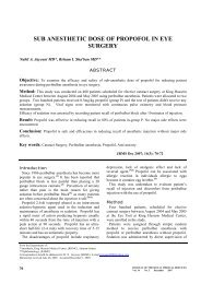
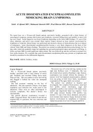
leukocytosis. Chest X-ray Fig. 1 showed right lung<br />
upper lobe shadow, which was confirmed by chest<br />
ultrasound. Chest ultrasound showed a soft tissue<br />
mass 2.5x1.8cm extending deep to ribs of mixed<br />
echogenicity with necrotic areas and increased<br />
vascularity. Abdominal ultrasound was normal.<br />
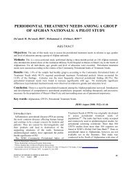
Chest CT scan Fig. 2 showed a large soft tissue<br />
mass of mixed density measuring 5.5x5x4 cm in<br />
dimensions, showing inhomogeneous enhancement<br />
related to the right upper chest wall with extrathoracic<br />
component and large intra-thoracic<br />
component extending to the mediastinum. It encased<br />
the branches of the aorta and downward to the right<br />
hilar region where it compressed the right upper lobe<br />
bronchus and probably the intermediate bronchus.<br />
Associated destruction of the third and fourth ribs<br />
was noted. In addition multiple different sizes of soft<br />
tissue nodules were noted in both lungs.<br />
From the Departments of:<br />
* Pediatrics, Queen Alia Military Hospital, Amman-Jordan.<br />
** Pediatric Surgery, Queen Alia Military Hospital, Amman-Jordan.<br />
Correspondence should be addressed to Dr. A. <strong>Goussous</strong>. P. O. Box 2125 Amman 11953 Jordan, E-mail: arwamurad@hotmail.com<br />
Manuscript received June 3, 2004. Accepted September 2, 2004.<br />
JOURNAL OF THE ROYAL MEDICAL SERVICES<br />
Vol. 14 No. 3 December 2007<br />
57
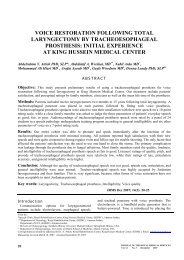
Fig. 1. Chest X-ray showing right upper lobe lung<br />
opacity<br />
Fig. 2. Chest CT-Scan showing right upper lobe lung<br />
mass with bony destruction and extension to the<br />
anterior chest wall<br />
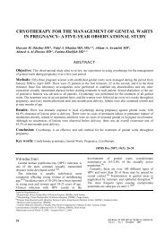
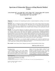
Fig. 3. Right upper lung lobectomy Fig. 4. Bone isotope scans showing 3 rd rib<br />
osteomyelitis<br />
Appearances suggested soft tissue sarcoma or<br />
Ewings sarcoma with secondary metastatic<br />
pulmonary deposits. Incisional biopsy was taken<br />
with pus collection drainage. The specimen was<br />
inconclusive, therefore an excisional biopsy was<br />
decided, and a right lung upper lobectomy was done<br />
(Fig. 3). Pathologic examination reported<br />
necrotizing granulomatous pneumonitis with<br />
fungal forms, and no evidence of malignancy.<br />
Microbiological study of the specimens revealed<br />
hyphae of Aspergillus Spp. Ziehl Nielsen stain for<br />
acid fast bacilli was negative. Pus collection culture<br />
showed Aspergillus Spp. The diagnosis of invasive<br />
pulmonary aspergillosis (IPA) was made. A bone<br />
isotope (Fig. 4) scan showed third rib osteomyelitis.<br />
Brain and abdominal CT scans were normal.<br />
Echocardiogram showed a normal heart.<br />
According to this diagnosis, the possibility of<br />
primary immunodeficiency disease was raised and an<br />
immunological screen was done and showed elevated<br />
IgG of 2126.9 mg/dl (700-1600), normal levels of<br />
IgA, IgM, and IgE. T can B cell markers showed<br />
normal distribution of T and B-lymphocytes and NK<br />
cells. Sweat chloride was 22 meq/l. Nitroblue<br />
Tetrazolium Test (NBT) showed 0% (both<br />
unstimulated and stimulated) (N.R. for unstimulated<br />
cells was 2-17% positive cells), HIV was negative,<br />
and flow cytometry to measure oxidative burst was<br />
unavailable in our institution. Based on the clinical<br />
picture, investigations and the NBT test result, the<br />
diagnosis of Chronic Granulomatous Disease was<br />
made in this patient on April 2003. The patient was<br />
treated with Amphotericin B and Trimethoprimsulfamethoxazole<br />
(TMP-SMX) for 6 weeks. After<br />
treatment the patient was discharged in a good<br />
general condition on a prophylactic daily oral dose of<br />
Itraconazole (5mg/kg) and TMP-SMX (10mg/kg<br />
TMP). The parents are 2 nd degree relatives and there<br />
is no family history of immunodeficiency disease.<br />
Discussion<br />
Patients with CGD characteristically have<br />
lymphadenopathy, hypergammaglobulinemia,<br />
hepato-splenomegaly, dermatitis, failure to thrive,<br />
58<br />
JOURNAL OF THE ROYAL MEDICAL SERVICES<br />
Vol. 14 No. 3 December 2007
anemia, chronic diarrhea, and abscesses. (1)<br />
Nevertheless this was not the mode of presentation in<br />
our case. Our patient was thriving well and presented<br />
with a chest mass with invasive aspergillus<br />
involvement of the lung and pleura that had caused<br />
rib osteomyelitis and chest wall abscess with lack of<br />
any other organ system involvement. Invasive<br />
aspergillus infection usually involves pulmonary,<br />
sinus, cerebral, or cutaneous sites. Rarely,<br />
endocarditis, osteomyelitis, meningitis, infection of<br />
the eye or orbit, and esophagitis occur. (4)<br />
Segal and Holland (3) have looked into a national<br />
database describing the spectrum of infections in 368<br />
patients with CGD. They found that the commonest<br />
pathogens include the Aspergillus sp. (pneumonia),<br />
S. aureus (suppurative adenitis, subcutaneous<br />
infections, and liver abscesses), Serratia sp<br />
(osteomyelitis and pneumonia), Nocardia sp., and B<br />
cepacia (pneumonia and sepsis). (3) Aspergillosis is<br />
the most important cause of death in patients with<br />
CGD. (3)<br />
In the case presented here, based on the<br />
radiological findings, the presumptive diagnosis of<br />
malignancy was made. Nevertheless the final<br />
diagnosis of IPA which had led to the diagnosis of<br />
CGD in this patient was made after excisional<br />
biopsy. Open or Thoracoscopic lung biopsies are<br />
generally the "gold standard" in the diagnosis of<br />
pulmonary problems in immuno-compromised<br />
patients. (5) The diagnosis of IPA is best made by<br />
demonstrating the presence of hyphae in the lung<br />
tissue sample along with culture that is positive for<br />
Aspergillus from the same side. Methenamine silver<br />
nitrate and periodic acid-Schiff stains are the usual<br />
stains to demonstrate the characteristic hyphae. (5) The<br />
National Institute of Immunology, Allergy, and<br />
Infectious Diseases has provided a working case<br />
definition. The diagnosis of IPA is definite when<br />
tissue histopathology shows the hyphae, with or<br />
without a positive culture for Aspergillus from the<br />
same site, or a positive culture from tissue obtained<br />
by an invasive procedure such as transbronchial<br />
biopsy, percutaneous needle aspiration, or open-lung<br />
biopsy. (5) Serologic studies have no established value<br />
in the diagnosis of invasive pulmonary<br />
aspergillosis. (4) Other studies showed that the<br />
Aspergillus galactomannan enzyme immunoassay<br />
(GM EIA) may be a useful diagnostic tool for IA, but<br />
its sensitivity is variable. Results demonstrated that<br />
decreasing the index cutoff for positively to 0.5<br />
increased its sensitivity with minimal loss of<br />
specificity. The low cutoff increased the duration of<br />
test positively before diagnoses by clinical means.<br />
Therefore 0.5 cutoffs may allow for better<br />
performance as an early diagnostic test. 6)<br />
For screening of CGD, the Nitroblue Tetrazolium<br />
(NBT) dye test is still widely used, but its rapidly<br />
being replaced by the more accurate flow cytometry<br />
test using dihydrorhodamine-123 fluorescence (DHR<br />
test). (1) DHR detects oxidant production because it<br />
increases florescence when oxidized by H 2 O 2 .<br />
Surgical excision has been successful for some<br />
cases<br />
of pulmonary infection. Some clinicians emphasize<br />
prompt surgery as a modality for centrally located<br />
lesion (near the mediastinum) because of the higher<br />
likelihood of catastrophic hemorrhage. There is<br />
suggestion that surgical resection of isolated single<br />
lesions is associated with better survival; however,<br />
the presence of a single lesion itself is indicative of<br />
early diagnosis and improved outcome compared<br />
with multiple foci of disease. (7) In our case, surgery<br />
has been beneficial, and has improved the prognosis<br />
of the patient.<br />
The largest therapeutic experience is with<br />
amphotericin B deoxycholate, which should be given<br />
at maximum tolerated doses. (8) Artiago FB (9)<br />
described a case in which pleural involvement was<br />
effectively treated with intrapleural instillation of<br />
Amphoericin B. Responses to antifungal agents are<br />
variable, and clinical response and overall mortality<br />
are highly dependent on the hosts underlying<br />
immune deficit at the time of diagnosis, clinical<br />
manifestations, and whether immune-reconstruction<br />
occurs during therapy. (7) TMP-SMX was added to<br />
the regimen as a prophylaxis, (1) as well as<br />
Itraconazole, which appears to be an effective and<br />
well-tolerated treatment that reduces the frequency of<br />
fungal infections in chronic granulomatous disease (10)<br />
and because of its good penetration into bone. (8)<br />
Treatment options for CGD in our patient are bone<br />
marrow transplantation (BMT) and IFN-gamma<br />
injections. (1) In a multicentric, randomized study<br />
involving prophylactic IFN-gamma (50mug/m²<br />
subcutaneously three times weekly) the number of<br />
severe infections was reduced by more than 70% and<br />
was beneficial in both the X-linked and autosomal<br />
recessive types of CGD. (3) Bone marrow<br />
transplantation is the only known cure for CGD. (1)<br />
Some patients with CGD have been treated<br />
successfully with bone marrow transplantation. (11)<br />
Others may be treated by careful hygiene, preventive<br />
antibiotics and injections if IFN-gamma. (11) The<br />
mortality and morbidity rates associated with BMT<br />
have discouraged its routine use the CGD patients.<br />
BMT is most useful in patients who have had<br />
recurrent severe infection despite antibiotics and<br />
IFN-gamma prophylaxis. (3)<br />
Invasive pulmonary aspergillosis is a serious and<br />
rare entity, and the presentation of Chronic<br />
Granulomatous Disease with a chest mass is even<br />
JOURNAL OF THE ROYAL MEDICAL SERVICES<br />
Vol. 14 No. 3 December 2007<br />
59
more unusual. Therefore CGD should be considered<br />
in the differential diagnosis of chest wall mass and<br />
failure to do so can lead to a delay in diagnosis and<br />
prolongs morbidity. The patient is being followed up.<br />
She is thriving well, with no major<br />
immunodeficiency symptoms and is tolerating<br />
treatment well. Her most recent follow up date was<br />
on June 2004.<br />
Acknowledgement<br />
We would like to thank Dr. Adel Al-Wahadneh,<br />
pediatric immunologist, for his help and support.<br />
References<br />
1. Behrman RE, Kliegman RM, Jenson HB.<br />
Leukopenia. In: Behrman RE, Kliegman RM,<br />
Jenson HB (eds). Nelson Textbook of Pediatrics,<br />
17 th ed. Philadelphia, WB Saunders. 2004; 121:<br />
715-717.<br />
2. Watane A, Jain A, Milligan T. Pediatrics in<br />
review. Am Acad Pediatr 2000; 21(6): 205-208.<br />
3. Segal BH, Holland SM. Primary phagocytic<br />
disorders of childhood. Pediatr Clin North Am<br />
2000; 47(6):1311-1312.<br />
4. Abramson JS, Halsey NA. Red Book report of<br />
the committee on infectious diseases. Am Acad<br />
Pediatr 2003, 208-210.<br />
5. Soubani AO, Chandrasekar PH. The<br />
Clinical Spectrum of Pulmonary Aspergillosis.<br />
Chest 2002; 121(6):1988-1999.<br />
6. Marr KA, Balajee SA, McLaughlin L. Detection<br />
of galactomannan antigenemia by enzyme<br />
immunoassay for the diagnosis of invasive<br />
aspergillosis: variables that affect performance. J<br />
Infect Dis 2004; 190(3): 641-649.<br />
7. Marr KA, Patterson T, Denning D.<br />
Aspergillosis: Pathogenesis, clinical<br />
manifestations, and therapy. Infect Dis Clin North<br />
Am 2002; 16(4): 875-894.<br />
8. Stevens DA, Kan VL, Judson MA, et al. Practice<br />
Guidelines for Diseases Caused by Aspergillus.<br />
Clin Infect Dis 2000; 30(4): 669-709.<br />
9. Baquero-Artiago F, Garcia-Miguel MJ,<br />
Hernandez F, et al. Combined systemic and<br />
intrapleural treatment of Aspergillus pulmonary<br />
empyema after invasive aspergillosis. J Pediatr<br />
Infect Dis 2003; 22(5): 471-473.<br />
10. Gallin J1, Alling Dw, Malech HL. Itraconazole<br />
to prevent fungal infection in chronic<br />
granulomatous disease. N Engl J Med 2003;<br />
348(24): 2416-2422.<br />
11. Bonillia FA, Geha RS. Primary<br />
Immunodeficiency diseases. J Allergy Clin Imm<br />
2003; 111(2): 267-268.<br />
60<br />
JOURNAL OF THE ROYAL MEDICAL SERVICES<br />
Vol. 14 No. 3 December 2007