Management of Boerhaave's Syndrome: Report of Three ... - rjge.ro
Management of Boerhaave's Syndrome: Report of Three ... - rjge.ro
Management of Boerhaave's Syndrome: Report of Three ... - rjge.ro

You also want an ePaper? Increase the reach of your titles
YUMPU automatically turns print PDFs into web optimized ePapers that Google loves.
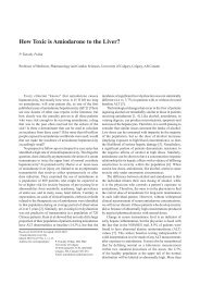
84 Tsalis et al<br />
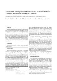
Fig.3 Patient 3. Contrast enhanced CT scan, demonstrating<br />
significant mediastinal free air, and extended left sided<br />
pleural effusion.<br />
the sudden onset <st<strong>ro</strong>ng>of</st<strong>ro</strong>ng> a sharp, post emetic, mid epigastric<br />
pain, radiating to the left shoulder. On the basis <st<strong>ro</strong>ng>of</st<strong>ro</strong>ng> classic<br />
esophageal rupture history, the diagnosis was readily made<br />
by means <st<strong>ro</strong>ng>of</st<strong>ro</strong>ng> erect chest x-ray and CT (Fig.3). A confirmative<br />
subsequent hypaque swallow study demonstrated small<br />
extravasation <st<strong>ro</strong>ng>of</st<strong>ro</strong>ng> contrast medium f<strong>ro</strong>m a distal esophageal<br />
perforation. After a short resuscitation period, the patient<br />
was transferred to the Operating Theatres, where the<br />
rupture site, located on the left esophageal wall next to the<br />
gast<strong>ro</strong>esophageal junction, was app<strong>ro</strong>ached with midline<br />
lapa<strong>ro</strong>tomy. A primary esophageal closure in two layers with<br />
additional fundoplication was performed. Furthermore, a<br />
small left thoracotomy was performed and the thoracic cavity<br />
was copiously irrigated. Finally the chest was closed with<br />
a large (32-gauge) thoracostomy tube in situ. On the third<br />
postoperative day the patient was returned to the main ward<br />
where repeated esophageal contrast medium studies verified<br />
a successful outcome. On postoperative day 15 the patient<br />
began oral feeding. Five weeks after surgery, the patient was<br />
discharged in good general condition. At the time <st<strong>ro</strong>ng>of</st<strong>ro</strong>ng> writing,<br />
6 years after the initial operation, he continues to do well.<br />
Discussion<br />
The presentation <st<strong>ro</strong>ng>of</st<strong>ro</strong>ng> Boerhaave’s synd<strong>ro</strong>me is usually nonspecific<br />
and may mimic many other clinical disorders [11].<br />
Our diagnostic tools were chest X-ray, CT, esophagography,<br />
and esophagoscopy, but principally a high index <st<strong>ro</strong>ng>of</st<strong>ro</strong>ng> suspicion<br />
is required for timely diagnosis. In our cases, CT scans were<br />
done immediately after oral contrast administration, to detect<br />
the level and size <st<strong>ro</strong>ng>of</st<strong>ro</strong>ng> perforation and define the sur<strong>ro</strong>unding<br />
tissue inflammation, which helped as in deciding on the most<br />
app<strong>ro</strong>priate therapy [12,13].<br />
The management <st<strong>ro</strong>ng>of</st<strong>ro</strong>ng> the synd<strong>ro</strong>me remains cont<strong>ro</strong>versial<br />
since treatment can be surgical or non-surgical, and indications<br />
vary according to the functional state <st<strong>ro</strong>ng>of</st<strong>ro</strong>ng> the esophagus, the<br />
presence <st<strong>ro</strong>ng>of</st<strong>ro</strong>ng> associated lesions and the habits <st<strong>ro</strong>ng>of</st<strong>ro</strong>ng> the different<br />
medical teams [1,9,14]. Today, it is accepted that the method<br />
<st<strong>ro</strong>ng>of</st<strong>ro</strong>ng> treatment plays an important <strong>ro</strong>le in the mortality rate<br />
and although surgery has been the most common app<strong>ro</strong>ach,<br />
the selection criteria for conservative treatment reported by<br />
Altorjay et al [15] (intramural perforation, benign defects,<br />
and the absence <st<strong>ro</strong>ng>of</st<strong>ro</strong>ng> sepsis) in 1977, and Came<strong>ro</strong>n et al [16]<br />
in 1979 (disruption contained in the mediastinum; the cavity<br />
draining back into the esophagus; minimal symptoms; and<br />
minimal signs <st<strong>ro</strong>ng>of</st<strong>ro</strong>ng> sepsis), are still valid and should be taken<br />
into account. With reference to the above, perforations<br />
and pleural contamination, once cont<strong>ro</strong>lled by adequate<br />
drainage, simply become an esophagocutaneous fistula and<br />
will heal the same as most gast<strong>ro</strong>intestinal fistulas [17].<br />
Recently, some authors claimed that rapid closure <st<strong>ro</strong>ng>of</st<strong>ro</strong>ng> the<br />
esophageal leak and drainage, could also be achieved by<br />
the minimal invasive endoscopic app<strong>ro</strong>ach by inserting an<br />
endop<strong>ro</strong>sthesis, followed by interventional drainage and/or<br />
thoracoscopic irrigation <st<strong>ro</strong>ng>of</st<strong>ro</strong>ng> the contaminated thoracic cavity<br />
[18-20]. Nevertheless, we believe that this app<strong>ro</strong>ach can be<br />
applied only for iat<strong>ro</strong>genic and early detected perforations.<br />
A self-expandable covered metallic esophageal stent<br />
was placed in one <st<strong>ro</strong>ng>of</st<strong>ro</strong>ng> our patients in order to treat a distal<br />
esophageal post perforation stricture. Esophageal stenting<br />
for non-malignant strictures is cont<strong>ro</strong>versial. The covered<br />
self-expanding metallic stent was originally used for the<br />
stricture or fistula caused by malignant diseases [18]. On the<br />
other hand, many authors investigated the application <st<strong>ro</strong>ng>of</st<strong>ro</strong>ng> this<br />
alternative method to the management <st<strong>ro</strong>ng>of</st<strong>ro</strong>ng> benign conditions<br />
[21]. In our case, stent placement was used mainly because<br />
<st<strong>ro</strong>ng>of</st<strong>ro</strong>ng> the patient’s refusal for further invasive p<strong>ro</strong>cedure, as well<br />
as considering his moderate medical condition.<br />
There is still cont<strong>ro</strong>versy about the most app<strong>ro</strong>priate<br />
type <st<strong>ro</strong>ng>of</st<strong>ro</strong>ng> surgery for patients with esophageal perforation.<br />
Some surgeons performed primary repair, regardless <st<strong>ro</strong>ng>of</st<strong>ro</strong>ng> the<br />
interval between the perforation and intervention, resulting<br />
in a diverse outcome [8,10]. Our opinion on primary<br />
repair is that it is better to be avoided in delayed patients.<br />
Conversely, if the inflammatory p<strong>ro</strong>cess is limited, primary<br />
repair is a reasonable option and may result in an excellent<br />
outcome [10]. Some others suggest that esophagectomy<br />
may be better f<strong>ro</strong>m primary repair for patients with delayed<br />
perforation, because <st<strong>ro</strong>ng>of</st<strong>ro</strong>ng> the high risk <st<strong>ro</strong>ng>of</st<strong>ro</strong>ng> leakage [1,22].<br />
We believe that primary reconstruction must be the first<br />
treatment option in stable, nonseptic patients. Debridement<br />
and drainage with or without continuous lavage [23, 24] is<br />
another option, especially if the patient’s general condition<br />
is impaired or p<strong>ro</strong>gressive sepsis is apparent. We applied this<br />
method in one <st<strong>ro</strong>ng>of</st<strong>ro</strong>ng> our patients with satisfactory results. If the<br />
interval between injury and intervention exceeds 24h or CT<br />
shows signs <st<strong>ro</strong>ng>of</st<strong>ro</strong>ng> p<strong>ro</strong>gressive periesophageal inflammation,<br />
reinforcement <st<strong>ro</strong>ng>of</st<strong>ro</strong>ng> the esophageal repair by viable regional<br />
tissue is recommended [11].<br />
If delayed reconstruction is being considered, it is<br />
possible to bring up the stomach, the small intestine or<br />
the colon to join the cervical esophagus. The timing <st<strong>ro</strong>ng>of</st<strong>ro</strong>ng><br />
reconstruction must be based on the patient’s condition or/and<br />
recovery. Reconstruction <st<strong>ro</strong>ng>of</st<strong>ro</strong>ng> the esophagus can be performed<br />
simultaneously if there is no severe systemic inflammatory<br />
response. Otherwise, delayed reconstruction (2-4 months)<br />
is possibly the best option [1]. The new esophagus can be