RJGE 3/2001 - Journal of Gastrointestinal and Liver Diseases
RJGE 3/2001 - Journal of Gastrointestinal and Liver Diseases
RJGE 3/2001 - Journal of Gastrointestinal and Liver Diseases

Create successful ePaper yourself
Turn your PDF publications into a flip-book with our unique Google optimized e-Paper software.
<strong>RJGE</strong> VOL.10, NR.3 , <strong>2001</strong><br />
<strong>RJGE</strong> 3/<strong>2001</strong><br />
ETIOLOGICAL PROFILE OF CHRONIC HEPATITIS AND LIVER CIRRHOSIS IN ROMANIA<br />
- A MULTICENTRE STUDY<br />
Mircea Grigorescu, Corina Radu, Oliviu Pascu, Alex<strong>and</strong>ru Oproiu, Ana Ionita, Carol<br />
Stanciu, Marioara Stan, Vasile Drug, Florea Voinea, Irinel Voinescu, Marius<br />
Georgescu, Ana Bratu, Cristian Munteanu<br />
Original papers<br />
ACUTE BACTERIAL INFECTIONS - RISK FACTORS FOR UPPER DIGESTIVE BLEEDINGS<br />
IN CIRRHOTIC PATIENTS<br />
Ana Orban-Schiopu, Rodica Rotaru<br />
HEMODYNAMIC CHANGES DURING LIVER TRANSPLANTATION IN DIFFERENT LIVER<br />
DISEASES<br />
Ramona E. Nicolau-Raducu, Lennart Eleborg, Daniela Damian, Mihai Nicolau-Raducu<br />
CASE REPORTS<br />
THE EFFECTS OF INFLIXIMAB ON A PATIENT WITH CROHN'S DISEASE AND DILATED<br />
CARDIOMYOPATHY<br />
Silviu Iobagiu, Oliviu Pascu, Cornelia Popovici, Adriana Petrica<br />
EARLY DIAGNOSIS AND THERAPEUTIC APPROACH IN GARDNER'S SYNDROME - A<br />
REPORT OF THREE CASES<br />
Aurel Mironiuc, Doru Milas<br />
SIMULTANEOUS THROMBOSIS OF HEPATIC VEINS AND VENA CAVA INFERIOR DUE<br />
TO INHERITED RESISTANCE TO ACTIVATED PROTEIN C<br />
Slobodan Kazic, Vladimir Vukcevic, Predrag Miljic, Dragutin Savic, Ivo Elezovic,<br />
Mirjana Perisic, Branka Dapcevic<br />
DIAGNOSTIC FEATURES OF PERITONEAL MALIGNANT MESOTHELIOMA<br />
Valeriu Sbârcea, Anca Rosu, C. Searpe, Mihaela Tenovici<br />
TECHNIQUE/CASE REPORT<br />
EXPANDABLE ESOPHAGEAL "ULTRAFLEX" STENT FIOR THE PALLIATION OF GASTRIC<br />
CANCER<br />
Vasile L. Drug, R Timmer<br />
CLINICAL IMAGING<br />
THREE-DIMENSIONAL ULTRASONOGRAPHY OF THE LOWER GASTROINTESTINAL<br />
TRACT - A NEW ULTRASOUND EXAMINATION TECHNIQUE OR AN ALTERNATIVE TO<br />
ENDOSCOPY?<br />
Radu Badea, T. Vasile, Andrada Seiceanu<br />
You can ask a free issue editor_rjge@email.ro 1
<strong>RJGE</strong> VOL.10, NR.3 , <strong>2001</strong><br />
ETIOLOGICAL PROFILE OF CHRONIC HEPATITIS AND LIVER CIRRHOSIS IN<br />
ROMANIA - A MULTICENTRE STUDY<br />
Mircea Grigorescu1, Corina Radu1, Oliviu Pascu1, Alex<strong>and</strong>ru Oproiu2, Ana Ionita2,<br />
Carol Stanciu3, Marioara Stan3, Vasile Drug3, Florea Voinea4, Irinel Voinescu4,<br />
Marius Georgescu5, Ana Bratu6, Cristian Munteanu7<br />
1) "Iuliu Hatieganu" University <strong>of</strong> Medicine <strong>and</strong> Pharmacy, Cluj-Napoca. 2) "Carol<br />
Davila" University <strong>of</strong> Medicine <strong>and</strong> Pharmacy, Bucharest. 3) University <strong>of</strong> Medicine<br />
<strong>and</strong> Pharmacy, Iasi. 4) Faculty <strong>of</strong> Medicine, Constanta. 5) University <strong>of</strong> Medicine <strong>and</strong><br />
Pharmacy, Craiova. 6) University <strong>of</strong> Medicine <strong>and</strong> Pharmacy, Tg. Mures. 7) Faculty <strong>of</strong><br />
Medicine, Sibiu<br />
Abstract<br />
A prospective multicentre study including 2022 patients <strong>and</strong> covering all the<br />
geographical regions <strong>of</strong> Romania was performed with the aim <strong>of</strong> establishing the<br />
etiological pr<strong>of</strong>ile <strong>of</strong> chronic hepatitis <strong>and</strong> liver cirrhosis in Romania. The diagnosis<br />
was based on clinical, functional <strong>and</strong> morphological data, <strong>and</strong> the etiological pr<strong>of</strong>ile<br />
was established by determining viral markers, autoimmune markers <strong>and</strong> by<br />
metabolic screening. The main etiological factor <strong>of</strong> chronic hepatitis was represented<br />
by viral infections (90.8%), in a decreasing order: C virus (64%), B virus (15.7%), D<br />
virus (9.61%), double B+C (6.94%) <strong>and</strong> treble B+C+D (3.7%) associations. In the<br />
case <strong>of</strong> hepatitis B, the antigen HBe negative or anti HBe positive forms represented<br />
91.67% <strong>of</strong> the cases, the infection with the mutant pre-C virus being possible.<br />
Regarding the etiological pr<strong>of</strong>ile <strong>of</strong> liver cirrhosis, viral cirrhosis represented 48.3%,<br />
alcoholic 19.5%, viral <strong>and</strong> alcoholic 16.2%, <strong>and</strong> those <strong>of</strong> indefinite etiology 11.2%.<br />
In the case <strong>of</strong> viral etiology, C viral infection represented 59.79%, B viral infection<br />
15.83%, the double B+D 16.6%, B+C 5.98%, <strong>and</strong> treble association B+C+D 5.79%.<br />
You can ask a free issue editor_rjge@email.ro 2
<strong>RJGE</strong> VOL.10, NR.3 , <strong>2001</strong><br />
ACUTE BACTERIAL INFECTIONS - RISK FACTORS FOR UPPER DIGESTIVE<br />
BLEEDINGS IN CIRRHOTIC PATIENTS<br />
Ana Orban-Schiopu, Rodica Rotaru<br />
Elias Hospital, Bucharest<br />
Abstract<br />
Although acute variceal bleeding in cirrhotic patients is a severe complication<br />
sometimes lethal, the trigger factors are still unknown. The aim <strong>of</strong> this study was to<br />
find out if acute bacterial infection enhanced the risk for upper digestive bleedings in<br />
cirrhotic patients. We also intended to find out whether acute bacterial infections<br />
modify the coagulation pattern <strong>and</strong> whether these alterations are related to upper<br />
digestive bleedings.<br />
This prospective study is based on 59 patients with liver cirrhosis (Child B)<br />
hospitalized with acute pyelonephritis (38 patients), acute lung diseases (18<br />
patients) <strong>and</strong> acute enterocolitis (3 patients). All these cases were subject to<br />
complex investigation, including both coagulation pr<strong>of</strong>ile <strong>and</strong> Doppler<br />
ultrasonography <strong>of</strong> the portal circulation. In 49 cases, which developed upper<br />
digestive bleedings after admission to hospital, the esogastroduodenoscopic<br />
evaluation was done.<br />
The results <strong>of</strong> our study demonstrated the aggravation <strong>of</strong> diffuse intravascular<br />
coagulation <strong>and</strong> the considerable increase <strong>of</strong> the hepatic congestion index in all cases<br />
with upper digestive bleedings. The early treatment <strong>of</strong> acute bacterial infections in<br />
cirrhotic patients is necessary, in order to prevent upper digestive bleedings.<br />
You can ask a free issue editor_rjge@email.ro 3
<strong>RJGE</strong> VOL.10, NR.3 , <strong>2001</strong><br />
HEMODYNAMIC CHANGES DURING LIVER TRANSPLANTATION IN DIFFERENT<br />
LIVER DISEASES<br />
Ramona E. Nicolau-Raducu1, Lennart Eleborg2, Daniela Damian3, Mihai Nicolau-<br />
Raducu4<br />
1) Dept. <strong>of</strong> Anesthesiology <strong>and</strong> Intensive Care, Municipal University Hospital,<br />
Timisoara. 2) Dept. <strong>of</strong> Anesthesiology <strong>and</strong> Intensive Care, Huddinge University<br />
Hospital, Karolinska Institute, Stockholm, Sweden. 3) Dept. <strong>of</strong> Anesthesiology <strong>and</strong><br />
Intensive Care, County University Hospital. 4) Dept. <strong>of</strong> General Surgery, Municipal<br />
University Hospital, Timisoara<br />
Abstract<br />
We highlight the hemodynamic changes evidenced during liver transplantation,<br />
according to the operation phases related to liver disease etiology, anesthetic <strong>and</strong><br />
vasoactive drugs requirement. We analyzed 20 patients, grouped as follows:<br />
cholestatic liver diseases (n=5), cirrhosis (n=12), familial amyloidotic<br />
polyneuropathy - FAP (n=3). We compared the hemodynamic parameters recorded<br />
one hour after the beginning <strong>of</strong> the surgery, one hour after removal <strong>of</strong> the recipient's<br />
old liver <strong>and</strong> one hour after the reperfusion <strong>of</strong> the grafted liver. During the<br />
preanhepatic phase, the patients from the cholestatic <strong>and</strong> cirrhosis groups had a<br />
hyperdynamic status while in the patients from the FAP group, normal cardiac output<br />
<strong>and</strong> low systemic vascular resistance was seen. A significant decrease in the<br />
systemic arterial pressure was recorded in the cirrhosis patients, immediately after<br />
reperfusion, more significant than in the FAP group. The fentanyl requirement was<br />
low in the patients with FAP. The need for vasoactive drugs increased gradually from<br />
the preanhepatic to the anhepatic phase, reaching its highest point after reperfusion,<br />
during the postanhepatic phase. In conclusion, the hemodynamic changes during<br />
liver transplantation are related to the etiology <strong>of</strong> liver disease.<br />
You can ask a free issue editor_rjge@email.ro 4
<strong>RJGE</strong> VOL.10, NR.3 , <strong>2001</strong><br />
Case reports<br />
THE EFFECTS OF INFLIXIMAB ON A PATIENT WITH CROHN'S DISEASE AND<br />
DILATED CARDIOMYOPATHY<br />
Silviu Iobagiu, Oliviu Pascu, Cornelia Popovici, Adriana Petrica<br />
The 3rd Medical Clinic, University <strong>of</strong> Medicine <strong>and</strong> Pharmacy Cluj-Napoca<br />
Abstract<br />
Crohn's disease is an inflammatory bowel disorder with unknown etiology. In the<br />
pathogenesis <strong>of</strong> this disease, TNF-alpha has an important role, justifying the<br />
treatment with infliximab, an anti-TNF-alpha, in the forms resistant at the usual<br />
treatment <strong>and</strong> in those complicated with fistulae. TNF-alpha is also incriminated in<br />
the pathogenesis <strong>of</strong> other diseases, <strong>and</strong> for this reason, infliximab treatment can<br />
induce a marked improvement in these diseases also. We describe a patient with<br />
Crohn's disease <strong>and</strong> perianal fistulas, unmanageable with the usual treatment, <strong>and</strong><br />
with idiopathic dilated cardiomyopathy <strong>and</strong> cardiac failure who, after infliximab<br />
treatment, evidenced a clear improvement <strong>of</strong> the digestive disorder along with the<br />
closing <strong>of</strong> the perianal fistulas. The cardiac parameters, established through<br />
echocardiography, <strong>and</strong> the signs <strong>of</strong> the cardiac failure also demonstrated a<br />
spectacular improvement.<br />
You can ask a free issue editor_rjge@email.ro 5
<strong>RJGE</strong> VOL.10, NR.3 , <strong>2001</strong><br />
EARLY DIAGNOSIS AND THERAPEUTIC APPROACH IN GARDNER'S<br />
SYNDROME - A REPORT OF THREE CASES<br />
Aurel Mironiuc1, Doru Milas2<br />
1) 2nd Surgical Clinic, University <strong>of</strong> Medicine <strong>and</strong> Pharmacy. 2) 2nd Medical Clinic,<br />
County Hospital, Cluj-Napoca<br />
Abstract<br />
Colonic polyposis in general <strong>and</strong> familial adenomatous polyposis (FAP) in particular<br />
are rare in our geographical area: 3.36% polyposis, 1% FAP, 0,16% Gardner's<br />
syndrome from colonic tumors. Three cases with Gardner's syndrome are presented,<br />
the diagnostic criteria <strong>and</strong> the treatment being discussed. The treatment was surgical<br />
<strong>and</strong> consisted <strong>of</strong> total colectomy with ileo-rectal anastomosis.<br />
The histological examination <strong>of</strong> surgical specimens showed tubulovillous<br />
adenomatous aspect. The patients were followed-up at 3, 6, 9 months<br />
postoperatively, <strong>and</strong> a decrease in the number <strong>and</strong> volume <strong>of</strong> remaining rectal<br />
polyps was found. One <strong>of</strong> the patients, aged 40, presented a malignant rectal lesion<br />
3 months postoperatively that was resected (well differentiated adenocarcinoma).<br />
You can ask a free issue editor_rjge@email.ro 6
<strong>RJGE</strong> VOL.10, NR.3 , <strong>2001</strong><br />
SIMULTANEOUS THROMBOSIS OF HEPATIC VEINS AND VENA CAVA<br />
INFERIOR DUE TO INHERITED RESISTANCE TO ACTIVATED PROTEIN C<br />
Slobodan Kazic1, Vladimir Vukcevic1, Predrag Miljic2, Dragutin Savic3, Ivo Elezovic1,<br />
Mirjana Perisic4, Branka Dapcevic1<br />
1) Center for Gastroenterology <strong>and</strong> Hepatology, Zvezdara Hospital. 2) Institute <strong>of</strong><br />
Haematology, Clinical Center <strong>of</strong> Serbia. 3) Department <strong>of</strong> Radiology, Clinic for<br />
Cardiovascular <strong>Diseases</strong> "Dedinje". 4) Department <strong>of</strong> Gastroenterology, Clinical<br />
Center <strong>of</strong> Serbia, Belgrade<br />
Abstract<br />
We report the case <strong>of</strong> a 21-year old girl who was referred to us because <strong>of</strong> malaise,<br />
dyspnea <strong>and</strong> the appearance <strong>of</strong> visible venous blood vessels on the abdominal wall.<br />
Under-taken investigation, which included splenoportography <strong>and</strong> cavography,<br />
revealed a complete occlusion <strong>of</strong> both hepatic veins <strong>and</strong> the inferior caval vein just<br />
above the junction <strong>of</strong> common iliac veins. Haematological testing revealed the<br />
presence <strong>of</strong> inherited resistance to activated protein C which is a well recognized<br />
factor for the development <strong>of</strong> venous thrombosis. Her twin sister, although she had<br />
the same mutation, remained asymptomatic, implying the important role <strong>of</strong> nongenetic<br />
factors in the pathogenesis <strong>of</strong> thrombosis. Despite the fact that inherited<br />
resistance to activated protein C represents a relatively common cause <strong>of</strong> venous<br />
thrombosis, simultaneous occlusion <strong>of</strong> both hepatic <strong>and</strong> inferior caval vein in the<br />
same patient due to this sort <strong>of</strong> thrombophylia has not been reported before.tc "<br />
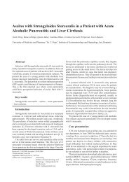
<strong>Liver</strong> metastases from a thyroid cancer are very rare <strong>and</strong> are present in only 0.57%<br />
<strong>of</strong> all patients with thyroid cancer. We consider that it merits presenting two cases<br />
with large liver metastases from a thyroid cancer. In the first case the primary occult<br />
thyroid tumor was revealed one year after the liver tumor resection; a thyroidectomy<br />
was performed. The second patient underwent total thyroidectomy <strong>and</strong> liver<br />
resection in a simultaneous operation. Both patients had a good outcome at 44<br />
months <strong>and</strong> 14 months, respectively. Diagnostic <strong>and</strong> therapeutic aspects are<br />
discussed."<br />
You can ask a free issue editor_rjge@email.ro 7
<strong>RJGE</strong> VOL.10, NR.3 , <strong>2001</strong><br />
DIAGNOSTIC FEATURES OF PERITONEAL MALIGNANT MESOTHELIOMA<br />
Valeriu Sbârcea1, Anca Rosu1, C. Searpe1, Mihaela Tenovici2<br />
1) Clinic <strong>of</strong> Internal Medicine <strong>and</strong> Gastroenterology, CFR Clinical Hospital, University<br />
<strong>of</strong> Medicine <strong>and</strong> Pharmacy. 2) Department <strong>of</strong> Histopathology, CFR Clinical Hospital,<br />
Craiova<br />
Abstract<br />
We present the case <strong>of</strong> a 71-year-old female patient admitted to the hospital with<br />
ascites, which started to develop three months after a pleurisy was diagnosed as<br />
being <strong>of</strong> tuberculotic. After ruling out peritoneal carcinomatosis, the diagnosis <strong>of</strong><br />
peritoneal malignant mesothelioma was established based on the clinical <strong>and</strong><br />
imaging findings <strong>and</strong> confirmed by histopathological <strong>and</strong> immunohistochemical tests.<br />
This case was the starting point <strong>of</strong> a review <strong>of</strong> this pathological condition.<br />
You can ask a free issue editor_rjge@email.ro 8
<strong>RJGE</strong> VOL.10, NR.3 , <strong>2001</strong><br />
TECHNIQUE/CASE REPORT<br />
EXPANDABLE ESOPHAGEAL "ULTRAFLEX" STENT FIOR THE PALLIATION OF<br />
GASTRIC CANCER<br />
Vasile L. Drug1, R Timmer2<br />
1) 2nd Medical Clinic, University Hospital "Sf. Spiridon", Iasi. 2) Department <strong>of</strong><br />
Gastroenterology, "St. Antonious Ziekenhuis", Nieuwegein, The Netherl<strong>and</strong>s<br />
Abstract<br />
Patients with gastric cancer <strong>and</strong> no indication for curable resection may be submitted<br />
to palliative surgery, but the mortality <strong>and</strong> morbidity rate is significantly high.<br />
Exp<strong>and</strong>able metal stents are currently used to relieve malignant obstructions from<br />
the oesophagus, bile ducts, colon, duodenum <strong>and</strong> small intestine <strong>and</strong> there recently<br />
have been published case reports <strong>and</strong> short articles on gastric cancer stenting. We<br />
report the case <strong>of</strong> an 85 year old man, diagnosed with gastric cancer <strong>and</strong> with<br />
symptoms <strong>of</strong> gastrointestinal obstruction. Cardio-respiratory severe diseases <strong>and</strong><br />
local large extension were considered contraindicative for curative surgery. An<br />
"Ultraflex" exp<strong>and</strong>able metal stent was used for the palliation <strong>of</strong> obstruction. The<br />
result was good, allowing the patient to continue with a semi-solid diet.<br />
You can ask a free issue editor_rjge@email.ro 9
<strong>RJGE</strong> VOL.10, NR.3 , <strong>2001</strong><br />
CLINICAL IMAGING<br />
THREE-DIMENSIONAL ULTRASONOGRAPHY OF THE LOWER<br />
GASTROINTESTINAL TRACT - A NEW ULTRASOUND EXAMINATION<br />
TECHNIQUE OR AN ALTERNATIVE TO ENDOSCOPY?<br />
Radu Badea1, T. Vasile2, Andrada Seiceanu1<br />
1) 3rd Medical Clinic. 2) Radiology Clinic, University <strong>of</strong> Medicine <strong>and</strong> Pharmacy, Cluj-<br />
Napoca<br />
Abstract<br />
Three-dimensional ultrasound examination (3D ultrasonography) represents a new<br />
diagnostic technique. It provides high quality data, on perpendicular axes, <strong>and</strong> it is<br />
reproducible. The digestive mucosa may be visualized <strong>and</strong> the features related to<br />
tumoral lesions may be investigated (tumour type, degree <strong>of</strong> pr<strong>of</strong>usion, the existence<br />
<strong>of</strong> adenopathies). In order to achieve a high quality 3D US a special preparation <strong>of</strong><br />
the patient <strong>and</strong> relatively sophisticated equipment are required. The technique has a<br />
significant error coefficient, namely it cannot distinguish between food debris <strong>and</strong><br />
tumoral formations. Additional studies are necessary to assess its utility in a clinical<br />
environment.<br />
You can ask a free issue editor_rjge@email.ro 10