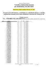
MAKETA 5/3
MAKETA 5/3
MAKETA 5/3

You also want an ePaper? Increase the reach of your titles
YUMPU automatically turns print PDFs into web optimized ePapers that Google loves.
28<br />
A C T A M E D I C A M A R T I N I A N A 2 0 0 5 5/3<br />
receptor status in our study there was a prevalence of cases with both of the receptors (ER, PR)<br />
being positive, or at least one of them positive, which made up 51.5 %. ER only was positive in 48<br />
cases (49.5 %) and PR only in 26 cases (26.8 %). The relatively low incidence of ER and PR positive<br />
cases can be explained by the prevalence of poorly differentiated DIC and high grade DCIS<br />
(VNC 3). The positive ER and PR status is statistically significantly correlated with VNC DCIS categories.<br />
Positive receptors were found mainly in non-high grade cases, while in the group of high<br />
grade DCIS there was an increase of phenotypes with both receptors negative. The most striking<br />
statistically significant differences in correlation with monitored parameters were noticed between<br />
the groups of VNC 1 and VNC 3, and VNC 2 and VNC 3, respectively. However, when evaluating<br />
VI, NPI, hormone-receptor phenotypes, there was not any statistically significant difference<br />
found between the groups of VNC 1 and VNC 2. VNC 2 forms up a group, which substantially<br />
does not differ from VNC 1, but shows different biological behavior against VNC 3.<br />
VNC is correlated with the disease free interval and consecutive development of an invasive<br />
carcinoma (9). VNC was used also in other studies (32,33). Kessar et al. (32) showed that most<br />
of the mammographically detected cases in screening or non-screening programs are detected<br />
already in stage VNC 3 DCIS.<br />
Problems of the VNC are the criteria considering necrosis (22). The central zone of eosinophil<br />
necrotic debris containing 5 or more pycnotic nuclei in whatever architectonic sample of DCIS<br />
serves as a minimum requirement for necrosis (2,14). According to the architectonic classification<br />
we consider comedo-type necrosis those ones, which are centrally localized and represent at<br />
least 50 % of the diameter of the afflicted lumen (18).<br />
One of the positive aspects of the VNC is its possible application in Van Nuys prognostic<br />
index, on the basis of which it is possible to define 3 risk groups of DCIS and subsequent therapeutic<br />
process. This index has been created from the results of retrospective studies of the 3<br />
basic indicators, like the size of DCIS, VNC and the margins of the surgical specimen (34), which<br />
were attested in other studies, as well (35).<br />
It is known, that DCIS is a precursor of invasive carcinoma (3). The heterogeneity of genetic alterations<br />
found in intraductal and invasive breast carcinomas supports the hypothesis of different<br />
genetic mechanisms in clonal evolution of the breast cancer (36,37). From the latest observations it<br />
can be assumed that cribriform / solid DCIS and comedo-type DCIS represent different subtypes of<br />
DCIS expressing 2 different ways of progression: to low and high grade. The evolution of DCIS advances<br />
from normal epithelium through hyperplasia and atypical hyperplasia to comedo-type DCIS in<br />
high grade lesions. Low grade lesions progress from normal epithelium through hyperplasia to cribriform<br />
and solid DCIS (38). Correlations between the evaluated characteristics of DCIS and DIC support<br />
indirectly by phenotype current hypothesis about clonal evolution of the breast cancer.<br />
On the basis of our results and review of literature, it is possible to evaluate VNC as a simple<br />
and clinically relevant system clearly defining prognostic groups, what is indicated by dependencies<br />
on some prognostic indices in invasive carcinomas. In comparison to architectonical<br />
classification of DCIS the VNC shows less heterogeneity.<br />
REFERENCES<br />
1. Moreno A, Lloveras B, Figueras A, et al. Ductal carcinoma in situ of the breast: correlation between histologic classifications<br />
and biologic markers. Mod Pathol 1997; 10: 1088 - 1092<br />
2. Silverstein MJ. Ductal carcinoma in situ of the breast. BMJ 1998; 317: 734 - 739<br />
3. Tavassoli FA. Ductal Carcinoma In situ: Introduction of the concept of ductal intraepithelial neoplasia. Mod Pathol 1998; 11:<br />
140 - 154<br />
4. Glöckner S, Lehmann U, Wilke N, Kleeberger W, Länger F, Kreipe H. Amplification of growth regulatory genes in intraductal<br />
breast cancer is associated with higher nuclear grade but not with the progression to invasiveness. Lab Invest 2001; 81: 565<br />
- 571<br />
5. Lennington WJ, Jensen RA, Dalton LW, Page DL. Ductal carcinoma in situ of the breast. Heterogeneity of individual lesions.<br />
Cancer 1994; 73: 118 - 124<br />
6. Patchefsky AS, Schwartz GF, Finkelstein SD, et al. Heterogenity of intraductal carcinoma of the breast. Cancer 1989; 63: 731<br />
- 741<br />
7. National Coordinating Group for Breast Screening Pathology. Pathology Reporting in Breast Cancer Screening. 2nd Ed. 1995;<br />
23 - 27<br />
8. Holland R, Peterse JL, Millis RR, et al. Ductal carcinoma in situ: a proposal for a new classification. Semin Diagn Pathol 1994;<br />
11: 167 - 180