MAKETA 5/3
MAKETA 5/3
MAKETA 5/3

You also want an ePaper? Increase the reach of your titles
YUMPU automatically turns print PDFs into web optimized ePapers that Google loves.
22<br />
A C T A M E D I C A M A R T I N I A N A 2 0 0 5 5/3<br />
pital in Martin, Slovakia. 106 cases of DIC with the evaluation of DCIS component were randomly<br />
selected in this study. Lobular invasive breast cancers, mixed ducto-lobular invasive cancers,<br />
and special rare forms of invasive breast cancer (e.g. metaplastic) where the DCIS component<br />
was present were excluded. The examined case samples were obtained from women operated<br />
on the above mentioned departments at the age of 33 – 89 years (mean 57 years). The histological<br />
grade of DIC was evaluated by Elston and Ellis modification of histological grading system<br />
(1991) (12). All histological samples were stained by hematoxyline eosine.<br />
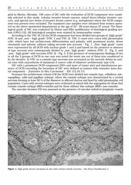
According to the VNC (9,13) the DCIS component has been divided into groups of „high grade“<br />
(VNC 3) and „non - high grade“ (VNC 1 and VNC 2). VNC 3 cases were cases with pleomorphic<br />
cells, which have lost architectonic differentiation and polarity, with prominent nuclei, dense<br />
chromatin and mitosis, without taking necrosis into account - Fig. 1. „Non- high grade“ cases<br />
were represented by all DCIS with nuclear grade 1 and 2 and based on the presence or absence<br />
of type necrosis were subsequently divided to „non- high grade“- without (VNC 1) - Fig. 2, and<br />
„non - high grade“ with necrosis (VNC 2) - Fig. 3. If the presence of contemporary findings of two<br />
or all the 3 groups of DCIS in one case was noted the worst one out of them was considered to<br />
be the decisive. In VNC as a comedo type necrosis was accounted as the necrotic debris in central<br />
zone with caryorrhexis of minimum 5 cancer cells of whatever architectonic type (14).<br />
DIC with a prominent DCIS component (25% and more of tumor size) and simultaneous presence<br />
of DCIS exceeding the boundary of DIC were defined as tumors with extensive intra-ductal<br />
component - „Extensive In Situ Carcinoma“ - EIC (15,16,17).<br />
Pursuant the architectonic criteria (18) the DCIS were divided into comedo type, cribriform, micropapillary,<br />
solid and papillary subtype, where the comedo subtype was characterized by a central<br />
necrosis forming at least 50 % of the diameter in affected ductus and lined by solid pleomorphic cell<br />
population. Cases with several subtypes were evaluated as mixed with the segregation for those that<br />
contain comedo subtype (MIX+comedo) and for those without this subtype (MIX+ non-comedo).<br />
The vascular invasion (VI) was assessed as the presence of vascular emboli in lymphatic vessels<br />
Figure 1: High grade ductal carcinoma in situ with central comedo necrosis – Van Nuys Classification 3