MAKETA 5/3
MAKETA 5/3
MAKETA 5/3

You also want an ePaper? Increase the reach of your titles
YUMPU automatically turns print PDFs into web optimized ePapers that Google loves.
ISSN 1335-8421 Acta Med Mart 2005, 5(3)<br />
ACTA MEDICA<br />
MARTINIANA<br />
Journal for Biomedical Sciences,<br />
Clinical Medicine and Nursing<br />
Contents<br />
3<br />
Skin angiogenesis and its significance in some skin diseases<br />
Desanka Výbohová, Katarína Adamicová, Yvetta Mellová, Gabriela Hešková,<br />
Lenka Kunertová, Magdaléna Marčeková, Milan Mello<br />
9<br />
Postnatal prophylaxis of early-onset group B streptococcal infections<br />
in newborns – a pilot study<br />
Lucia Štillová, Mirko Zibolen, Ingrid Krausová, Zuzana Strechová<br />
15<br />
Evaluation of somatic growth in children with juvenile idiophatic arthritis<br />
Darja Jarošová, Vladimír Mihál, Radomíra Čermáková<br />
21<br />
Assessment of the in situ component in ductal invasive carcinomas<br />
by Van Nuys classification<br />
Karol Kajo, Pavol Žúbor, Katarína Macháleková, Silvester Galo<br />
30<br />
Neurolysis of brachial plexus and peripheral nerves of upper extremities<br />
Viktor Matejčík<br />
Published by the Jessenius Faculty of Medicine in Martin,<br />
Comenius University in Bratislava, Slovakia
A C T A M E D I C A M A R T I N I A N A 2 0 0 5 5/3<br />
E d i t o r - i n - C h i e f :<br />
Javorka, K., Martin, Slovakia<br />
I n t e r n a t i o n a l E d i t o r i a l B o a r d :<br />
Belej, K., Martin, Slovakia<br />
Buchanec, J., Martin, Slovakia<br />
Honzíková, N., Brno, Czech Republic<br />
Kliment, J., Martin, Slovakia<br />
Lehotský, J., Martin, Slovakia<br />
Lichnovský, V., Olomouc, Czech Republic<br />
Mareš, J., Praha, Czech Republic<br />
Plank, L., Martin, Slovakia<br />
Stránsky, A., Martin, Slovakia<br />
Tatár, M., Martin, Slovakia<br />
Żwirska-Korczala, K., Zabrze-Katowice, Poland<br />
E d i t o r i a l O f f i c e :<br />
Acta Medica Martiniana<br />
Jessenius Faculty of Medicine, Comenius University<br />
(Dept. of Physiology)<br />
Malá Hora 4<br />
037 54 Martin<br />
Slovakia<br />
Instructions for authors: http:|www.jfmed.uniba.sk (Acta Medica Martiniana)<br />
T l a č :<br />
P+M Turany<br />
© Jessenius Faculty of Medicine, Comenius University, Martin, Slovakia, 2005
A C T A M E D I C A M A R T I N I A N A 2 0 0 5 5/3 3<br />
SKIN ANGIOGENESIS AND ITS SIGNIFICANCE IN SOME SKIN DISEASES<br />
DESANKA VÝBOHOVÁ 1 , KATARÍNA ADAMICOVÁ 2 , YVETTA MELLOVÁ 1 , GABRIELA HEŠKOVÁ 1 ,<br />
LENKA KUNERTOVÁ 1 , MAGDALÉNA MARČEKOVÁ 1 , MILAN MELLO 1<br />
1<br />
Department of Anatomy, Comenius University, Jessenius Faculty of Medicine and 2 Department of Pathology, Comenius<br />
University, Jessenius Faculty of Medicine, Martin, Slovakia<br />
A b s t r a c t<br />
Angiogenesis is a strict controlled sequence of events that results in a new blood vessels formation and remodelling<br />
of the preexisting vessels. Angiogenesis occurs in the skin during physiologic processes such as anagen stage of the<br />
hair cycle, during the optimal wound healing and also in some cutaneous diseases like psoriasis, rosacea, Kaposi’s sarcoma<br />
and other skin tumors. Especially angioproliferation in skin tumors and psoriasis was studied in details. Tumor<br />
angiogenesis is one of important biological features that is closely related to tumor progression and patient’s prognosis.<br />
Pathological angiogenesis in cutaneous melanoma, basal cell carcinoma and hemangioma are discussed. Though psoriasis<br />
is primarily a lymphocyte-driven disease, the prominent expansion of the superficial vascular plexus (tortuosity,<br />
dilatation, increased permeability, endothelial cells proliferation) in lesional skin suggests that psoriasis can be added to<br />
angiogenesis dependent diseases. Research into the angiogenesis process can contribute to the development of new strategies<br />
in antiangiogenic therapy, within the antitumor and antipsoriatic therapy.<br />
K e y w o r d s : skin angiogenesis, angiogenic diseases, skin tumors, psoriasis<br />
1. INTRODUCTION<br />
Angiogenesis (neovascularisation) is a process of new blood vessels development from preexisting<br />
vessels. It is a part of many physiologic processes but it can also be a part or a mechanism<br />
in the pathogenesis of numerous diseases. Vascular changes and angiogenesis, both excessive<br />
and insufficient one, occur also in some skin diseases. Authors bring a review of present<br />
knowledge on angiogenesis and skin diseases in which pathological, especially excessive angiogenesis,<br />
play an important role.<br />
2. ANGIOGENESIS IN THE SKIN DURING THE INTRAUTERINE DEVELOPMENT<br />
Vasculogenesis and angiogenesis realize the formation of vascular network in embryo. Vasculogenesis<br />
gives rise to the heart and the first primitive vascular plexus inside the embryo, in<br />
the surrounding membranes and the yolk sac circulation. Angiogenesis is responsible for the<br />
remodelling and the formation of new vessels from preexisting vessels (1).<br />
Light microscopy and electron microscopy studies using computer reconstruction have described<br />
the initial indications of the vascular development in the skin in the 35th – 40th day of the<br />
intrauterine development. The first skin vessels are simple endothelial structures, which are differentiated<br />
within the mesenchyme. New-formed capillaries arise from these vessels simultaneously<br />
with the skin development. Skin capillaries ingrow into the developing dermal papillae.<br />
Basal lamina is incomplete and forms only amorphous deposits. At the end of the first trimester<br />
basic organisation of the fetal skin blood supply is built. The other layers of the vascular wall,<br />
especially arterial, are formed during the third month of the intrauterine development (2, 3).<br />
3. MECHANISM OF ANGIOGENESIS<br />
Angiogenesis is a strict controlled series of the events, that results in new blood vessels formation<br />
and remodelling of the preexisting vessels. Angiogenesis is stimulated by the proangio-<br />
Address for correspondence:<br />
MUDr. Desanka Výbohová, Department of Anatomy JFM CU,Malá Hora Str. N. 4<br />
037 54 Martin, Slovakia<br />
Phone: ++421434131427, e-mail:vybohova@jfmed.uniba.sk
4<br />
A C T A M E D I C A M A R T I N I A N A 2 0 0 5 5/3<br />
genic – angiogenesis stimulating factors ( VEGF - vascular endothelial growth factor, FGF-fibroblast<br />
growth factor, TGF – transforming growth factor, PDGF – palted derived growth factor,<br />
angiogenin, angiopoetin 1, MMPs – matrix metalloproteinases). These factors are produced by<br />
the cells of injuried and disordered tissues e.g.: the inflammatory cells, tumor cells, keratinocytes<br />
of the psoriatic plaques, endothelial cells. Production of the proangiogenic factors is stimulated<br />
also by hypoxia. Angiogenic factors disperse into the surrounding tissue and bind to the<br />
endothelial cells. Even though a lot of proangiogenic factors are known, VEGF is still one of the<br />
major endothelial cell – specific stimulatory factors both in physiological and also in pathological<br />
stages (1).<br />
Angiogenic process starts with capillary vasodilation and hyperpermeability with subsequent<br />
extravasation of plasma proteins into the extracellular matrix. Before the proliferation and<br />
migration of endothelial cells local enzymatic degradation of the basement membrane is required<br />
(4). For the degradation of the basement membrane and for the continuation of the<br />
sprouts forming activation of the matrix metalloproteinase production is needed. Matrix metalloproteinases<br />
are zinc - dependent endopeptidases capable of disintegrating extracellular matrix<br />
components. They are produced by different cells in the skin especially by fibroblasts, keratinocytes,<br />
endothelial cells. Their production is not continual but induced by the various cytokines<br />
and growth factors, matrix and cell interactions (5). Proliferation and migration of endothelial<br />
cells come after the basement membrane degradation. Endothelial cells – leaders leave<br />
parent vessels and start to migrate through the degraded matrix into the surrounding tissue.<br />
Migrating endothelial cells - leaders are followed by proliferating endothelial cells. The extracellular<br />
matrix in front of the tip of formating vascular sprouts is dissolved by matrix metalloproteinases<br />
(6).<br />
Migration and proliferation of the endothelial cells in growing vascular sprouts is mediated<br />
through the matrix via the angiogenic enzymes, growth factors and their receptors and also by<br />
cell adhesion molecules - especially integrins (αvβ3, αvβ5). Integrins serve as grappling hooks to<br />
help pull the sprouting vessel forward. They are expressed in small levels on quiescent blood vessels<br />
and under the exposure to VEGF stimuli they are upregulated on the surface of the endothelial<br />
cells of new developing vessels. Not only integrins but also another cell adhesion molecules<br />
like adherins and selectins are involved in angiogenesis process (7).<br />
Lumen of newly formed vessel is formed by the curvature of each endothelial cell (6).<br />
The initial endothelial plexus consists of homogenous endothelial cell tubes, which are remodelled<br />
into a mature network. Remodelling involves the creation of the small and large vessels,<br />
the association with the pericytes and smooth muscle cells and the final regulation of the vascular<br />
density. Initially the endothelial plexus is created in excess so reduction of vascular density<br />
- vascular pruning is needed (8).<br />
During the final step of angiogenesis pericytes play an important role. It is suggested that<br />
pericytes – pluripotent perivascular cells, are generated by in situ differentiation of mesenchymal<br />
precursors at the time of endothelial sprouting. These cells express alpha – smooth muscle<br />
actin, therefore their contractile function is supposed (9).<br />
Some in vitro studies supposed and confirmed that pericytes and smooth muscle cells influence<br />
the endothelial cells proliferation and migration (10, 11). The main role of mural cells – pericytes<br />
and smooth muscle cells is stabilization of newly formed vessels. The aquisition of a coating<br />
with pericytes means the end of plasticity window in which the vascular architecture answers<br />
for the oxygen need of the tissue (8).<br />
Pericytes interaction with the endothelial cells is realized through the long cytoplasmic processes<br />
(12). Disruption of the endothelial – pericyte association can lead to the excessive regression<br />
of vascular loops and abnormal remodelling (8). The process of pericyte recruitment is probably<br />
facilitated by VEGF. The role of VEGF in accelerating pericyte coverage is a novel function,<br />
however, the mechanism has not been clear yet (13). Recruitment of pericytes and generation<br />
of new basement membrane is followed by the latest steps of the angiogenic process: the<br />
fusion of the newly formed vessels and the initiation of the blood flow (14).
A C T A M E D I C A M A R T I N I A N A 2 0 0 5 5/3 5<br />
4. CONTROL OF ANGIOGENESIS, PHYSIOLOGICAL AND PATHOLOGICAL ANGIOGENESIS<br />
Neovascularisation is a tightly regulated process. Control of angiogenesis is mediated through<br />
the angiogenesis inducers and angiogenesis inhibitors.<br />
Angiogenesis inducers can be divided into the groups. The group including VEGF (vascular<br />
endothelial growth factor) and angiopoetins (Ang 1, Ang 2) acts specifically on the endothelial<br />
cells. Another group contains cytokines, chemokines, angiogenic enzymes which all act directly<br />
on the endothelial cells and also on wide range of other target cells (inflammatory cells and<br />
fibrous tissue cells which initialize the angiogenesis process). The best known member of this<br />
group is bFGF (fibroblast growth factor). Apart from the mentioned ones the group consisting of<br />
indirectly acting factors, which effect on macrophages, endothelial and tumor cells to release<br />
angiogenic factors is known. PDGF (plated-derived growth factor), TGF (transforming growth factor),<br />
interleukin-8, integrins belong to this group (14, 15,16).<br />
Angiostatin, thrombospondin, vasculostatin, vasostatin belong to the endogenous angiogenesis<br />
inhibitors. These factors probably act on the endothelial cells and induce their apoptosis (14,<br />
15, 16).<br />
Angiogenesis continuation and the end of angiogenesis process depend on the mutual ratio<br />
of angiogenesis growth factors and inhibitors. However, generally can be said that angiogenesis<br />
is influenced more by the production of angiogenesis inhibitors than stimulators (16).<br />
A physiological angiogenesis is characterized by the balance expression of endothelial survival<br />
and death signals. During the initial phases increased level of proangiogenic factors is coincidental<br />
with the reduced level of angiogenesis inhibitors. In the moment when metabolic<br />
demands of the tissue are satisfied, the level of proangiogenic factors decreases and the level of<br />
inhibitors stars to increase (16).<br />
During the pathological angiogenesis, reduced level of the angiogenesis inhibitors with relatively<br />
or absolutely increased level of proangiogenic factors results in prolonged upregulation and<br />
excessive angiogenesis (16,17).<br />
Major differences between pathological and physiological angiogenesis are: „ turn off “ mechanisms,<br />
which regulate angiogenesis process and they are repressed and another difference -<br />
newly formed vessels have different morphological characteristics - they are highly disorganized<br />
with numerous openings within the vessel walls (17).<br />
5. SKIN DISEASES WITH PATHOLOGICAL ANGIOGENESIS<br />
Angiogenesis occurs in the skin during physiologic processes such as anagen stage of the hair<br />
cycle, during optimal wound healing and also in some cutaneous diseases (18).<br />
The most typical well studied angiogenesis - dependent diseases, in which excessive angiogenesis<br />
occurs, are some skin tumors and psoriasis.<br />
Skin tumors<br />
Current results indicate that tumor angiogenesis is one of important biological features that<br />
is closely related to tumor growth and metastasis. Avascular tumors, rarely larger than 1-2 mm 3 ,<br />
are restricted in their growth because they are short of oxygen and nutrients.<br />
The angiogenic phenotype depends on a net balance between proangiogenic and antiangiogenic<br />
factors produced and released by the tumor cells. If tumor cells make the so called angiogenic<br />
switch - they undergo phenotypic change and become potentially lethal. The tumor mass can<br />
be expanded and spreads into the circulation (20). VEGF and bFGF are the most often found<br />
angiogenic proteins in tumors. Their activities seem to be synergistic (21).<br />
Cutaneous melanoma<br />
Already as early as thirty years ago cutaneous malignant melanoma was first time described<br />
as to induce new blood vessels formation (22). The latest quantitative morphologic analysis
6<br />
A C T A M E D I C A M A R T I N I A N A 2 0 0 5 5/3<br />
aimed on a microvascular density of primary cutaneous melanoma shows, that tumor vascularity<br />
is a significant prognostic factor. An increasing degree of vascularity of melanoma has been<br />
associated with incresed rates of relapse, including regional nodal and distant metastasis and<br />
reduced relapse free and overall survival (22, 23). Increasing tumor vascularity has been associated<br />
with increased incidence of ulceration in the primary tumor, possibly helping to explain the<br />
biologic basis of this known prognostic factor (23). Studies have shown a significantly higher<br />
microvascular density in nodular melanomas than in superficially spreading melanomas. The<br />
highest microvascular density was significantly associated with melanomas in Clark_s level 5,<br />
from what we can assume the important role of angiogenesis in vertical melanoma progression<br />
(24, 25). Besides the supposed role of angiogenesis in vertical growth we have knowledge about<br />
the supposed importance of angiogenesis in melanoma metastatic process. Patients with distant<br />
metastases have higher microvessel count in primary tumor compared to patients with lymph<br />
node metastases (26). Other studies even have reported positive correlation between increased<br />
serum concentration of angiogenic factors ( VEGF, FGF, IL-8) and the stage of disease, tumor<br />
progression and survival in malignant melanoma patient (27). Due to these results we can suppose<br />
that angiogenesis or increased intratumor microvascularity could be a new potent predictive<br />
factor of disease prognosis and selection for the therapy, also for antiangiogenic therapy,<br />
in individual melanoma patients.<br />
Basal cell carcinoma<br />
The prognostic significance of tumor angiogenesis is known also in basal cell carcinoma.<br />
Basal cell carcinoma shows a relatively benign course (BBC type 1) but some basal cell carcinomas<br />
can infiltrate deeper structures and metastasize, they apear „ aggressive“ in behavior (BBC<br />
type 2). BBC type 2 samples usually show higher microvessel count than BBC type 1. Therefore,<br />
we can assume that angiogenesis within the tumor can correlate with aggressive behavior in<br />
basal cell carcinomas (28).<br />
Cutaneous hemangioma<br />
In hemangiomas of infancy pathological angiogenesis can start already during the intrauterine<br />
development. A characteristic manifestation of cutaneous hemangioma has a proliferating<br />
phase and involuting phase. The proliferative phase is associated with increased mitotic rate of<br />
the endothelial cells, extensive pockets of rapidly proliferating endothelial cells, with mast cell<br />
proliferation in the basement membrane. During the early involuting phase mitoses are diminished<br />
and apoptosis of endothelial cells predominates (29). Hemangioma - derived cells differ in<br />
proliferation and migration from normal endothelial cells in in vitro studies. Boye et al. (30) have<br />
supposed that hemangiomas constitute clonal expansion and altered behavior of endothelial<br />
cells, what is probably caused by somatic mutations in genes regulating endothelial cell proliferation<br />
(30). Other immunohistochemical studies showed that proliferating hemangioma endothelial<br />
cells express high levels of VEGF and bFGF (31). Furthermore, the patients with active<br />
proliferative hemangiomas have significantly higher urine levels of bFGF and normal levels<br />
during involuting phase and after the therapy (32). The presence of higher urine levels of bFGF<br />
could be useful in differentiating between hemangiomas and vascular malformation (33).<br />
Psoriasis<br />
Present information puts psoriasis into the group of primarily lymphocyte - driven diseases<br />
but the prominent expansion of the superficial vascular plexus in lesional skin suggests that<br />
psoriasis can be added to angiogenesis - dependent diseases (34). Tortuosity, dilatation, increased<br />
permeability, endothelial cells proliferation within the venous limb of the capillary loops are<br />
characteristic microvascular changes in psoriatic lesions (35). Ultrastructural studies show that<br />
capillary loops within the psoriatic dermal papillae displays morphologic features of the venous<br />
capillary with bridge fenestrations and multilayered basement membrane. It is because the<br />
venous limb is the source of the cells for the elongation of the loop. Thus venous limb is enlar-
A C T A M E D I C A M A R T I N I A N A 2 0 0 5 5/3 7<br />
ged and arterial limb is consequently shortened until the whole capillary loop has a venous configuration,<br />
what is more with the tortuosity of the apical segment (35,36). The microvascular<br />
expansion is limited only to superficial vascular plexus (36). Precise trigger of angiogenesis in psoriasis<br />
has not yet been clear. T cells and hypoxia are disscused as probable triggers (37). It is supposed<br />
that psoriatic lesions provoke angiogenesis by multiple mechanisms. Lesional keratinocytes<br />
produce proangiogenic cytokines especially VEGF, IL-8, thymidine phosphorylase and endothelial<br />
cells proliferative factor. Bhushan et al. (38) compared levels of VEGF and endothelial cell<br />
stimulating angiogenesis factor in lesional skin, non-lesional skin of psoriatic patients and skin<br />
of normal control subjects. Observation showed the highest levels of these factors in lesional skin<br />
and the lowest levels in the skin of normal control subjects (38). Proangiogenic signals affect on<br />
the superficial vascular plexus in the upper dermis and start endothelial cells proliferation.<br />
Because integrins play an important role in cell – matrix interaction and endothelial cells activation,<br />
increased expression of αvβ3 integrin is another proangiogenic factor (39). Upregulation of<br />
angiopoetin 1, angiopoetin 2 (they act directly on endothelial cells) and specific endothelial -Tie 2<br />
receptor are also closely associated with excessive pathological angiogenesis in psoriatic plaques.<br />
Angiopoetin 1 is produced by stromal cells within the dermal papillae. Angiopoetin 2 is secreted<br />
by keratinocytes. Angiopoetins- Tie 2 system probably co-acts with VEGF and bFGF (40).<br />
Superficial vascular plexus expansion is critical for hyperplastic keratinocytes and their<br />
metabolic needs and it also provides enlarged endothelial surface area for the inflammatory cells<br />
displacement (37).<br />
6. CONCLUSIONS<br />
Clinical applications of research on angiogenesis include diagnostic, prognostic and mainly<br />
therapeutic applications. The quantification of angioproliferation in cancer biopsy specimens can<br />
predict progression and metastasis. Therapeutic applications could be contributing both for the<br />
treatment of neoplastic and non-neoplastic diseases. Detailed observation and understanding of<br />
angiogenesis helped the development of antiangiogenic drugs. These molecules act by the inhibition<br />
of endothelial cells, blocking activators of angiogenesis and extracellular matrix breakdown<br />
or directly destroy immature neovasculature (41). Antiangiogenic therapy could become<br />
a new strategy in the treatment of tumors and other angiogenesis - dependent diseases.<br />
7. REFERENCES<br />
1. Risau W. Mechanisms of angiogenesis. Nature 1997; 386:631-42<br />
2. Johnson CL, Holbrook KA. Development of human embryonic and fetal dermal vasculature. J Invest Dermatol<br />
1989; 93: 10S- 17S<br />
4. Moore KL, Persaud TVN. Zrození člověka. Praha: ISV nakladatelství, 2002<br />
5. Folkman J, Shing Y. Angiogenesis. J Biol Chem 1992; 267: 10931-4<br />
6. Kahari VM, Saarialho- Kere U. Matrix metalloproteinases in skin. Exp Dermatol 1997; Oct 6(5): 199-213<br />
7. Folkman J, Haudenschild C. Angiogenesis in vitro. Nature 1980; 288: 551-6<br />
8. Senger DR, Ledbetter SR, Claffey KP et al. Stimulation of endothelial cell migration by vascular permeability factor/vascular<br />
endothelial growth factor through cooperative mechanisms involving the αvβ3 integrin, osteopontin, and<br />
thrombin. Am J Pathol 1996; Jul 149(1): 293-305<br />
9. Benjamin LE, Hemo I, Keshet E. A plasticity window for blood vessels remodelling is defined by pericyte coverage of<br />
the preformed endothelial network and is regulated by PDGF-B and VEGF. Development 1998; 125: 1591-1598<br />
10. Nehls V, Denzer K, Drenckhahn D. Pericyte involvement in capillary sprouting during angiogenesis in situ. Cell Tissue<br />
Res 1992; 270: 469 – 474<br />
11. Orlidge A, D Amore PA. Inhibition of capillary endothelial cell growth by pericytes and smooth muscle cells. J Cell<br />
Biol 1987; 105: 1455-1462<br />
12. Sato Y, Rifkin DB. Inhibition of endothelial cell movement by pericytes and smooth muscle cells. J Cell Biol<br />
1989;109: 309-315<br />
13. Doherty MJ, Canfield AE. Gene expresion during vascular pericyte differentiation. Crit Rev Eukaryot Gene Exp 1999;<br />
9: 1-17<br />
14. Takagi H, King GL, Aiello LP. Identification and characterization of VEGF receptor (Flt) in bovine retinal pericytes.<br />
Diabetes 1996; 45:1016-1023
8<br />
A C T A M E D I C A M A R T I N I A N A 2 0 0 5 5/3<br />
15. Liekens S, De Clerq E, Neyts J. Angiogenesis: regulators and clinical applications. Biochemical Pharmacology 2001;<br />
61: 253-270<br />
16. Tonini T, Rossi F, Claudio PP.Molecular basis of angiogenesis and Cancer. Oncogene 2003; 22: 6549-6556<br />
17. Nor JE, Polverini PJ. Role of endothelial cell survival and death signals in angiogenesis. Angiogenesis 1999; 3: 101-<br />
106<br />
18. Ferrara N. The role of vascular endothelial growth factor in pathological angiogenesis. Breast Cancer Res Treat 1995;<br />
36:127-137<br />
19. Mecklenburg L, Tobin DJ, Muller-Rover S et al. Active hair growth (anagen) is associated with angiogenesis. J Invest<br />
Dermatol 2000; 114: 909-916<br />
20. Hubler WR, Wolf JE. Melanoma, tumor angiogenesis and human Neoplasia. Cancer 1976; 38: 187-192<br />
21. Pepper MS, Ferrara N, Orci L et al. Potent synergism between vascular endothelial growth factor and basic fibroblast<br />
growth factor in the induction of angiogenesis in vitro. Biochem Biophys Res 1992; 189: 824-831<br />
22. Hannah D, Folkman J. Patterns and emerging mechanisms of the angiogenic switch during tumorigenesis. Cell<br />
1996; 86: 353-364<br />
23. Marcoval J, Moreno A, Graelis J et al. Angiogenesis and malignant melanoma – angiogenesis is related to the development<br />
of vertical tumorigenic phase. J Cutan Pathol 1997; 24: 212-218<br />
24. Kashani-Sabet M, Sagebiel RW, Ferreira CMM et al. Tumor vascularity in the prognostic assessment of primary cutaneous<br />
melanoma. J Clin Oncol 2Q002, 20 (7): 1826-1831<br />
25. Jonjic N, Zamolo G, Stifter S et al. Cytomorphological variations, proliferation and angiogenesis in the prognosis of<br />
cutaneous melanoma. Clin and Exp Dermatol 2003; 28: 310-314<br />
26. Straume O, Salvesen HB, Akslen LA. Angiogenesis is important in vertical growth phase melanomas. Intern J Oncol<br />
1999; 15: 595-599<br />
27. Neitzel LT, Neitzel CD, Magee KL et al. Angiogenesis correlates with metastasis in melanoma. Ann Surg Oncol 1999;<br />
6 (1): 70-74<br />
28. Urgurel S, Rappl G, Tilgen W, Reinhold U. Increased serum concentration of angiogenic factors in malignant<br />
melanoma patients correlates with tumor progression and survival. J Clin Oncol 2001;19 (2): 577-583<br />
29. Staibano S, Boscaino A, Salvatore G et al. The prognostic significance of tumor angiogenesis in nonaggressive and<br />
aggressive basal cell carcinoma of the human skin. Hum Pathol 1996; 27 (7): 695-700<br />
30. Razon MJ, Kraling BM, MullikenJB at al. Increased apoptosis coincides with onset of involution in infantile hemangioma.<br />
Microcirculation 1998; 5: 189-195<br />
31. Boye E, Yu Y, Paranya G et al. Clonality and altered behavior of endothelial cells from hemangiomas. J Clin Invest<br />
2001; 107: 745-752<br />
32. Beck L, D’Amore PA. Vascular development: cellular and molecular regulation. FASEB J 1997; 11 (5): 365-373<br />
33. Dosquet C, Coudert MC, Wassef M et al. Importance of bFGF for diagnosis and treatment of hemangiomas. Ann Dermatol<br />
Venereol 1998; 125(5): 313-316<br />
34. Barker JNWN. Pathophysiology of psoriasis. Lancet 1991; 338:227-230<br />
35. Braverman IM, Yen A. Ultrastructure of the capillary loops in the dermal papillae of psoriasis. J Invest Dermatol<br />
1977; 68: 53-60<br />
36. Braverman IM, Simbley BA. Role of microcirculation in the treatment and pathogenesis of psoriasis. J Invest Dermatol<br />
1982; 78: 12 -17<br />
37. Creamer D, Sullivan D, Bicknell R et al. Angiogenesis in psoriasis. Angiogenesis 2002; 5: 231-236<br />
38. Bushan M, McLaughlin B, Weiss JB et al. Levels of endothelial cell - stimulating angiogenesis factor and VEGF are<br />
elevated in psoriatic epidermis. Br J Dermatol 1999; 141: 1054-1060<br />
39. Creamer D, Allen M, Sousa A at al. Altered vascular endothelium integrin expression in psoriasis. Am J Pathol<br />
1995; 147: 1661-1667<br />
40. Kuroda K, Sapadi A, Shoji T et al. Altered expression of angiopoetins and Tie 2 receptor in psoriasis. J Invest Dermatol<br />
2001; 116:713-720<br />
41. Rosen L. Antiangiogenic Strategies and agents in clinical trials. The Oncologist 2005; 5: 20-27<br />
Received: August, 8, 2005<br />
Accepted: November, 2, 2005
A C T A M E D I C A M A R T I N I A N A 2 0 0 5 5/3 9<br />
POSTNATAL PROPHYLAXIS OF EARLY-ONSET GROUP B STREPTOCOCCAL<br />
INFECTIONS IN NEWBORNS – A PILOT STUDY<br />
LUCIA ŠTILLOVÁ, MIRKO ZIBOLEN, INGRID KRAUSOVÁ, ZUZANA STRECHOVÁ<br />
Department of Neonatology, Jessenius Faculty of Medicine, Comenius University, Faculty Hospital of Martin, Martin,<br />
Slovakia<br />
A b s t r a c t<br />
Recently, Streptococcus agalactiae or group B Streptococcus (GBS), is the leading cause of early-onset infections in<br />
newborns. Neonatal Early-Onset Group B Streptococcal Disease (EOGBSD) is manifested mostly as sepsis, and it remains<br />
the most frequent cause of morbidity and mortality in neonatal period. Maternal intrapartum GBS colonization is a<br />
major risk factor for EOGBSD.<br />
Maternal intrapartum antibiotic prophylaxis (IAP) does not prevent all cases of the disease. Management of asymptomatic<br />
neonates of GBS colonized mothers is problematic. Evidence supports the use of postnatal intramuscular penicillin<br />
in newborns to prevent EOGBSD. The authors of the article prefer IAP combined with postnatal antibiotic prophylaxis<br />
(PAP) in a defined group of newborns at risk of the disease, using strict indication criteria.<br />
The aim of the pilot study is to design an optimal strategy for administering PAP. The authors have created a protocol<br />
for management of a full-term infant born to a GBS colonized mother. The study is prospective, the study sample<br />
consists of 100 full-term newborns with adequate postnatal adaptation.<br />
Out of 100 infants, PAP was administered to 9 infants. In 8 of them the indication criteria included leucocytosis, in<br />
1 case additional obstetrical risk factors. In the study group there was no case of clinically manifest infection, and no<br />
case of sepsis, either suspected or proven. The authors suggest that the strategy of selective PAP using penicillin, may<br />
be an effective and safe method in order to reduce morbidity and mortality from streptococcal infections. They recommend<br />
a combination of IAP and selective PAP.<br />
K e y w o r d s : neonatal sepsis, early-onset infection, Streptococcus agalactiae, Group B Streptococcus (GBS), penicillin<br />
prophylaxis<br />
INTRODUCTION<br />
Today, Streptococcus agalactiae or Lancefield group B Streptococcus (GBS), is identified as<br />
the leading cause of invasive bacterial infections in neonates (1). At our clinic there were 11.6 %<br />
of preterm newborns affected with early-onset infection and the culture showed that the most<br />
frequent pathogen found in mothers was GBS (16.2 %) (2). Sepsis remains the most frequent<br />
cause of morbidity and mortality in neonatal period. Incidence of sepsis is the highest in neonatal<br />
period in comparison to other periods of life (3, 4). Early-onset neonatal sepsis is defined if<br />
clinical signs are developed within the first 72 hours of life (5). It is associated with severe, mostly<br />
fulminant course and high case fatality rate. Still, there is no appropriate laboratory marker<br />
for early diagnosing of neonatal sepsis (3).Although neonatal Early-Onset Group-B Streptococcal<br />
Disease (EOGBSD) is rare, it is the most common cause of serious infection in newborn babies.<br />
The EOGBSD or perinatal form of GBS disease, typically occurs within the first 24 hours of life<br />
in most of the patients (6), with fulminant sepsis or pneumonia and less often with meningitis.<br />
The attack rates for the EOGBSD range from 0.5 or 1 to 4 cases per 1,000 live births (7, 8). Earlyonset<br />
disease accounts for approximately 80 % of GBS cases (9). The reported case fatality rate<br />
is 5 - 20 %, even 20 – 60 % (10, 8).<br />
Maternal intrapartum GBS colonization is a major risk factor for EOGBSD in infants. Women<br />
with prenatal GBS colonization were 25 times more likely than women with negative prenatal<br />
cultures to deliver infants with the disease (1). The prevalence of recto-vaginal colonization<br />
among pregnant women ranges from 15 to 40 % (11). The colonization is dynamic, and it is usually<br />
asymptomatic. Carriers are identified by bacteriological testings. Colonization of newborns<br />
Address for correspondence:<br />
Lucia Štillová, Gen. Svobodu 84, 03601 Martin, Slovakia<br />
Phone: +421 904 445 271 e-mail: lucanovalucia@zoznam.sk
10<br />
A C T A M E D I C A M A R T I N I A N A 2 0 0 5 5/3<br />
results from transmission either in utero by ascending spread or, mostly, during passage<br />
through the birth canal. The rate of vertical transmission in neonates born to women colonized<br />
with GBS at time of delivery ranges from 30 to 70 % (7, 6). Although most newborns of colonized<br />
mothers become colonized on skin or mucous membranes, they remain asymptomatic;<br />
among them, 1 to 4 % rapidly develop a clinical infection.<br />
Because of the persistent high incidence and severity of GBS disease, several preventive strategies<br />
have been evaluated. The reference recommendations were published by the CDC in 1996<br />
(12), reevaluated and updated in 2002 (1). Maternal intrapartum antibiotic prophylaxis (IAP) is<br />
currently considered the most effective strategy to prevent neonatal GBS disease and it is the<br />
method of choice. Thus the recommended strategy for the prevention of perinatal GBS disease is<br />
based on a universal prenatal screening-approach with the integration of risk-based options<br />
when necessary.<br />
However, maternal intrapartum chemoprophylaxis will not prevent all cases of early-onset<br />
GBS disease (9). In case that intrapartum antibiotics are not given despite an indication (e.g.,<br />
delivery occurred precipitously before antibiotics could be administered to a GBS-positive<br />
woman), insufficient data are available on which to recommend a single management strategy<br />
for the newborn (1). Giving an injection of penicillin immediately after birth to newborn babies<br />
routinely has been proposed as another way of preventing infection (11). Some centers provide<br />
intramuscular penicillin to asymptomatic infants within 1 hour of birth, based on results of<br />
observational studies showing declines in early-onset GBS disease (1). The complete absence of<br />
EOGBSD was reported as an unexpected benefit of a policy of routine administration of intramuscular<br />
penicillin to prevent gonococcal ophthalmia (13). However, it is important to become<br />
aware of the fact, that routine use of antimicrobial prophylaxis for newborns whose mothers<br />
received IAP is not recommended (1, 9).<br />
Most of the infants affected are full-term infants, even if the attack rate is higher in premature<br />
and low weight neonates (7). Term infants account for approximately 70 % of cases of earlyonset<br />
GBS septicemia in neonates (9). Our objective is to determine whether the administration<br />
of intramuscular penicillin at birth to a defined group of full-term newborns of colonized mothers<br />
is an effective and safe method to prevent morbidity and mortality from EOGBSD. The aim<br />
of the pilot study is to design an optimal strategy for administering postnatal antibiotic prophylaxis<br />
(PAP).<br />
METHODS<br />
There are no national guidelines for the EOGBSD prevention currently available in many<br />
European countries (7). Nevertheless, obstetric program in our hospital already includes a GBS<br />
prevention policy based on a universal prenatal screening-based strategy with the integration of<br />
risk-based options when necessary. In order to prevent further neonatal GBS disease, we pay<br />
attention to obstetric risk factors and information about administering IAP, and we identify a<br />
group of colonized mothers’ newborns at higher risk to administer PAP.<br />
We have chosen GBS positive culture at 35 to 37 weeks of gestation, and, at the same time,<br />
either the total number of newborns’ leucocytes at birth more than 25, or less than 8 x 10 9 per<br />
liter, or the presence of more obstetric risk factors, as the indication criteria for PAP. For the postnatal<br />
prophylaxis we use intramuscular penicillin in a daily dose of 80, 000 units per kilogram<br />
of newborn’s weight, applied in two doses per day. After the third dose of penicillin, the blood<br />
count is checked, and if it does not return to normal levels, penicillin is continued. If there is a<br />
positive blood culture result, the prophylaxis has to be changed into therapy. If sepsis is suspected,<br />
based on clinical and laboratory evaluations, empiric antibiotic therapy, using ampicillin<br />
and gentamicin, should be started.<br />
We have created a protocol for management of a full-term infant born to a GBS colonized mother.<br />
We analyze weight at birth, Apgar score, maternal risk factors such as intrapartum fever (38<br />
o<br />
C or higher), prolonged rupture of membranes (18 hours or more), previous infant with invasive<br />
GBS infection, and urinary tract infection during current pregnancy, then administrating of
A C T A M E D I C A M A R T I N I A N A 2 0 0 5 5/3 11<br />
IAP and its duration, blood cell count at birth and after the third dose of postnatal penicillin, clinical<br />
signs, amniotic water culture and newborn’s outer ear canal culture results.<br />
The study is prospective. The pilot study was started in September 2004, and in July 2005<br />
the study sample consisted of 100 newborns. The study group accounts for 11.3 % of all fullterm<br />
infants born within that time period. The mean birth weight of the study group was 3. 394<br />
grams (± SD = 490 grams). The mean gestational age was 40.2 gestational weeks (± SD = 1.1<br />
weeks). Postnatal adaptation of the newborns was adequate, only in one infant with Apgar score<br />
5/7/8 it was more difficult.<br />
RESULTS<br />
There were 11.3 % of all full-term newborns born to GBS colonized mothers. Firstly, we<br />
assess the risk for GBS sepsis according to the Swiss Society of Neonatology. Full-term infants<br />
of GBS colonized mothers, without any additional risk factors and without IAP, are considered<br />
at risk for GBS sepsis 6 / 1, 000. If there are any additional risk factors, the risk grows 12.5<br />
times. If duration of IAP is longer than 4 hours, it reduces the risk 33 times (the risk is<br />
0.18 / 1 000) (14). It is not known how much postnatal antimicrobial prophylaxis reduces the risk<br />
for the disease. In our study group there were 73 newborns at risk for sepsis 6 / 1 000. Twenty<br />
two neonates were at risk 0.18 / 1 000, because of administration of IAP. There were 4 neonates<br />
at risk 2.25 / 1 000, because of additional risk factors and IAP at the same time. Only one newborn<br />
was at risk 75 / 1 000, because of prolonged rupture of membranes and unsatisfactory<br />
duration of IAP. In this infant, to whom PAP was not administered because of adequate blood<br />
count, there were no signs of infection found and there were negative culture results. (Fig.1)<br />
Fig. 1 Distribution of risk for GBS sepsis in the study group<br />
Out of 100 infants, PAP was administered in 9 cases. In 8 cases the indication criteria included<br />
leucocytosis, in 1 case it included additional risk factors such as prolonged rupture of membranes<br />
and intrapartum fever in mother (Fig.2). In 8 newborns, the total number of leucocytes<br />
went back to normal after three doses of penicillin. In 1 newborn the PNP had to be prolonged.<br />
Out of the 9 newborns with PNP, in 5 of their mothers was not administered any IAP. According<br />
to our criteria, PAP was administered to 4 infants, although their mothers received ampicillin<br />
prophylaxis and the infants were at risk 0.18 / 1000 (Fig.3). We keep in mind that IAP does not<br />
prevent all EOGBSD cases.
12<br />
A C T A M E D I C A M A R T I N I A N A 2 0 0 5 5/3<br />
Amniotic water culture results were GBS positive in 21 % of newborns, 85 % out of them were<br />
infants at high risk for sepsis (6 / 1000). Ear culture results were positive in 27 % of newborns,<br />
88 % out of them were at high risk for sepsis (Fig.4). We can conclude that higher risk of GBS<br />
sepsis is associated with higher incidence of positive microbiological testings.<br />
Fig. 2 Administration of postnatal antibioprophylaxis (PAP) and the indication criteria (RF – risk factors, Leu - leucocytosis)<br />
Fig. 3 Distribution of administration of intrapartum antibioprophylaxis in mothers (IAP) and postnatal antibioprophylaxis<br />
in their newborns (PAP)<br />
Fig. 4 Microbiological examination: amniotic water culture results and newborns’ outer ear canal culture results
A C T A M E D I C A M A R T I N I A N A 2 0 0 5 5/3 13<br />
It is difficult to make a reliable diagnosis, because of stereotypical way in which a newborn<br />
reacts to various insults (4). EOGBSD is mostly manifested as sepsis or pneumonia and less<br />
often with meningitis. Therefore, a complete clinical evaluation is needed and it is important to<br />
assess the clinical signs in a complex way. Infection is defined as an inflammatory response to<br />
the presence of microorganisms or to their invasion to normally sterile sites of the host; sepsis<br />
as a systemic inflammatory response on existing infection (15). In the study group, there were<br />
three infants with temporary muscle hypotonus, five infants with transient hyperreactivity. In<br />
one newborn there was a temporary tachypnea (72 / min) at birth but no other clinical or laboratory<br />
signs of infection. Abnormalities of blood cell count were found in 8 infants- leucocytosis<br />
was a part of indication criteria for PNP in them. There was found no leucopenia, no trombocytosis<br />
or trombocytopenia. The mean count of leucocytes in the study group was 18.3 x 10 9 per<br />
liter (± SD = 5.2 x 10 9 per liter), the mean count of thrombocytes was 254.4 x 10 9 per liter (± SD<br />
= 57.2 x 10 9 per liter).<br />
In the study group there was no case of clinically manifest infection. There was no sepsis suspected<br />
or proven by clinical or laboratory tests.<br />
DISCUSSION<br />
If management of symptomatic neonates is well defined, the management of asymptomatic<br />
neonates of GBS colonized mothers is problematic. There is only an approach for empiric management<br />
of infants with suspected sepsis born to women who received or should have received IAP<br />
(1, 9). It is important to begin empiric antimicrobial therapy as soon as possible if sepsis is suspected<br />
(16). On the other hand, because of non-typical way of responding to various insults in<br />
neonatal period, it is not satisfactory to rely on clinical signs and just observe an infant at risk<br />
for sepsis. Prognosis depends mainly on the time when therapy has begun, and on the complexity<br />
of intensive care (6).<br />
Current strategies used to prevent EOGBSD, are focused upon maternal antibiotic prophylaxis.<br />
In the era of intrapartum antibiotic prophylaxis there has been found a significant<br />
decrease in the overall rate of EOGBSD without an increase in rate of antibiotic-resistant<br />
EOGBSD (17). Attention should now be directed to prevent further neonatal EOGBSD. Observational<br />
studies have suggested that the administration of intramuscular penicillin to the<br />
newborn immediately following delivery (within four hours of birth) may be an effective and<br />
safe method to prevent morbidity and mortality from EOGBSD. Although a Cochrane review<br />
focused on a large randomized controlled trial (18) does not support the routine use of intramuscular<br />
penicillin to prevent EOGBSD in newborn infants (11), a significantly lower incidence<br />
of GBSD in the penicillin groups was reported, and there were no hypersensitivity reactions<br />
observed. In summary, the evidence from uncontrolled, retrospective and non-randomized<br />
controlled prospective studies supports the use of postnatal intramuscular penicillin<br />
in newborns to prevent EOGBSD (11, 19, 20, 21). We do accept that routine administration<br />
of antibiotics for all newborns born to mothers who received intrapartum chemoprophylaxis<br />
to prevent GBS disease is not recommended (9, 1). There are three concerns which underlie<br />
the need to limit the target neonatal population for chemoprophylaxis: the potential for serious<br />
adverse reactions to penicillin, the possible emergence of antibiotic-resistant bacteria,<br />
and cost (9). Therefore, we prefer postnatal chemoprophylaxis in a defined group of newborns<br />
at risk for the disease, using strict indication criteria. For the future, immunoprophylaxis<br />
seems to be the most promising method for prevention of neonatal streptococcal infection. At<br />
present, there is still no vaccine available for clinical use.<br />
Taking into account the incidence of EOGBSD, our pilot study group is not a large one.<br />
At this time, it seems to us that the strategy of selective PAP may be an effective and safe<br />
method in order to reduce morbidity and mortality from streptococcal infections. We recommend<br />
a combination of intrapartum chemoprophylaxis and selective postnatal antibioprophylaxis.
14<br />
A C T A M E D I C A M A R T I N I A N A 2 0 0 5 5/3<br />
REFERENCES<br />
1. Schrag S, Gorwitz R, Fultz-Butts K, Schuchat A. Prevention of Perinatal Group B Streptococcal Disease. Revised<br />
guidelines from Centers for Disease Control and Prevention. MMWR Recomm Rep 2002; 51: 1-22.<br />
2. Hudecová J, Biringer K. Výskyt včasných infekcií u predčasne narodených novorodencov na Neonatologickej klinike<br />
v Martine v r. 1998-2003. In: Aktuality v neonatológii. Martin: JLF UK; 2004. p. 14-17.<br />
3. Zbojan J. Novorodenecká sepsa. In: Zibolen M, Zbojan J, Dluholucký S. Praktická neonatológia. Martin: Osveta;<br />
2001. p. 355-360.<br />
4. Holečková K. Infekcie v najmladších vekových skupinách. In: Bálint O et al. Infektológia a antiinfekčná terapia.<br />
Martin: Osveta; 2000. p. 360-370.<br />
5. Zibolen M. Sepsis neonatorum. In: Buchanec J et al. Repetitórium pediatra. Martin: Osveta; 1994. p.827-829.<br />
6. Bálint O, Trupl J. Streptokokové a enterokokové infekcie. In: Bálint O et al. Infektológia a antiinfekčná terapia.<br />
Martin: Osveta; 2000. p. 143-156.<br />
7. Prevention of perinatal group B streptococcal infections. Guidelines from the Belgian Health Council, 2003<br />
(SHC.7721). Available from: URL: http:// www. health. fgov. be/ CSH_ HGR/ English/ Brochures/GBS_EN2003.pdf<br />
8. Martius J. Infekce streptokoky sérologické skupiny B. In: Martius G, Breckwoldt M, Pfleiderer A. Gynekologie<br />
a porodnictví. Martin: Osveta; 1997. p. 161-162.<br />
9. American Academy of Pediatrics. Revised guidelines for prevention of early onset group B streptococcal infection.<br />
Pediatrics 1997; 99: 489-496.<br />
10. Baker CJ. Group B streptococcal infections. In: Stoll BJ, Weisman LE: Clinics in perinatology. Infections in perinatology.<br />
Vol. 24. Philadelphia: WB Saunders; 1997. p.59-70.<br />
11. Woodgate P, Flenady V, Steer P. Intramuscular penicillin for the prevention of early onset group B streptococcal infection<br />
in newborn infants. The Cochrane Database of Systematic Reviews 2004, Issue 2. Art.No.: CD003667.pub2.<br />
DOI: 10.1002/14651858.CD003667.pub2.<br />
12. Prevention of perinatal group B streptococcal disease: a public health perspective. Centers for Disease Control and<br />
Prevention. MMWR Recomm Rep 1996; 45: 1-24.<br />
13. Steigman AJ, Bottone EJ, Hanna BA. Intramuscular penicillin administration at birth: prevention of early-onset<br />
group B streptococcal disease. Pediatrics 1978; 62: 842-4.<br />
14. Kind C. Betreuung der Neugeborenen von Muttern, die mit Streptokokken der Gruppe B kolonisiert sind. Guidelines.<br />
Swiss Society of Neonatology. (Cited 2005). Available from: URL: http:// www. neonet.ch<br />
15. Minárik M. Sepsa a septický šok. In: Fedor M, Minárik M, Kunovský M et al. Intenzívna starostlivosť v pediatrii.<br />
Martin: Osveta; 2002. p.69-75.<br />
16. Zbojan J. Sepsis neonatorum. In: Buchanec J et al. Vademékum pediatra. Martin: Osveta; 2001. p.935-937.<br />
17. Chen KT et al. No increase in rates of early-onset neonatal sepsis by antibiotic-resistant group B Streptococcus in<br />
the era of intrapartum antibiotic prophylaxis. American Journal of Obstetrics and Gynecology 2005; 192: 1167-<br />
1171.<br />
18. Pyati SP, Pildes RS, Jacobs NM et al. Penicillin in infants weighing two kilograms or less with early onset group B<br />
streptococcal disease. N Eng J Med 1983; 308: 1383-1389.<br />
19. Gerard P et al. Group B streptococcal colonization of pregnant women and their neonates. Epidemiological study and<br />
controlled trial of prophylactic treatment of the newborn. Acta Paediatrica Scandinavica 1979; 68: 819-823.<br />
20. Patel DM, Rhodes PG, LeBlanc MH, Graves GR, Glick C, Morrison J. Role of postnatal penicillin prophylaxis in prevention<br />
of neonatal group B streptococcus infection. Acta Paediatrica 1999; 88: 874-879.<br />
21. Siegel JD, McCracken GH Jr et al. Single-dose penicillin prophylaxis of neonatal group-B streptococcal disease: conclusion<br />
of a 41 month controlled trial. Lancet 1982; 1: 1426-1430.<br />
Received: September, 19, 2005<br />
Accepted: November, 25, 2005<br />
This work was presented at Student Scientific Conference April, 2005
A C T A M E D I C A M A R T I N I A N A 2 0 0 5 5/3 15<br />
EVALUATION OF SOMATIC GROWTH IN CHILDREN WITH JUVENILE<br />
IDIOPHATIC ARTHRITIS<br />
DARJA JAROŠOVÁ, VLADIMÍR MIHÁL 1 , RADOMÍRA ČERMÁKOVÁ 2<br />
Medico-Social Faculty, Ostrava University, 1 Paediatric Clinic, Faculty Hospital Olomouc, 2 Paediatric Clinic,<br />
Faculty Hospital Ostrava, Czech Republic<br />
A b s t r a c t<br />
In 1999 our research was carried out in 81 children with juvenile idiopathic arthritis (JIA). Nine somatic parameters<br />
value (body height, body weight, sitting body height, thigh length, foot length, foot width, hand length, hand width,<br />
wrist circumference) were compared with the results of corresponding age and sex groups stated in Anthropometric Studies<br />
of the Czechoslovak Population in 1985. Other comparisons and evaluations were based on differences of JIA types,<br />
corticosteroids administration, age at the onset of the disease and disease duration. We found slightly decreased values<br />
of body height, body weight, right and left foot, left wrist circumference and BMI with respect to mean z-scores. Student_s<br />
t-test showed significant difference in 63% of somatic parameters between patients treated with corticoids and those treated<br />
with other non-steroid drugs. Negative influence of cumulative dosing and long administration of different corticoids<br />
was found in systemic type of JIA treated with methylprednisolon. Systemic disease showed statistically significant<br />
differences from oligoarticular type and polyarticular type in most somatic parameters. The age at the onset of the disease<br />
and the disease duration did not show any influence on body growth. Significant differences in somatic parameters<br />
were found between children treated with glucocorticoids and children treated with other drugs. The greatest negative<br />
influence on growth was found in systemic type of JIA and during the methylprednisolone therapy.<br />
K e y w o r d s : juvenile idiopathic arthritis, corticosteroids, growth disturbances, anthropometry<br />
INTRODUCTION<br />
Somatic growth reflects the quality of the child’s health. It is so closely connected with all<br />
functions of the growing body that it can serve as a measure for evaluating the total health status<br />
of a human being, the child. Growth is often negatively influenced by congenital or chronic<br />
diseases, one of them is juvenile idiopathic arthritis (JIA).<br />
Chronic inflammation of one or even more joints is the most common symptom of JIA, inflammation<br />
can even spread to other body organs and systems. As Jacobs (1) has reported the disease<br />
usually starts when the child is 3 or 4 years old, or just before the beginning of puberty. JIA is more<br />
typical for girls and we can distinguish three basic types: systemic, oligoarticular and polyarticular.<br />
Growth disorders, various body growth deformations, which are characteristic manifestations<br />
of chronic arthritis in children, were described by G. F. Still (2). The most serious manifestation<br />
of the disease is the limitation of the total growth, as mentioned by several authors (1-9),<br />
which can be demonstrated by acceleration or retardation of the growing process. Consequences<br />
are either temporary or permanent, affected body parts are shortened, prolonged, widened, narrowed<br />
or deformed. Knees, wrist joints, elbow joints, hand and foot joints are affected most, as<br />
described by several authors (1, 9, 10).<br />
The aim of this study was to evaluate the growth of children with juvenile idiopathic arthritis.<br />
The differences in growth between the various subtypes of arthritis were studied and the<br />
influence of glucocorticoid therapy on growth was evaluated.<br />
MATERIAL AND METHODS<br />
A group of 81 children (37 boys and 44 girls) aged 6-17 years was treated for juvenile idiopathic<br />
arthritis. Disease duration was 4 years, at minimum. All basic data were obtained from<br />
Address for correspondence:<br />
Ass.Prof. Darja Jarošová, MSc. PhD., Ostrava University, Medico-Social Faculty, Syllabova 19,<br />
70300 Ostrava-Zábřeh, Czech Republic<br />
Phone: +420/597460534, e-mail: Darja.Jarosova@osu.cz
16<br />
A C T A M E D I C A M A R T I N I A N A 2 0 0 5 5/3<br />
hospital records of Rheumatological Department, Paediatric Clinic of the Faculty Hospital at<br />
Ostrava. For the purpose of statistical evaluation, analyses were carried out in groups with the<br />
same sex, corresponding corticosteroid therapy and JIA subtype.<br />
Anthropometric measurements were performed in children during 1999, in spring and<br />
autumn. We were interested in 9 somatic parameters - body height, sitting body height, body<br />
weight, thigh length, hand length and width, foot length and width, and wrist circumference. The<br />
reason why the stated basic somatic parameters were used in the study was that they are known<br />
for their growth disorders in JIA. All parameters were used for precise comparison with the Czech<br />
reference values. Special standard method of anthropometric measurement corresponding to the<br />
Czech population by Martin and Saller – modification by Bláha et al. (11) was used. We were<br />
interested in personal data, in age at the onset of the disease, disease duration, and indicated<br />
therapy (corticosteroid dose and duration of treatment). For statistical analysis of anthropometric<br />
data, the values were expressed as z-scores according to the formula: (X1-X2)/DS, where X1<br />
= subject’s measurement, X2 = mean of the reference population for age and sex, and SD = SD<br />
of the mean for the reference population.<br />
We were interested if measured bodies were influenced by the age at the onset of disease,<br />
disease duration, dose and duration of corticosteroid administration. Data were statistically analysed<br />
by statistic software SPSS V 7.5 (mean, median, SD, range). The statistical comparisons<br />
were performed with two-tailed unpaired t test (corticosteroid treatment) and analysis of variance<br />
ANOVA (types of JIA). Pearson’s correlation coefficients were calculated to assess the relationships<br />
between somatic parameters and main demographic and clinical characteristics. The<br />
α level was set at 0.05 for all statistical tests.<br />
RESULTS<br />
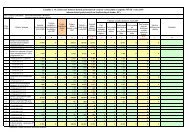
A group contained 37 boys and 44 girls at the average age of 12.97 years (Table 1). The ratio<br />
of male to female was 1 : 1.2. Fifty two children (64 %) had oligoarticular, 21 (26%) had polyarticular<br />
and 8 children (10%) had the systemic type of the disease. The average age at the onset<br />
of the disease was 7.73 years in boys, 7.59 years in girls. JIA started most often at the age of 7<br />
and 10 years in boys, and at the age of 4.8 and 9 years in girls. The average disease duration<br />
was 5.51 years in boys, and 6.06 years in girls. Glucocorticoids were administered to 14 boys<br />
and 12 girls (32%). We found out 100% indication for corticosteroid medication in systemic JIA,<br />
50% indication in polyarticular type, and 16% indication in oligoarticular type.<br />
Comparing z-scores of somatic parameters of girls and boys did not reveal any differences<br />
between the sexes (Figure 1). With the exception of two parameters, all average values of measured<br />
body parameters between +0.5 and –0.5 z-score were considered normal.<br />
Table. 1. Main clinical characteristics of JIA patients
A C T A M E D I C A M A R T I N I A N A 2 0 0 5 5/3 17<br />
Fig. 1.<br />
Structural morphogram - mean standard deviation scores of children with JIA (boys, girls and group)<br />
The age at the onset of the disease was considered to be a possible factor influencing somatic<br />
parameters, but its correlation did not reveal any significant results. Relation of time of disease<br />
manifestation to the width of right hand in polyarticular patients was statistically significant on 1%<br />
level (p = 0.009). Correlation of z-scores in oligoarticular and system diseases were not significant.<br />
The disease duration had no negative influence on somatic parameters of children, either.<br />
Correlation coefficients did not reveal any statistically significant dependencies.<br />
A great difference was found when comparing children treated with corticoids (n = 27) to<br />
children treated with other drugs (n = 54). Significant differences were found in 10 out of 16 soma-<br />
Fig. 2. Structural morphogram – mean standard deviation scores of children with JIA treated with glucocorticoids and<br />
children treated with other drugs
18<br />
A C T A M E D I C A M A R T I N I A N A 2 0 0 5 5/3<br />
tic parameters, some of them were on 1% level of significance (sitting body height, left hand length<br />
and left foot length). Statistically significant results were found in both height parameters (body<br />
height, sitting body height), and in all hand and foot parameters (width and length). All z-scores<br />
of somatic parameters in children treated with corticoids were low and situated in the negative<br />
field of structural morphogram (Figure 2). The greatest differences between the two groups were<br />
found in both body height parameters (difference up to 1 SDS) and in foot length parameters.<br />
Cumulative doses of individual glucocorticoids were calculated from hospital records. Relationships<br />
between body parameters and cumulative dose of glucocorticoids were evaluated. The<br />
highest number of statistically significant correlations was found in oligoarticular JIA. Significant<br />
relations between cumulative dose of prednisone and body weight (p = 0.029), both wrist<br />
circumferences (p = 0.043 and p = 0.016) and BMI (p = 0.049) were found. Correlation of cumulative<br />
dose of methylprednisolone was statistically significant only in the right hand width (p =<br />
0.011) and left hand width (0.043). In triamcinolone, which was used least of all, only one negative<br />
correlation was found, in left wrist circumference (p = 0.034). Polyarticular JIA revealed only<br />
two significant correlations; during treatment with methylprednisolone in left hand length (p =<br />
0.049) and during treatment with triamcinolone in left foot length (p = 0.04). In systemic disease<br />
statistically significant relations of cumulative doses of prednisone were found in sitting body<br />
height (p = 0.042) and in cumulative doses of methylprednisolone five negative correlations were<br />
found in body height (p = 0.027), sitting body height (p = 0.028), left foot length (p = 0.047), left<br />
foot width (p = 0.011) and left wrist circumference (p = 0.039). Between cumulative dose of triamcinolone<br />
and z-score of somatic parameters no statistically significant relation was found.<br />
We were interested how duration of corticosteroids therapy influenced somatic parameters.<br />
Correlation coefficients were calculated for each kind of drugs and each JIA type, not considering<br />
age and sex. In oligoarticular subtype four positive and one negative statistically significant relations<br />
were found. Duration of prednisone treatment influenced body weight (p = 0.048) and left<br />
wrist circumference (p = 0.016). Duration of methylprednisolone treatment significantly influenced<br />
both hands width (p = 0.015 and p = 0.048). No significant correlation was found between<br />
duration of prednisone treatment and duration of methylprednisolone treatment and somatic<br />
parameters in polyarticular JIA. The highest number of correlations of somatic parameters was<br />
found in systemic JIA and methylprednisolone. Statistically significant relations to duration of<br />
methylprednisolone treatment were found in all somatic parameters (p = 0.0001 to 0.049) except<br />
right thigh length and right wrist circumference and BMI. In this type of disease there was statistical<br />
correlation between duration of triamcinolone treatment and length of right (p = 0.041) and<br />
left thigh (p = 0.023) and BMI (p = 0.021). No statistical dependency was found in prednisone.<br />
For evaluating differences of mean z-scores of somatic parameters between three basic JIA<br />
types the analysis of variance was used. Systemic disease differed significantly from oligoarticular<br />
and polyarticular in 13 mean z-scores of somatic parameters (body height, body weight, sitting<br />
body height, right and left thigh length, left and right foot length and width, left and right<br />
hand length and width). Only in left and right wrist circumference and BMI no differences were<br />
found between the subtypes. In children treated with corticoids we found statistically significant<br />
differences between oligoarticular and systemic types of JIA in mean scores of left and right thigh<br />
length, left and right foot length and width and right hand length. Polyarticular JIA differed from<br />
systemic in mean z-scores of body height, sitting body height, left and right thigh length, left and<br />
right foot length, right foot width, right hand length and left hand width.<br />
DISCUSSION<br />
Between sexes there were no differences in somatic parameters. High values of z-scores revealed<br />
similarity.<br />
Growth retardation was first mentioned by Still (2) in the his description of JIA. Still expressed<br />
the idea that growth was influenced more if the disease started at the child’s early age. The<br />
correlation between age at the onset of JIA and somatic parameters was found out only in one<br />
significant correlation (right hand width in polyarticular patients).
A C T A M E D I C A M A R T I N I A N A 2 0 0 5 5/3 19<br />
Results of various studies on the influence of disease duration on the growth of children with<br />
JIA are not easy to interpret. Ansell (3) and Laaksonen (12) considered disease duration to be a<br />
factor which influenced the children’s growing significantly and in a very negative way. Zerin et<br />
al. (10) focused on hand growing retardation caused by disease duration. Polito et al. (7) found<br />
statistically significant correlation between the disease duration and z-score of body height. In<br />
our study correlations did not prove any dependency of somatic parameters on disease duration.<br />
Study group contained 52 children with oligoarticular form of JIA. In this kind of patients<br />
linear growing is usually influenced to a lower degree, which may explain absence of correlation<br />
between the child’s age of illness manifestation and illness duration .<br />
Glucocorticoids are often indicated for treatment of systemic disease of bindweb in children<br />
and adults. Some authors (1,2,13,14,15) considered growth retardation and osteoporosis to be<br />
the most serious side effects of long-term therapy. Corticosteroid administration was indicated<br />
in 27 children of our group. When comparing these children to those non-treated with corticoids,<br />
statistically significant differences were found in 10 out of 16 somatic parameters. Children<br />
treated with corticoids showed negative and lower values of all standardised somatic parameters,<br />
these values were even lower than average values of the Czech reference group by Bláha (11). Z-<br />
score values of children without corticoids were positive and above average values. Z-scores of<br />
children treated with corticoids were slightly higher only in BMI. Statistically significant differences<br />
were found only in height parameters (body height, sitting body height) and in all parameters<br />
of hand and foot. We suppose that these results were influenced by a high number of<br />
children with systemic disease (8 children) and polyarticular disease (11 children) in our group.<br />
Total number was 27 children treated with corticoids. Two mentioned types of JIA are known to<br />
cause most of total and local growth deformities. Children at the age from 11 to 14 years treated<br />
with corticoids were affected most. Similar results were published by Laaksonen (12) who<br />
during a 10-year-research on 544 children with JIA found out that the growth deformities occurred<br />
most often just before puberty and just on its start.<br />
The greatest significant difference between children treated with glucocorticoids and children<br />
treated with other drugs was found in sitting body height. The average body height of children<br />
with non-steroid therapy was nearly the same as the standards for the Czech population of children<br />
of the same age. On the other side the average body height of children treated with corticoids<br />
differed in the most statistically significant way from the reference values of Czech standards.<br />
Body growth retardation of children with JIA was mentioned for the first time in 1932 (16), even<br />
before introducing treatment with corticoids. Correlation results confirm that the linear growth<br />
in the group of children treated with corticoids is influenced in a negative way also by the used<br />
drugs in all length and width parameters of foot and arm. Nevertheless, the deviations found on<br />
foot can be influenced by various other factors: tissue swellings, irregular burdening as a result<br />
of overgrowth of the thigh bone. Results of hand measurement in the active stage of illness can<br />
be influenced by swellings. We did not find statistically significant differences in body weight and<br />
BMI between children treated with corticoids and children treated with other drugs. BMI is not<br />
easy to interpret, as it can be influenced by a great variety of factors: side effects of corticoids,<br />
disease activity, or patient’s actual nutritional regime.<br />
Corticoids are used for treating more severe JIA forms. Retardation of the growing process is<br />
thought to be caused both by the illness itself and the cumulative doses of drugs. Growth is<br />
influenced by the total amount of glucocorticoids.<br />
In our study significant correlations of somatic parameters prevailed in cumulative doses of<br />
corticoids. It was interesting to find out that more dependencies were found in oligoarticular JIA<br />
than in polyarticular and systemic JIA - especially in circumference and width parameters of<br />
hand. Minor joints deformations of hand and foot are known to occur in JIA. We found similar<br />
results. During active stage of disease there were swellings of joints, or even swellings of the whole<br />
wrist, which could influence measured values. In our study the highest number (eight) of somatic<br />
parameters was influenced with cumulative doses of methylprednisone, especially in systemic<br />
JIA. But the fact is that systemic JIA influences the body growth in the most negative way even<br />
without the administration of corticoids. Cumulative doses of triamcinolone influenced parame-
20<br />
A C T A M E D I C A M A R T I N I A N A 2 0 0 5 5/3<br />
ters when comparing to other three drugs, least of all. We enrolled in our transversal study all<br />
registered patients with JIA who met the basic research requirements. Six of them were treated<br />
with triamcinolone before. Cumulative doses and duration of each drug administration were studied<br />
retrospectively. This is the reason why also patients treated with this drug were enrolled.<br />
Duration of corticosteroid therapy influenced somatic parameters in a very interesting way.<br />
Children treated with prednisone were expected to be influenced negatively most of all, but only<br />
two significant correlations of duration of prednisone therapy were found in oligoarticular patients.<br />
The most significant dependences of somatic parameters on duration of administration<br />
were found in children with systemic JIA form treated with methylprednisone.<br />
For comparing somatic parameters of individual JIA forms within the whole group of 81 children<br />
the analysis of variance was used. Systemic JIA was statistically different from oligoarticular<br />
and polyarticular in thirteen somatic parameters (81%). Only in wrist circumference and BMI<br />
no statistically significant differences were found.<br />
Statistically significant differences between JIA types in children treated with corticoids were<br />
found in ten somatic parameters (62.5%), always in relation to the systemic disease. Our results<br />
show that in children treated with corticoids there were no statistically significant differences<br />
among JIA types in six somatic parameters only. We found out, that systemic JIA differed from<br />
oligoarticular and polyarticular in all other parameters.<br />
The results of our study confirm the negative influence of corticoids on the body growth of<br />
children with JIA. No statistically significant differences in somatic parameters were found between<br />
boys and girls. Neither the child’s age of illness manifestation, nor the illness duration<br />
influenced somatic parameters of our patients with JIA. The most negative influence was proved<br />
in methylprednisone and systemic JIA form.<br />
REFERENCES<br />
1. Jacobs JC. Pediatric rheumatology for the practitioner. New York: Springer-Verlag; 1993.<br />
2. Still GF. On a form of chronic joint disease in children. Med Chir Trans 1897; 80: 47.<br />
3. Ansell BM, Bywaters EGL. Growth in Still_s disease. Ann Rheum Dis 1956; 15: 295-319.<br />
4. Bacon MC, White PH, Raiten DJ, Craft N, Margolis S, Levander OA. Nutritional status and growth in juvenile<br />
rheumatoid arthritis. Seminars in Arthritis and Rheumatism 1990; 20: 97-106.<br />
5. Bennett AE, Silverman ED, Miller JJ, Hinty R. Insulin - like growth factor I and II in children with systematic onset<br />
juvenile arthritis. J Rheumatol 1998; 15: 655-668.<br />
6. Cassidy JT, Hillman LS. Abnormalities in skeletal growth in children with juvenile rheumatoid arthritis. Pediatr<br />
Rheumatol 1997; 23: 499-522.<br />
7. Polito C, Strano CG, Olivieri AN, Alessio M, Iammarrone CS, Todisco N, Papale MR. Growth retardation in nonsteroid<br />
treated juvenile rheumatoid arthritis. Scan J Rheumatol 1997; 26: 99-103.<br />
8. Saha MT, Verronen P, Laippala P. Growth of prepubertal children with juvenile chronic arthritis. Acta Paediatr<br />
1999; 88: 724-28.<br />
9. Truckenbrodt H, Häfner R. Allgemeine und lokale Wachstumsstörungen bei chronischer Arthritis im Kindesalter.<br />
Schweiz Med Wschr 1991; 121: 608-20.<br />
10. Zerin JM, Rockwell DT, Garn SM, Schlesinger AE, Sullivan DB. Carpo-metacarpal growth disturbance and the<br />
assessment of carpal narrowing in children with juvenile rheumatoid arthritis. Invest Radiol 1991; 26: 727-33.<br />
11. Bláha P. et al. Antropometrie československé populace od 6 do 55 let. Československá spartakiáda 1985. Praha: ÚNZ<br />
VS; 1986.<br />
12. Laaksonen AL. A prognostic study of juvenile rheumatoid arthritis. Acta Pediatr Scan 1966; 55: Supl.<br />
13. Kotaniemi A. Growth retardation and bone loss as determinants of axial osteopenia in juvenile chronic arthritis.<br />
Scan J Rheumatol 1993; 26: 14-18.<br />
14. Kotaniemi A, Savolainen A, Kautiainen H, Kröger H. Estimation of central osteopenia in children with chronic polyarthritis<br />
treated with glucocorticoids. Pediatrics 1993; 91: 1127-29.<br />
15. Reeve J, Loftus J, Hesp R, Ansell BM, Wright DJ, Woo PMM. Biochemical prediction of changes in spinal bone mass<br />
in juvenile chronic (or rheumatoid) arthritis treated with glucocorticoids. J Rheumatol 1993; 20: 1189-95.<br />
16. Kuhns JG, Swaim LT. Disturbances of growth in chronic arthritis in children. Am J Dis Child 1932; 43: 1118.<br />
Received:August, 8, 2005<br />
Accepted: October, 27, 2005
A C T A M E D I C A M A R T I N I A N A 2 0 0 5 5/3 21<br />
ASSESSMENT OF THE IN SITU COMPONENT IN DUCTAL INVASIVE<br />
CARCINOMAS BY VAN NUYS CLASSIFICATION<br />
KAROL KAJO 1 , PAVOL ŽÚBOR 2 , KATARÍNA MACHÁLEKOVÁ 1 , SILVESTER GALO 2<br />
1<br />
Department of Pathology, Jessenius Faculty of Medicine, Comenius University and Martin Faculty Hospital, 2 Department<br />
of Obstetrics and Gynaecology, Jessenius Faculty of Medicine, Comenius University and Martin Faculty Hospital,<br />
A b s t r a c t<br />
The purpose of this study was to assess the in situ component in ductal invasive carcinomas by Van Nuys classification<br />
– VNC and to provide correlations of ductal carcinoma in situ - DCIS together with other prognostic indices in ductal<br />
invasive carcinoma - DIC (histological grade, vascular invasion, hormone receptor status and Nottingham prognostic<br />
index - NPI).<br />
106 cases of DIC with DCIS were analyzed in the study. DCIS were evaluated by the Van Nuys classification and divided<br />
into three groups (VNC1-3). We have taken the architectonic classification in consideration (comedo type, cribriform,<br />
solid, micropapillary type) when we were making enlistments.<br />
General agreement between VNC groups of DCIS and ductal invasive carcinoma grades was in 79 cases (75%). DIC<br />
cases with high grade (n G3<br />
=54) achieved this agreement in 93%. Differences between VNC groups and grade, vascular<br />
invasion, NPI and hormone receptors reached the level of statistical significance (p
22<br />
A C T A M E D I C A M A R T I N I A N A 2 0 0 5 5/3<br />
pital in Martin, Slovakia. 106 cases of DIC with the evaluation of DCIS component were randomly<br />
selected in this study. Lobular invasive breast cancers, mixed ducto-lobular invasive cancers,<br />
and special rare forms of invasive breast cancer (e.g. metaplastic) where the DCIS component<br />
was present were excluded. The examined case samples were obtained from women operated<br />
on the above mentioned departments at the age of 33 – 89 years (mean 57 years). The histological<br />
grade of DIC was evaluated by Elston and Ellis modification of histological grading system<br />
(1991) (12). All histological samples were stained by hematoxyline eosine.<br />
According to the VNC (9,13) the DCIS component has been divided into groups of „high grade“<br />
(VNC 3) and „non - high grade“ (VNC 1 and VNC 2). VNC 3 cases were cases with pleomorphic<br />
cells, which have lost architectonic differentiation and polarity, with prominent nuclei, dense<br />
chromatin and mitosis, without taking necrosis into account - Fig. 1. „Non- high grade“ cases<br />
were represented by all DCIS with nuclear grade 1 and 2 and based on the presence or absence<br />
of type necrosis were subsequently divided to „non- high grade“- without (VNC 1) - Fig. 2, and<br />
„non - high grade“ with necrosis (VNC 2) - Fig. 3. If the presence of contemporary findings of two<br />
or all the 3 groups of DCIS in one case was noted the worst one out of them was considered to<br />
be the decisive. In VNC as a comedo type necrosis was accounted as the necrotic debris in central<br />
zone with caryorrhexis of minimum 5 cancer cells of whatever architectonic type (14).<br />
DIC with a prominent DCIS component (25% and more of tumor size) and simultaneous presence<br />
of DCIS exceeding the boundary of DIC were defined as tumors with extensive intra-ductal<br />
component - „Extensive In Situ Carcinoma“ - EIC (15,16,17).<br />
Pursuant the architectonic criteria (18) the DCIS were divided into comedo type, cribriform, micropapillary,<br />
solid and papillary subtype, where the comedo subtype was characterized by a central<br />
necrosis forming at least 50 % of the diameter in affected ductus and lined by solid pleomorphic cell<br />
population. Cases with several subtypes were evaluated as mixed with the segregation for those that<br />
contain comedo subtype (MIX+comedo) and for those without this subtype (MIX+ non-comedo).<br />
The vascular invasion (VI) was assessed as the presence of vascular emboli in lymphatic vessels<br />
Figure 1: High grade ductal carcinoma in situ with central comedo necrosis – Van Nuys Classification 3
A C T A M E D I C A M A R T I N I A N A 2 0 0 5 5/3 23<br />
Figure 2: Non-high grade ductal carcinoma in situ without necrosis – Van Nuys Classification 1<br />
Figure 3: Non-high grade ductal carcinoma in situ with necrosis – Van Nuys Classification 2
24<br />
A C T A M E D I C A M A R T I N I A N A 2 0 0 5 5/3<br />
or invasion into blood vessels into the peritumoral space. The Nottingham prognostic index (NPI)<br />
was set by the following criteria: 0.2 x tumor size (cm) + lymph node stage (1 - 3) + histological<br />
grade (1 - 3) (19). Cases were divided into three groups: 1.) with score ≤ 3.4; 2.) score 3.4-5.4; and<br />
3.) score > 5.4 in accordance with achieved final score. The estrogen (ER) and progesterone receptors<br />
(PR) were examined immunohistochemically in the majority of cases (n ERPR<br />
= 97). Cases with<br />
> 10 % expression of malignant cells have been considered positive. For their determination we<br />
used monoclonal antibodies against ER (clone ER1D5, Immunotech) and against PR (clone<br />
PR10A9, Immunotech) diluted 1:50. The streptavidin-biotin-peroxidase detection system (Biogenex)<br />
and chromogen 3-amino-9-ethylcarbazol - AEC (Sigma) were used for visualization. ER and PR status<br />
was evaluated semi-quantitatively and cases in which more than 10 % of tumor cells (in DCIS<br />
or invasive component) showed adequate antigen expression were considered positive (20,21).<br />
Statistical analysis was performed using SPSS 12.0 ® (SPSS Inc., Chicago, IL) statistical software<br />
for Windows XP Professional. Chi - square test (χ 2 ) was used to compare values in observed<br />
criteria. p value < 0.05 was considered to be statistically significant.<br />
RESULTS<br />
In this study were DIC (n G3<br />
= 55) with grade 3 the most often diagnosed cases, which represented<br />
52 % of all examined cases. Similarly, the DCIS component was most frequently present<br />
in the form of high grade (n VNC3<br />
= 54), what stands for 51 % cases. In 17 cases (16.0 %) an extensive<br />
intraductal component (EIC) was detected in 25-99 % of tumor mass extent.<br />
Correlations between VNC of DCIS and DIC grade are shown in Table 1. These results point<br />
out the significant correlation between the selected groups of DCIS and histological grade of DIC<br />
(p < 0.0001).<br />
Table 1 Correlation between VNC of DCIS and grade of DIC<br />
DCIS….ductal carcinoma in situ<br />
VNC….Van Nuys classification<br />
Correspondence between the groups of VNC and grading of invasive breast carcinoma is nearly<br />
in 75 %. In the categories of VNC 1, VNC 2 and VNC 3 was revealed this correspondence in<br />
36 %, 70 % and 94 %, respectively. In VNC 1 and VNC 3 groups has not been observed extreme<br />
discrepancy, what means that in VNC 1 there were not detected poorly differentiated forms of<br />
DIC and in VNC 3 invasive ductal component within histological grade 1. The highest differences<br />
were found in comparison of VNC 1 versus VNC 3 and VNC 2 versus VNC 3, respectively<br />
(both p_ 0.0001). The less profound, but still statistically significant differences were between the<br />
groups of VNC 1 versus VNC 2 (p< 0.05).<br />
Subtypes of DCIS divided by architectonic criteria and correlations with three VNC groups<br />
were observed in this study. The most frequent subtype of DCIS in VNC 3 group was the comedo-type<br />
and in VNC 1 the cribriform one (Table 2).
A C T A M E D I C A M A R T I N I A N A 2 0 0 5 5/3 25<br />
Table 2 Distribution of DCIS into subtypes and VNC groups<br />
DCIS….ductal carcinoma in situ<br />
Only in 39 (36.8 %) of the cases one architectonic subtype was noted and in the rest of the<br />
67 cases (63.2 %) the combination of two or three subtypes was present. In 44 cases (41.5 %)<br />
a comedo subtype of DCIS was detected and in 23 (21.7 %) non-comedo subtypes have been diagnosed.<br />
The pure cribriform type in „non-high grade“ categories was present in 17 cases, with<br />
dominance in VNC 1 group (n=13; 76.5 %). Despite this the pure comedo-type, such as mixed<br />
subtypes with comedo-type have been mostly revealed in the VNC 3 category (n=11; 92 %, and<br />
n=33; 75 %). Only one case of DCIS micropapillary subtype was diagnosed in the VNC 3 group.<br />
Vascular invasion (VI) was diagnosed in 43 (40.6 %) cases and it was most prevalent in the<br />
VNC 3 of DCIS group (57.4 %). The distribution of the DCIS cases in VNC 1, VNC 2, and VNC 3<br />
groups between VI+ and VI– groups is statistically significant (p < 0.01). In reciprocal categorization,<br />
differences in VNC 2 versus VNC 3 (p< 0.01) are higher than in VNC 1 versus VNC 2 (p<<br />
0.05). We did not find significant differences in correlation of VNC 1 versus VNC 3.<br />
Table 3 Correlation between the vascular invasion and DCIS groups by VNC<br />
DCIS….ductal carcinoma in situ<br />
VNC….Van Nuys classification<br />
In the examining procedure we have evaluated the NPI in 96 cases (table 4). The most frequent<br />
conclusions of NPI were in score range 3.4 to 5.4 (n = 46; 47.9 %). The finding of DCIS component<br />
with VNC 3 was found only in cases with NPI higher than 3.4. There were found 3 cases
26<br />
A C T A M E D I C A M A R T I N I A N A 2 0 0 5 5/3<br />
of DIC with VNC 1 DCIS (12.5 %) in the group with NPI over 5.4. Statistically significant differences<br />
were present between categories of VNC 1 versus VNC 3, and VNC 2 versus VNC 3 (p <<br />
0.0001, respectively). We did not find significant differences in correlation between VNC 1 versus<br />
VNC 2.<br />
The hormone receptor status in association with VNC categories of DCIS has been determined<br />
only in 97 cases. Cases both ER and PR positive or with one positive receptor type have been<br />
predominantly present (51.5 %). ERs were positive in 48 (49.5 %) and PRs in 26 (26.8 %) cases<br />
(table 5). The obvious dominance of receptor-positive forms in categories of VNC 1 and VNC 2<br />
has been diagnosed. The VNC 3 group of DCIS was associated with the increased rate of the<br />
forms, where both receptors were negative. Statistical correlations between groups of VNC 1 versus<br />
VNC 3 (p < 0.0001) and VNC 2 versus VNC 3 (p < 0.001) reached the high level of significance.<br />
There were no significant differences between VNC 1 and VNC 2.<br />
Table 4. Correlations between VNC categories and NPI<br />
DCIS….ductal carcinoma in situ<br />
NPI….Nottingham prognostic index<br />
VNC….Van Nuys classification<br />
Table 5. Correlations between hormone receptor phenotypes and VNC categories of DCIS<br />
DCIS….ductal carcinoma in situ<br />
VNC….Van Nuys classification<br />
ER….estrogen receptors<br />
PR….progesterone receptors
A C T A M E D I C A M A R T I N I A N A 2 0 0 5 5/3 27<br />
DISCUSSION<br />
DCIS represents a spectrum of histological and cytological changes, which reflect the differences<br />
in biological behavior (1,18). Despite the fact that histological heterogeneity of the DCIS<br />
is known already from the 1960’s, it has not been sub-classified until the 1980’s. From clinical<br />
aspect the DCIS was evidenced as a self entity, and mastectomy was the method of choice (5).<br />
DCIS had later been divided into subtypes on the basis of intraductal architectonics: comedo-type,<br />
solid, cribriform, micropapillary and papillary types (18). Such a classification system<br />
was based on empiric observations and it did not predict biological behavior (22). It was found<br />
that dividing DCIS on the basis of architectonic samples has its limitations, because the majority<br />
of DCIS are mixed, that means consisting of more subtypes. Furthermore, the correlation<br />
between the architectonic classification and nuclear grading, but also the reproducibility are low<br />
(23). In our set of patients we found an architectonical “pure” subtype of DCIS only in 39 cases,<br />
which equaled to 36.8 % from the entire group of patients. Similar results (32-39 %) were reported<br />
from other studies (13,24), which evidences for high heterogeneity of classifications based<br />
solely on architectonical criteria. Although the importance of the architectonical classification is<br />
limited, classification into specific subtypes fits some biological characteristics, such as comedocarcinomas<br />
which have higher incidence of recurrence and invasiveness than other subtypes<br />
(18,25,26,27), pure micropapillary carcinomas, regardless of nuclear grading, have got a tendency<br />
to appear in several breast quadrants (6,17), while the solid subtype is more likely to be<br />
completely extirpated (18). Although architectonical characteristics are currently not considered<br />
prognostically significant, they are recommended when describing bioptic findings (3,28,29).<br />
Lagios (25) has divided DCIS on the basis of architectonic signs, nuclear grading and the presence<br />
of necrosis into four groups. In the first two groups – comedo-type and cribriform with<br />
papillary subtype, presence of necrosis and high nuclear grade he proved more frequent incidence<br />
of hidden invasiveness than in the other 2 groups representing DCIS with cribriform and<br />
micropapillary architectonics, medium or low nuclear grade and without necrosis. Two basic<br />
characteristics of this classification were strongly predictive for local recurrence: the high nuclear<br />
grade and comedo-type necrosis. The evaluation of these two has become a basis for several<br />
classifications. In the 1990_s several morphologic classification schemes were formed in<br />
order to evaluate the biological potential of DCIS, thereby predicting the risk of local recurrence<br />
after conservative therapy. One of those is the VNC (9), which was established in 1995. The VNC<br />
is based on two statistically significant predictors of local recurrence, which are the presence or<br />
absence of high-grade and comedo-type necrosis (13). Both of these factors reflect cancer biology<br />
and they are well sampled by practical pathologists. All high grade DCIS, regardless of the<br />
presence or absence of comedo-type necrosis, are in the worst prognostic group. The great<br />
advantage of this classification is the fact that nuclear grade 2 itself must not be identified<br />
(14,30), the evaluation of which is accompanied with greatest disagreements between evaluating<br />
pathologists. The results of our analyses indicate that differences between VNC 1 and VNC 2 are<br />
obscure, which confirms the existence of a single common group of „non-high grade“ DCIS. Therefore,<br />
in the VNC the most important criterion is to confirm or exclude „high-grade“ carcinoma.<br />
There is a known correlation between the differentiation of invasive carcinomas and DCIS with<br />
other biological markers (1,20). It was proved, that the nuclear grade of DCIS (31) and VNC are<br />
correlated with the degree of differentiation of DIC (30). In our study we could not demonstrate<br />
cases of VNC 3 DCIS in good differentiated forms of DIC, neither VNC 1 in poorly differentiated<br />
DIC, respectively. If the histological grading is considered as an independent prognostic indicator<br />
of recurrence and of the total survival of patients with breast cancer (12), then a statistically significant<br />
correlation between VNC of DCIS and the histological grading of DIC reflects the prognostic<br />
importance of the VNC. This is supported by the results correlating NPI of invasive carcinomas,<br />
which represent a complex index combined from prognostically relevant morphological<br />
indices (tumor size, LN status and histological grading). DCIS with VNC 3 is more aggressive and<br />
is associated with a high probability of consecutive invasive cancer (31). The next index with a significant<br />
negative correlation with VNC is the hormone receptor status. In evaluating of hormone
28<br />
A C T A M E D I C A M A R T I N I A N A 2 0 0 5 5/3<br />
receptor status in our study there was a prevalence of cases with both of the receptors (ER, PR)<br />
being positive, or at least one of them positive, which made up 51.5 %. ER only was positive in 48<br />
cases (49.5 %) and PR only in 26 cases (26.8 %). The relatively low incidence of ER and PR positive<br />
cases can be explained by the prevalence of poorly differentiated DIC and high grade DCIS<br />
(VNC 3). The positive ER and PR status is statistically significantly correlated with VNC DCIS categories.<br />
Positive receptors were found mainly in non-high grade cases, while in the group of high<br />
grade DCIS there was an increase of phenotypes with both receptors negative. The most striking<br />
statistically significant differences in correlation with monitored parameters were noticed between<br />
the groups of VNC 1 and VNC 3, and VNC 2 and VNC 3, respectively. However, when evaluating<br />
VI, NPI, hormone-receptor phenotypes, there was not any statistically significant difference<br />
found between the groups of VNC 1 and VNC 2. VNC 2 forms up a group, which substantially<br />
does not differ from VNC 1, but shows different biological behavior against VNC 3.<br />
VNC is correlated with the disease free interval and consecutive development of an invasive<br />
carcinoma (9). VNC was used also in other studies (32,33). Kessar et al. (32) showed that most<br />
of the mammographically detected cases in screening or non-screening programs are detected<br />
already in stage VNC 3 DCIS.<br />
Problems of the VNC are the criteria considering necrosis (22). The central zone of eosinophil<br />
necrotic debris containing 5 or more pycnotic nuclei in whatever architectonic sample of DCIS<br />
serves as a minimum requirement for necrosis (2,14). According to the architectonic classification<br />
we consider comedo-type necrosis those ones, which are centrally localized and represent at<br />
least 50 % of the diameter of the afflicted lumen (18).<br />
One of the positive aspects of the VNC is its possible application in Van Nuys prognostic<br />
index, on the basis of which it is possible to define 3 risk groups of DCIS and subsequent therapeutic<br />
process. This index has been created from the results of retrospective studies of the 3<br />
basic indicators, like the size of DCIS, VNC and the margins of the surgical specimen (34), which<br />
were attested in other studies, as well (35).<br />
It is known, that DCIS is a precursor of invasive carcinoma (3). The heterogeneity of genetic alterations<br />
found in intraductal and invasive breast carcinomas supports the hypothesis of different<br />
genetic mechanisms in clonal evolution of the breast cancer (36,37). From the latest observations it<br />
can be assumed that cribriform / solid DCIS and comedo-type DCIS represent different subtypes of<br />
DCIS expressing 2 different ways of progression: to low and high grade. The evolution of DCIS advances<br />
from normal epithelium through hyperplasia and atypical hyperplasia to comedo-type DCIS in<br />
high grade lesions. Low grade lesions progress from normal epithelium through hyperplasia to cribriform<br />
and solid DCIS (38). Correlations between the evaluated characteristics of DCIS and DIC support<br />
indirectly by phenotype current hypothesis about clonal evolution of the breast cancer.<br />
On the basis of our results and review of literature, it is possible to evaluate VNC as a simple<br />
and clinically relevant system clearly defining prognostic groups, what is indicated by dependencies<br />
on some prognostic indices in invasive carcinomas. In comparison to architectonical<br />
classification of DCIS the VNC shows less heterogeneity.<br />
REFERENCES<br />
1. Moreno A, Lloveras B, Figueras A, et al. Ductal carcinoma in situ of the breast: correlation between histologic classifications<br />
and biologic markers. Mod Pathol 1997; 10: 1088 - 1092<br />
2. Silverstein MJ. Ductal carcinoma in situ of the breast. BMJ 1998; 317: 734 - 739<br />
3. Tavassoli FA. Ductal Carcinoma In situ: Introduction of the concept of ductal intraepithelial neoplasia. Mod Pathol 1998; 11:<br />
140 - 154<br />
4. Glöckner S, Lehmann U, Wilke N, Kleeberger W, Länger F, Kreipe H. Amplification of growth regulatory genes in intraductal<br />
breast cancer is associated with higher nuclear grade but not with the progression to invasiveness. Lab Invest 2001; 81: 565<br />
- 571<br />
5. Lennington WJ, Jensen RA, Dalton LW, Page DL. Ductal carcinoma in situ of the breast. Heterogeneity of individual lesions.<br />
Cancer 1994; 73: 118 - 124<br />
6. Patchefsky AS, Schwartz GF, Finkelstein SD, et al. Heterogenity of intraductal carcinoma of the breast. Cancer 1989; 63: 731<br />
- 741<br />
7. National Coordinating Group for Breast Screening Pathology. Pathology Reporting in Breast Cancer Screening. 2nd Ed. 1995;<br />
23 - 27<br />
8. Holland R, Peterse JL, Millis RR, et al. Ductal carcinoma in situ: a proposal for a new classification. Semin Diagn Pathol 1994;<br />
11: 167 - 180
A C T A M E D I C A M A R T I N I A N A 2 0 0 5 5/3 29<br />
9. Silverstein MJ, Poller DN, Waisman JR, et al. Prognostic classification of breast ductal carcinoma-in-situ. Lancet 1995; 345:<br />
1154 - 1157<br />
10. Poller DN, Silverstein MJ, Galea M, et al. Ideas in pathology. Ductal carcinoma in situ of the breast: a proposal for a new simplified<br />
histological classification association between cellular proliferation and c-erbB-2 protein expression. Mod Pathol 1994;<br />
7: 257 - 262<br />
11. Douglas-Jones AG, Schmid KW, Bier B, et al. Metallothionein expression in duct carcinoma in situ of the breast. Hum Pathol<br />
1995; 26: 217 - 222<br />
12. Elston CW, Ellis IO. Pathological prognostic factors in breast cancer. I. The value of histological grade in breast cancer: experience<br />
from a large study with long-term follow up. Histopathology 1991; 19: 403 - 410<br />
13. Silverstein MJ, Lagios MD. Use of predictors of recurrence to plan therapy for DCIS of the breast. Oncology 1997; 11: 393 -<br />
410<br />
14. Douglas-Jones AG, Gupta SK, Attanoos RL, Morgan JM, Mansel RE. A critical appraisal of six modern classifications of ductal<br />
carcinoma in situ of the breast (DCIS): correlation with grade of associated invasive carcinoma. Histopathology 1996; 29:<br />
397 - 409<br />
15. Schnitt SJ, Connolly JL. Processing and evaluation of breast excision specimens. A clinically oriented approach. Am J Clin<br />
Pathol 1992; 98: 125 - 137<br />
16. Sainsbury JR, Anderson TJ, Morgan DA, Dixon JM. ABC of breast diseases: Breast cancer. BMJ 1994; 309: 1150 - 1153<br />
17. Connolly JL, Boyages J, Nixon AJ, et al. Predictors of breast recurrence after conservative surgery and radiation therapy for<br />
invasive breast cancer. Mod Pathol 1998; 11: 134 - 139<br />
18. Bellamy CO, McDonald C, Salter DM, Chetty U, Anderson TJ. Noninvasive ductal carcinoma of the breast: the relevance of<br />
histologic categorization. Hum Pathol 1993; 24: 16 - 23<br />
19. Galea MH, Blamey RW, Elston CE, Ellis IO. The Nottingham Prognostic Index in primary breast cancer. Breast Cancer Res<br />
Treat 1992; 22: 207 - 219<br />
20. Bose S, Lesser ML, Norton L, Rosen PP. Immunophenotype of intraductal carcinoma. Arch Pathol Lab Med 1996; 120: 81 - 85<br />
21. Thürlimann B. The international St Gallen consensus 1998: an update. OIP 1998; 3: 4 - 7<br />
22. Quinn CM, Ostrowski JL, Parkin GJ, Horgan K, Benson EA. Ductal carcinoma in situ of the breast: the clinical significance<br />
of histological classification. Histopathology 1997; 30: 113 - 119<br />
23. Sloane JP, Amendoeira I, Apostolikas N, et al. Consistency achieved by 23 European pathologists in categorizing ductal carcinoma<br />
in situ of the breast using five classifications. European Commission Working Group on Breast Screening Pathology.<br />
Hum Pathol 1998; 29: 1056 -1062<br />
24. Harrison M, Coyne JD, Gorey T, Dervan PA. Comparison of cytomorphological and architectural heterogenity in mammographically-detected<br />
ductal carcinoma in situ. Histopathology 1996; 28: 445 - 450<br />
25. Lagios MD. Duct carcinoma in situ. Pathology and treatment. Surg Clin North Am 1990; 70: 853 - 871<br />
26. Solin LJ, Yeh IT, Kurtz J, et al. Ductal carcinoma in situ (intraductal carcinoma) of the breast treated with breast-conserving<br />
surgery and definitive irradiation. Correlation of pathologic parameters with outcome of treatment. Cancer 1993; 71: 2532 -<br />
2542<br />
27. Fisher ER, Costantino J, Fisher B, Palekar AS, Redmond C, Mamounas E. Pathologic findings from the National Surgical<br />
Adjuvant Breast Project (NSABP) Protocol B-17. Intraductal carcinoma (ductal carcinoma in situ). The National Surgical Adjuvant<br />
Breast and Bowel Project Collaborating Investigators. Cancer 1995; 75: 1310 - 1319<br />
28. Association of Directors of Anatomic and Surgical Pathology. Recommendations for the Reporting of Breast Carcinoma. Mod<br />
Pathol 1996; 9: 77-81<br />
29. The Consensus Conference Committee. Consensus conference on the classification of ductal carcinoma in situ. Cancer 1997;<br />
80: 1798 - 1802<br />
30. Rudnicka L, Stachura J. Correlation between type and grade of ductal carcinoma in situ and concomitant invasive ductal<br />
carcinoma of the breast. Pol J Pathol 2000; 51: 137 - 143<br />
31. Lampejo OT, Barnes DM, Smith P, Millis RR. Evaluation of infiltrating ductal carcinomas with a DCIS component: Corrrelation<br />
of the histologic type of the in situ component with grade of infiltrating component. Semin Diagn Pathol 1994; 11: 215 - 222<br />
32. Kessar P, Perry N, Vinnicombe SJ, Hussain HK, Carpenter R, Wells CA. How significant is detection of ductal carcinoma in<br />
situ in a breast screening programme? Clin Radiol 2002; 57: 807 - 814<br />
33. de Roos MA, Pijnappel RM, Post WJ, de Vries J, Baas PC, Groote LD. Correlation between imaging and pathology in ductal<br />
carcinoma in situ of the breast. World J Surg Oncol 2004; 2: 4-7<br />
34. Silverstein MJ, Lagios MD, Craig PH, et al. A prognostic index for ductal carcinoma in situ of the breast. Cancer 1996; 77:<br />
2267 - 2274<br />
35. Jenkins J, Wilson CR, Mallon EA, George WD, Doughty JC. Assessment and validation of Van Nuys prognostic index in<br />
a series of patients with carcinoma in situ (DCIS) of the breast. Br J Surg 2002; 89: 9 (Abstract)<br />
36. Buerger H, Otterbach F, Simon R, et al. Comparative genomic hybridization of ductal carcinoma in situ of the breast-evidence<br />
of multiple genetic pathways. J Pathol 1999; 187: 396 - 402<br />
37. Buerger H, Otterbach F, Simon R, et al. Different genetic pathways in the evolution of invasive breast cancer are associated<br />
with distinct morphological subtypes. J Pathol 1999; 189: 521 - 526<br />
38. Mariuzzi L, Mombello A, Granchelli G, et al. Quantitative study of breast cancer progression: different pathways for various<br />
in situ cancers. Mod Pathol 2002; 15: 18 - 25<br />
Acknowledgments<br />
The authors would like to thank professor Lukáš Plank, head of the Department of Pathology and professor Ján Danko, head of<br />
the Department of the Obstetrics and Gynaecology, both at Jessenius Medical Faculty, Comenius University in Martin, for<br />
their valuable hints and critical comments, which improved the manuscript.<br />
Received: May, 5, 2005<br />
Accepted: September, 22,2005
30<br />
A C T A M E D I C A M A R T I N I A N A 2 0 0 5 5/3<br />
NEUROLYSIS OF BRACHIAL PLEXUS AND PERIPHERAL NERVES<br />
OF UPPER EXTREMITIES<br />
VIKTOR MATEJČÍK<br />
Department of Neurosurgery, Faculty of Medicine, Comenius University, Bratislava, Slovakia<br />
A b s t r a c t<br />
Outcome: The work presents results of neurolysis of brachial plexus and peripheral nerves of upper extremities performed<br />
in the Neurosurgical Clinic of the Faculty Hospital Bratislava during the 18-year period from 1985 to 2003.<br />
Patient group and methods: 59 patients were subjected to surgical therapy by neurolysis, including 10 treatments of<br />
brachial plexus and 49 treatments of the upper limb nerves. The average age in the patient group was 35 years. Although<br />
indications for the treatment were complete or severe motor deficit persisting over 2-3 months, several objective facts<br />
caused that the average period between the surgery and injury was 6.9 months. Extremely long period over 12 months<br />
occurred in 3 cases. The treatment was performed in patients with complete neurological motor and sensitive deficit. The<br />
external neurolysis was performed in 59 patients and this treatment was supplemented also by internal neurolysis in<br />
two cases. In both patients where an intense pain not responding to medications was present, even though the loss of<br />
function was only partial. Cases requiring resection of fascicles and reconstruction with autotransplants and cases where<br />
reconstruction surgery had been already performed in the past have not been included into the study. The analysis of<br />
results was performed with respect to the period between the injury and operation, patient age, the type of injured nerve<br />
and the degree of fibrotic changes that have been identified peroperationally.<br />
Results: The evaluation of results was performed according to Seddon classification (1975). The best results were<br />
observed in the youngest patient group aged below 20 years, in cases of early therapy onset, in patients with distal traumatic<br />
lesions and for neurolyses of n. radialis.<br />
Conclusion: Based on our experience we believe that the early surgical revision is the critical factor determining the<br />
outcome of the operations in indicated cases.<br />
K e y w o r d s : traumatic lesions of peripheral nerves, neurolysis<br />
INTRODUCTION<br />
The movements of head or shoulder require motion of brachial plexus with respect to the surrounding<br />
tissues. Analogous condition is valid also for peripheral nerves during the movements<br />
of extremities. Such unrestricted movements are assured by the free connective tissue – adventitia,<br />
which spreads between the epifascicular epineurium and connects the neural trunk with<br />
the surrounding tissues such as tendons, arteries, fascial surface and periosteum. In normal<br />
nerves, mesoneurium is fine and transparent, holding arteries and veins related to the vascular<br />
system of the nerve. It is usually thinned above and below each lesion site.<br />
Fibrotic changes of adventitia that emerge after the injury are initiated by the formation of<br />
oedema or haematoma and they are aggravated in cases accompanied by infection. They result<br />
in the adherence of various parts of plexus to surrounding tissue and are subject to compression<br />
and traction as a result of progressive scarring. These changes have particularly severe consequences<br />
in regions where mobility is important, especially in regions specific for potential strait<br />
syndromes. Chronic compression may lead to changes in small vessels of endo- and perineurium<br />
and later to perineural and endoneural fibroses. The more severe the injury, the more probable<br />
is the formation of cicatricial tissue in the nerve.<br />
To protect the fascicles from compression, the neural trunk is able to change its shape. Individual<br />
fascicles of the neural trunk are able to move independently and to adjust to the pressure<br />
and movement. In this work we present our experience with the neurolyses of brachial plexus<br />
and peripheral nerves.<br />
In the available literature we have encountered several reports dealing exclusively with the<br />
Address for correspondence:<br />
V. Matejcík, MD, CSc., Dept. of Neurosurgery, Faculty of Medicine, Comenius Univ., Limbova Str.N.5, 833 05 Bratislava,<br />
Slovakia<br />
Phone: +421- 2-5954 2532, Fax: +421-2-5954 3532
A C T A M E D I C A M A R T I N I A N A 2 0 0 5 5/3 31<br />
problem of neurolyses after the injuries of peripheral nerves of upper extremities and brachial<br />
plexus (1,2,3).<br />
PATIENTS AND METHODS<br />
Neurolysis has been performed in 49 patients with injured peripheral nerves of upper limbs:<br />
neurolysis of n. radialis in 19 patients (39 %), neurolysis of n. ulnaris in 13 patients (26 %), neurolysis<br />
of n. medianus in 11 patients (22 %), neurolysis of n. axilaris in 3 patients (6 %). In 3<br />
cases (6 %) neurolysis included more than a single nerve (n. medianus and n. ulnaris). Neurolysis<br />
of the brachial plexus has been performed in 10 patients. Fibrosis of the grade IA, IIA and<br />
IIIA with changes in epifascicular epineurium was detected in 8 of them while 2 patients had<br />
fibrosis of the grade IB, IIB and IIIB. The results of external neurolysis were affected by the severity<br />
of nerve damage.<br />
Nerve damage was usually secondary injury caused mainly by fractures or by iatrogenic injuries,<br />
capture of the nerve in the suture, improperly fixed plaster splint, compression by fixation<br />
material or by fibrotic changes in its proximity. Only in two cases after anticoagulation therapy<br />
we encountered with compression after haemorrhages in soft tissue.<br />
All patients passed a close clinical investigation before the treatment. The indications for the<br />
treatment was complete motor deficit with the absence of spontaneous improvement during 2-3<br />
months. Indications of surgical treatment by internal neurolysis included also cases where the<br />
loss of function was only partial or where an intense pain not responding to medication was present.<br />
All operations were performed by the microtechnique.<br />
Pre-operational EMG examination revealed the denervation in 24 patients (49 %) with injuries<br />
of peripheral nerves of upper limbs that have been treated by external neurolysis and pre-operation<br />
EMG activity in deep cervical muscles was present in all 10 patients with injured brachial<br />
plexus.<br />
Repeated post-operational EMG examination was performed in all patients. The follow-up<br />
period lasted from 1 to 18 years. Unfortunately, the purpose of NMR does not appear sufficient.<br />
All patients passed a set of post-operational examinations that were aimed at determination<br />
the degree of restoration of sensibility and motor activity. The results of neurolysis in upper<br />
limbs were evaluated according to Seddon classification (1975) (4). Motor improvement was evaluated<br />
by a 5-grade scale (Table 1) and the degree of improvement of sensibility by a 4-grade<br />
scale (Table 2). As excellent and very good were evaluated functional grades M4 and S3 (or higher),<br />
as good were estimated the grades M3 and S2. The results evaluated in the grades M0-M2<br />
and S0-S1 were considered to be unsatisfactory.<br />
Table 1. Classification of motor functions
32<br />
A C T A M E D I C A M A R T I N I A N A 2 0 0 5 5/3<br />
Table 2. Classification of sensitive functions<br />
Table 3. Classification of fibrosis according to the localisation
A C T A M E D I C A M A R T I N I A N A 2 0 0 5 5/3 33<br />
The evaluation of the degree of improvement included also the peroperational finding of the<br />
connective tissue fibrosis of peripheral nerves. We followed the classification of Millesi H. et al.<br />
(1993) (2) that categorizes the fibrosis according to localization (Table 3) and its degree (Table 4).<br />
The peroperational evaluation of the degree of fibrotic changes was difficult and often only provisional<br />
approximate. These relatively new results we have attempted often to evaluate on findings<br />
during surgery, sometimes even 18 years old, with the aim, they can bring maybe definite,<br />
perhaps heterogeneous sight for young beginning surgeons. Patients were organized into two<br />
groups. The first group included 57 patients treated by external neurolysis and represented the<br />
grades IA, IIA and IIIA of fibrotic changes. The second group included 2 patients treated also by<br />
internal neurolysis with fibrotic changes classified as IB, IIB and IIIB grade.<br />
Table 4 Classification of fibrosis according to the grade<br />
The analysis of the results of post-operational examinations was performed with respect to the<br />
period between the injury and surgical treatment (Table 5) and with respect to the age (Table 6).<br />
RESULTS<br />
The complete recovery of mobility, strength and EMG finding were observed in 25 patients<br />
(42 %) classified in the M5 grade, out of the total 59 patients. The classification grade M3 and higher<br />
was attributed to 52 patients (88 %). Neurolysis improved also the restoration of sensibility.<br />
This effect was dependent on the period between the injury and surgical treatment, patient age
34<br />
A C T A M E D I C A M A R T I N I A N A 2 0 0 5 5/3<br />
and on the type of injured nerve. The utile degree of reconstitution was observed in 16 cases of<br />
treatment of n. radialis (94 %). As we have mentioned very good results of n.radialis neurolysis<br />
can be according to worldwide and resident literature associated with spontaneous regeneration<br />
result, which could be developed also without external neurolysis (3).<br />
The first signs of improvement were observed sometimes after hours or days past deliberation<br />
surgery of upper limbs. In the cases of treatment of brachial plexus, this effect was observed<br />
after weeks or months, in two cases even after more than one year (with significant improvement<br />
after 2-3 years). The complete recovery of sensitive functions was not observed in any of the patients<br />
aged over 40 years.<br />
In some cases the objective examination and subjective feeling of patients indicated an improvement<br />
while EMG examination did not reveal any change compared to previous examination.<br />
The recovery of sensitive and motor functions depends on the period between the injury and operation.<br />
The best results were obtained for the early surgery up to three months after the injury<br />
where all 40 patients (100 %) were classified with very good or good grade (M3 and higher). The<br />
results were getting worse with increasing length of this period (Table 5). In the age group of<br />
young patients (under 20 years) all results were classified as excellent (Table 6).<br />
Haematoma as the most frequent complication occurred in two cases and it required a timely<br />
re-operation. Dehiscence of the wound appeared in one case.<br />
We did not observe any pains after external or subsequent internal neurolysis. Relapse after<br />
the operation was only of transient nature.<br />
Table 5. Dependence of the outcome of external neurolysis of brachial plexus and peripheral nerves on the period<br />
between the injury and surgical treatment<br />
*<br />
Values are expressed as the number of treated cases / number of cases with functional status M3 and more<br />
Table 6. Dependence of the outcome of external neurolysis of brachial plexus and peripheral nerves on patient age<br />
DISCUSSION<br />
Peripheral nerves can be comprimed on two levels of cicatrization: superficially by surrounding<br />
tissues and intrafascicularly by a fibrotic compression of axons.<br />
Exact and timely treatment performed most often by external neurolysis is the initial step in<br />
the majority of operations. It is indicated in cases of posttraumatic fibrosis. It creates the elementary<br />
conditions for the reduction of the period required for the functional recovery of nerves<br />
after posttraumatic axonotmesis. In the period after surgical treatment, the fibrotic tissue around<br />
the nerve proliferates and sometimes acquires even cartilaginous consistence interfering thereby
A C T A M E D I C A M A R T I N I A N A 2 0 0 5 5/3 35<br />
with the identification of the specific nerve. We recognized in our patient group that the formation<br />
of cicatrized tissue was less pronounced and less compressive in children compared to adults.<br />
We did not find such observation in the available literature. A thickened neuroma was frequently<br />
observed after the release of the nerve. In such cases, the presence of soft neuroma with preserved<br />
fascicular pattern was the indication for internal neurolysis. The presence of a hard stony<br />
neuroma was always the ground for its resection and subsequent reconstitution surgery.<br />
Deliberation operations provided optimal conditions for the adjustment of motor and sensory<br />
functions even after prolonged time intervals. They are based on the release of the nerve from<br />
the surrounding ligamentous tissue. Preparation is localized in the fibrotic tissue, former adventitia,<br />
and in the upper layers of epifascicular epineurium. It can be performed in the “sharp” way<br />
by scalpel or in the “blunt” way by fine surgical scissors. It is better to proceed from the proximal<br />
site distally as such a preparation gives a higher chance of preservation of outgoing branches.<br />
The success of the treatment depends on the ability of nerve tissue to generate a new type<br />
of “gliding tissue” (analogy of the adventitia). This is possible in the absence of post-operational<br />
complications such as haematomas or infections.<br />
Injury grades IA, IIA and IIIA can improve spontaneously. If this does not occur or if the recovery<br />
is only partial, fibrotic compression of axons prevents further improvement. In such cases<br />
neurolysis is required for sustained improvement. External neurolysis is performed only if the<br />
intactness of internal layers of the epifascicular epineurium is preserved. Unfortunately, it is<br />
unlikely that fibrosis would remain limited to perifascicular epineurium. The presence of fibrosis<br />
of interfascicular epineurium complicates determination of the degree of injury, determination<br />
of the site for epineurotomy as well as selection of the cases where internal neurolysis is required.<br />
If the peroperational inspection reveals a severe damage to a part of the nerve (injury grade<br />
IIIC), remaining fascicles may be evaluated by electrophysiological per-operation examinations,<br />
which is important for per-operation decision mainly in case of continual neuroma. Damaged<br />
fascicles may be resected and repaired by neural graft. In the cases of suspected grade IV injury,<br />
we performed the reconstruction surgery by means of an autotransplant. Such cases were<br />
not included into this study.<br />
Surgical treatment was indicated in cases where absolutely no improvement was observed 2<br />
months after the injury of brachial plexus and the deep pressure sensibility was absent on all<br />
fingers. On the other hand, if the recovery is persistent and all nerve roots show certain signs of<br />
continuity, surgical reconstruction may be postponed. The results of neurolysis of brachial plexus<br />
roots were dependent on the root types. Selective neurolysis of one or several parts of brachial<br />
plexus may be indicated if the recovery ceases and the residual deficit is still significant.<br />
In contrast to the treatment of roots C5 and C6, neurolysis of lower roots (C8 and Th1) did<br />
not result in the expected recovery to grades M4 or M5. The reason is probably slow regeneration<br />
and division length, so even after the most efficient operation effective peripheral reinnervation<br />
cannot be expected. This may be caused also by complexity of the functions of hand related<br />
to C8, Th1 compared to simple musculature of shoulder and elbow.<br />
The evaluation of the neurolysis outcome is difficult since it is not clear which improvements<br />
could be related to the surgical treatment and which result from the spontaneous recovery of the<br />
preserved nerve parts. Inferior results of internal neurolysis are explained not only by damaged<br />
fascicles present in the nerve as a complex bunch but also by the reaction of the intraneural connective<br />
tissue to the new trauma resulting in the formation of even more pronounced accretions.<br />
CONCLUSION<br />
External neurolysis is indicated in cases of posttraumatic fibrosis. The indication for the treatment<br />
were mobility, strength and sensibility recovery ceases. Based on our experience we believe<br />
that the early surgical revision is the critical factor determining the outcome of the operations<br />
in these cases.
36<br />
A C T A M E D I C A M A R T I N I A N A 2 0 0 5 5/3<br />
REFERENCES<br />
1. Kline DG, Hudson AR. Nerve injuries. Operative results for major nerve entrapment, and tumors. Philadelphia: WB<br />
Saunders; 1995.<br />
2. Seddon HJ. Surgical disorders of the peripheral nerves. London: Churchill Livingstone; 1975.<br />
3. Zvěřina E, Stejskal I. Poranení periférních nervů. Praha: Avicenum; 1979.<br />
4. Millesi H, Rath T, Reihsner R, Zöch G. Microsurgical neurolysis: its anatomical and physiological basis and its classification.<br />
Microsurgery 1993; 14: 430-9.<br />
Received: January, 27, 2005<br />
Accepted: May, 13, 2005