Efficacy of 2 types of silicone hydrogel bandage contact lenses after ...
Efficacy of 2 types of silicone hydrogel bandage contact lenses after ...
Efficacy of 2 types of silicone hydrogel bandage contact lenses after ...

Create successful ePaper yourself
Turn your PDF publications into a flip-book with our unique Google optimized e-Paper software.
ARTICLE<br />
<strong>Efficacy</strong> <strong>of</strong> 2 <strong>types</strong> <strong>of</strong> <strong>silicone</strong> <strong>hydrogel</strong> <strong>bandage</strong><br />
<strong>contact</strong> <strong>lenses</strong> <strong>after</strong> photorefractive keratectomy<br />
Michael A. Grentzelos, MD, Sotiris Plainis, MSc, PhD, Nikolaos I. Astyrakakis, OD,<br />
Vasilios F. Diakonis, MD, George D. Kymionis, MD, PhD, Panagiotis Kallinikos, PhD,<br />
Ioannis G. Pallikaris, MD, PhD<br />
PURPOSE: To compare the efficacy <strong>of</strong> 2 <strong>types</strong> <strong>of</strong> <strong>silicone</strong> <strong>hydrogel</strong> <strong>bandage</strong> <strong>contact</strong> <strong>lenses</strong> with high<br />
oxygen transmissibility <strong>after</strong> photorefractive keratectomy (PRK).<br />
SETTING: Institute <strong>of</strong> Vision and Optics, Faculty <strong>of</strong> Medicine, University <strong>of</strong> Crete, Heraklion, Crete,<br />
Greece.<br />
METHODS: In this prospective study, 1 eye <strong>of</strong> patients having bilateral PRK was randomly fitted with<br />
a <strong>bandage</strong> <strong>contact</strong> lens <strong>of</strong> lotrafilcon A (Night & Day) and the fellow eye, with a <strong>bandage</strong> <strong>contact</strong> lens<br />
<strong>of</strong> lotrafilcon B (O 2 Optix). The patients and the examiner were masked to which <strong>bandage</strong> <strong>contact</strong><br />
lens type was in which eye. Patients were examined on the day <strong>of</strong> surgery and 1, 3, and 5 days postoperatively.<br />
Postoperative examinations included uncorrected distance visual acuity and slitlamp<br />
biomicroscopy to assess epithelial defect size. Subjective evaluation <strong>of</strong> pain and vision was recorded<br />
1, 2, 3, and 4 days postoperatively.<br />
RESULTS: The study enrolled 44 patients (88 eyes). The mean epithelial defect size immediately<br />
<strong>after</strong> surgery was 47.0 mm 2 with both <strong>types</strong> <strong>of</strong> <strong>bandage</strong> <strong>contact</strong> <strong>lenses</strong>. There was no statistically<br />
significant difference in epithelial defect size between the 2 <strong>lenses</strong> at any postoperative visit. Three<br />
days postoperatively, reepithelialization was complete in 75.0% <strong>of</strong> eyes in the lotrafilcon A group<br />
and 72.7% <strong>of</strong> the eyes in the lotrafilcon B group.<br />
CONCLUSION: There were no differences in corneal reepithelialization or subjective measurements<br />
<strong>after</strong> PRK between the 2 <strong>types</strong> <strong>of</strong> <strong>silicone</strong> <strong>hydrogel</strong> <strong>bandage</strong> <strong>contact</strong> <strong>lenses</strong>.<br />
J Cataract Refract Surg 2009; 35:2103–2108 Q 2009 ASCRS and ESCRS<br />
Although laser in situ keratomileusis (LASIK) is the<br />
most popular refractive procedure today, photorefractive<br />
keratectomy (PRK) is an older, well-established<br />
flapless procedure that has been performed for more<br />
than 20 years. 1 However, eyes with PRK have more<br />
postoperative pain than eyes that have had LASIK as<br />
well as delayed visual recovery. 2 It is also well established<br />
that s<strong>of</strong>t <strong>contact</strong> <strong>lenses</strong> can be used as a <strong>bandage</strong><br />
<strong>after</strong> PRK or laser-assisted subepithelial keratectomy<br />
(LASEK). 3–5 This treatment was initially used to<br />
reduce postoperative pain. 6,7 However, subsequent<br />
studies found that application <strong>of</strong> a <strong>bandage</strong> <strong>contact</strong><br />
lens increased the time and quality <strong>of</strong> epithelial healing<br />
and preserved the epithelial flap <strong>after</strong> LASIK. 8–10<br />
To achieve optimum biocompatibility for overnight<br />
wear, <strong>bandage</strong> <strong>contact</strong> <strong>lenses</strong> should allow enough<br />
oxygen flow to maintain corneal aerobic metabolism.<br />
Silicone <strong>hydrogel</strong> <strong>contact</strong> <strong>lenses</strong> use materials with<br />
high oxygen permeability and thus meet this need. 11<br />
Even so, conventional <strong>contact</strong> <strong>lenses</strong> from <strong>hydrogel</strong><br />
Q 2009 ASCRS and ESCRS<br />
Published by Elsevier Inc.<br />
materials remain the most common type <strong>of</strong> <strong>bandage</strong><br />
<strong>contact</strong> lens <strong>after</strong> corneal refractive surgery; however,<br />
because <strong>of</strong> their low oxygen transmissibility, they do<br />
not meet the criterion for overnight wear. Recent studies<br />
9,10 report that in most patients, <strong>silicone</strong> <strong>hydrogel</strong><br />
<strong>contact</strong> <strong>lenses</strong> <strong>of</strong> lotrafilcon A provide significantly<br />
faster corneal reepithelialization and reduced patient<br />
discomfort <strong>after</strong> PRK. This lens (Night & Day, Ciba Vision)<br />
received U.S. Food and Drug Administration<br />
(FDA) approval for 30 days <strong>of</strong> continuous wear and<br />
for use as a <strong>bandage</strong> <strong>contact</strong> lens for therapeutic<br />
applications. 12<br />
During the past 5 years, other <strong>contact</strong> <strong>lenses</strong> <strong>of</strong><br />
newer <strong>silicone</strong> <strong>hydrogel</strong> materials, such as lotrafilcon<br />
B, have been introduced on the market. Although the<br />
lotrafilcon B lens (O 2 Optix, Ciba Vision) received<br />
FDA approval for 6 days <strong>of</strong> continuous wear, a period<br />
satisfying a criterion for epithelial healing, it has not<br />
been approved for use as a <strong>bandage</strong> lens for therapeutic<br />
purposes.<br />
0886-3350/09/$dsee front matter 2103<br />
doi:10.1016/j.jcrs.2009.07.015
2104 SILICONE HYDROGEL BANDAGE CONTACT LENSES AFTER PRK<br />
The aim <strong>of</strong> the current study was to compare the<br />
post-PRK performance <strong>of</strong> lotrafilcon A and lotrafilcon<br />
B <strong>hydrogel</strong> <strong>contact</strong> <strong>lenses</strong> from the same manufacturer.<br />
Outcome measures were postoperative vision, pain,<br />
epithelial defect size, and speed <strong>of</strong> reepithelialization.<br />
PATIENTS AND METHODS<br />
This prospective double-masked comparative clinical study<br />
enrolled patients who had bilateral PRK for the correction<br />
<strong>of</strong> myopia at the Institute <strong>of</strong> Vision and Optics, University<br />
<strong>of</strong> Crete. All patients provided written consent before enrollment.<br />
The research conformed to the tenets <strong>of</strong> the Declaration<br />
<strong>of</strong> Helsinki and followed a protocol approved by the<br />
University <strong>of</strong> Crete Research Board.<br />
Exclusion criteria included previous refractive surgery,<br />
myopic refractive error higher than 8.00 diopters (D), anisometropia<br />
higher than 2.00 D, and ocular or systemic disease<br />
that could affect epithelial healing. All patients were asked to<br />
cease <strong>contact</strong> lens wear at least 3 weeks before the preoperative<br />
examination.<br />
Surgical Technique<br />
The surgical procedure was performed under sterile conditions<br />
using topical anesthesia <strong>of</strong> proxymetacaine hydrochloride<br />
0.5% eyedrops. The epithelium was removed with<br />
a s<strong>of</strong>t rotating brush. Stromal ablation was performed with<br />
a 400 Hz Allegretto laser platform (WaveLight AG). After ablation,<br />
mitomycin-C 0.02% was applied for up to 15 seconds.<br />
At the end <strong>of</strong> the procedure, 1 eye <strong>of</strong> each patient, randomly<br />
determined by a coin flip, was fitted with a lotrafilcon A <strong>bandage</strong><br />
<strong>contact</strong> lens (Night & Day) and the fellow eye, with a lotrafilcon<br />
B <strong>bandage</strong> <strong>contact</strong> lens (O 2 Optix). Table 1 shows the<br />
specifications <strong>of</strong> the <strong>lenses</strong>. Patients were masked to which<br />
type <strong>of</strong> <strong>bandage</strong> <strong>contact</strong> lens was in which eye. The same clinician<br />
evaluated the fit <strong>of</strong> the <strong>bandage</strong> <strong>contact</strong> <strong>lenses</strong> using<br />
slitlamp biomicroscopy.<br />
The postoperative medication regimen was the same for<br />
both eyes and included dicl<strong>of</strong>enac sodium 0.1% for 2 days<br />
and tobramycin–dexamethasone drops until the <strong>bandage</strong><br />
<strong>contact</strong> lens was removed. Patients were encouraged to use<br />
artificial tears every 30 minutes until the day <strong>of</strong> complete<br />
Submitted: February 26, 2009.<br />
Final revision submitted: June 23, 2009.<br />
Accepted: July 18, 2009.<br />
From the Institute <strong>of</strong> Vision and Optics, Faculty <strong>of</strong> Medicine,<br />
University <strong>of</strong> Crete, Crete, Greece.<br />
No author has a financial or proprietary interest in any material or<br />
method mentioned.<br />
Presented in part at the annual meeting <strong>of</strong> the American Academy <strong>of</strong><br />
Optometry, Anaheim, California, USA, October 2008.<br />
Joanna Moschandreas helped in the statistical design <strong>of</strong> the study.<br />
AMVIS Hellas SA, Athens, Greece, supplied the <strong>contact</strong> <strong>lenses</strong>.<br />
Corresponding author: Michael A. Grentzelos, MD, University <strong>of</strong><br />
Crete, Faculty <strong>of</strong> Medicine, Institute <strong>of</strong> Vision and Optics (IVO),<br />
71003, Heraklion, Crete, Greece. E-mail: mgrentz@gmail.com.<br />
Table 1. Contact lens characteristics.<br />
Bandage Contact Lens<br />
Parameter Night & Day O 2 Optix<br />
Material<br />
Name Lotrafilcon A Lotrafilcon B<br />
Type Silicone <strong>hydrogel</strong> Silicone <strong>hydrogel</strong><br />
Dk/t 140 110<br />
Water content (%) 24 33<br />
Diameter (mm) 13.8 14.2<br />
Back vertex<br />
Plano<br />
Plano<br />
power (D)<br />
Back optic<br />
zone radius (mm)<br />
8.40 8.60<br />
Dk/t Z oxygen transmissibility coefficient<br />
reepithelialization. For pain control, patients could take 1<br />
to 2 tablets <strong>of</strong> oral nimesulide 100 mg.<br />
Postoperative Follow-up<br />
All patients were examined 1, 3, and 5 days postoperatively.<br />
Each postoperative examination included slitlamp biomicroscopy<br />
and assessment <strong>of</strong> uncorrected distance visual<br />
acuity measured using logMAR ETDRS charts at 4 m.<br />
Slitlamp biomicroscopy was used to evaluate the integrity<br />
<strong>of</strong> the corneal media and objectively assess epithelial healing.<br />
An ophthalmologist who was unaware <strong>of</strong> the type <strong>of</strong> <strong>bandage</strong><br />
<strong>contact</strong> lens in each eye performed the assessments.<br />
The epithelial defect size was calculated from the remaining<br />
area (A) <strong>of</strong> the epithelial defects using the following<br />
equation:<br />
AZp½ðaþbÞ=4Š 2<br />
where a is the shortest dimension <strong>of</strong> the defect and b is the<br />
longest dimension. Other researchers have used this equation<br />
to determine the remaining epithelial defect size <strong>after</strong><br />
PRK. 10 Each <strong>bandage</strong> <strong>contact</strong> lens was removed when there<br />
was no observable remaining epithelial defect. After removal<br />
<strong>of</strong> the <strong>bandage</strong> <strong>contact</strong> lens, fluorescein was instilled to confirm<br />
the absence <strong>of</strong> an epithelial defect. The day <strong>of</strong> epithelial<br />
healing was recorded in each case.<br />
Subjective evaluation <strong>of</strong> pain (discomfort) and vision was<br />
also recorded on every postoperative day. Pain scores were<br />
evaluated on a scale <strong>of</strong> 0 to 4 as follows: 0 Z no discomfort<br />
or pain; 1 Z mild discomfort; 2 Z moderate burning pain;<br />
3 Z burning pain requiring oral medication (nimesulide<br />
100 mg); 4 Z severe constant or sharp pain not mitigated<br />
with oral medication. Vision scores were evaluated on a scale<br />
<strong>of</strong> 0 to 4, with 0 representing blurred vision and 4, vision simulating<br />
the patient’s corrected distance visual acuity.<br />
Statistical Analysis<br />
Based on pilot data and previously published studies, 10<br />
the maximum standard deviation <strong>of</strong> the difference in epithelial<br />
defect size between the lotrafilcon A <strong>contact</strong> lens and the<br />
lotrafilcon B <strong>contact</strong> lens 1 day and 3 days postoperatively is<br />
G5. Thus, using a sample size <strong>of</strong> 40, the study was designed<br />
to detect a difference in epithelial defect size <strong>of</strong> 2.3 with 80%<br />
J CATARACT REFRACT SURG - VOL 35, DECEMBER 2009
SILICONE HYDROGEL BANDAGE CONTACT LENSES AFTER PRK<br />
2105<br />
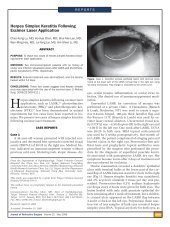
Figure 1. Slitlamp biomicroscopic images <strong>of</strong> the same eye at<br />
day 1 (left) and day 3 (right) postoperatively.<br />
power at a significance level <strong>of</strong> 5%. This value corresponds to<br />
the average defect size 1 day before complete reepithelialization<br />
reported by Engle et al. 10<br />
Statistical analysis was performed using StatView<br />
s<strong>of</strong>tware (version 5.0, SAS Institute, Inc.) on a Macintosh<br />
platform (Apple Inc.). Subjective and objective outcomes<br />
were compared using paired Student t and chi-square<br />
tests, where appropriate. Because the number <strong>of</strong> discordantpairsineachcasewassmall(!10),<br />
paired proportions<br />
were compared using tests based on exact<br />
binomial probabilities.<br />
RESULTS<br />
Forty-four patients (88 eyes) were enrolled the study.<br />
The mean age <strong>of</strong> the 22 men and 22 women was 28.5<br />
years (range 20 to 45 years). Slitlamp biomicroscopy<br />
at the end <strong>of</strong> surgery showed the fit <strong>of</strong> the <strong>bandage</strong><br />
<strong>contact</strong> lens was satisfactory in all eyes.<br />
Figure 1 shows characteristic slitlamp biomicroscopic<br />
images <strong>of</strong> the cornea in the same eye 1 day<br />
and 3 days postoperatively. The corneal epithelial defect<br />
was mostly healed by the third postoperative day.<br />
The mean attempted spherical equivalent was 3.90 D<br />
G 1.82 (SD) in the lotrafilcon A group and 3.88 G<br />
1.81 D in the lotrafilcon B group; the difference was not<br />
statistically significant (P Z .69). The mean epithelial<br />
defect size immediately <strong>after</strong> surgery was 47.0 mm 2<br />
in both groups; the range <strong>of</strong> the defect was 29.7<br />
to 78.8 mm 2 in the lotrafilcon A group and 31.2 to<br />
70.8 mm 2 in the lotrafilcon B group. Figure 2 shows<br />
the correlation for epithelial defect size between the<br />
2 <strong>bandage</strong> <strong>contact</strong> <strong>lenses</strong> immediately <strong>after</strong> surgery.<br />
On average, eyes in the lotrafilcon B group had<br />
slightly larger areas <strong>of</strong> epithelial defect than eyes in<br />
the lotrafilcon A group. The difference was not statistically<br />
significant at 1 day (26.0 mm 2 versus 25.7 mm 2 ;<br />
P Z .67), 3 days (2.2 mm 2 versus 1.9 mm 2 ; P Z .64), or<br />
5 days (0.2 mm 2 versus 0.3 mm 2 ; P Z .56) (Figure 3).<br />
Three days postoperatively, reepithelialization was<br />
complete in 33 eyes (75.0%) in the lotrafilcon A group<br />
and 32 eyes (72.7%) in the lotrafilcon B group. At 5<br />
days, reepithelialization was complete in 42 eyes<br />
(95.5%) and 41 eyes (93.2%), respectively. The<br />
difference between groups was not statistically significant<br />
on either day (P Z 1.00).<br />
Figure 4 compares the mean subjective pain and vision<br />
scores between the 2 groups at all time points.<br />
There were no statistically significant differences<br />
between the 2 <strong>bandage</strong> <strong>contact</strong> <strong>lenses</strong> at any time<br />
(P O.10).<br />
DISCUSSION<br />
Although the therapeutic use <strong>of</strong> s<strong>of</strong>t <strong>contact</strong> <strong>lenses</strong> was<br />
reported almost 40 years ago, 13–15 their use has increased<br />
significantly in recent years as a result <strong>of</strong> the<br />
introduction <strong>of</strong> surgical techniques designed to permanently<br />
correct refractive errors. S<strong>of</strong>t <strong>contact</strong> <strong>lenses</strong><br />
used as a <strong>bandage</strong> <strong>after</strong> refractive surgery aid in corneal<br />
protection and pain relief and accelerate the healing<br />
process 16 by preserving the epithelial flap <strong>after</strong><br />
Figure 2. Correlation <strong>of</strong> the area <strong>of</strong> epithelial defect between the 2<br />
<strong>silicone</strong> <strong>hydrogel</strong> <strong>lenses</strong> immediately <strong>after</strong> surgery. The dashed<br />
line represents the least-square regression fit (r 2 Z 0.94).<br />
J CATARACT REFRACT SURG - VOL 35, DECEMBER 2009
2106 SILICONE HYDROGEL BANDAGE CONTACT LENSES AFTER PRK<br />
Figure 3. Plot <strong>of</strong> the mean epithelial defect size on all postoperative<br />
days. The error bars represent G1 SD. Values at day 5 correspond to<br />
11 eyes and 12 eyes (lotrafilcon A and lotrafilcon B, respectively)<br />
(Day 0 Z day <strong>of</strong> surgery).<br />
LASIK or by promoting reepithelialization <strong>after</strong> PRK<br />
or LASEK. 5,9,10<br />
Contact <strong>lenses</strong> interact mechanically with the cornea<br />
and modify the physiologic processes <strong>of</strong> corneal tissue,<br />
reducing corneal function. The most frequent complications<br />
<strong>of</strong> <strong>contact</strong> lens wear are the direct result <strong>of</strong><br />
Figure 4. Subjective pain (top) and vision (bottom) on all postoperative<br />
days (Day 0 Z day <strong>of</strong> surgery).<br />
impaired oxygen supply to the cornea. Rigid gas--<br />
permeable and s<strong>of</strong>t <strong>contact</strong> lens materials with high<br />
oxygen permeability have driven the success <strong>of</strong> continuous<br />
wear because they have eliminated the hypoxic<br />
effects previously associated with extended-wear<br />
<strong>lenses</strong>. 17,18 Essential to this was the establishment <strong>of</strong><br />
corneal oxygen requirements and the development<br />
<strong>of</strong> materials and manufacturing techniques to meet<br />
those requirements. The introduction in the late<br />
1990s <strong>of</strong> <strong>silicone</strong> <strong>hydrogel</strong> <strong>contact</strong> <strong>lenses</strong> designed for<br />
continuous wear was beneficial to the field <strong>of</strong> refractive<br />
surgery. The high oxygen transmissibility <strong>of</strong> these<br />
<strong>lenses</strong> alleviates hypoxic-induced complications,<br />
enhancing wound healing and epithelial cell reproduction<br />
<strong>after</strong> refractive surgery.<br />
Photorefractive keratectomy, a well-established<br />
flapless refractive procedure, has been performed for<br />
more than 20 years. 1 However, significant drawbacks<br />
<strong>of</strong> PRK remain; that is, it causes greater postoperative<br />
pain and more delayed visual recovery than LASIK.<br />
The therapeutic value <strong>of</strong> the lotrafilcon A <strong>bandage</strong><br />
<strong>contact</strong> lens has been shown, 19 as has its efficacy in<br />
achieving faster corneal reepithelialization and in reducing<br />
discomfort <strong>after</strong> PRK. 10 Therefore, we evaluated<br />
whether another <strong>silicone</strong> <strong>hydrogel</strong> <strong>bandage</strong><br />
<strong>contact</strong> lens with different technical characteristics<br />
produced by the same manufacturer would have the<br />
same efficacy. Our hypothesis was that a <strong>bandage</strong> <strong>contact</strong><br />
lens <strong>of</strong> lotrafilcon B, which has FDA approval for 6<br />
days <strong>of</strong> continuous wear (versus 30 days for the lotrafilcon<br />
A lens), would be as efficacious as the lotrafilcon<br />
A lens in reepithelialization <strong>after</strong> PRK.<br />
Recently, Gil-Cazorla et al. 5 evaluated the efficacy <strong>of</strong><br />
2 <strong>types</strong> <strong>of</strong> <strong>silicone</strong> <strong>hydrogel</strong> <strong>contact</strong> <strong>lenses</strong> used as<br />
a <strong>bandage</strong> <strong>after</strong> LASEK. The 2 <strong>lenses</strong> differed significantly<br />
in oxygen transmissibility, water content, surface<br />
treatment, and initial modulus. The authors<br />
found no significant differences between the 2 <strong>lenses</strong><br />
in vision, corneal epithelial status, conjunctival and<br />
limbal hyperemia, or lens movement. However, the<br />
results are not directly comparable to those in our<br />
study because <strong>of</strong> the significant differences between<br />
LASEK and PRK.<br />
The present study, in which each patient wore both<br />
<strong>types</strong> <strong>of</strong> <strong>lenses</strong>, was a direct comparison between lotrafilcon<br />
A and lotrafilcon B lens <strong>after</strong> PRK. We controlled<br />
factors that could influence the epithelial healing process,<br />
such as the environment and physiologic healing<br />
response, and patients and examiners were masked to<br />
which type <strong>of</strong> lens was in which eye. Although, on average,<br />
eyes with a lotrafilcon A lens had slightly smaller<br />
areas <strong>of</strong> epithelial defect than eyes with a lotrafilcon<br />
B lens, there were no statistically significant differences<br />
between the 2 <strong>lenses</strong> at any postoperative examination.<br />
There was also no difference in the number <strong>of</strong><br />
J CATARACT REFRACT SURG - VOL 35, DECEMBER 2009
SILICONE HYDROGEL BANDAGE CONTACT LENSES AFTER PRK<br />
2107<br />
days required for complete reepithelialization or in the<br />
subjective evaluation <strong>of</strong> pain or vision between the 2<br />
<strong>lenses</strong> at any time. Engle et al. 10 found significantly<br />
faster corneal reepithelialization and reduced patient<br />
discomfort during the first 48 hours <strong>after</strong> PRK in the<br />
eyes with a lotrafilcon A <strong>bandage</strong> <strong>contact</strong> lens than<br />
in eyes with a <strong>hydrogel</strong> <strong>bandage</strong> <strong>contact</strong> lens. Although<br />
there was no significant correlation between<br />
discomfort and defect size on any postoperative day<br />
with either <strong>bandage</strong> <strong>contact</strong> lens, our study found<br />
that larger epithelial defects at 3 days were associated<br />
with higher levels <strong>of</strong> pain with both lens <strong>types</strong>. It is not<br />
clear why Engle et al. did not find a significant correlation<br />
because larger epithelial defects should cause<br />
greater discomfort as a result <strong>of</strong> greater sensory nerve<br />
exposure. Moreover, in our study, patients reported<br />
increased discomfort postoperatively, although complete<br />
reepithelialization may have already occurred.<br />
This might be because the oral medication for pain<br />
control, which would likely result in underestimation<br />
<strong>of</strong> subjective evaluation <strong>of</strong> pain, had been discontinued<br />
by that time. However, in addition to the major<br />
role <strong>of</strong> corneal sensory nerves, other factors might<br />
play a part in pain sensation.<br />
Furthermore, in the subjective vision evaluation, all<br />
patients reported that vision on the third postoperative<br />
day was worse than on the earlier 2 postoperative<br />
days. This was not unexpected because the epithelial<br />
healing process takes place at the center <strong>of</strong> the cornea<br />
at that time.<br />
Although the 2 <strong>types</strong> <strong>of</strong> <strong>bandage</strong> <strong>contact</strong> <strong>lenses</strong> used<br />
in our study differ in the back optic zone radius and<br />
diameter, there were no significant differences between<br />
the 2 <strong>lenses</strong> fitted in post-blink lens movement<br />
and centration, corneal coverage, or tear-film thickness<br />
<strong>after</strong> the lens was removed (all assessed using specular<br />
reflection). The basic fitting philosophy, as previously<br />
suggested, 20 was to provide adherence and stabilization,<br />
allowing for less than normal mobility. On the<br />
other hand, some tear exchange should be conserved<br />
to provide sufficient oxygen to the corneal periphery<br />
for metabolic changes to occur in the regenerating corneal<br />
epithelium. Seo et al. 21 showed that a flat-fitted<br />
<strong>bandage</strong> <strong>contact</strong> lens <strong>after</strong> LASEK results in shorter epithelial<br />
healing time than a steep-fitted lens. However,<br />
it is important to differentiate between the role <strong>of</strong> a <strong>bandage</strong><br />
<strong>contact</strong> lens fitted <strong>after</strong> LASEK and the role <strong>after</strong><br />
PRK. After LASEK, the key action <strong>of</strong> the <strong>bandage</strong> <strong>contact</strong><br />
lens is to accelerate the wound-healing response <strong>of</strong><br />
the corneal epithelium. After PRK, the <strong>bandage</strong> <strong>contact</strong><br />
lens should promote stabilization and complete corneal<br />
reepithelialization. Based on this, we fit <strong>bandage</strong><br />
<strong>contact</strong> <strong>lenses</strong> tighter than normal to prevent excessive<br />
lens movement that could cause further trauma to the<br />
central cornea or patient discomfort.<br />
In conclusion, as have previous studies, our study<br />
found that <strong>contact</strong> <strong>lenses</strong> <strong>of</strong> <strong>silicone</strong> elastomer materials<br />
can be used as an effective and well-tolerated<br />
<strong>bandage</strong> <strong>after</strong> refractive surgery. 5,9,10 Furthermore,<br />
we show for the first time that in addition to the lotrafilcon<br />
A <strong>contact</strong> lens with its established efficacy,<br />
the lotrafilcon B <strong>contact</strong> lens can be used as an effective<br />
<strong>bandage</strong> <strong>after</strong> PRK because <strong>of</strong> the limited time<br />
(4 to 5 days) usually required for complete corneal<br />
reepithelialization.<br />
REFERENCES<br />
1. Ghadhfan F, Al-Rajhi A, Wagoner MD. Laser in situ keratomileusis<br />
versus surface ablation: visual outcomes and complications.<br />
J Cataract Refract Surg 2007; 33:2041–2048<br />
2. El-Maghraby A, Salah T, Waring GO III, Klyce S, Ibrahim O.<br />
Randomized bilateral comparison <strong>of</strong> excimer laser in situ keratomileusis<br />
and photorefractive keratectomy for 2.50 to 8.00 diopters<br />
<strong>of</strong> myopia. Ophthalmology 1999; 106:447–457<br />
3. Shahinian L Jr. Laser-assisted subepithelial keratectomy for low<br />
to high myopia and astigmatism. J Cataract Refract Surg 2002;<br />
28:1334–1342<br />
4. Anderson NJ, Beran RF, Schneider TL. Epi-LASEK for the correction<br />
<strong>of</strong> myopia and myopic astigmatism. J Cataract Refract<br />
Surg 2002; 28:1343–1347<br />
5. Gil-Cazorla R, Teus MA, Hernández-Verdejo JL, de Benito-<br />
Llopis L, García-Gonzalez M. Comparative study <strong>of</strong> two <strong>silicone</strong><br />
<strong>hydrogel</strong> <strong>contact</strong> <strong>lenses</strong> used as <strong>bandage</strong> <strong>contact</strong> <strong>lenses</strong> <strong>after</strong><br />
LASEK. Optom Vis Sci 2008; 85:884–888<br />
6. Cherry PMH. The treatment <strong>of</strong> pain following excimer laser photorefractive<br />
keratectomy: additive effect <strong>of</strong> local anesthetic<br />
drops, topical dicl<strong>of</strong>enac, and <strong>bandage</strong> s<strong>of</strong>t <strong>contact</strong>. Ophthalmic<br />
Surg Lasers 1996; 27:S477–S480<br />
7. McDonald MB, Deitz MR, Frantz JM, Kraff MC, Krueger RR,<br />
Salz JJ, Kraff CR, Maguen E, Matta CS, Nesburn AB,<br />
Piebenga LW. Photorefractive keratectomy for low-to-moderate<br />
myopia and astigmatism with a small-beam, tracker-directed excimer<br />
laser. Ophthalmology 1999; 106:1481–1488; discussion<br />
by RD Stulting, 1488–1489<br />
8. Demers P, Thompson P, Bernier RG, Lemire J, Laflamme P. Effect<br />
<strong>of</strong> occlusive pressure patching on the rate <strong>of</strong> epithelial<br />
wound healing <strong>after</strong> photorefractive keratectomy. J Cataract Refract<br />
Surg 1996; 22:59–62<br />
9. Szaflik JP, Ambroziak AM, Szaflik J. Therapeutic use <strong>of</strong> a lotrafilcon<br />
A <strong>silicone</strong> <strong>hydrogel</strong> s<strong>of</strong>t <strong>contact</strong> lens as a <strong>bandage</strong> <strong>after</strong> LA-<br />
SEK surgery. Eye Contact Lens 2004; 30:59–62<br />
10. Engle AT, Laurent JM, Schallhorn SC, Toman SD,<br />
Newacheck JS, Tanzer DJ, Tidwell JL. Masked comparison <strong>of</strong><br />
<strong>silicone</strong> <strong>hydrogel</strong> lotrafilcon A and etafilcon A extended-wear<br />
<strong>bandage</strong> <strong>contact</strong> <strong>lenses</strong> <strong>after</strong> photorefractive keratectomy. J<br />
Cataract Refract Surg 2005; 31:681–686<br />
11. Foulks GN. Prolonging <strong>contact</strong> lens wear and making <strong>contact</strong><br />
lens wear safer. Am J Ophthalmol 2006; 141:369–373<br />
12. Nilsson SEG. Seven-day extended wear and 30-day continuous<br />
wear <strong>of</strong> high oxygen transmissibility s<strong>of</strong>t <strong>silicone</strong> <strong>hydrogel</strong> <strong>contact</strong><br />
<strong>lenses</strong>: a randomized 1-year study <strong>of</strong> 504 patients. CLAO<br />
J 2001; 27:125–136<br />
13. Sedlácek J. [Possibilities <strong>of</strong> application <strong>of</strong> eye drugs with the aid<br />
<strong>of</strong> gel-<strong>contact</strong> <strong>lenses</strong>]. [Czechoslovakian] Cesk Oftalmol 1965;<br />
21:509–512<br />
14. Gasset AR, Kaufman HE. Therapeutic uses <strong>of</strong> hydrophilic <strong>contact</strong><br />
<strong>lenses</strong>. Am J Ophthalmol 1970; 69:252–259<br />
J CATARACT REFRACT SURG - VOL 35, DECEMBER 2009
2108 SILICONE HYDROGEL BANDAGE CONTACT LENSES AFTER PRK<br />
15. Gould HL. Therapeutic <strong>contact</strong> <strong>lenses</strong>. Int Ophthalmol Clin<br />
1970; 10(1):131–141<br />
16. Bergenske PD, Caroline PJ, Smythe JK. Contact lens as an adjunct<br />
in refractive surgery practice. Contact Lens Spectrum<br />
2002; 17(3):30–37. Available at http://www.clspectrum.com/<br />
article.aspx?articleZ12116. Accessed September 14, 2009<br />
17. Cavanagh HD, Ladage PM, Li SL, Yamamoto K, Molai M,<br />
Ren DH, Petroll WM, Jester JV. Effects <strong>of</strong> daily and overnight<br />
wear <strong>of</strong> a novel hyper oxygen-transmissible s<strong>of</strong>t <strong>contact</strong> lens<br />
on bacterial binding and corneal epithelium; a 13-month clinical<br />
trial. Ophthalmology 2002; 109:1957–1969<br />
18. Ren DH, Yamamoto K, Ladage PM, Molai M, Li L, Petroll WM,<br />
Jester JV, Cavanagh HD. Adaptive effects <strong>of</strong> 30-night wear <strong>of</strong><br />
hyper-O 2 transmissible <strong>contact</strong> <strong>lenses</strong> on bacterial binding and<br />
corneal epithelium; a 1-year clinical trial. Ophthalmology 2002;<br />
109:27–39; invited comment by PC Donshik, 39–40<br />
19. Montero J, Sparholt J, Mély R, Long B. Retrospective case series<br />
<strong>of</strong> therapeutic applications <strong>of</strong> lotrafilcon A <strong>silicone</strong> <strong>hydrogel</strong><br />
s<strong>of</strong>t <strong>contact</strong> <strong>lenses</strong>. Eye Contact Lens 2003; 29:72–75<br />
20. de Oliveira PR, Bailey MD. Therapeutic <strong>contact</strong> <strong>lenses</strong>. In:<br />
Mannis MJ, Zadnik K, Coral-Ghanem C, Kara-José N, eds, Contact<br />
Lenses in Ophthalmic Practice. New York, NY, Springer,<br />
2004; 197–203<br />
21. Seo JH, Wee WR, Lee JH, Kim MK. Effect <strong>of</strong> base curve radius <strong>of</strong><br />
therapeutic <strong>lenses</strong> on epithelial healing <strong>after</strong> laser-assisted subepithelial<br />
keratectomy. Korean J Ophthalmol 2007; 21:85–89. Available<br />
at http://synapse.koreamed.org/Synapse/Data/PDFData/<br />
0065KJO/kjo-21-85.pdf. Accessed September 14, 2009<br />
First author:<br />
Michael A Grentzelos, MD<br />
Institute <strong>of</strong> Vision and Optics,<br />
Faculty <strong>of</strong> Medicine, University <strong>of</strong><br />
Crete, Heraklion, Crete, Greece<br />
J CATARACT REFRACT SURG - VOL 35, DECEMBER 2009