Herpes Simplex Keratitis Following Excimer Laser Application
Herpes Simplex Keratitis Following Excimer Laser Application
Herpes Simplex Keratitis Following Excimer Laser Application

Create successful ePaper yourself
Turn your PDF publications into a flip-book with our unique Google optimized e-Paper software.
REPORTS<br />
<strong>Herpes</strong> <strong>Simplex</strong> <strong>Keratitis</strong> <strong>Following</strong><br />
<strong>Excimer</strong> <strong>Laser</strong> <strong>Application</strong><br />
Chao-Kung Lu, MD; Ko-Hua Chen, MD; Shui-Mei Lee, MD;<br />
Wen-Ming Hsu, MD; Jui-Yang Lai, MS; Yen-Shien Li, MS<br />
ABSTRACT<br />
PURPOSE: To report two cases of herpes simplex keratitis following<br />
excimer laser application.<br />
METHODS: Two immunocompetent patients with no history of<br />
ocular viral infection developed ulcers after LASIK and phototherapeutic<br />
keratectomy (PTK), respectively.<br />
RESULTS: Antiviral treatment was administered, and the lesions<br />
healed within 14 days.<br />
CONCLUSIONS: These two cases suggest that herpes simplex<br />
virus was associated with the use of the excimer laser. [J Refract<br />
Surg. 2006;22:509-511.]<br />
<strong>Herpes</strong> simplex keratitis following excimer laser<br />
application, such as LASIK, 1-4 photorefractive<br />
keratectomy (PRK), 5 and phototherapeutic keratectomy<br />
(PTK), 6,7 has been demonstrated in animal<br />
experiments but few cases have been reported in humans.<br />
We present two cases of herpes simplex keratitis<br />
following excimer laser treatment.<br />
CASE REPORTS<br />
CASE 1<br />
A 44-year-old woman presented with injected conjunctiva<br />
and decreased best spectacle-corrected visual<br />
acuity (BSCVA) of 20/25 in the right eye. Medical history<br />
indicated an immunocompetent woman without<br />
previous cold sore, blistering rash, atopic disease, dry<br />
From the Department of Ophthalmology, Taipei Veterans General<br />
Hospital (Lu, Chen, Lee, Hsu, Li), Taipei; National Yang-Ming<br />
University (Chen, Lee, Hsu); Division of Medical Engineering, National<br />
Health Research Institute (Chen, Lai, Li), Taipei; and the Department<br />
of Chemical Engineering, National Tsing Hua University (Lai),<br />
Hsinchu, Taiwan, ROC.<br />
This study was supported by a grant from Taipei Veterans General<br />
Hospital (VGH-92-376-7).<br />
The authors have no proprietary interest in the materials presented<br />
herein.<br />
Correspondence: Ko-Hua Chen, MD, Dept of Ophthalmology,<br />
Taipei Veterans General Hospital, #201, Section 2, Shih-Pai Rd,<br />
Taipei, 11217, Taiwan, ROC. Tel: 886 2 28757325, 886 2 28714633;<br />
Fax: 886 2 28761351; E-mail: khchen@vghtpe.gov.tw<br />
Received: April 10, 2005<br />
Accepted: November 15, 2005<br />
Journal of Refractive Surgery Volume 22 May 2006<br />
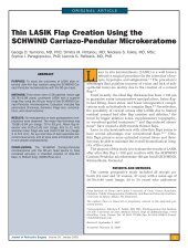
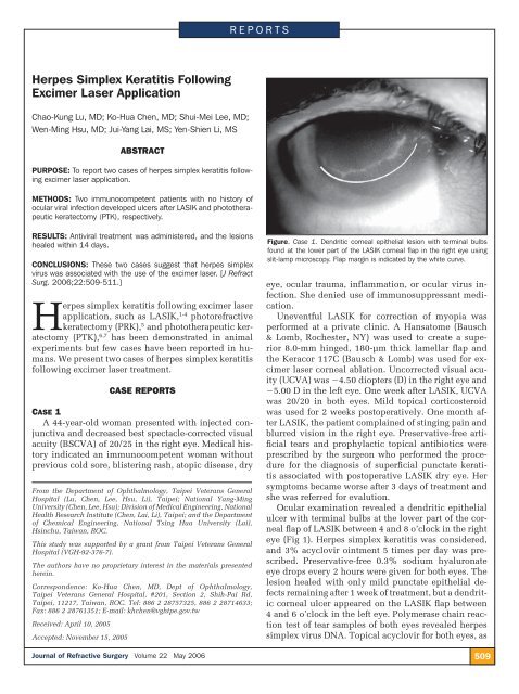
Figure. Case 1. Dendritic corneal epithelial lesion with terminal bulbs<br />
found at the lower part of the LASIK corneal flap in the right eye using<br />
slit-lamp microscopy. Flap margin is indicated by the white curve.<br />
eye, ocular trauma, inflammation, or ocular virus infection.<br />
She denied use of immunosuppressant medication.<br />
Uneventful LASIK for correction of myopia was<br />
performed at a private clinic. A Hansatome (Bausch<br />
& Lomb, Rochester, NY) was used to create a superior<br />
8.0-mm hinged, 180-µm thick lamellar flap and<br />
the Keracor 117C (Bausch & Lomb) was used for excimer<br />
laser corneal ablation. Uncorrected visual acuity<br />
(UCVA) was 4.50 diopters (D) in the right eye and<br />
5.00 D in the left eye. One week after LASIK, UCVA<br />
was 20/20 in both eyes. Mild topical corticosteroid<br />
was used for 2 weeks postoperatively. One month after<br />
LASIK, the patient complained of stinging pain and<br />
blurred vision in the right eye. Preservative-free artificial<br />
tears and prophylactic topical antibiotics were<br />
prescribed by the surgeon who performed the procedure<br />
for the diagnosis of superficial punctate keratitis<br />
associated with postoperative LASIK dry eye. Her<br />
symptoms became worse after 3 days of treatment and<br />
she was referred for evalution.<br />
Ocular examination revealed a dendritic epithelial<br />
ulcer with terminal bulbs at the lower part of the corneal<br />
flap of LASIK between 4 and 8 o’clock in the right<br />
eye (Fig 1). <strong>Herpes</strong> simplex keratitis was considered,<br />
and 3% acyclovir ointment 5 times per day was prescribed.<br />
Preservative-free 0.3% sodium hyaluronate<br />
eye drops every 2 hours were given for both eyes. The<br />
lesion healed with only mild punctate epithelial defects<br />
remaining after 1 week of treatment, but a dendritic<br />
corneal ulcer appeared on the LASIK flap between<br />
4 and 6 o’clock in the left eye. Polymerase chain reaction<br />
test of tear samples of both eyes revealed herpes<br />
simplex virus DNA. Topical acyclovir for both eyes, as<br />
509
Reports<br />
well as oral acyclovir 200 mg 5 times a day was prescribed<br />
for 10 days following appearance of the second<br />
ulcer. The dendritic lesion in the left eye healed after<br />
1 week of treatment. Best spectacle-corrected visual<br />
acuity was 20/20 in both eyes 1 month later. At 1-year<br />
follow-up, there was no evidence of herpes simplex<br />
keratitis recurrence.<br />
CASE 2<br />
A 76-year-old immunocompetent woman was referred<br />
due to pain in the left eye of 4 months’ duration.<br />
She had a history of glaucoma in both eyes and underwent<br />
trabeculectomy and cataract surgery in the left eye<br />
10 years prior to presentation. Macular edema developed<br />
5 years postoperatively and she underwent unsuccessful<br />
grid laser photocoagulation. There was no history<br />
of cold sore, blistering rash, atopic disease, dry eye,<br />
ocular trauma, inflammation, or ocular viral infection.<br />
She denied use of immunosuppressant medications.<br />
Ocular examination revealed severe corneal edema<br />
with bullous formation and cornea epithelial defect<br />
in the left eye. Best spectacle-corrected visual acuity<br />
in the left eye was count-fingers at 30 cm. The diagnosis<br />
was pseudophakic bullous keratopathy with recurrent<br />
corneal erosion. Phototherapeutic keratectomy<br />
was performed, in addition to therapeutic contact lens<br />
treatment, to relieve recurrent corneal epithelium erosion.<br />
Chloramphenical 0.25% eye drops 4 times a day<br />
and preservative-free lubricant every 2 hours were prescribed<br />
to the left eye postoperatively. Three weeks<br />
after PTK, the patient presented with irritation and<br />
conjunctival injection in the left eye. Slit-lamp microscopy<br />
revealed a dendritic epithelial ulcer with terminal<br />
bulbs at the paracentral cornea between 5 and 9<br />
o’clock in the left eye. Polymerase chain reaction test of<br />
the tear sample of the left eye revealed herpes simplex<br />
virus DNA. <strong>Herpes</strong> simplex keratitis following PTK<br />
was considered and 3% acyclovir ointment 5 times<br />
a day was prescribed immediately. After 1 week of<br />
treatment, the corneal ulcer healed and only mild superficial<br />
punctate keratitis was seen. During 6-month<br />
follow-up, herpes simplex keratitis did not recur.<br />
DISCUSSION<br />
In the cases presented, typical dendritic lesions of<br />
herpes simplex keratitis occurred within 1 month after<br />
excimer laser surgery. Polymerase chain reaction<br />
test demonstrated herpes simplex virus DNA in tear<br />
samples of the lesion eyes. Symptomotology worsened<br />
following preservative-free lubricant treatment and resolved<br />
with antiviral treatment. Thus, herpes simplex<br />
keratitis associated with LASIK and PTK procedures<br />
was suggested.<br />
510<br />
Many people without clinical herpes simplex infection<br />
have latent virus. Worldwide, 60% to 90% of the<br />
adult population is positive for HSV-1 antibody and<br />
only 1% to 6% of primary infections are clinically recognized.<br />
8 Asymptomatic people can shed herpes simplex<br />
virus in tears. 8 This report suggests that herpes<br />
simplex virus infection in individuals without clinical<br />
signs and symptoms was activated by excimer laser<br />
surgery.<br />
The association of excimer laser corneal ablation and<br />
activation of herpes simplex virus has been demonstrated<br />
in rabbits and mice, 4,6 but only a few human cases<br />
of herpes simplex keratitis associated with LASIK<br />
and PRK have been reported. 1-3,5 These patients had<br />
a history of herpes simplex keratitis, systemic herpes<br />
simplex virus infection, or multiple corneal surgeries<br />
before PRK was performed. 1-3 Some patients were<br />
treated with bilateral LASIK, but postoperative herpes<br />
simplex keratitits occurred only in one eye. We present<br />
the first case with bilateral herpes simplex keratitis<br />
after bilateral LASIK without previous corneal surgery<br />
or history of herpes simplex.<br />
Systemic prophylactic antiviral agents in patients<br />
with a history of herpes simplex keratitis before PRK<br />
or LASIK has been suggested. 1,3,5,9 Factors such as<br />
preoperative emotional stress, postoperative tear dysfunction,<br />
ultraviolet radiation exposure, and surgical<br />
trauma (eg, lamellar keratoplasty) have been related to<br />
reactivation of herpes simplex virus. Further, herpes<br />
simplex keratitis may be secondary to postoperative<br />
use of topical corticosteroid (case 1). Although few<br />
cases have been reported, the relationship between<br />
herpes simplex keratitis and LASIK, PRK, or PTK cannot<br />
be excluded and patients should be informed of<br />
this possibility before surgery.<br />
We report a case of herpes simplex keratitis associated<br />
with bilateral LASIK in a healthy woman and<br />
a patient with herpes simplex keratitis after PTK for<br />
treatment of pseudophakic bullous keratopathy. We<br />
suggest the importance of obtaining a detailed patient<br />
history before surgery and administering prophylactic<br />
antiviral treatment in patients with previous herpes<br />
simplex virus infection.<br />
REFERENCES<br />
1. Levy J, Lapid-Gortzak R, Klemperer I, Lifshitz T. <strong>Herpes</strong> simplex<br />
virus keratitis after laser in situ keratomileusis. J Refract<br />
Surg. 2005;21:400-402.<br />
2. Perry HD, Doshi SJ, Donnenfeld ED, Levinson DH, Cameron CD.<br />
<strong>Herpes</strong> simplex reactivation following laser in situ keratomileusis<br />
and subsequent corneal perforation. CLAO J. 2002;28:69-<br />
71.<br />
3. Davidorf JM. <strong>Herpes</strong> simplex keratitis after LASIK. J Refract<br />
Surg. 1998;14:667.<br />
journalofrefractivesurgery.com
Reports<br />
4. Dhaliwal DK, Romanowski EG, Yates KA, Hu D, Goldstein M,<br />
Gordon YJ. Experimental laser-assisted in situ keratomileusis<br />
induces the reactivation of latent herpes simplex virus. Am J<br />
Ophthalmol. 2001;131:506-507.<br />
5. Wulff K, Fechner PU. <strong>Herpes</strong> simplex keratitis after photorefractive<br />
keratectomy. J Refract Surg. 1997;13:613.<br />
6. Pepose JS, Laycock KA, Miller JK, Chansue E, Lenze EJ, Gans LA,<br />
Smith ME. Reactivation of latent herpes simplex virus by excimer<br />
laser photokeratectomy. Am J Ophthalmol. 1992;114:45-50.<br />
7. Vrabec MP, Anderson JA, Rock ME, Binder PS, Steinert RF,<br />
Durrie DS, Chase DS. Electron microscopic findings in a cornea<br />
with recurrence of herpes simplex keratitis after excimer laser<br />
phototherapeutic keratectomy. CLAO J. 1994;20:41-44.<br />
8. Kaufman HE, Azcuy AM, Varnell ED, Sloop GD, Thompson<br />
HW, Hill JM. HSV-1 DNA in tears and saliva of normal adults.<br />
Invest Ophthalmol Vis Sci. 2005;46:241-247.<br />
9. Dhaliwal DK, Romanowski EG, Yates KA, Hu D, Mah FS,<br />
Fish DN, Gordon YJ. Valacyclovir inhibition of recovery of<br />
ocular herpes simplex virus type 1 after experimental reactivation<br />
by laser in situ keratomileusis. J Cataract Refract Surg.<br />
2001;27:1288-1293.<br />
Dry Eye After Photorefractive<br />
Keratectomy With Adjuvant Mitomycin C<br />
George D. Kymionis, MD, PhD; Nikolaos S. Tsiklis, MD;<br />
Harilaos Ginis, PhD; Vasilios F. Diakonis, MD;<br />
Ioannis Pallikaris, MD, PhD<br />
ABSTRACT<br />
PURPOSE: To report a patient with dry eye after bilateral photorefractive<br />
keratectomy (PRK) with mitomycin C treatment in one eye.<br />
METHODS: A 29-year-old woman underwent PRK for moderate<br />
myopia. The left eye was randomly assigned and intraoperative<br />
topical mitomycin C was administered. The right (control) eye was<br />
treated with intraoperative corticosteroid only.<br />
RESULTS: The patient developed dry eye symptoms and superfi -<br />
cial punctuate keratopathy in the eye treated with mitomycin C.<br />
Fifteen months after surgery no improvement was noted.<br />
CONCLUSIONS: Photorefractive keratectomy with mitomycin C<br />
treatment could induce or exacerbate dry eye. [J Refract Surg.<br />
2006;22:511-513.]<br />
From the Department of Ophthalmology (Kymionis, Pallikaris) and<br />
the Institute of Vision and Optics (Kymionis, Tsiklis, Ginis, Diakonis,<br />
Pallikaris), University of Crete, Heraklion, Crete, Greece.<br />
The authors have no proprietary interest in the materials presented<br />
herein.<br />
Correspondence: George D. Kymionis, MD, PhD, University of<br />
Crete, Institute of Vision and Optics, Medical School, Dept of<br />
Ophthalmology, 71110 Heraklion, Crete, Greece. Tel: 30 2810 371800;<br />
Fax: 30 2810 394653; E-mail: kymionis@med.uoc.gr<br />
Received: June 7, 2005<br />
Accepted: October 3, 2005<br />
Journal of Refractive Surgery Volume 22 May 2006<br />
During the past several years, changes have been<br />
made in refractive surgery trends. Although<br />
LASIK is a widely used treatment technique,<br />
there is increasing interest in excimer surface ablation<br />
procedures such as photorefractive keratectomy (PRK),<br />
laser subepithelial keratomileusis (LASEK), and epi-<br />
LASIK.<br />
Recently, encouraging results have been reported<br />
in reducing haze after PRK by administering a single<br />
intraoperative application of mitomycin C. 1 These<br />
studies suggest that the prophylactic use of a diluted<br />
mitomycin C 0.02% solution produced lower haze<br />
rates, better uncorrected visual acuity (UCVA) and best<br />
spectacle-corrected visual acuity (BSCVA) results, and<br />
more accurate refractive outcomes than those achieved<br />
in the control group. 1,2 Despite these results, there is<br />
considerable evidence of mitomycin C toxicity in the<br />
literature and concern is increasing regarding the side<br />
effects and long-term complications. 3,4 Although, mitomycin<br />
C use in refractive surgery differs from pterygium<br />
and glaucoma surgery, there is increased awareness<br />
of its possible complications. We report a patient<br />
who developed dry eye symptoms in the left eye after<br />
PRK with mitomycin C application, whereas the fellow<br />
eye treated with PRK and corticosteroids did not<br />
develop similar symptoms.<br />
CASE REPORT<br />
A 29-year-old woman with no history of dry eye<br />
underwent bilateral PRK for moderate myopia. Preoperative<br />
examination was normal. She was informed of<br />
possible intra- and postoperative complications, and<br />
was included in a prospective, double-masked, randomized<br />
clinical trial for the safety and efficacy of topical<br />
mitomycin C use after PRK in myopic eyes. One<br />
eye was randomly assigned to receive intraoperative<br />
topical mitomycin C application. Randomization was<br />
performed by opening a sealed envelope that contained<br />
the treatment information. The patient gave written<br />
informed consent in accordance with institutional<br />
guidelines and the Declaration of Helsinki. Preoperative<br />
corneal sensitivity (Cochet-Bonnet esthesiometer)<br />
and Schirmer tests were not performed, as they were<br />
not included in the initial study protocol design.<br />
Preoperative corneal thickness was 500 µm in both<br />
eyes. Epithelium was removed using a rotating soft<br />
brush at the 8-mm zone. Ablation was performed using<br />
the Allegretto Wavelight (WaveLight AG Technologie,<br />
Erlangen, Germany) 200 Hz excimer laser system. Attempted<br />
correction was 5.50 0.50 170 in the left<br />
eye and 5.00 1.00 180 in the right eye (mitomycin<br />
C eye and control eye, respectively). Immediately<br />
after laser treatment, the left eye received a single topi-<br />
511
Reports<br />
cal application of mitomycin C 0.02% diluted in balanced<br />
salt solution. This was administered by placing<br />
a mitomycin C–soaked sponge (7 mm in diameter) over<br />
the ablated stroma. The sponge was kept in place for 2<br />
minutes, then the corneal surface and entire conjunctival<br />
fornix were vigorously irrigated with 20 cc of<br />
balanced salt solution to remove residual mitomycin<br />
C. The right eye received a sponge soaked in balanced<br />
salt solution applied in the same manner. The procedure<br />
was completed without complication. At the end<br />
of surgery, a combination of steroids and antibiotic<br />
drops (Tobradex; SA Alcon, Couvreur NV, Belgium)<br />
was administered in both eyes 4 times daily, and a<br />
bandage soft contact lens was applied until full corneal<br />
re-epithelialization occurred. After re-epithelialization,<br />
the left eye received a placebo solution (artificial<br />
tears) whereas the control eye was treated with fluorometholone<br />
sodium 2% (FML; Allergan, Westport,<br />
County Mayo, Ireland) 4 times daily for 2 weeks. The<br />
dosage of corticosteroid and placebo was tapered one<br />
drop every second week. No additional steroids were<br />
used during follow-up.<br />
The postoperative period was uneventful. At 1 month,<br />
the patient reported dryness in both eyes treated with<br />
frequent preservative-free artificial tears. Uncorrected<br />
visual acuity (UCVA) was 20/32 in both eyes. Examination<br />
revealed mild superficial punctate keratopathy<br />
of both corneas. Three months after surgery, the patient<br />
reported that the sensation of dryness improved<br />
in the control eye but remained stable in the mitomycin<br />
C eye. Superficial punctuate keratopathy did not<br />
improve in the mitomycin C eye whereas no evidence<br />
was noted in the control eye, except for mild haze.<br />
Uncorrected visual acuity was 20/20 in both eyes. Fifteen<br />
months postoperatively, UCVA was 20/20 in both<br />
eyes with an improvement in haze (trace) formation in<br />
the control eye. Slit-lamp microscopy of the left eye<br />
revealed no improvement of superficial punctate keratopathy<br />
and the patient reported ocular irritation and<br />
foreign body sensation. Tear film status tests (Schirmer<br />
test without anesthetic eyedrops [Schirmer I] and with<br />
anesthetic eyedrops [Schirmer II]) were performed, revealing<br />
an excessive decrease in tear secretion in the<br />
left eye (Schirmer I and II, 3 mm and 2 mm, respectively)<br />
in comparison with the control eye (Schirmer<br />
I and II, 8 mm and 6 mm, respectively). Preservativefree<br />
artificial tears (Vismed; TRB Chemedica AG, Haar/<br />
München, Germany) and ointment (Dacrio Gel, SA<br />
Alcon) were administered.<br />
512<br />
DISCUSSION<br />
Photorefractive keratectomy can induce or exacerbate<br />
dry eye after surgery. The cause involves decreased<br />
corneal sensation, resulting in reduced tear<br />
production. In this report, because preoperative measurement<br />
of tear flow was not available, the presentation<br />
of increased dry eye symptoms and postoperative<br />
decreased tear flow confirmed by Schirmer tests, which<br />
were worse in the left eye compared to the control eye,<br />
indicates that adjuvant mitomycin C application in<br />
PRK may affect tear production. A possible explanation<br />
of this complication may be the increased extent<br />
and duration of corneal hypesthesia associated with<br />
mitomycin C application, in addition to the PRK ablation<br />
damage to the corneal nerve plexus, which did<br />
not resolve after 15 months of follow-up (nerve plexus<br />
usually recovers completely after 12 months 5 ).<br />
Furthermore, a direct decrease in aqueous tear production,<br />
increased tear osmolarity and evaporation, and<br />
toxic effect in lacrimal glands and goblet cell density 6<br />
could be additional factors that contribute to mitomycin<br />
C–induced dry eye. We suggest that the adverse effect of<br />
mitomycin C to the surrounding tissue could be minimized<br />
by eliminating application to the ablation site.<br />
Surgical experience with LASEK indicates it is feasible<br />
to expose only part of the cornea to a liquid substance (ie,<br />
alcohol). Similarly, irrigation could be performed using a<br />
cone- or cylinder-like container that seals against the corneal<br />
surface and uses an aspiration port to safely remove<br />
the excessive mitomycin C from the corneal surface.<br />
It is possible that all of these explanations are partly<br />
correct, and each may contribute to the symptoms experienced<br />
postoperatively. Although dry eye could be<br />
more common and more severe in patients with preexisting<br />
tear flow abnormality, preoperative evaluation<br />
of tear film status is necessary to identify patients with<br />
tear film deficiency.<br />
Photorefractive keratectomy with mitomycin C application<br />
may induce tear deficiency or exacerbate preexisting<br />
dry eye disease. This side effect can be present<br />
up to 15 months after surgery. Candidates for surgery<br />
should be screened preoperatively so that patients<br />
with borderline tear secretion can be advised of possible<br />
mitomycin C–related side effects.<br />
REFERENCES<br />
1. Gambato C, Ghirlando A, Moretto E, Busato F, Midena E. Mitomycin<br />
C modulation of corneal wound healing after photorefractive<br />
keratectomy in highly myopic eyes. Ophthalmology.<br />
2005;112:208-218.<br />
2. Carones F, Vigo L, Scandola E, Vacchini L. Evaluation of the<br />
prophylactic use of mitomycin-C to inhibit haze formation<br />
after photorefractive keratectomy. J Cataract Refract Surg.<br />
2002;28:2088-2095.<br />
3. Rubinfeld RS, Pfister RR, Stein RM, Foster CS, Martin NF,<br />
Stoleru S, Talley AR, Speaker MG. Serious complications of<br />
topical mitomycin C after pterygium surgery. Ophthalmology.<br />
1992;99:1647-1654.<br />
journalofrefractivesurgery.com
Reports<br />
4. Dougherty PJ, Hardten DR, Lindstrom RL. Corneoscleral melt<br />
after pterygium surgery using a single intraoperative application<br />
of mitomycin-C. Cornea. 1996;15:537-540.<br />
5. Perez-Santonja JJ, Sakla HF, Cardona C, Chipont E, Alio JL.<br />
Corneal sensitivity after photorefractive keratectomy and laser<br />
in situ keratomileusis for low myopia. Am J Ophthalmol.<br />
1999;127:497-504.<br />
6. Francis BA, Du LT, Najafi K, Murthy R, Kurumety U, Rao N,<br />
Minckler DS. Histopathologic features of conjunctival filtering<br />
blebs. Arch Ophthalmol. 2005;123:166-170.<br />
Decentration and Cataract Formation<br />
10 Years <strong>Following</strong> Posterior Chamber<br />
Silicone Phakic Intraocular Lens<br />
Implantation<br />
Samar A. Al-Swailem, MD; Ali A. Al-Rajhi, FRCS, FRCOphth<br />
ABSTRACT<br />
PURPOSE: To report a 10-year follow-up for bilateral implantation<br />
of a Chiron Adatomed silicone posterior chamber phakic intraocular<br />
lens (PIOL).<br />
METHODS: A 32-year-old man presented with bilateral blurred<br />
vision and monocular diplopia in the left eye of 2 years’ duration.<br />
RESULTS: Slit-lamp microscopy showed bilateral anterior subcapsular<br />
cataract and temporal PIOL decentration, and no visible<br />
space between the PIOL and crystalline lens in the right eye. After<br />
explantation of the posterior chamber PIOL, lens aspiration, and<br />
IOL implantation, uncorrected visual acuity improved to 20/15 in<br />
the right eye. Scanning electron microscopy examination showed<br />
denser deposits on the central portion of the back surface when<br />
compared with the edges.<br />
CONCLUSIONS: Long-term follow up of certain designs of posterior<br />
chamber PIOLs may reveal late occurrence of complications. Cataract<br />
formation may be related to direct contact between the implanted<br />
and crystalline lenses. [J Refract Surg. 2006;22:513-515.]<br />
In 1986, Fyodorov introduced the idea of implanting<br />
a negative silicone intraocular lens (IOL) in the<br />
posterior chamber, just anterior to the surface of the<br />
From the Anterior Segment Division, Department of Ophthalmology<br />
(Al-Swailem, Rajhi) and the Research Department (Rajhi), King<br />
Khaled Eye Specialist Hospital, Riyadh, Saudi Arabia.<br />
The authors have no proprietary interest in the materials presented<br />
herein.<br />
Correspondence: Samar A. Al-Swailem, MD, Anterior Segment<br />
Division, King Khalid Eye Specialist Hospital, PO Box 7191, Riyadh<br />
11462, Saudi Arabia. Tel: 966 1 482 1234; Fax: 966 1 482 9311;<br />
E-mail: angelofsa@yahoo.com<br />
Received: June 28, 2005<br />
Accepted: November 16, 2005<br />
Journal of Refractive Surgery Volume 22 May 2006<br />
crystalline lens for the correction of myopia in phakic patients.<br />
1,2 Chiron Adatomed GmbH (Ratingen, Germany)<br />
produced another silicone posterior chamber phakic<br />
intraocular lens (PIOL) in 1992 that was first implanted<br />
in Germany. 2,3 Since then, various designs of silicone<br />
myopic PIOLs have been reported— Fyodorov,<br />
Russian plate (Mikof Company, Russia), 1 and top-hat<br />
style. 3,4 However, production of these lenses was soon<br />
abandoned because of high complication rate. 2<br />
One hundred twenty-five implanted silicone-generation<br />
Chiron Adatomed posterior chamber PIOLs have<br />
been reported in three studies. 3,5,6 We report the longest<br />
follow-up for this design.<br />
CASE REPORT<br />
A 32-year-old man presented with complaints of bilateral<br />
gradual blurring of vision and monocular diplopia<br />
in the left eye of 2 years’ duration. Ten years earlier,<br />
he had bilateral implantation of posterior chamber<br />
PIOLs for the correction of high myopia. He reported<br />
no history of ocular disease.<br />
Uncorrected visual acuity (UCVA) was 20/30 in the<br />
right eye and 20/40 in the left eye, improving with pinhole<br />
to 20/25. Refraction was 0.25 1.50 91 in the<br />
right eye and 2.27 1.37 62 in the left eye. Intraocular<br />
pressure was normal in both eyes. Slit-lamp microscopy<br />
showed bilateral superior limbal conjunctival<br />
scarring, patent bilateral superior surgical peripheral<br />
iridectomy, and bilateral anterior subcapsular opacification.<br />
When compared with the left eye, the right<br />
eye had denser cataract, no visible space between the<br />
PIOL and crystalline lens, and no visible nasal optical<br />
edge prior to dilation. After dilation, moderate temporal<br />
decentration was seen in the left eye. Funduscopy<br />
revealed bilateral myopic changes and a cup-to-disc<br />
ratio of 0.5. Axial length was 26.58 mm in the right eye<br />
and 26.26 mm in the left eye.<br />
The Chiron Adatomed PIOL was explanted from<br />
the right eye using a 5.5-mm clear corneal temporal<br />
approach in which it was dislocated into the anterior<br />
chamber, followed by lens aspiration using a<br />
Sovereign system with whiteStar power modulation<br />
(Advanced Medical Optics, Santa Ana, Calif) . It was<br />
a single, boat-shaped piece with an overall diameter<br />
of 12.0 mm. The optic had a biconcave configuration<br />
with a diameter of 5.25 mm. Scanning electron<br />
microscopy examination showed diffuse deposits<br />
of unknown origin over the entire front surface. It<br />
was denser on the central portion of the back surface<br />
when compared with the edges (Figs 1 and 2).<br />
A 10.0-diopter (D), foldable, three-piece AcrySof<br />
posterior chamber IOL (MA60BM; Alcon Laboratories<br />
Inc, Ft Worth, Tex), optic diameter 6.0 mm,<br />
513
Reports<br />
Figure 1. Explanted IOL (scanning electron microscopy, original magnification<br />
11) with diffuse opacities on the front surface (white arrows). These<br />
opacities were denser at the area of contact between the concave back<br />
surface of the IOL and crystalline lens centrally (black arrows). Two surgically<br />
induced cracks during explantation are located at the inferior margin.<br />
Figure 2. Higher magnification of the abnormal opacity deposits (scanning<br />
electron microscopy, original magnification 5500). These deposits are of<br />
unknown etiology and may represent dead cells or fiber accumulation.<br />
was implanted. The Holladay formula 7 was used to<br />
calculate IOL power, aiming for emmetropia. At 2<br />
months postoperatively, UCVA improved to 20/15<br />
and refraction was 0.25 3.0 96.<br />
DISCUSSION<br />
The main advantages of posterior chamber PIOL<br />
use for correction of high myopia are that surgery is<br />
relatively simple, reversible, carries no risk of corneal<br />
endothelium contact, and does not depend on the vagaries<br />
of corneal wound healing or sacrifice of the crystalline<br />
lens and its accommodative ability. 8,9<br />
The case reported demonstrates three unique findings.<br />
First, it presents the longest follow-up (10 years)<br />
for the Chiron Adatomed IOL design compared to other<br />
studies in which follow-up ranged from 6 to 32 months<br />
(mean 17.2 months) 3 and 3 to 24 months. 5,6 The longest<br />
follow-up reported for older-design silicone-generation<br />
PIOLs was 7 years. 4 Second, it demonstrates late occurrence<br />
of cataract formation 10 years postoperatively<br />
when compared with the Fechner et al 3 and Brauweiler<br />
et al 6 reports. In the former, the incidence of cataract<br />
was 18% and onset ranged from 12 to 24 months postoperatively,<br />
whereas in the latter, the incidence was<br />
81.1% with onset ranging from 3 to 24 months. Third,<br />
it highlights the first scanning electron microscopy examination<br />
of an explanted Chiron Adatomed IOL.<br />
Several potential pathophysiological mechanisms<br />
for cataractogenesis have been proposed. The fact that<br />
an anterior subcapsular cataract is the most dominant<br />
pattern of cataract in all cases, and the observation<br />
of its formation as early as 3 months postoperatively<br />
in two patients of the Brauweiler et al series, 6 would<br />
514<br />
strongly suggest direct trauma to the crystalline<br />
lens as a mechanism of cataractogenesis. Fechner et<br />
al 3 observed that the space between the IOL posterior<br />
surface and crystalline lens, defined as the lens<br />
vault, may be advantageous, as none of the opacified<br />
crystalline lenses had a visible space. In contrast, the<br />
crystalline lens in eyes with forward-buckled IOLs<br />
have remained clear. In 1999, Fechner stated that<br />
the Chiron Adatomed IOL used in his study was not<br />
constructed to provide enough space. 10 In fact, 58%<br />
of 40 eyes reported had implantations of small IOLs<br />
0.5 mm longer than the horizontal corneal diameter<br />
white-to-white distance. 3 Others claim the predictable<br />
measurement of the posterior chamber width is the<br />
ciliary sulcus diameter rather than the white-to-white<br />
distance because the latter has an irregular contour<br />
causing internal diameter variations and may be obtained<br />
using variable techniques (surgical caliber versus<br />
topography-based calibers). 10 Furthermore, small<br />
optical diameter was claimed by Wiechens to induce<br />
cataract 7 years after implanting a top-hat style IOL.<br />
Thus, constant or intermittent contact from increased<br />
crystalline lens curvature, either during accommodation<br />
or gradual enlargement of anterior-posterior lens<br />
diameter, could explain cataract formation. 3 This<br />
rationale was supported by the finding of a circular<br />
contact zone of an acellular substance on the back<br />
surface of IOLs by scanning electron microscropy. 4<br />
Fechner recommended altering the lens vault by implanting<br />
oversized IOLs (adding 1.0 mm to the whiteto-white<br />
diameter), rationalizing that a larger IOL will<br />
be forced to buckle forward, thereby increasing the<br />
lens vault, or by shortening the posterior IOL surface<br />
journalofrefractivesurgery.com
Reports<br />
radius to lift the center away from the anterior surface<br />
of the crystalline lens. 3,10<br />
In contrast, the report by Marinho et al 5 did not support<br />
the hypothesis of a short IOL diameter causing cataract<br />
formation as none of their patients developed cataract<br />
up to 24 months of follow-up, although 13% had IOL<br />
implantations 1.0 mm shorter than the horizontal whiteto-white<br />
diameter. However, this study reported shorter<br />
follow-up (86.8% and 76.5% eyes, mean 12 and 6<br />
months, respectively) 5 when compared with the report<br />
by Fechner et al (15.9% eyes, mean 6 months). 3<br />
Another mechanism of cataractogenesis is that a<br />
posterior chamber PIOL can induce metabolic changes<br />
in the crystalline lens by altering its oxygen transport<br />
and nutrition transmission, either through a decrease<br />
in the lens vault from constant or intermittent trauma<br />
or through subclinical inflammation. 2,9,11 However, the<br />
composition of new generation IOLs has undergone<br />
change, incorporating new materials, such as collagen,<br />
to increase their hydrophilia, oxygen permeability,<br />
and biocompatibility. 8 Four designs of IOLs (ICM V1<br />
to V4 and ICH V1 to V4, minus and plus power, respectively)<br />
have been developed by STAAR Surgical AG<br />
(Nidau, Switzerland/Monrovia, Calif). Variation in the<br />
incidence of cataract formation following implantation<br />
may be due to deviation in definition of cataract or<br />
opacity, follow-up period, surgical technique, and lens<br />
design. 12 The highest incidence (16.6%) was observed<br />
with a non-vaulted flat V3 design. It is no longer used<br />
and is replaced by a better vaulted design, V4. Cataracts<br />
were of non-progressive, anterior subcapsular<br />
type with the V4 and were more likely to be associated<br />
with age 50 years, progressive corneal endothelial<br />
cell loss, intraoperative trauma, and low-central<br />
implantable contact lens vault. 8,9 Chiron Adatomed<br />
IOL decentration has been reported in only one series<br />
(5.8%) necessitating explantation of the small diameter<br />
(10.5 mm) IOL. 3 Combined optic decentration and<br />
cataract formation in our patient strongly suggests a<br />
slightly undersized IOL diameter, relative to the whiteto-white<br />
diameter, was implanted.<br />
Our findings of late complication occurrence support<br />
long-term follow-up in patients with PIOLs. Use<br />
of ultrasound biomicroscopy or optical coherence tomography<br />
is helpful in choosing the appropriate IOL<br />
size to reduce or eliminate complications.<br />
REFERENCES<br />
1. Erturk H, Ozcetin H. Phakic posterior chamber intraocular lenses<br />
for the correction of high myopia. J Refract Surg. 1995;11:388-<br />
391.<br />
2. Jimenez-Alfaro I, Benitez del Castillo JM, Garcia-Feijoo J, Gil de<br />
Bernabe JG, Serrano de La Iglesia JM. Safety of posterior chamber<br />
phakic intraocular lenses for the correction of high myopia:<br />
anterior segment changes after posterior chamber phakic intraocular<br />
lens implantation. Ophthalmology. 2001;108:90-99.<br />
3. Fechner PU, Haigis W, Wichmann W. Posterior chamber myopia<br />
lenses in phakic eyes. J Cataract Refract Surg. 1996;22:178-182.<br />
4. Wiechens B, Winter M, Haigis W, Happe W, Behrendt S, Rochels<br />
R. Bilateral cataract after phakic posterior chamber top hat-style<br />
silicone intraocular lens. J Refract Surg. 1997;13:392-397.<br />
5. Marinho A, Neves MC, Pinto MC, Vaz F. Posterior chamber silicone<br />
phakic intraocular lens. J Refract Surg. 1997;13:219-222.<br />
6. Brauweiler PH, Wehler T, Busin M. High incidence of cataract<br />
formation after implantation of a silicone posterior<br />
chamber lens in phakic, highly myopic eyes. Ophthalmology.<br />
1999;106:1651-1655.<br />
7. Holladay JT, Prager TC, Chandler TY, Musgrove KH, Lewis JW,<br />
Ruiz RS. A three-part system for refining intraocular lens power<br />
calculation. J Cataract Refract Surg. 1988;14:17-24.<br />
8. Sanchez-Galeana CA, Smith RJ, Sanders DR, Rodriguez FX,<br />
Litwak S, Montes M, Chayet AS. Lens opacities after posterior<br />
chamber phakic intraocular lens implantation. Ophthalmology.<br />
2003;110:781-785.<br />
9. Lackner B, Pieh S, Schmidinger G, Simader C, Franz C, Dejaco-<br />
Ruhswurm I, Skorpik C. Long-term results of implantation of<br />
phakic posterior chamber intraocular lenses. J Cataract Refract<br />
Surg. 2004;30:2269-2276.<br />
10. Fechner PU. Cataract formation with a phakic IOL. J Cataract<br />
Refract Surg. 1999;25:461-462.<br />
11. Fink AM, Gore C, Rosen E. Cataract development after implantation<br />
of the Staar Collamer posterior chamber phakic<br />
lens. J Cataract Refract Surg. 1999;25:278-282.<br />
12. Menezo JL, Peris-Martinez C, Cisneros A, Martinez-Costa R.<br />
Posterior chamber phakic intraocular lenses to correct high<br />
myopia: a comparative study between Staar and Adatomed<br />
models. J Refract Surg. 2001;17:32-42.<br />
Journal of Refractive Surgery Volume 22 May 2006<br />
515