Microwave-enhanced enzyme reaction for protein mapping by mass ...
Microwave-enhanced enzyme reaction for protein mapping by mass ...
Microwave-enhanced enzyme reaction for protein mapping by mass ...

Create successful ePaper yourself
Turn your PDF publications into a flip-book with our unique Google optimized e-Paper software.
<strong>Microwave</strong>-<strong>enhanced</strong> <strong>enzyme</strong> <strong>reaction</strong> <strong>for</strong> <strong>protein</strong><br />
<strong>mapping</strong> <strong>by</strong> <strong>mass</strong> spectrometry: A new approach<br />
to <strong>protein</strong> digestion in minutes<br />
BIRENDRA N. PRAMANIK, 1 UROOJ A. MIRZA, 1 YAO HAIN ING, 1 YAN-HUI LIU, 1<br />
PETER L. BARTNER, 1 PATRICIA C. WEBER, 1 AND AJAY K. BOSE 2<br />
1<br />
Schering-Plough Research Institute, Kenilworth, New Jersey 07033, USA<br />
2<br />
George Barasch Bioorganic Research Laboratory, Department of Chemistry and Chemical Biology, Stevens<br />
Institute of Technology, Hoboken, New Jersey 07030 USA<br />
(RECEIVED May 3, 2002; FINAL REVISION July 19, 2002; ACCEPTED July 28, 2002)<br />
Abstract<br />
Accelerated proteolytic cleavage of <strong>protein</strong>s under controlled microwave irradiation has been achieved.<br />
Selective peptide fragmentation <strong>by</strong> endoproteases trypsin or lysine C led to smaller peptides that were<br />
analyzed <strong>by</strong> matrix-assisted laser desorption ionization (MALDI) or liquid chromatography-electrospray<br />
ionization (LC-ESI) techniques. The efficacy of this technique <strong>for</strong> <strong>protein</strong> <strong>mapping</strong> was demonstrated <strong>by</strong> the<br />
<strong>mass</strong> spectral analyses of the peptide fragmentation of several biologically active <strong>protein</strong>s, including cytochrome<br />
c, ubiquitin, lysozyme, myoglobin, and interferon �-2b. Most important, using this novel approach<br />
digestion of <strong>protein</strong>s occurs in minutes, in contrast to the hours required <strong>by</strong> conventional methods.<br />
Keywords: <strong>Microwave</strong> irradiation; accelerated <strong>protein</strong> digestion; <strong>mass</strong> spectrometry; selective endoprotease<br />
<strong>reaction</strong>s<br />
Advances in recombinant DNA technology have resulted in<br />
the production of a variety of therapeutic <strong>protein</strong>s. Recent<br />
progress in genomics and proteomics research has also identified<br />
many new <strong>protein</strong>s (Hanash 2000; Pandey and Mann<br />
2000; Washburn and Yates 2000; Washburn et al. 2001).<br />
Two ionization techniques—electrospray ionization (ESI)<br />
and matrix-assisted laser desorption ionization (MALDI)<br />
ionization—have greatly expanded the role of <strong>mass</strong> spectrometry<br />
(MS) in molecular weight determination of intact<br />
<strong>protein</strong>s as well as their post-translationally modified variants,<br />
even with molecular <strong>mass</strong>es exceeding 500 kDa (Fenn<br />
et al. 1989; Hillenkamp et al. 1991; Chait and Kent 1992;<br />
Mirza et al. 2000).<br />
To obtain detailed structural in<strong>for</strong>mation, <strong>protein</strong>s are selectively<br />
cleaved into smaller polypeptide fragments <strong>by</strong><br />
Reprint requests to: Birendra N. Pramanik, Schering-Plough Research<br />
Institute, Galloping Hill Road, Kenilworth, NJ 07033; e-mail: birendra.<br />
pramanik@spcorp.com; fax: 908-740-3916.<br />
Article and publication are at http://www.<strong>protein</strong>science.org/cgi/doi/<br />
10.1110/ps.0213702.<br />
2676<br />
controlled chemical or enzymatic <strong>reaction</strong>s. The resulting<br />
mixture is then analyzed <strong>by</strong> MALDI-MS or LC-ESI-MS<br />
(Gibson and Biemann 1984; Griffin et al. 1991; Davis and<br />
Terry 1992). This method of <strong>protein</strong> analysis is known as<br />
peptide <strong>mapping</strong> (Chowdhury et al. 1990; Yates et al. 1993;<br />
Nguyen et al. 1995). A useful source <strong>for</strong> gaining a broad<br />
understanding of the application of LC-ESI-MS to the<br />
analysis of peptides and <strong>protein</strong>s is described in the book<br />
Applied Electrospray Mass Spectrometry (Pramanik et al.<br />
2002).<br />
Each polypeptide fragment can be further studied<br />
<strong>by</strong> tandem <strong>mass</strong> spectrometry (MS/MS) <strong>for</strong> the verification<br />
of the amino acid sequence and the determination<br />
of any post-translational modification of the original<br />
<strong>protein</strong>. In addition, the peptide map data or the amino<br />
acid sequence in<strong>for</strong>mation generated in this manner<br />
can be used in a genome or <strong>protein</strong> database search<br />
<strong>for</strong> <strong>protein</strong> identification. Thus, enzymatic cleavage <strong>for</strong> producing<br />
smaller fragments of the analyte is an important<br />
step in the characterization of the structure of <strong>protein</strong>s.<br />
Protein Science (2002), 11:2676–2687. Published <strong>by</strong> Cold Spring Harbor Laboratory Press. Copyright © 2002 The Protein Society
In recent years microwave irradiation has been shown to<br />
accelerate organic <strong>reaction</strong>s (Gedye et al. 1986; Giguere et<br />
al.1986; Bose et al 1990, 1997, 2002b; Erickson 1998; Lidstrom<br />
et al. 2001; Richter et al. 2001; Larhed et al. 2002).<br />
For example, <strong>reaction</strong>s that require several hours under conventional<br />
conditions can be completed in a few minutes. In<br />
an ef<strong>for</strong>t to avoid the risk of possible explosions, Bose and<br />
coworkers (1997, 2000) under the name of microwave-induced<br />
organic <strong>reaction</strong> enhancement (MORE) chemistry<br />
have carried out <strong>reaction</strong>s in an open flask using limited<br />
amounts of higher boiling solvents such as acetonitrile, N,Ndimethyl<br />
<strong>for</strong>mamide (DMF), and chlorobenzene. <strong>Microwave</strong><br />
irradiation has been applied <strong>by</strong> synthetic chemists to<br />
improve chemical processes or in modifying chemo-, regio-,<br />
or stereo-selectivity. A comprehensive review of this subject<br />
was published (Caddick 1995; Hong et al. 2001). Additional<br />
reports are cited in the literature illustrating the<br />
breadth of microwave chemistry, which cover such areas as<br />
heterocycles, natural products, peptides, catalytic <strong>reaction</strong>s,<br />
and other medicinal compounds (Suib 1998; Erdelyi and<br />
Gogoll 2001; Kabalka et al. 2001).<br />
In the area of peptide biochemistry, the use of microwave-assisted<br />
<strong>reaction</strong>s has been very limited. Chen and<br />
coworkers (1991) used microwave irradiation to accelerate<br />
the hydrolysis of peptides and <strong>protein</strong>s with 6 M HCl in a<br />
sealed tube. In a recent report, microwave-<strong>enhanced</strong> modification<br />
of the Akabori <strong>reaction</strong> (Akabori et al. 1952) <strong>for</strong> the<br />
identification of the carboxy-terminal amino acid residue of<br />
peptides (and also the amino acid sequence of some segments<br />
of the peptide starting at the amino terminus) has<br />
been developed in our laboratory (Bose et al. 2002a). The<br />
key hydrazinolysis step of the Akabori <strong>reaction</strong> can be completed<br />
in 5 min–10 min under microwave irradiation in an<br />
open vessel, whereas the conventional technique requires<br />
heating in a sealed tube <strong>for</strong> several hours. In this report, we<br />
describe the use of microwave technology <strong>for</strong> the enzymatic<br />
digestion of <strong>protein</strong>s. Under microwave irradiation, rapid<br />
and selective enzymatic proteolysis of several <strong>protein</strong>s was<br />
achieved.<br />
Results and Discussion<br />
<strong>Microwave</strong>-<strong>enhanced</strong> trypsin digestion<br />
of bovine cytochrome c<br />
Initial studies of microwave digestion of <strong>protein</strong>s were carried<br />
out on bovine cytochrome c, a globular <strong>protein</strong> relatively<br />
resistant to enzymatic cleavage under nondenaturing<br />
conditions (Mirza et al. 1993). The <strong>protein</strong> was treated with<br />
trypsin at a 1:25 protease-to-<strong>protein</strong> ratio (<strong>by</strong> weight) and<br />
the solution was subjected to microwave irradiation <strong>for</strong> various<br />
time intervals as described in Materials and Methods.<br />
The temperature of the apparatus was set at 37°C throughout<br />
the digestion. The <strong>reaction</strong> was quenched with 0.1%<br />
<strong>Microwave</strong>-<strong>enhanced</strong> enzymatic <strong>protein</strong> digestion in minutes<br />
trifluoroacetic acid and the products were analyzed <strong>by</strong><br />
MALDI-MS. Dramatically, within 10 min of microwave<br />
irradiation, extensive cleavage products of the <strong>protein</strong> were<br />
generated as shown in the MALDI <strong>mass</strong> spectrum of the<br />
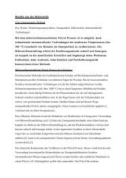
digested sample (Fig. 1). Most of the expected tryptic peptides<br />
were observed in the spectrum, except <strong>for</strong> the fragment<br />
corresponding to sequences 1–7. Very low abundance signals<br />
corresponding to singly and doubly charged molecular<br />
ions were observed in the spectrum (data not shown). No<br />
attempt was made to determine the amount of the undigested<br />
<strong>protein</strong>.<br />
Figure 1B shows the coverage of cytochrome c obtained<br />
based on Figure 1A. Mass of each tryptic fragment in Figure<br />
1A (which corresponds to a part of the cytochrome c sequence)<br />
is shown in the <strong>for</strong>m of a horizontal line in Figure<br />
1B. Tryptic fragments discussed in this paper are denoted <strong>by</strong><br />
T 1,T 2,T 3,...T n. All those peaks that match the <strong>protein</strong><br />
sequence are shown in the <strong>for</strong>m of horizontal lines; they<br />
represent the extent of the coverage of the <strong>protein</strong> from the<br />
amino-terminus to the carboxyl -terminus in Figure 1B. It is<br />
important to note that a number of signals corresponding to<br />
incomplete cleavages were observed in the MALDI spectrum<br />
(Fig. 1 and Table 1). We have found previously that<br />
this type of cleavage occurs where two contiguous cleavage<br />
sites are present (K-K, R-R, K-R) or those cleavage sites are<br />
in close proximity (Pramanik et al. 1991).<br />
A parallel experiment was conducted using a trypsin-tocytochrome<br />
c ratio of 1:25; this time the digestion mixture<br />
was maintained at a temperature of 37°C <strong>for</strong> 6 h (classic<br />
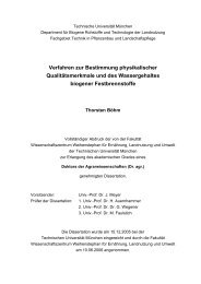
approach) (Lee and Shively 1990). Figure 2A shows the<br />
MALDI <strong>mass</strong> spectrum of the resulting tryptic fragments<br />
and Figure 2B shows the corresponding coverage of the<br />
<strong>protein</strong>. Almost 88% of the <strong>protein</strong> coverage was achieved<br />
from this digestion; only two peptide fragments corresponding<br />
to sequences 1–8 and 23–27 were not observed (Table<br />
2). A comparison of these two approaches of trypsin digestion<br />
of cytochrome c demonstrates that microwave irradiation<br />
<strong>for</strong> 10 min produced similar results to the classic<br />
method in 6 h of digestion.<br />
Additional studies were conducted to compare the extent<br />
of tryptic peptides generation from cytochrome c in 12 min<br />
at 37°C under microwave irradiation versus the classic<br />
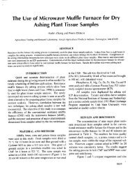
method at 37°C. Figure 3A shows the MALDI <strong>mass</strong> spectrum<br />
of the 12-min microwave-assisted trypsin digest of<br />
cytochrome c (1:25, w/w) at 37°C. Polypeptide signals exhibited<br />
in the spectrum provided 90% sequence coverage of<br />
cytochrome c. The signals correspond to singly charged<br />
(m/z 12,234) and the doubly charged (m/z 6,117) ions in<br />
spectrum (Fig. 3A) represent the presence of intact cytochrome<br />
c in the sample. Figure 3B describes the MALDI<br />
data <strong>for</strong> a 12-min trypsin digestion at 37°C without microwave<br />
<strong>for</strong> cytochrome c (1:25, w/w). This experiment shows<br />
no hydrolysis products of the <strong>protein</strong>. The observed ion<br />
peak at m/z 1,281 and the other weak lower <strong>mass</strong> ions do not<br />
www.<strong>protein</strong>science.org 2677
Pramanik et al.<br />
Fig. 1. (A) MALDI <strong>mass</strong> spectrum of tryptic fragments of cytochrome c after 10 min of microwave irradiation at 1:25 protease-to<strong>protein</strong><br />
ratio <strong>by</strong> weight and (B) tryptic fragments in A are represented in the <strong>for</strong>m of horizontal lines, showing the total sequence<br />
coverage of cytochrome c.<br />
correspond to the tryptic fragments of cytochrome c. The<br />
two abundant ions at higher <strong>mass</strong>es (m/z 12,236 and 6,118)<br />
corresponded to the intact cytochrome c. Thus, microwave<br />
irradiation greatly accelerates the digestion process, providing<br />
complete <strong>mapping</strong> of the <strong>protein</strong> in minutes, whereas the<br />
classic method provides no cleavage products.<br />
In another experiment, cytochrome c was subjected to<br />
microwave irradiation in the absence of a protease. After 20<br />
min of microwave irradiation of the <strong>protein</strong> solution,<br />
MALDI-MS did not yield signals in the lower <strong>mass</strong> region<br />
corresponding to peptide fragments. Instead, the intact <strong>protein</strong><br />
was detected (data not shown). This result confirms that<br />
microwave irradiation only assists and enhances the enzymatic<br />
digestion of <strong>protein</strong>s; it does not induce degradation/<br />
breakdown of the <strong>protein</strong> in the absence of a protease.<br />
<strong>Microwave</strong>-assisted trypsin digestion<br />
of bovine ubiquitin<br />
The study was continued with another <strong>protein</strong>, bovine ubiquitin,<br />
which is a tightly folded <strong>protein</strong> that is extremely<br />
2678 Protein Science, vol. 11<br />
resistant to denaturation. The stability of the <strong>protein</strong> has<br />
been attributed to the pronounced hydrophobic core and that<br />
90% of the residues in the polypeptide chain appear to be<br />
involved in intramolecular hydrogen bonding. In this study,<br />
microwave experiments were per<strong>for</strong>med at various ratios of<br />
<strong>enzyme</strong>-to-<strong>protein</strong> concentrations. These results were compared<br />
with the data obtained from the classic methods.<br />
Figure 4A shows the coverage of bovine ubiquitin after<br />
the <strong>protein</strong> was subjected to microwave-<strong>enhanced</strong> trypsin<br />
digestion (1:5, w/w) <strong>for</strong> 10 min at 37 °C. The <strong>protein</strong> coverage<br />
was established <strong>by</strong> the observation of the expected<br />
peptide signals in the LC-ESI <strong>mass</strong> spectrum. This result<br />
was further verified <strong>by</strong> MALDI-MS analysis. Figure 4B<br />
shows the ubiquitin coverage after 24 h of trypsin digestion<br />
<strong>by</strong> the classic methods when the sample was heated on a hot<br />
plate at 37°C. These results suggested that a similar coverage<br />
was obtained from the two different experiments (Fig.<br />
4). Thus, microwave irradiation enhances the enzymatic digestion;<br />
within 10 min almost complete primary structural<br />
in<strong>for</strong>mation was obtained. In addition, the microwave tryp-
Table 1. Peptides with <strong>mass</strong> values observed in MALDI <strong>mass</strong> spectrum of<br />
cytochrome c after 10 min of trypsin digestion with microwave irradiation<br />
Peptide sequence<br />
sin digestions of ubiquitin were carried <strong>for</strong> various time<br />
intervals (10 min, 30 min, and 60 min) using lower ratios of<br />
<strong>enzyme</strong>-to-<strong>protein</strong> concentrations (1:25–1:200, w/w). The<br />
<strong>mass</strong> spectral data yielded satisfactory coverage, but increasing<br />
the <strong>reaction</strong> time (30 min and 60 min) resulted in<br />
only minor improvements. This data suggest that at longer<br />
irradiation times (>30 min) the <strong>enzyme</strong> loses its activity.<br />
However, earlier time points such as 10 min, 30 min, and 60<br />
min at 37°C using the classic approach did not have any<br />
effect on the <strong>protein</strong>, except <strong>for</strong> a small amount of cleavage<br />
observed between amino acid residues 74 and 75.<br />
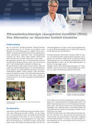
Another <strong>protein</strong>, horse heart myoglobin (molecular <strong>mass</strong><br />
16,951 Da), was also used in the study. Figure 5 shows the<br />
MALDI <strong>mass</strong> spectrum of trypsin digest obtained from 10<br />
min of microwave irradiation. The spectral data demonstrates<br />
a complete coverage of the <strong>protein</strong>. This study further<br />
establishes the efficacy of the microwave methodology.<br />
Dithiothreitol (DTT) reduced chicken egg lysozyme (molecular<br />
<strong>mass</strong> 14,306 Da) was similarly subjected to microwave-assisted<br />
trypsin digestion (1:5, w/w). The MALDI<br />
<strong>mass</strong> spectrum (Fig. 6A) of this sample displayed the expected<br />
tryptic peptides with the exception of the fragment<br />
corresponding to sequences 74–96. When the digestion experiment<br />
was conducted at 37°C <strong>for</strong> 10 min in the absence<br />
of microwave irradiation, no tryptic fragments of lysozyme<br />
were detected in the MALDI spectrum (Figure 6B); instead,<br />
abundant ions representing the singly and doubly charged<br />
molecular ions of lysozyme were exhibited in the <strong>mass</strong><br />
spectrum. Thus, these control experiments further confirm<br />
<strong>Microwave</strong>-<strong>enhanced</strong> enzymatic <strong>protein</strong> digestion in minutes<br />
Expected<br />
<strong>mass</strong> values Observed peptide with <strong>mass</strong> values<br />
T 1 GDVEK 546.6<br />
T 2 GK 203.2<br />
T 3 K 146.2 T 3–7 � 2273<br />
T 4 IFVQK 633.8<br />
T 5 CAQCHTVEK 1018.2<br />
T 6 GGK 260.3 T 6–8 � 1674.6 T 6–9 � 1802.7<br />
T 7 HK 283.3<br />
T 8 TGPNLHGLFGR 1168.3 T 8 � 1168.3, T 8–9 � 1296.3<br />
T 9 K 146.2 T 9–11 � 1825.6 T 9–13 � 3947.7<br />
T 10 TGQAPGFSYTDANK 1456.5 T 10–12 � 3690.4 T 10–13 � 3816.0<br />
T 11 NK 260.3 T 11–15 � 3798.5<br />
T 12 GITWGEETLMEYLENPK 2010.2 T 12 � 2010 T 12–14 � 2796<br />
T 13 K 146.2 T 13 � 805.4<br />
T 14 YIPGTK 677.8 T 10–14 � 4475 T 9–14 � 4603.0<br />
T 15 MIFAGIK 779.0 T 15 � 788.9, T 15–16 � 906.4,<br />
T 16 K 146.2 T 16–19 � 1561.2<br />
T 17 K 146.2 T 17–21 � 1976.8<br />
T 18 GER 360.4 T 18–19 � 1305.3<br />
T 19 EDLIAYLK 964.1 T 15–19 � 2322<br />
T 20 K 146.2 T 16–20 � 1689.6<br />
T 21 ATNE 433.4 T 15–21 � 2865.2, T 16–21 � 2104.9<br />
the effect of microwave in accelerating the enzymatic digestion<br />
of <strong>protein</strong>s.<br />
Application of microwave-assisted trypsin<br />
digestion of recombinant human interferon �-2b<br />
(rh-IFN �-2b) <strong>protein</strong><br />
The peptide <strong>mapping</strong> of rh-IFN �-2b, an antiviral/anticancer<br />
agent marketed <strong>by</strong> Schering-Plough, demonstrates the<br />
utility of microwave-assisted enzymatic digestion <strong>for</strong> a<br />
larger <strong>protein</strong>. This <strong>protein</strong> has an average molecular <strong>mass</strong><br />
of 19,266 Da and contains four cysteines at positions 1, 29,<br />
98, and 138; these cysteine residues are responsible <strong>for</strong> the<br />
presence of the two disulfide bonds in the <strong>protein</strong>. We have<br />
previously characterized this <strong>protein</strong> including the confirmation<br />
of cysteines 1–98 and 29–138 disulfide linkages<br />
(Pramanik et al. 1991).<br />
The microwave-assisted trypsin digest mixture of rh-IFN<br />
�-2b using various irradiation intervals were analyzed <strong>by</strong><br />
on-line LC-ESI-MS, using a C18 reversed phase 1-mm/15cm<br />
column. Figure 7 represents the total ion chromatogram<br />
(TIC) after 10 min of irradiation. The individual tryptic<br />
peptides of rh-IFN �-2b are denoted <strong>by</strong> T 1,T 2,T 3,...T n.<br />
These data agree with the cDNA-determined amino acid<br />
sequence of rh-IFN �-2b. Several peptide signals arising<br />
from incomplete cleavages were also observed in the spectrum;<br />
we have found in our earlier studies that incomplete<br />
cleavage <strong>by</strong> trypsin often occurs under normal digestion<br />
www.<strong>protein</strong>science.org 2679
Pramanik et al.<br />
Fig. 2. (A) MALDI <strong>mass</strong> spectrum of tryptic peptides of cytochrome c generated after 6 h of digestion (1:25, w/w) at 37°C (classic<br />
approach) and (B) tryptic fragments in A is represented in the <strong>for</strong>m of horizontal lines showing the total sequence coverage of<br />
cytochrome c.<br />
conditions. As discussed above, the rh-IFN �-2b molecule<br />
has four cysteines (at positions 1, 29, 98, and 138), which<br />
are contained in the tryptic peptides T 1,T 5,T 10, and T 17,<br />
respectively (Fig. 7). The peptide signals T 1-SS-T 10, T 5-SS-<br />
T 17, T 5-SS-T 16,17 , and T 1-SS-T 9,10 observed in the TIC<br />
corresponded to disulfide-linked peptides at m/z 4,616,<br />
2,118, 2,246, and 6,049, respectively.<br />
In a separate experiment, the reduced <strong>for</strong>m of rh-IFN<br />
�-2b (obtained <strong>by</strong> treatment with DTT) was digested under<br />
microwave irradiation conditions as described above, and<br />
the sample mixture was analyzed <strong>by</strong> LC-ESI-MS. As expected,<br />
the MS data showed complete disappearance of the<br />
disulfide-containing peptide signals (m/z 4,616, 2,118,<br />
2,246, and 6,049), whereas new intense signals due to the<br />
released peptides T 1,T 5,T 10, and T 17 appeared in the spectrum<br />
(data not shown).<br />
Another specific <strong>enzyme</strong>, endoprotease lysine-C, has also<br />
been successfully applied with microwave irradiation to the<br />
digestion of <strong>protein</strong>s, within 10 min of microwave irradiation,<br />
>90% of the digested products of these <strong>protein</strong>s (cytochrome<br />
c, lysozyme, ubiquitin, and myoglobin) were observed<br />
in the MALDI spectra (data not shown). These re-<br />
2680 Protein Science, vol. 11<br />
sults demonstrate that the application of microwave is not<br />
restricted to one specific <strong>enzyme</strong>.<br />
Application of microwave-assisted digestion was also applied<br />
to <strong>protein</strong>s (<strong>for</strong> example, myoglobin, SA 436, YAE-1)<br />
separated on SDS-PAGE gels. The in-gel digestion of <strong>protein</strong>s<br />
with trypsin using microwave irradiation was successfully<br />
tried, and complete peptide maps of <strong>protein</strong>s were<br />
obtained (data not shown). Briefly, coo<strong>mass</strong>ie blue-stained<br />
<strong>protein</strong> band was excised and washed with 50% methanolic<br />
solution. The washed band was subjected to microwave<br />
irradiation <strong>for</strong> destaining in the presence of 30 �Lof10mM<br />
ammonium acetate buffer. Fifteen minutes of microwave<br />
irradiation with 30% power (144 W) at 37°C resulted in the<br />
complete destaining of the gel band. The colorless gel was<br />
then transferred into a new 300-�L Eppendorf tube <strong>for</strong> trypsin<br />
digestion. The gel was cut into smaller pieces and suspended<br />
in 10 mM ammonium bicarbonate solution be<strong>for</strong>e it<br />
was treated with trypsin. A similar procedure as described<br />
above was followed <strong>for</strong> in-gel digestion of <strong>protein</strong> using<br />
microwave irradiation. After 15 min of digestion, the <strong>protein</strong><br />
solution was quenched with 0.1% TFA and subjected to<br />
<strong>mass</strong> spectrometric analysis.
Table 2. Peptides with <strong>mass</strong> values observed in MALDI <strong>mass</strong> spectrum of<br />
cytochrome c after 6hoftrypsin digestion at 37°C (classic method)<br />
Peptide sequence<br />
Kinetic studies of the trypsin digestion of IFN �-2b<br />
under microwave irradiation<br />
A series of experiments were conducted to determine the<br />
<strong>reaction</strong> rate of the microwave-assisted trypsin digestion of<br />
the <strong>protein</strong>, IFN �-2b. Data obtained from a microwave<br />
assisted digestion experiment, in which an instrumental setting<br />
of 37°C was used (Star-6 microwave apparatus, CEM<br />
Corp.), was then compared to the results achieved <strong>by</strong> the<br />
classic method at 37°C (Fig. 8). Digestion products were<br />
sampled at intervals of 0 min, 5 min, 10 min, 20 min, and 30<br />
min, and the resulting aliquots were then analyzed <strong>by</strong> LC-<br />
MS. At 5 min, the microwave-assisted <strong>reaction</strong> was found to<br />
be 11% complete, 17% at 10 min, 27% at 20 min, and ∼ 32%<br />
at 30 min. Two more data points were acquired at 1hand<br />
2 h, respectively, indicating no further increase in the rate of<br />
the <strong>reaction</strong>. In comparison, the classic tryptic digestion<br />
method did not show <strong>reaction</strong> products until the 20-min<br />
mark, 20% at 20 min, 36% at 30 min. After 30 min the rate<br />
of digestion <strong>by</strong> the classic method continued to increase but<br />
at a slower rate, reaching 50% in 2 h and 78% in ∼ 6 h (data<br />
not shown). In the case of the microwave-assisted <strong>reaction</strong>,<br />
the loss of enzymatic activity (at ∼ 30 min) is possibly due to<br />
the denaturation of the <strong>protein</strong> at elevated temperature.<br />
<strong>Microwave</strong>-assisted tryptic digestion experiments of IFN<br />
�-2b were also carried out at instrumental temperature settings<br />
of 45°C and 55°C, and aliquots were taken at 0 min,<br />
5 min, 10 min, 20 min, and 30 min. These results were<br />
<strong>Microwave</strong>-<strong>enhanced</strong> enzymatic <strong>protein</strong> digestion in minutes<br />
Expected<br />
<strong>mass</strong> values Observed peptide with <strong>mass</strong> values<br />
T 1 GDVEK 546.6<br />
T 2 GK 203.2<br />
T 3 K 146.2<br />
T 4 IFVQK 633.8 T 4–5 � 1633.5<br />
T 5 CAQCHTVEK 1018.2<br />
T 6 GGK 260.3<br />
T 7 HK 283.3 T 7–8 � 1433.6<br />
T 8 TGPNLHGLFGR 1168.3 T 8 � 1168.3, T 8–9 � 1296.3<br />
T 9 K 146.2 T 9–13 � 3947.7<br />
T 10 TGQAPGFSYTDANK 1456.5 T 10–12 � 3690.4<br />
T 11 NK 260.3<br />
T 12 GITWGEETLMEYLENPK 2010.2 T 12 � 2010, T 12,13 � 2010<br />
T 13 K 146.2 T 13–14 � 805.4<br />
T 14 YIPGTK 677.8 T 14–15 � 1438.9<br />
T 15 MIFAGIK 779.0 T 15 � 778.5, T 15–21 � 2866.7<br />
T 16 K 146.2 T 16–18 � 616.5, T 16–20 � 1781.5<br />
T 17 K 146.2 T 17–19 � 1433.6, T 17–20 � 1634.6<br />
T 18 GER 360.4 T 18–19 � 1305.3<br />
T 19 EDLIAYLK 964.1<br />
T 20 K 146.2<br />
T 21 ATNE 433.4<br />
analyzed <strong>by</strong> LC-ESI-MS to determine the amount of <strong>protein</strong><br />
digested <strong>for</strong> each time interval. These results are plotted in<br />
Figure 8. At 5-min intervals, both digestion experiments<br />
showed ∼ 60% of the <strong>protein</strong> digested, reaching 67%–70%<br />
digestion in 10 min–20 min. At this point, no further increase<br />
in the yield was observed, suggesting that the activity<br />
of trypsin had been destroyed. Fresh <strong>enzyme</strong> (3�g of trypsin)<br />
was then added to the two samples from the 45°C<br />
experiment that had been collected at 10 min and 20 min.<br />
These samples were then subjected to further microwave<br />
irradiation of 10 min–20 min resulting in 80%–90%<br />
completion of the digestion (data not shown). With elevated<br />
temperature at 45°C and 55°C, the amount of digestion<br />
produced in 10 min has been quadrupled compared to the<br />
data <strong>for</strong> the 37°C setting. The identical rate plots <strong>for</strong> 45°C<br />
and 55°C <strong>reaction</strong>s indicate that a temperature of 45°C optimizes<br />
the microwave-assisted tryptic digestion of IFN<br />
�-2b. In conclusion, we have shown that microwave-assisted<br />
<strong>reaction</strong>s can produce high yields in minutes, which<br />
would require hours <strong>by</strong> classic methods.<br />
Observations on the rate of acceleration<br />
of the enzymatic digestion<br />
To investigate the mechanism involved in the observed rate<br />
acceleration of the enzymatic cleavage of <strong>protein</strong>s under<br />
microwave irradiation, trypsin digestion of rh-IFN �-2b was<br />
www.<strong>protein</strong>science.org 2681
Pramanik et al.<br />
Fig. 3. MALDI <strong>mass</strong> spectra of cytochrome c after 12 min of trypsin digest (1:25, w/w)(A) with microwave irradiation and (B) without<br />
microwave irradiation.<br />
carried out at three different microwave temperature settings,<br />
37°C, 45°C, and 55°C (Fig. 8). Reaction samples<br />
were taken at intervals of 0 min, 5 min, 10 min, 20 min, and<br />
30 min. The temperatures of the <strong>reaction</strong> solutions were<br />
Fig. 4. Total sequence coverage of ubiquitin (1:5, w/w) (A) after 10 min of<br />
microwave-assisted tryptic digestion and (B) after 24 h of trypsin digest at<br />
37°C (classic approach).<br />
2682 Protein Science, vol. 11<br />
measured at each sampling point using a thermocouple and<br />
were found to be 49°C, 65°C, and 74°C, respectively. Each<br />
measurement was made as quickly as possible to minimize<br />
a decrease in the solution temperature. On the basis of our<br />
experiments, these temperatures were reached in ∼ 5 min of<br />
microwave irradiation. Significantly, the temperatures of<br />
these <strong>reaction</strong> solutions were found to be much higher than<br />
their microwave settings. It is important to note that the<br />
rapid elevation of the solution temperature to >60°C greatly<br />
<strong>enhanced</strong> the digestion <strong>reaction</strong>.<br />
In an ef<strong>for</strong>t to explore the feasibility that the rapid increase<br />
in temperature is responsible <strong>for</strong> the accelerated rates<br />
observed in the microwave, the above enzymatic <strong>reaction</strong><br />
was further studied without microwave irradiation, using a<br />
heating block to maintain the desired temperature. A fresh<br />
<strong>reaction</strong> mixture was introduced into the preheated block at<br />
60°C to simulate the rapid heating process observed in the<br />
microwave. The rates of enzymatic cleavage measured in<br />
this study fairly resembled the results obtained from the<br />
microwave experiment (Fig. 8). This suggests that the rapid<br />
increase in the <strong>reaction</strong> temperature is at least partially responsible<br />
<strong>for</strong> the large acceleration seen under microwave<br />
conditions (Lidstrom et al. 2001; Larhed et al. 2002).
Factors influencing proteolytic cleavage<br />
of <strong>protein</strong>s with <strong>enzyme</strong>s<br />
Many enzymatic <strong>reaction</strong>s—including the proteolytic cleavage<br />
of <strong>protein</strong>s with trypsin and lysine c—are traditionally<br />
conducted in aqueous solution at 37°C <strong>for</strong> a few hours. The<br />
rationale appears to be (1) the slow rate of denaturation of<br />
the <strong>enzyme</strong> and the substrate <strong>protein</strong>, and (2) a reasonable<br />
rate <strong>for</strong> the recognition of ligation sites (e.g., next to an<br />
arginine or lysine component <strong>for</strong> trypsin) on the substrate<br />
<strong>protein</strong> <strong>by</strong> the <strong>enzyme</strong> resulting in selective peptide bond<br />
scission. The sheath of water molecules around <strong>protein</strong>s and<br />
intramolecular hydrogen bonding between different parts of<br />
a <strong>protein</strong> molecule play important roles in the dynamic behavior<br />
of <strong>protein</strong>s. There is growing recognition that folding<br />
and unfolding of <strong>protein</strong>s may influence the chemical interaction<br />
of <strong>protein</strong>s with ligands.<br />
There are two schools of thought about microwave energy<br />
transfer to a <strong>reaction</strong>. One set of research workers<br />
believe that microwaves are just a convenient way of heating<br />
(Kuhnert 2002); other research workers (Mayo et al.<br />
2002) have produced experimental data that point to a nonthermal<br />
microwave effect. It is interesting to note, however,<br />
that no microwave-assisted <strong>reaction</strong> is slower than the corresponding<br />
conductivity/convection-heated conventional <strong>reaction</strong>.<br />
Early in the development of microwave technology,<br />
it was observed that microwaves would produce notable<br />
increase in <strong>reaction</strong> rates <strong>for</strong> <strong>reaction</strong>s that are slow under<br />
conventional conditions.<br />
We have studied enzymatic cleavage of <strong>protein</strong>s using<br />
microwave energy and compared our findings with conven-<br />
<strong>Microwave</strong>-<strong>enhanced</strong> enzymatic <strong>protein</strong> digestion in minutes<br />
Fig. 5. MALDI <strong>mass</strong> spectrum of the tryptic digest of myoglobin after 10 min of microwave irradiation.<br />
tional heating of the <strong>reaction</strong> mixture. <strong>Microwave</strong>s are nonionizing<br />
radiation that interact in the liquid phase with ion<br />
pairs and with organic molecules that have dipole moment<br />
(such as amides, esters, and water, but not hydrocarbons<br />
such as benzene or hexane). <strong>Microwave</strong> energy is directly<br />
transferred to these species, and the energy profile is different<br />
than that involved in traditional conduction/convection<br />
heating. However, both types of enzymatic <strong>reaction</strong><br />
could produce nearly comparable results in spite of the different<br />
energy profiles and thus allow MALDI and ESI-MS<br />
methods to be used on the <strong>protein</strong> hydrolysate produced <strong>by</strong><br />
either method <strong>for</strong> correct <strong>protein</strong> <strong>mapping</strong>.<br />
Our experimental observations show that ∼ 60°C is an<br />
optimum temperature <strong>for</strong> rapid proteolysis and a reasonably<br />
low rate of denaturation. That bulk temperature <strong>for</strong> the <strong>reaction</strong><br />
mixture can be reached <strong>by</strong> heating on a hot plate or<br />
<strong>by</strong> microwave irradiation <strong>for</strong> 1 min–2 min.<br />
Tightly folded <strong>protein</strong>s are known to require many hours<br />
<strong>for</strong> adequate proteolysis <strong>by</strong> <strong>enzyme</strong>s under conventional<br />
conditions. It is expected that these very <strong>protein</strong>s will give<br />
greatly <strong>enhanced</strong> proteolysis rates under microwave irradiation.<br />
We plan to test this possibility. Our current observations<br />
on the action of trypsin on bovine ubiquitin is in agreement<br />
with this expectation.<br />
Conclusions<br />
Accelerated enzymatic digestion of <strong>protein</strong>s under controlled<br />
microwave irradiation has been demonstrated. This<br />
approach accelerates the digestion process to minutes versus<br />
www.<strong>protein</strong>science.org 2683
Pramanik et al.<br />
Fig. 6. MALDI <strong>mass</strong> spectrum of the DTT-reduced lysozyme after 10 min of trypsin digest (1:5, w/w) (A) with microwave irradiation<br />
and (B) without microwave irradiation.<br />
hours <strong>by</strong> traditional methods. Kinetic study experiments indicated<br />
that the microwave-assisted process is optimized at<br />
an instrumental setting of 45°C (actual bulk temperature<br />
60°C) resulting in ∼ 70% completion of the <strong>protein</strong> digestion<br />
in 10 min. In the case of trypsin or lysine digest, most of the<br />
expected peptide fragments were generated with >80% coverage<br />
of the <strong>protein</strong>. No nonspecific cleavage product was<br />
detected in the mixture. This <strong>mapping</strong> strategy combined<br />
with MALDI or LC-ESI-MS provides sequence and disulfide<br />
bond in<strong>for</strong>mation, as well as the location of any modification<br />
sites with high speed and accuracy. We have successfully<br />
demonstrated the usefulness of this new method<br />
<strong>for</strong> probing primary structure of ubiquitin, cytochrome c,<br />
myoglobin, lysozyme, and a few other <strong>protein</strong>s such as IFN<br />
�-2b.<br />
Preliminary work shows that even inexpensive domestic<br />
microwave ovens can be programmed <strong>for</strong> this novel approach<br />
to proteolytic cleavage of <strong>protein</strong>s. Further studies<br />
<strong>for</strong> a better understanding of microwave-assisted enzymatic<br />
<strong>reaction</strong>s are in progress. We believe that the use of microwaves<br />
in <strong>protein</strong> identification will be an important advancement<br />
in biotechnology and proteome research.<br />
2684 Protein Science, vol. 11<br />
Materials and methods<br />
<strong>Microwave</strong> operation<br />
Digestion of <strong>protein</strong>s was conducted in a specialized, temperaturecontrolled,<br />
single beam microwave applicator (Prolabo Synthewave<br />
402) with a maximum power of 300 W; only 10% (30 W) of<br />
the available power was used. The sample vial was placed in a<br />
three-hole Teflon insertion rack, which in turn was lowered into<br />
the irradiation chamber. The temperature of the apparatus was<br />
programmed to increase gradually from 40°C to 50°C. The actual<br />
temperature of the sample solution was measured with a Fisher<br />
thermocouple (Fisherbrand Dual-Channel Thermometer with Offsets,<br />
Fisher Scientific, Pittsburgh, PA) immediately after microwave<br />
irradiation and was found to be ∼ 50°C.<br />
The digestion of <strong>protein</strong>s was also conducted with a single<br />
beam, multi-inlet Star-6 <strong>Microwave</strong> Apparatus (CEM Corporation,<br />
Matthews, NC). This microwave has a maximum power output of<br />
480 W. For our experiments, this equipment was set at 30% of the<br />
available power or 144 W. Sample vials were placed in a four-hole<br />
Teflon insert rack, which could be lowered into the glass insertion<br />
chamber. Enzyme digestion experiments were carried out at fixed<br />
instrumental temperature settings of 37°C, 45°C, and 55°C. These<br />
targeted microwave temperature settings were kept constant <strong>by</strong> the<br />
sensor-controlled on/off duty cycle of the magnetron. The infrared<br />
sensor continuously monitored the surface of the glass wall of the
insertion chamber in the vicinity of the sample vial. A fan near the<br />
glass insertion chamber switched on to cool the reactants when the<br />
duty cycle was in the off position. <strong>Microwave</strong> irradiation was<br />
delayed 1 min be<strong>for</strong>e allowing the microwave-assisted <strong>reaction</strong> to<br />
begin to ensure that the instrument had reached its targeted temperature.<br />
The actual <strong>reaction</strong> solution temperatures after microwave<br />
irradiation were measured with the Fisher thermocouple. It<br />
was determined that the microwave-targeted temperature settings<br />
of 37°C, 45°C, and 55°C generated actual solution temperatures of<br />
49°C, 65°C, and 74°C, respectively. Sample aliquots were collected<br />
at intervals of 0 min, 5 min, 10 min, 20 min, and 30 min.<br />
Enzymatic digestion was also carried out in a heating block<br />
controlled <strong>by</strong> a Pierce Reacti-Therm heating controller (Pierce<br />
Chemical Co, Rock<strong>for</strong>d, IL). Temperatures were monitored <strong>by</strong> a<br />
mercury thermometer placed in an adjacent sample well (block).<br />
Materials<br />
Ammonium bicarbonate (Sigma A-6141), cytochrome c from bovine<br />
heart (Sigma 0-2037), chicken egg lysozyme (Sigma L-6876),<br />
horse heart myoglobin (Sigma M-1882), and ubiquitin from bovine<br />
red blood cells (Sigma U-6253Y) were purchased and used as<br />
obtained.<br />
Trypsin (sequence grade, Boehringer Mannheim Corp., Cat.<br />
1418475), purchased from Boehringer Mannheim Corp. (Indianapolis,<br />
IN) and was used without further purification.<br />
Recombinant human interferon �-2b (rh-IFN �-2b) was purified<br />
from Escherichia coli (Schering-Plough Research Institute, Union,<br />
NJ).<br />
<strong>Microwave</strong>-<strong>enhanced</strong> enzymatic <strong>protein</strong> digestion in minutes<br />
Fig. 7. Total ion chromatogram of trypsin digest of interferon �-2b after 10 min of microwave irradiation.<br />
Mass spectrometry and analysis <strong>by</strong> MALDI-MS<br />
MALDI spectra were obtained with Voyager-DE STR reflectron<br />
time-of-flight <strong>mass</strong> spectrometer (PerSeptive Biosystems,<br />
Framingham, Massachusetts, USA) equipped with nitrogen laser<br />
(337 nm, 3-ns output pulse) and a high current detector. Spectral<br />
data were acquired in the linear mode with an acceleration voltage<br />
of 25 kV. Each <strong>mass</strong> spectrum was generated from the accumulated<br />
data of 100 laser shots. Alpha-cyano 4-hydroxy cinnamic<br />
acid (Aldrich 47,687.0) was used as a MALDI matrix, and a mixture<br />
of a 50% solution of 0.1% trifluoroacetic acid and acetonitrile<br />
was used as a matrix solution. A 3-�L aliquot of the sample<br />
solution was mixed with an equal volume of the matrix. A 0.7-�L<br />
of the resulting mixture was used <strong>for</strong> MALDI-MS analysis. The<br />
MALDI system was calibrated with an external standard.<br />
LC-ESI-MS analysis<br />
A PE-Sciex API 365 triple quadrupole <strong>mass</strong> spectrometer was<br />
applied <strong>for</strong> LC-ESI-MS analysis. The HPLC analyses were carried<br />
out on Shimadzu LC-100 Pumps, with reverse-phase HPLC Phenomenex<br />
C18 Jupiter column (5 �m, 300 Å, 15 cm/1 mm).<br />
Enzymatic digestion of cytochrome c (MW 12,229 Da),<br />
myoglobin (MW 16,951 Da), lysozyme (MW 14,306<br />
Da), and ubiquitin (MW 8,565.0 Da)<br />
Digestion experiments were per<strong>for</strong>med in 350-�L Eppendorf<br />
tubes. The concentration of the <strong>protein</strong> was maintained at 20 �M<br />
www.<strong>protein</strong>science.org 2685
Pramanik et al.<br />
Fig. 8. Plots showing the rate of trypsin digestion of IFN �-2b in the first 30 min under microwave irradiation and classic condition.<br />
in 75 mM ammonium bicarbonate solution. A variable ratio of<br />
protease-to-<strong>protein</strong>, 1:200 to 1:5 (w/w) concentration was used.<br />
Samples were irradiated <strong>for</strong> 5 min, 10 min, 15 min, 20 min, 30<br />
min, or 60 min with microwave at 30% (144 W) of the maximum<br />
power setting. After a preselected interval, the sample was<br />
quenched <strong>by</strong> the addition of 0.1% TFA solution and was stored in<br />
dry ice <strong>for</strong> <strong>mass</strong> spectrometric analysis.<br />
Enzymatic digestion of rh-IFN �-2b (MW 19,269)<br />
A 10-�L rh-IFN �-2b <strong>protein</strong> solution (concentration of 3 �g/�L)<br />
was dissolved in 90 �L of ammonium bicarbonate buffer in an<br />
Eppendorf tube, and three sets of this solution were prepared <strong>for</strong><br />
trypsin digestion. Trypsin was added to each of the 100-�L solution<br />
at an <strong>enzyme</strong>-to-<strong>protein</strong> ratio of 1:50, 1:25, and 1:5 (w/w),<br />
respectively. The solution was then irradiated under microwave <strong>for</strong><br />
5 min and 10 min. A separate microwave experiment was conducted<br />
using the disulfide reduced <strong>for</strong>m of rh-IFN �-2b where the<br />
reduction of rh-IFN �-2b was per<strong>for</strong>med <strong>by</strong> adding 20-fold molar<br />
excess of DTT.<br />
Acknowledgments<br />
We are indebted to the National Science Foundation (CHE-<br />
9910242), Union Mutual Foundation, and the George Barasch Research<br />
Fellowship funds <strong>for</strong> partial support of this research. We<br />
thank Stevens Institute of Technology <strong>for</strong> laboratory facilities and<br />
the Schering-Plough Research Institute <strong>for</strong> use of advanced <strong>mass</strong><br />
spectrometric instrumentation. We acknowledge Dr. John Piwinski<br />
<strong>for</strong> his support of this research project.<br />
The publication costs of this article were defrayed in part <strong>by</strong><br />
2686 Protein Science, vol. 11<br />
payment of page charges. This article must there<strong>for</strong>e be here<strong>by</strong><br />
marked “advertisement” in accordance with 18 USC section 1734<br />
solely to indicate this fact.<br />
References<br />
Akabori, S., Ohno, K., and Narita, K. 1952. On the hydrazinolysis of <strong>protein</strong>s<br />
and peptides: A method <strong>for</strong> the characterization of carboxy-terminal amino<br />
acids in <strong>protein</strong>s. Bull. Chem. Soc. Japan 25: 214–218.<br />
Bose, A.K., Manhas, M.S., Ghosh, M., Raju, V.S., Tabei, K., and Urbanczyk,<br />
L.Z. 1990. Highly accelerated <strong>reaction</strong>s in microwave oven: Synthesis of<br />
heterocycles. Heterocycles 30: 741–744.<br />
Bose, A.K., Banik, B.K., Lavlinskaia, N., Jayaraman, M., and Manhas, M.S.<br />
1997. MORE chemistry in a microwave. Chemtech 27: 18–24.<br />
Bose, A.K., Banik, B.K., Mathur, C., Wagle, D.R., and Manhas, M.S. 2000.<br />
Polyhydroxy amino acide derivatives via �-lactams using enantiospecific<br />
approaches and microwave techniques. Tetrahedron 56: 5603–5619.<br />
Bose, A.K., Ing, Y-H., Pramanik, B.N., Bartner, P.L., Liu, Y.-H., Heimark, L.,<br />
Lavlinskaia, N., and Sareen, C. 2002a. <strong>Microwave</strong> <strong>enhanced</strong> akabori <strong>reaction</strong><br />
<strong>for</strong> peptide analysis. J. Am. Soc. Mass Spectrom. 13: 839–850.<br />
Bose, A.K., Manhas, M.S., Ganguly, S.N., Sharma, A.H., and Banik, B.K.<br />
2002b. MORE chemistry <strong>for</strong> less pollution: Applications <strong>for</strong> process development.<br />
Synthesis (in press).<br />
Caddick, S. 1995. <strong>Microwave</strong>-assisted organic <strong>reaction</strong>s. Tetrahedron 51:<br />
10403–10432.<br />
Chait, B.T. and Kent, S.B.H. 1992. Weighing naked <strong>protein</strong>s: Practical, highaccuracy<br />
<strong>mass</strong> measurement of peptides and <strong>protein</strong>s. Science 257: 1885–<br />
1894.<br />
Chen, S.T., Chiou, S.H., and Wang, K.T. 1991. Enhancement of chemical<br />
<strong>reaction</strong>s <strong>by</strong> microwave irradiation. J. Chinese Chemical Soc. 38: 85–91.<br />
Chowdhury, S.K., Katta, V., and Chait, B.T. 1990. Electrospray ionization <strong>mass</strong><br />
spectrometric peptide <strong>mapping</strong>: A rapid, sensitive technique <strong>for</strong> <strong>protein</strong><br />
structure analysis. Biochem. Biophys. Res. Commun. 167: 686–692.<br />
Davis, M.T. and Terry, D.L. 1992. Analysis of peptide mixtures <strong>by</strong> capillary<br />
high per<strong>for</strong>mance liquid chromatography: A practical guide of small-scale<br />
separations. Protein Sci. 1: 935–944.
Erdelyi, M. and Gogoll, A. 2001. Rapid homogeneous-phase Sonogashira coupling<br />
<strong>reaction</strong>s using controlled microwave heating. J. Org. Chem. 66:<br />
4165–4169.<br />
Erickson, B. 1998. Standardizing the world with microwaves. Anal. Chem. 70:<br />
467A–471A.<br />
Fenn, J.B., Mann, M., Meng, C.K., Wong, S.F., and Whitehouse, C.M. 1989.<br />
Electrospray ionization of large biomolecules. Science 246: 64–71.<br />
Gedye, R., Smith, F., Westaway, K., Ali, H., Baldisera, L., Laberge L., and<br />
Rousell, J. 1986. The use of microwave ovens <strong>for</strong> rapid organic synthesis.<br />
Tetrahedron Lett. 27: 279–282.<br />
Gibson, B.W. and Biemann, K. 1984. Strategy <strong>for</strong> the <strong>mass</strong> spectrometric verification<br />
and correction of the primary structures of <strong>protein</strong>s deduced from<br />
their DNA sequence. Proc. Natl. Acad. Sci. 81: 1956–1960.<br />
Giguere, R.J., Bray, T.L., Duncan, S.C., and Majetich, G. 1986. Application of<br />
commercial microwave ovens to organic synthesis. Tetrahedron Lett. 27:<br />
4945–4948.<br />
Griffin, P.R., Coffman, J.A., Hood, L.E., and Yates III, J.R. 1991. Structural<br />
analysis of <strong>protein</strong>s <strong>by</strong> capillary HPLC electrospray tandem <strong>mass</strong> spectrometry.<br />
Int. J. Mass Spectrom. Ion Process 11: 131–149.<br />
Hanash, S.M. 2000. Biomedical application of two-dimensional electrophoresis<br />
using immobilized pH gradients: Current status. Electrophoresis 21: 1202–<br />
1209.<br />
Hillenkamp, F., Karas, M., Beavis, R.C., and Chait, B.T. 1991. Matrix-assisted<br />
laser desorption/ionization <strong>mass</strong> spectrometry of biopolymers. Anal. Chem.<br />
63: 1193A–1203A.<br />
Hong, B.C., Shr, Y.J., Liao, J.H., Jiang, Y.F., and Kumar, B.S. 2001. <strong>Microwave</strong>-assisted<br />
[6+4]-cycloaddition of fulvenes and a-pyrones to azuleneindoles:<br />
Facile synthesis of angioplatic agents. Bioorganic & Medicinal<br />
Chemistry Letters 11: 1981–1984.<br />
Kabalka, G.W., Wang, L., and Pagni, R.M. 2001. <strong>Microwave</strong> <strong>enhanced</strong> glaser<br />
coupling under solvent-free conditions. Synlett. 1: 108–110.<br />
Kuhnert, N. 2002. <strong>Microwave</strong>-assisted <strong>reaction</strong> in organic synthesis—Are there<br />
any nonthermal microwave effects? Angew. Chem. Int. Ed. 41: 1863–1866.<br />
Larhed, M., Moberg, C., and Hallberg, A. 2002. <strong>Microwave</strong>-accelerated homogeneous<br />
catalysis in organic chemistry. Acc. Chem. Res. (in press).<br />
Lee, T.D. and Shively, J.E. 1990. Enzymatic and chemical digestion of <strong>protein</strong>s<br />
<strong>Microwave</strong>-<strong>enhanced</strong> enzymatic <strong>protein</strong> digestion in minutes<br />
<strong>for</strong> <strong>mass</strong> spectrometry. In Methods Enzymol., Academic Press, New York,<br />
NY, 193: 361–374.<br />
Lidstrom, P., Tierney, J., Wathey, B., and Westman, J. 2001. <strong>Microwave</strong>-assisted<br />
organic synthesis. Tetrahedron 57: 9225–9283.<br />
Mayo, K.G., Nearhoof, E.H., and Kiddle, J.J. 2002. <strong>Microwave</strong>-accelerated<br />
ruthernium-catalyzed olefin metathesis. Org. Lett. 4: 1567–1570.<br />
Mirza, U.A., Cohen, S.L., and Chait, B.T. 1993. Heat-induced con<strong>for</strong>mational<br />
changes in <strong>protein</strong>s studied <strong>by</strong> electrospray ionization <strong>mass</strong> spectrometry.<br />
Anal. Chem. 65: 1–6.<br />
Mirza, U.A., Liu,Y.H., Tang, J.T., Porter, F., Bondoc, L., Chen, G., Pramanik,<br />
B.N., and Nagabhushan, T. 2000. Extraction and characterization of adenovirus<br />
<strong>protein</strong>s from sodium dodecylsulfate polyacrylamide gel electrophoresis<br />
<strong>by</strong> matrix-assisted laser desorption/ionization <strong>mass</strong> spectrometry. J.<br />
Am. Soc. Mass Spectrom. 11: 356–361.<br />
Nguyen, D.N., Becker, G.W., and Riggin, R.N. 1995. Protein <strong>mass</strong> spectrometry:<br />
Application to analytical biotechnology. J. Chromatography A 705:<br />
21–45.<br />
Pandey, A. and Mann, M. 2000. Proteomics to study genes and genomics.<br />
Nature 405: 837–846.<br />
Pramanik, B.N., Tsarbopoulos, A., Labdon, J.E., Trotta, P.P., and Nagabhushan,<br />
T.L. 1991. Structural analysis of biologically active peptides and recombinant<br />
<strong>protein</strong>s and their modified counterparts <strong>by</strong> <strong>mass</strong> spectrometry. J.<br />
Chromatography A 562: 377–389.<br />
Pramanik, B.N., Ganguly, A.K., and Gross, M.L. 2002. Applied electrospray<br />
<strong>mass</strong> spectrometry. Marcel Dekker, New York, NY.<br />
Richter, R.C., Link, D., and Kingston, H.M.S. 2001. <strong>Microwave</strong>-<strong>enhanced</strong><br />
chemistry. Anal. Chem. 1: 31A–37A.<br />
Suib, S.L. 1998. Applications of microwaves in catalysis. Cattech 1: 75–84.<br />
Washburn, M.P. and Yates III, J.R. 2000. Analysis of microbial proteome. Curr.<br />
Opin. Microbiol. 3: 292–297.<br />
Washburn, M.P., Wolter, S.D., and Yates III, J.R. 2001. Large-scale analysis of<br />
the yeast proteome <strong>by</strong> multi-dimensional <strong>protein</strong> identification technology.<br />
Nature Biotechnol. 19: 242–247.<br />
Yates III, J.R., Speicher, S., Griffin, P.R., and Hunkapiller, T. 1993. Peptide<br />
<strong>mass</strong> maps: A highly in<strong>for</strong>mative approach to <strong>protein</strong> identification. Anal.<br />
Biochem. 214: 397–408.<br />
www.<strong>protein</strong>science.org 2687