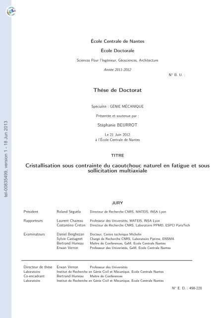
Cristallisation sous contrainte du caoutchouc naturel en fatigue et ...
Cristallisation sous contrainte du caoutchouc naturel en fatigue et ...
Cristallisation sous contrainte du caoutchouc naturel en fatigue et ...

You also want an ePaper? Increase the reach of your titles
YUMPU automatically turns print PDFs into web optimized ePapers that Google loves.
École C<strong>en</strong>trale de Nantes<br />
École Doctorale<br />
Sci<strong>en</strong>ces Pour l’Ingénieur, Géosci<strong>en</strong>ces, Architecture<br />
Année 2011-2012<br />
N o B. U. :<br />
Thèse de Doctorat<br />
Spécialité : GÉNIE MÉCANIQUE<br />
Prés<strong>en</strong>tée <strong>et</strong> sout<strong>en</strong>ue par :<br />
tel-00835499, version 1 - 18 Jun 2013<br />
Stéphanie BEURROT<br />
Le 21 Juin 2012<br />
à l’École C<strong>en</strong>trale de Nantes<br />
TITRE<br />
<strong>Cristallisation</strong> <strong>sous</strong> <strong>contrainte</strong> <strong>du</strong> <strong>caoutchouc</strong> <strong>naturel</strong> <strong>en</strong> <strong>fatigue</strong> <strong>et</strong> <strong>sous</strong><br />
sollicitation multiaxiale<br />
JURY<br />
Présid<strong>en</strong>t Roland Séguéla Directeur de Recherche CNRS, MATEIS, INSA Lyon<br />
Rapporteurs Laur<strong>en</strong>t Chazeau Professeur des Universités, MATEIS, INSA Lyon<br />
Costantino Cr<strong>et</strong>on Directeur de Recherche CNRS, Laboratoire PPMD, ESPCI ParisTech<br />
Examinateurs Daniel Berghezan Docteur, C<strong>en</strong>tre technique Michelin<br />
Sylvie Castagn<strong>et</strong> Chargé de Recherche CNRS, Laboratoire Pprime, ENSMA<br />
Bertrand Huneau Maître de Confér<strong>en</strong>ces, GeM, Ecole C<strong>en</strong>trale Nantes<br />
Erwan Verron Professeur des Univeristés, GeM, Ecole C<strong>en</strong>trale Nantes<br />
Directeur de thèse Erwan Verron Professeur des Universités<br />
Laboratoire Institut de Recherche <strong>en</strong> Génie Civil <strong>et</strong> Mécanique, Ecole C<strong>en</strong>trale Nantes<br />
Co-<strong>en</strong>cadrant Bertrand Huneau Maître de Confér<strong>en</strong>ces<br />
Laboratoire Institut de Recherche <strong>en</strong> Génie Civil <strong>et</strong> Mécanique, Ecole C<strong>en</strong>trale Nantes<br />
N o E. D. : 498-220
tel-00835499, version 1 - 18 Jun 2013
Remerciem<strong>en</strong>ts<br />
tel-00835499, version 1 - 18 Jun 2013<br />
Je ti<strong>en</strong>s à remercier tout d’abord les rapporteurs de c<strong>et</strong>te thèse, Messieurs Costantino<br />
Cr<strong>et</strong>on <strong>et</strong> Laur<strong>en</strong>t Chazeau, pour leur relecture de ce manuscrit. Je remercie égalem<strong>en</strong>t<br />
Madame Sylvie Castagn<strong>et</strong> <strong>et</strong> Messieurs Roland Séguéla <strong>et</strong> Daniel Berghezan pour leur<br />
participation à ma sout<strong>en</strong>ance de thèse <strong>en</strong> tant que membres <strong>du</strong> jury.<br />
Je remercie sincérem<strong>en</strong>t mes directeur <strong>et</strong> <strong>en</strong>cadrant de thèse, Messieurs Erwan Verron <strong>et</strong><br />
Bertand Huneau, pour leur <strong>en</strong>cadrem<strong>en</strong>t sci<strong>en</strong>tifique mais surtout l’amitié que nous avons<br />
développée au fil des ans. Leur pati<strong>en</strong>ce <strong>et</strong> leurs <strong>en</strong>couragem<strong>en</strong>ts ont été décisifs dans les<br />
mom<strong>en</strong>ts de doute <strong>et</strong> leur amitié a pleinem<strong>en</strong>t participé à ma motivation <strong>et</strong> à mon plaisir<br />
au travail.<br />
Je souhaite égalem<strong>en</strong>t exprimer ma gratitude <strong>en</strong>vers les personnes qui m’ont apporté<br />
leur aide technique <strong>et</strong> sci<strong>en</strong>tifique. Merci à Pierrick Guégan <strong>et</strong> Franck Pasco pour le temps<br />
passé à m<strong>et</strong>tre <strong>en</strong> place les essais <strong>et</strong> pour avoir supporté le bruit des machines p<strong>en</strong>dant<br />
de nombreux mois! Merci aussi à Adri<strong>en</strong> Leygue <strong>et</strong> Pierre Rublon pour les débats <strong>et</strong> les<br />
discussions autours <strong>du</strong> <strong>caoutchouc</strong>.<br />
Je remercie Steve Jerrams <strong>du</strong> Dublin Institute of Technology de m’avoir accueilli à<br />
Dublin <strong>du</strong>rant un an, <strong>et</strong> m’avoir ainsi permis de découvrir l’Irlande.<br />
Un grand merci aux membres de l’équipe Matériaux <strong>du</strong> GeM pour la bonne humeur<br />
qui régnait dans les couloirs. Je remercie particulièrem<strong>en</strong>t les collègues dev<strong>en</strong>us p<strong>et</strong>it à<br />
p<strong>et</strong>it amis : Arthur pour son chaleureux accueil, Marion <strong>et</strong> toutes nos discussions non<br />
sci<strong>en</strong>tifiques, Adri<strong>en</strong> pour son souti<strong>en</strong> moral <strong>et</strong> son appui logistique lors de mes visites<br />
nantaises, <strong>et</strong> Coco <strong>et</strong> sa constante bonne humeur.<br />
Pour finir, je remercie mes proches, famille <strong>et</strong> amis, pour m’avoir sout<strong>en</strong>u tout au long<br />
de ce proj<strong>et</strong>. Et un très grand merci à Bruno, qui sait me perm<strong>et</strong>tre de donner le meilleur<br />
de moi-même.<br />
iii
tel-00835499, version 1 - 18 Jun 2013
Table des matières<br />
Intro<strong>du</strong>ction générale 1<br />
1 Considérations préliminaires sur la méthode expérim<strong>en</strong>tale 5<br />
tel-00835499, version 1 - 18 Jun 2013<br />
I Échelle mésoscopique 33<br />
2 Mécanisme de propagation de fissure de <strong>fatigue</strong> 37<br />
II Caractérisation de la cristallisation <strong>sous</strong> <strong>contrainte</strong> <strong>du</strong> matériau 53<br />
3 <strong>Cristallisation</strong> <strong>sous</strong> <strong>contrainte</strong> <strong>du</strong> <strong>caoutchouc</strong> <strong>naturel</strong> non chargé <strong>et</strong> chargé<br />
au noir de carbone lors d’un cycle quasi-statique 57<br />
III Multiaxialité 75<br />
4 <strong>Cristallisation</strong> <strong>sous</strong> <strong>contrainte</strong> multiaxiale 79<br />
5 Eff<strong>et</strong> <strong>du</strong> chemin de chargem<strong>en</strong>t sur l’ori<strong>en</strong>tation <strong>et</strong> la désori<strong>en</strong>tation des<br />
cristallites 93<br />
IV Fatigue uniaxiale 103<br />
6 Eff<strong>et</strong> de la vitesse de déformation sur la cristallisation <strong>sous</strong> <strong>contrainte</strong><br />
lors d’un cycle de traction 107<br />
7 Caractéristiques de la cristallisation <strong>sous</strong> <strong>contrainte</strong> <strong>en</strong> <strong>fatigue</strong> 121<br />
8 Evolution de la cristallisation <strong>sous</strong> <strong>contrainte</strong> <strong>en</strong> <strong>fatigue</strong> 133<br />
Conclusion générale 145<br />
A Correction des clichés de diffraction 155<br />
v
tel-00835499, version 1 - 18 Jun 2013
Intro<strong>du</strong>ction générale<br />
tel-00835499, version 1 - 18 Jun 2013<br />
Le <strong>caoutchouc</strong> <strong>naturel</strong> est un matériau élastomère employé pour d’innombrables applications<br />
in<strong>du</strong>strielles : joints, systèmes anti-vibratoires, pneumatiques, <strong>et</strong>c. Dans beaucoup<br />
de ces systèmes, le <strong>caoutchouc</strong> <strong>naturel</strong> est choisi pour ses très bonnes propriétés mécaniques<br />
<strong>en</strong> grandes déformations <strong>et</strong> <strong>en</strong> <strong>fatigue</strong> car il est soumis à des chargem<strong>en</strong>ts cycliques,<br />
souv<strong>en</strong>t complexes. Pour c<strong>et</strong>te raison, la compréh<strong>en</strong>sion <strong>et</strong> la prédiction <strong>du</strong> comportem<strong>en</strong>t<br />
<strong>du</strong> <strong>caoutchouc</strong> <strong>naturel</strong> <strong>en</strong> <strong>fatigue</strong> multiaxiale est un <strong>en</strong>jeu majeur pour la conception de<br />
nouveaux pro<strong>du</strong>its.<br />
Dans la bibliographie, on distingue deux approches pour étudier la <strong>fatigue</strong> des élastomères.<br />
Historiquem<strong>en</strong>t, la première approche considérée a été l’étude de la <strong>du</strong>rée de vie <strong>du</strong><br />
matériau. Après des mesures de <strong>du</strong>rée de vie <strong>en</strong> <strong>fatigue</strong> (Cadwell <strong>et</strong> al., 1940; André <strong>et</strong> al.,<br />
1999), les auteurs se sont <strong>en</strong>suite intéressés à sa prédiction (Mars <strong>et</strong> Fatemi 2005; Saintier<br />
<strong>et</strong> al. 2006a,b; Verron <strong>et</strong> Andriyana 2008 par exemple). La deuxième approche consiste à<br />
s’intéresser à la propagation des fissures de <strong>fatigue</strong>. En 1953, Rivlin <strong>et</strong> Thomas ont étudié<br />
les aspects énergétiques de la propagation de fissures quasi-statiques dans les élastomères,<br />
puis Lake <strong>et</strong> al. (1965; 1995) <strong>en</strong>tre autres ont appliqué à la propagation <strong>en</strong> <strong>fatigue</strong> les outils<br />
alors developpés. Ces deux approches sont détaillées dans la revue bibliographique de Mars<br />
<strong>et</strong> Fatemi (2002) <strong>et</strong> dans les thèses de doctorat de l’Institut de Recherche <strong>en</strong> Génie Civil<br />
<strong>et</strong> Mécanique (Ostoja-Kuczynski, 2005; Le Cam, 2005; Andriyana, 2006; Aït-Bachir, 2010)<br />
auxquelles le lecteur peut se référer. Ces différ<strong>en</strong>tes études ont notamm<strong>en</strong>t montré que le<br />
<strong>caoutchouc</strong> <strong>naturel</strong> possède d’excell<strong>en</strong>tes propriétés <strong>en</strong> <strong>fatigue</strong> multiaxiale - grande <strong>du</strong>rée<br />
de vie <strong>et</strong> faible vitesse de propagation des fissures - mais sans pour autant parv<strong>en</strong>ir à les<br />
expliquer. Il est généralem<strong>en</strong>t admis dans la bibliographie que le phénomène de cristallisation<br />
<strong>sous</strong> <strong>contrainte</strong>, c’est-à-dire la capacité <strong>du</strong> matériau à cristalliser lorsqu’il est déformé,<br />
<strong>en</strong> est à l’origine, bi<strong>en</strong> que le li<strong>en</strong> <strong>en</strong>tre ces propriétés macroscopiques <strong>et</strong> la microstructure<br />
<strong>du</strong> matériau n’a jamais été établi.<br />
L’objectif de la prés<strong>en</strong>te thèse est de compr<strong>en</strong>dre l’origine des très bonnes propriétés<br />
<strong>en</strong> <strong>fatigue</strong> multiaxiale <strong>du</strong> <strong>caoutchouc</strong> <strong>naturel</strong>, <strong>en</strong> proposant de nouvelles approches à une<br />
échelle plus fine que l’échelle macroscropique des pièces <strong>et</strong> des éprouv<strong>et</strong>tes qui est généralem<strong>en</strong>t<br />
considérée par les mécanici<strong>en</strong>s. Ma thèse fait suite à celle de Le Cam (2005)<br />
qui a étudié la <strong>fatigue</strong> des élastomères à l’échelle des fissures <strong>et</strong> micro-fissures. Ici, nous<br />
avons choisi d’étudier le <strong>caoutchouc</strong> <strong>naturel</strong> vulcanisé <strong>et</strong> chargé au noir de carbone, qui est<br />
couramm<strong>en</strong>t utilisé dans l’in<strong>du</strong>strie.<br />
L’étude de la <strong>fatigue</strong> multiaxiale <strong>du</strong> <strong>caoutchouc</strong> <strong>naturel</strong> à une p<strong>et</strong>ite échelle est un<br />
problème très complexe <strong>et</strong> aucune étude à ce jour ne l’a abordé dans sa globalité. Nous<br />
avons id<strong>en</strong>tifié trois fac<strong>et</strong>tes <strong>du</strong> problème :<br />
– tout d’abord, l’échelle d’étude. Nous avons choisi d’étudier la <strong>fatigue</strong> à deux échelles<br />
différ<strong>en</strong>tes : une échelle que nous appelons mésoscopique, qui est celle des fissures <strong>et</strong><br />
micro-fissures, <strong>et</strong> l’échelle macromoléculaire qui perm<strong>et</strong> d’étudier les changem<strong>en</strong>ts de<br />
1
2<br />
phase <strong>et</strong> la cristallisation <strong>du</strong> matériau ;<br />
– <strong>en</strong>suite, les deux approches classiques de la <strong>fatigue</strong> : l’approche <strong>en</strong> <strong>du</strong>rée de vie d’une<br />
part <strong>et</strong> celle <strong>en</strong> propagation des fissures d’autre part peuv<strong>en</strong>t être adoptées;<br />
– <strong>en</strong>fin, le chargem<strong>en</strong>t mécanique est un aspect important. Compte t<strong>en</strong>u de la complexité<br />
<strong>du</strong> problème, nous nous sommes limités à deux types de chargem<strong>en</strong>t : la<br />
<strong>fatigue</strong> uniaxiale <strong>et</strong> la déformation quasi-statique multiaxiale <strong>en</strong> <strong>contrainte</strong>s planes.<br />
Il n’est bi<strong>en</strong> <strong>en</strong>t<strong>en</strong><strong>du</strong> pas possible d’aborder ce problème avec exhaustivité dans une seule<br />
étude ; nous avons donc choisi trois axes d’étude, qui sont schématisés sur la figure 1 :<br />
1. l’étude à l’échelle mésoscopique de la propagation des fissures de <strong>fatigue</strong> uniaxiale ;<br />
2. l’étude de la cristallisation <strong>sous</strong> <strong>contrainte</strong> <strong>en</strong> déformation multiaxiale ;<br />
3. <strong>et</strong> l’étude de la cristallisation <strong>sous</strong> <strong>contrainte</strong> <strong>en</strong> <strong>fatigue</strong> uniaxiale.<br />
<br />
¦¡¢¦¦<br />
#$ ! !<br />
tel-00835499, version 1 - 18 Jun 2013<br />
£¤ ¡¢¦<br />
¡¢£¤¥¦§¦¨©<br />
<br />
! ! "<br />
<br />
<br />
<br />
<br />
($ ¡ <br />
<br />
)$ ¡ <br />
<br />
Figure 1 – Représ<strong>en</strong>tation schématique des trois fac<strong>et</strong>tes <strong>du</strong> problème (<strong>en</strong> couleurs) <strong>et</strong><br />
des axes d’étude choisis (<strong>en</strong> noir).<br />
Pour les études de la cristallisation, c’est-à-dire à l’échelle macromoléculaire, j’ai effectué<br />
des essais de diffraction des rayons X au synchrotron Soleil (Gif-sur-Yv<strong>et</strong>te, France).<br />
% & '<br />
Le prés<strong>en</strong>t mémoire est prés<strong>en</strong>té <strong>sous</strong> la forme d’une série d’articles tels qu’ils ont été<br />
publiés ou soumis. L’état de l’art, la méthode expérim<strong>en</strong>tale <strong>et</strong> les résultats sont prés<strong>en</strong>tés<br />
<strong>et</strong> discutés, notamm<strong>en</strong>t au regard de la littérature, au fur <strong>et</strong> à mesure des articles. Cep<strong>en</strong>dant,<br />
certains travaux réalisés p<strong>en</strong>dant ma thèse n’apparaiss<strong>en</strong>t pas dans ces articles,<br />
<strong>en</strong> particulier ceux ayant permis d’établir la méthode expérim<strong>en</strong>tale ; un chapitre préliminaire<br />
est donc dédié aux trois étapes principales de c<strong>et</strong>te élaboration. Puis, les articles font<br />
chacun l’obj<strong>et</strong> d’un chapitre <strong>et</strong> sont regroupés <strong>en</strong> quatre parties.<br />
Dans la première partie, intitulée « Echelle mésoscopique » <strong>et</strong> compr<strong>en</strong>ant un seul chapitre,<br />
nous nous intéressons aux mécanismes de propagation des fissures dans le cas de la<br />
<strong>fatigue</strong> uniaxiale. L’objectif de c<strong>et</strong>te partie est de compr<strong>en</strong>dre les mécanismes de dissipation<br />
à l’échelle mésoscopique qui contribu<strong>en</strong>t aux excell<strong>en</strong>tes propriétés <strong>en</strong> <strong>fatigue</strong> <strong>du</strong> <strong>caoutchouc</strong><br />
<strong>naturel</strong>. A l’issue de c<strong>et</strong>te étude, nous obervons que l’échelle mésoscopique ne suffit pas<br />
pour relier les propriétés mécaniques <strong>du</strong> matériau <strong>et</strong> la cristallisation <strong>sous</strong> <strong>contrainte</strong> de<br />
manière satisfaisante.<br />
Dans la suite <strong>du</strong> manuscrit, nous nous intéressons donc à une échelle d’étude plus fine,<br />
l’échelle macromoléculaire qui est celle des mécanismes de cristallisation <strong>du</strong> matériau <strong>et</strong> qui
Intro<strong>du</strong>ction générale 3<br />
tel-00835499, version 1 - 18 Jun 2013<br />
a été étudiée par diffraction des rayons X de rayonnem<strong>en</strong>t synchrotron. Ainsi, la deuxième<br />
partie intitulée « Caractérisation de la cristallisation <strong>sous</strong> <strong>contrainte</strong> <strong>du</strong> matériau » comporte<br />
un chapitre qui s’intéresse <strong>en</strong> détails à la cristallisation <strong>sous</strong> <strong>contrainte</strong> <strong>du</strong> <strong>caoutchouc</strong><br />
<strong>naturel</strong> non chargé <strong>et</strong> chargé au noir de carbone <strong>sous</strong> chargem<strong>en</strong>t de traction quasi-statique<br />
uniaxiale. Ce type d’étude est aujourd’hui assez classique. Toutefois, ceci perm<strong>et</strong>tra de caractériser<br />
notre matériau <strong>et</strong> de fournir des repères pour les parties suivantes, mais ausi de<br />
dresser un bilan de l’état de l’art de la cristallisation <strong>sous</strong> <strong>contrainte</strong> <strong>du</strong> <strong>caoutchouc</strong> <strong>naturel</strong><br />
mesurée par diffraction des rayons X.<br />
Nous abordons <strong>en</strong>suite la cristallisation <strong>sous</strong> <strong>contrainte</strong> <strong>du</strong> <strong>caoutchouc</strong> <strong>naturel</strong> soumis<br />
à des déformations multiaxiales dans la troisième partie intitulée « Multiaxialité ». Celleci<br />
est composée de deux chapitres. En premier lieu, la cristallisation <strong>sous</strong> <strong>contrainte</strong> est<br />
étudiée <strong>en</strong> déformation uniaxiale <strong>et</strong> équibiaxiale. Ce chapitre montre que la multiaxialité<br />
de la déformation a un eff<strong>et</strong> important sur l’ori<strong>en</strong>tation des cristallites ; c<strong>et</strong> aspect de<br />
la cristallisation <strong>sous</strong> <strong>contrainte</strong> multiaxiale est détaillé <strong>en</strong>suite. Puis, dans un deuxième<br />
chapitre, nous nous intéressons à l’eff<strong>et</strong> <strong>du</strong> chemin de chargem<strong>en</strong>t sur l’ori<strong>en</strong>tation des<br />
cristallites.<br />
Dans la quatrième <strong>et</strong> dernière partie, intitulée « Fatigue uniaxiale », deux thèmes sont<br />
abordés. Tout d’abord, dans un premier chapitre, nous nous intéressons à la cristallisation<br />
<strong>sous</strong> <strong>contrainte</strong> au cours d’un seul cycle de traction uniaxiale, mais à grande vitesse de<br />
déformation, c’est-à-dire à une vitesse similaire à celles utilisées lors d’essais de <strong>fatigue</strong>.<br />
Ensuite, l’eff<strong>et</strong> de la répétition de ces cycles rapides est abordé, dans deux chapitres dédiés<br />
à la cristallisation <strong>sous</strong> <strong>contrainte</strong> <strong>en</strong> <strong>fatigue</strong> uniaxiale : l’un s’intéresse aux caractéristiques<br />
des cristallites <strong>et</strong> à leur évolution, l’autre se conc<strong>en</strong>tre sur l’évolution <strong>du</strong> degré de<br />
cristallinité au cours de la <strong>fatigue</strong> pour différ<strong>en</strong>ts niveaux de chargem<strong>en</strong>t.
tel-00835499, version 1 - 18 Jun 2013<br />
4
Chapitre 1<br />
Considérations préliminaires sur la<br />
méthode expérim<strong>en</strong>tale<br />
tel-00835499, version 1 - 18 Jun 2013<br />
Le format choisi pour ce manuscrit - une série d’articles - ne perm<strong>et</strong> pas d’exposer tous<br />
les travaux réalisés p<strong>en</strong>dant la thèse. En particulier, certaines difficultés expérim<strong>en</strong>tales qui<br />
ont été r<strong>en</strong>contrées n’y sont pas décrites. L’obj<strong>et</strong> de ce chapitre préliminaire est donc de<br />
prés<strong>en</strong>ter trois points importants de la méthode expérim<strong>en</strong>tale ayant nécessité une att<strong>en</strong>tion<br />
particulière :<br />
– la conception <strong>et</strong> la réalisation d’une machine de <strong>fatigue</strong> multiaxiale dédiée aux essais<br />
in-situ de diffraction des rayons X au synchrotron Soleil ;<br />
– la conception d’éprouv<strong>et</strong>tes de traction équibiaxiale pour des matériaux mous <strong>en</strong><br />
grandes déformations;<br />
– <strong>et</strong> la méthode d’analyse de clichés de diffraction obt<strong>en</strong>us à Soleil.<br />
Dans ce chapitre, nous utiliserons, par commodité, la notation anglo-saxone NR (pour<br />
natural rubber) pour désigner le <strong>caoutchouc</strong> <strong>naturel</strong>.<br />
1.1 Conception <strong>et</strong> réalisation d’une machine de <strong>fatigue</strong> multiaxiale<br />
L’objectif des essais réalisés au synchrotron Soleil (Gif-sur-Yv<strong>et</strong>te, Fance) lors de ma<br />
thèse était de mesurer la cristallisation <strong>sous</strong> <strong>contrainte</strong> <strong>du</strong> <strong>caoutchouc</strong> <strong>naturel</strong> par diffraction<br />
des rayons X lors d’essais in-situ de <strong>fatigue</strong> uniaxiale <strong>et</strong> de traction équibiaxiale. Il<br />
était donc nécessaire d’acquérir une machine de <strong>fatigue</strong> multiaxiale pouvant être utilisée<br />
sur la ligne DiffAbs <strong>du</strong> synchrotron Soleil. Il n’était pas possible d’ach<strong>et</strong>er une machine <strong>du</strong><br />
commerce, tant pour des raisons budgétaires que pour des raisons pratiques, aucune machine<br />
commercialisée ne correspondant parfaitem<strong>en</strong>t aux besoins. J’ai donc dû la concevoir<br />
<strong>et</strong> la réaliser dans le cadre de ma thèse.<br />
Il s’agit d’une machine de traction-compression plane bi-axiale, perm<strong>et</strong>tant la déformation<br />
d’éprouv<strong>et</strong>tes planes cruciformes <strong>et</strong> d’éprouv<strong>et</strong>tes uniaxiales « haltères » classiques.<br />
Les critères de conception de la machine étai<strong>en</strong>t nombreux :<br />
– adaptée à la ligne DiffAbs <strong>du</strong> synchrotron Soleil, c’est-à-dire respectant de très fortes<br />
<strong>contrainte</strong>s <strong>en</strong> termes d’<strong>en</strong>combrem<strong>en</strong>t <strong>et</strong> de poids;<br />
– perm<strong>et</strong>tant de réaliser des essais de <strong>fatigue</strong> <strong>en</strong> grandes déformations dans un temps<br />
« raisonnable », c’est-à-dire avec une fréqu<strong>en</strong>ce de quelques hertz, <strong>et</strong> <strong>du</strong>rant plusieurs<br />
heures sans interruption ;<br />
– perm<strong>et</strong>tant de réaliser des essais de traction équibiaxiale ;<br />
5
6 Chapitre 1<br />
tel-00835499, version 1 - 18 Jun 2013<br />
– perm<strong>et</strong>tant d’atteindre des niveaux de déformation suffisam<strong>en</strong>t élevés pour observer<br />
la cristallisation <strong>du</strong> <strong>caoutchouc</strong> ;<br />
– synchronisée avec les outils de contrôle de la ligne de diffraction, notamm<strong>en</strong>t pour<br />
assurer des temps de pause très courts <strong>du</strong>rant les essais de <strong>fatigue</strong> ;<br />
– garantissant une grande précision <strong>et</strong> une grande répétabilité des déformations imposées<br />
aux éprouv<strong>et</strong>tes, soit une parfaite synchronisation <strong>en</strong>tre les vérins <strong>et</strong> une grande<br />
précision des déplacem<strong>en</strong>ts;<br />
– perm<strong>et</strong>tant de garder la partie utile de l’éprouv<strong>et</strong>te fixe au cours des essais de traction<br />
;<br />
– polyval<strong>en</strong>te, pour des essais futurs : large gamme de vitesse, charge maximale des<br />
vérins élevée, pilotage des vérins programmable, utilisation avec différ<strong>en</strong>ts types<br />
d’éprouv<strong>et</strong>te, essais <strong>en</strong> traction <strong>et</strong> compression, <strong>et</strong>c. ;<br />
– pouvant être transportée facilem<strong>en</strong>t, donc démontable ;<br />
– <strong>et</strong> respectant le budg<strong>et</strong> déterminé.<br />
La machine de <strong>fatigue</strong> multiaxiale finalem<strong>en</strong>t réalisée, ainsi que le tableau électrique de<br />
commande, sont montrés sur la figure 1.1.<br />
Les différ<strong>en</strong>tes étapes de conception <strong>et</strong> de réalisation de c<strong>et</strong>te machine ont été les suivantes<br />
:<br />
1. Le choix des vérins <strong>et</strong> des différ<strong>en</strong>ts élém<strong>en</strong>ts électroniques de contrôle. Nous avons<br />
r<strong>et</strong><strong>en</strong>u un système de quatre vérins linéaires de traction - compression. La charge<br />
maximale des vérins est ±500 N <strong>et</strong> leur course est 70 mm. Chaque vérin est controlé<br />
par un variateur numérique indivi<strong>du</strong>el. Dans les systèmes classiques de pilotage, l’un<br />
des variateurs est généralem<strong>en</strong>t défini comme le variateur « père », par opposition<br />
aux autres variateurs dits « <strong>en</strong>fants ». Le variateur « père » est alors le seul à être<br />
programmé <strong>et</strong> piloté par un système extérieur (un opérateur, un ordinateur, le système<br />
de pilotage de la ligne DiffAbs, <strong>et</strong>c.), <strong>et</strong> celui-ci relaye les ordres aux variateurs<br />
« <strong>en</strong>fants ». Ce système, économique <strong>et</strong> simple, prés<strong>en</strong>te le risque d’un léger r<strong>et</strong>ard<br />
de phase des variateurs « <strong>en</strong>fants » sur le variateur « père ». La synchronisation des<br />
quatre vérins étant ess<strong>en</strong>tielle pour nos essais, nous avons finalem<strong>en</strong>t choisi d’ajouter<br />
une étape dans la hiérarchie de pilotage de la machine <strong>en</strong> utilisant un boîtier de commande<br />
électronique qui relaye les informations aux quatre variateurs simultaném<strong>en</strong>t.<br />
2. La conception <strong>du</strong> tableau électrique. Celui-ci est nécessaire pour assurer la communication<br />
<strong>en</strong>tre les différ<strong>en</strong>ts élém<strong>en</strong>ts de contrôle cités ci-dessus, mais égalem<strong>en</strong>t avec<br />
les systèmes de pilotage existant sur la ligne DiffAbs. De plus, des élém<strong>en</strong>ts de sécurité,<br />
comme un « coup de poing » d’arrêt d’urg<strong>en</strong>ce <strong>et</strong> des disjoncteurs, devai<strong>en</strong>t être<br />
ajoutés. Les détails de la conception <strong>et</strong> la réalisation <strong>du</strong> tableau ont été <strong>sous</strong>-traités<br />
à une <strong>en</strong>treprise spécialisée.<br />
3. La conception des mors. Dans le cadre d’essais de traction <strong>en</strong> grandes déformations<br />
sur des éprouv<strong>et</strong>tes plates, la principale difficulté lors de l’utilisation de machines<br />
standards est le glissem<strong>en</strong>t des éprouv<strong>et</strong>tes dans les mors. En eff<strong>et</strong>, lors d’un essai<br />
de traction uniaxiale, la variation d’épaisseur d’une éprouv<strong>et</strong>te est proportionnelle<br />
à la racine de l’ext<strong>en</strong>sion. Par exemple, si l’ext<strong>en</strong>sion de l’éprouv<strong>et</strong>te est λ = 4, ce<br />
qui est très courant pour des essais sur <strong>du</strong> <strong>caoutchouc</strong> <strong>naturel</strong>, alors l’épaisseur de<br />
l’éprouv<strong>et</strong>te est divisée par 2. Avec des mors classiques, c’est-à-dire deux plaques resserrées<br />
de part <strong>et</strong> d’autre de l’éprouv<strong>et</strong>te, celle-ci glisse. Pour résoudre ce problème,<br />
la meilleure solution est l’emploi de mors hydrauliques qui assur<strong>en</strong>t une pression<br />
constante des plaques sur l’éprouv<strong>et</strong>te, s’adaptant ainsi à la diminution de son épaisseur.<br />
L’inconvéniant majeur de ces mors est leur <strong>en</strong>combrem<strong>en</strong>t, incompatible avec<br />
les dim<strong>en</strong>sions imposées par la ligne de diffraction. J’ai donc plutôt choisi des mors
Considérations préliminaires sur la méthode expérim<strong>en</strong>tale 7<br />
tel-00835499, version 1 - 18 Jun 2013<br />
(a) Avant de la machine dans une<br />
configuration uniaxiale.<br />
(b) Arrière de la machine.<br />
(c) Tableau électrique.<br />
Figure 1.1 – Machine de <strong>fatigue</strong> multiaxiale conçue <strong>et</strong> réalisée dans le cadre de la thèse<br />
pour les essais de diffraction des rayons X au synchrotron Soleil.
8 Chapitre 1<br />
tel-00835499, version 1 - 18 Jun 2013<br />
mécaniques simples, mais avec une « gouttière » perm<strong>et</strong>tant l’utilisation d’éprouv<strong>et</strong>tes<br />
dont les extrémités sont plus épaisses, tel un bourel<strong>et</strong>, ce qui perm<strong>et</strong> de r<strong>et</strong><strong>en</strong>ir<br />
l’éprouv<strong>et</strong>te même si le contact <strong>en</strong>tre les mors <strong>et</strong> la surface de l’éprouv<strong>et</strong>te n’est plus<br />
assuré.<br />
4. La conception <strong>du</strong> support des vérins adapté au diffractomètre de la ligne DiffAbs,<br />
la réalisation des plans techniques de fabrication <strong>et</strong> le montage de l’<strong>en</strong>semble. Le<br />
dim<strong>en</strong>sionnem<strong>en</strong>t des différ<strong>en</strong>tes pièces a été réalisé avec un outil commercial de<br />
conception assistée par ordinateur (CAO) <strong>et</strong> une brève étude de vibrations. Quant<br />
au choix des matériaux, la plaque principale de support des vérins a été réalisée <strong>en</strong><br />
alliage d’aluminium afin de limiter le poids de la machine, <strong>et</strong> les autres pièces ont<br />
été réalisées <strong>en</strong> acier. Les vérins peuv<strong>en</strong>t être montés sur la plaque avec différ<strong>en</strong>tes<br />
positions, <strong>en</strong> fonction de la taille de l’éprouv<strong>et</strong>te testée.<br />
5. La programmation <strong>du</strong> système de contrôle des vérins. Le langage de programmation<br />
utilisé, appelé MotionPerfect R○, est propre au système. Il a donc fallu l’appr<strong>en</strong>dre<br />
avant de pouvoir programmer les différ<strong>en</strong>ts essais. C’est aussi lors de c<strong>et</strong>te étape qu’a<br />
été conçue la communication <strong>en</strong>tre la machine de <strong>fatigue</strong> <strong>et</strong> le système de pilotage de<br />
la ligne DiffAbs, afin d’assurer la meilleure synchronisation possible.<br />
La figure 1.2 montre la machine <strong>en</strong> place sur la ligne DiffAbs à Soleil <strong>et</strong> les fortes <strong>contrainte</strong>s<br />
liées à l’<strong>en</strong>combrem<strong>en</strong>t de la machine lors de sa conception.<br />
(a) Avant de la machine.<br />
(b) Arrière de la machine avec au premier<br />
plan le détecteur MAR345.<br />
Figure 1.2 – Machine de <strong>fatigue</strong> multiaxiale sur le ligne DiffAbs <strong>du</strong> synchrotron Soleil.<br />
1.2 Conception d’une éprouv<strong>et</strong>te biaxiale<br />
Il n’existe pas d’éprouv<strong>et</strong>tes standards pour réaliser des essais de traction équibiaxiale,<br />
comme c’est le cas pour les essais uniaxiaux, alors même que la géométrie de telles éprouv<strong>et</strong>tes<br />
n’est pas triviale. En eff<strong>et</strong>, si une simple éprouv<strong>et</strong>te plane carrée est fixée dans quatre<br />
mors standards, alors le système est trop contraint <strong>et</strong> l’éprouv<strong>et</strong>te ne peut pas être étirée<br />
<strong>en</strong> grandes déformations comme l’illustre le schéma de principe de la figure 1.3. Si une<br />
éprouv<strong>et</strong>te plane cruciforme simple est utilisée, alors la déformation au point c<strong>en</strong>tral de<br />
l’éprouv<strong>et</strong>te est bi<strong>en</strong> équibiaxiale, mais le niveau de déformation est très faible par rapport<br />
au niveau de déformation dans les bras de la croix. Une géométrie d’éprouv<strong>et</strong>te plus<br />
complexe est donc nécessaire pour réaliser des essais de traction équibiaxiale.
Considérations préliminaires sur la méthode expérim<strong>en</strong>tale 9<br />
Figure 1.3 – Schéma de principe d’une éprouv<strong>et</strong>te simple carrée. Celle-ci ne peut pas être<br />
étirée <strong>en</strong> grandes déformations avec 4 mors standards (représ<strong>en</strong>tés <strong>en</strong> noir).<br />
1.2.1 Critères de conception<br />
tel-00835499, version 1 - 18 Jun 2013<br />
Le but de l’étude était de déterminer une géométrie d’éprouv<strong>et</strong>te cruciforme répondant<br />
aux critères suivants :<br />
1. niveau de déformation au c<strong>en</strong>tre de l’éprouv<strong>et</strong>te le plus élevé possible <strong>et</strong> suffisant<br />
pour atteindre le seuil de cristallisation <strong>du</strong> <strong>caoutchouc</strong> ;<br />
2. pas de rupture de l’éprouv<strong>et</strong>te, notamm<strong>en</strong>t <strong>en</strong> dehors de la zone c<strong>en</strong>trale ;<br />
3. homogénéité de la déformation équibiaxiale sur une zone plus grande que la zone<br />
d’irradiation aux rayons X, c’est-à-dire pr<strong>en</strong>ant <strong>en</strong> compte la taille <strong>du</strong> faisceau synchrotron<br />
ainsi que son déplacem<strong>en</strong>t év<strong>en</strong>tuel au cours <strong>du</strong> temps;<br />
4. respect des <strong>contrainte</strong>s technologiques : <strong>en</strong>combrem<strong>en</strong>t, mise <strong>en</strong> œuvre, charge maximale<br />
des vérins;<br />
5. épaisseur au c<strong>en</strong>tre de la pièce suffisam<strong>en</strong>t p<strong>et</strong>ite pour obt<strong>en</strong>ir un cliché de diffraction<br />
faiblem<strong>en</strong>t bruité après 5 secondes d’exposition aux rayons X, <strong>et</strong> suffisam<strong>en</strong>t grande<br />
pour ne pas <strong>en</strong>dommager l’éprouv<strong>et</strong>te lors de c<strong>et</strong>te exposition.<br />
La principale difficulté lors de c<strong>et</strong>te étude a résidé dans les nombreuses inconnues liées<br />
aux critères cités ci-dessus. En eff<strong>et</strong>, pour des raisons pratiques, il a été nécessaire de<br />
déterminer la géométrie des éprouv<strong>et</strong>tes alors que la conception de la machine de traction<br />
était <strong>en</strong> cours, que les conditions expérim<strong>en</strong>tales précises d’essais au synchrotron n’étai<strong>en</strong>t<br />
pas <strong>en</strong>core déterminées <strong>et</strong> que le matériau n’avait pu être caractérisé. Ainsi, les inconnues<br />
étai<strong>en</strong>t <strong>en</strong>tre autres :<br />
– la loi de comportem<strong>en</strong>t <strong>du</strong> matériau ;<br />
– le seuil de cristallisation <strong>du</strong> matériau <strong>en</strong> traction uniaxiale <strong>et</strong> <strong>en</strong> traction équibiaxiale ;<br />
– la taille minimale de la zone homogène nécessaire.<br />
Pour c<strong>et</strong>te raison, le but de l’étude était de trouver un « bon » compromis respectant au<br />
mieux l’<strong>en</strong>semble des <strong>contrainte</strong>s, sans appliquer de critères discriminants quantifiés. De<br />
plus, dans un souci de simplicité, aucun calcul d’optimisation n’a été effectué. L’étude qui<br />
suit a <strong>en</strong> partie été m<strong>en</strong>ée <strong>en</strong> collaboration avec deux étudiants de Master <strong>du</strong>rant l’année<br />
2009-2010.<br />
1.2.2 Méthode expérim<strong>en</strong>tale<br />
Une étude bibliographique a permis de déterminer différ<strong>en</strong>tes solutions technologiques<br />
proposées par d’autres auteurs pour des essais sur métaux (Demmerle <strong>et</strong> Boehler, 1993;<br />
Hannon <strong>et</strong> Tiernan, 2008) <strong>et</strong> matériaux mous (Yu <strong>et</strong> al., 2002; Waldman <strong>et</strong> Lee, 2005;<br />
Helf<strong>en</strong>stein <strong>et</strong> al., 2010) ; ces solutions sont :<br />
– un amincissem<strong>en</strong>t <strong>du</strong> c<strong>en</strong>tre de l’éprouv<strong>et</strong>te ;<br />
– un évidem<strong>en</strong>ts des bras;
10 Chapitre 1<br />
– des bras de forme trapézoïdale, c’est-à-dire plus étroits vers le c<strong>en</strong>tre.<br />
Finalem<strong>en</strong>t, nous avons r<strong>et</strong><strong>en</strong>u une croix simple à bras de largeur régulière. Dans un premier<br />
temps, nous avons déterminé la longueur des bras perm<strong>et</strong>tant de satisfaire le critère n o 3,<br />
c’est-à-dire des bras suffisamm<strong>en</strong>t longs pour respecter le principe de Saint-V<strong>en</strong>ant. Dans<br />
un deuxième temps, nous avons aminci l’éprouv<strong>et</strong>te au c<strong>en</strong>tre afin de satisfaire les critères<br />
n o 1,2 <strong>et</strong> 5.<br />
La déformation des éprouv<strong>et</strong>tes a été calculée par élém<strong>en</strong>ts finis avec le logiciel Abaqus<br />
R○. L’approche est exclusivem<strong>en</strong>t cinématique, les conditions aux limites sont données<br />
<strong>en</strong> déplacem<strong>en</strong>t. Nous avons utilisé des élém<strong>en</strong>ts finis linéaires, une loi de comportem<strong>en</strong>t<br />
néo-Hooké<strong>en</strong>ne. Les plans de symétrie de l’éprouv<strong>et</strong>te perm<strong>et</strong>t<strong>en</strong>t de simuler un quart<br />
de celle-ci uniquem<strong>en</strong>t. Enfin, pour la première partie de l’étude, nous avons utilisé une<br />
représ<strong>en</strong>tation 2D de l’éprouv<strong>et</strong>te <strong>en</strong> <strong>contrainte</strong> plane ; dans la deuxième partie une représ<strong>en</strong>tation<br />
3D était nécessaire.<br />
1.2.3 Mise <strong>en</strong> œuvre <strong>et</strong> résultats<br />
tel-00835499, version 1 - 18 Jun 2013<br />
Longueur de bras<br />
Afin de choisir la plus p<strong>et</strong>ite longueur de bras perm<strong>et</strong>tant la meilleure homogénéité<br />
dans la zone c<strong>en</strong>trale, nous avons d’abord établi trois critères d’homogénéité :<br />
– biaxialité : la déformation est biaxiale si les termes hors-diagonaux <strong>du</strong> t<strong>en</strong>seur symétrique<br />
des déformations d’Euler-Almansi e (liés au cisaillem<strong>en</strong>t) sont faibles comparé<br />
aux termes de la diagonale (liés aux ext<strong>en</strong>sions) :<br />
√ (e12<br />
e 11<br />
) 2<br />
+<br />
(<br />
e12<br />
e 22<br />
) 2<br />
≪ ǫ, (1.1)<br />
– équibiaxialité : la déformation est équibiaxiale si les deux termes de la diagonale sont<br />
proches :<br />
√<br />
(e 11 −e 22 ) 2<br />
e 2 11 +e2 22<br />
≪ ǫ, (1.2)<br />
– niveau de déformation : la déformation est homogène si <strong>en</strong> tout point de la zone une<br />
mesure équival<strong>en</strong>te <strong>du</strong> niveau de déformation est proche de sa valeur au point c<strong>en</strong>tral<br />
de l’éprouv<strong>et</strong>te (unique point où la déformation est parfaitem<strong>en</strong>t équibiaxiale) :<br />
( ) √ e11 −e c<strong>en</strong>tral 2 ( )<br />
11 + e22 −e c<strong>en</strong>tral 2<br />
22<br />
( )<br />
e<br />
c<strong>en</strong>tral 2 ( )<br />
11 + e<br />
c<strong>en</strong>tral 2<br />
≪ ǫ, (1.3)<br />
22<br />
où e c<strong>en</strong>tral est la déformation au point c<strong>en</strong>tral de l’éprouv<strong>et</strong>te.<br />
Pour une longueur de bras donnée, l’homogénéité de la déformation au c<strong>en</strong>tre de l’éprouv<strong>et</strong>te<br />
dép<strong>en</strong>d <strong>du</strong> niveau de déformation imposé, donc des conditions limites. Ainsi, une<br />
première série de simulations a été réalisée afin de déterminer, pour cinq longueurs de bras<br />
différ<strong>en</strong>tes, les conditions aux limites nécessaires pour obt<strong>en</strong>ir les mêmes niveaux de déformation<br />
au c<strong>en</strong>tre des éprouv<strong>et</strong>tes. Ensuite, les trois critères d’homogénéité sont calculés<br />
pour les cinq géométries. La figure 1.4 (a) montre une éprouv<strong>et</strong>te dans l’état non déformé<br />
<strong>et</strong> les figures 1.4 (b) à (d) montr<strong>en</strong>t les niveaux d’homogénéité selon les trois critères (représ<strong>en</strong>tation<br />
dans l’état déformé). Enfin, la valeur maximale de chacun des trois critères
Considérations préliminaires sur la méthode expérim<strong>en</strong>tale 11<br />
(a) Etat non déformé.<br />
(b) Critère de biaxialité.<br />
tel-00835499, version 1 - 18 Jun 2013<br />
(c) Critère d’équibiaxialité.<br />
(d) Critère de niveau de déformation.<br />
Figure 1.4 – Exemple de calcul des critères d’homogénéité.<br />
(a) Critère de biaxialité.<br />
(b) Critère d’équibiaxialité.<br />
(c) Critère de niveau de déformation.<br />
Figure 1.5 – Valeur maximale des trois critères d’homogénéité dans une zone de rayon<br />
2 mm dans l’état non déformé au c<strong>en</strong>tre de l’éprouv<strong>et</strong>te, pour deux niveaux de déformation<br />
locale E, <strong>en</strong> fonction de la longueur de bras de l’éprouv<strong>et</strong>te cruciforme.
12 Chapitre 1<br />
d’homogénéité dans une zone de rayon 2 mm dans l’état non déformé au c<strong>en</strong>tre de l’éprouv<strong>et</strong>te<br />
a été relevé pour deux niveaux de déformation différ<strong>en</strong>ts. La figure 1.5 montre ces<br />
valeurs pour les différ<strong>en</strong>tes longueurs de bras.<br />
Finalem<strong>en</strong>t, la longeur de bras r<strong>et</strong><strong>en</strong>ue au terme de c<strong>et</strong>te première phase est la plus<br />
p<strong>et</strong>ite donnant de bons résultats, c’est-à-dire 10 mm.<br />
Amincissem<strong>en</strong>t de la zone c<strong>en</strong>trale<br />
Compte t<strong>en</strong>u de la forme des iso-valeurs d’homogénéité au c<strong>en</strong>tre de l’éprouv<strong>et</strong>te (voir<br />
fig. 1.4) <strong>et</strong> d’études précéd<strong>en</strong>tes sur les métaux (Demmerle <strong>et</strong> Boehler, 1993; Hannon <strong>et</strong><br />
Tiernan, 2008), trois amincissem<strong>en</strong>ts ont été étudiés :<br />
– amincissem<strong>en</strong>t circulaire de 25 % de l’épaisseur initiale,<br />
– amincissem<strong>en</strong>t circulaire de 50 % de l’épaisseur initiale,<br />
– amincissem<strong>en</strong>t cruciforme de 50 % de l’épaisseur initiale.<br />
La figure 1.6 montre deux des géométries étudiées. Pour chacun de ces amincissem<strong>en</strong>ts, le<br />
tel-00835499, version 1 - 18 Jun 2013<br />
(a) Amincissem<strong>en</strong>t<br />
circulaire de<br />
50 %.<br />
(b) Amincissem<strong>en</strong>t<br />
cruciforme de 50 %.<br />
Figure 1.6 – Deux amincissem<strong>en</strong>ts étudiés, dans l’état non déformé.<br />
niveau de déformation au point c<strong>en</strong>tral de l’éprouv<strong>et</strong>te est calculé <strong>en</strong> fonction des conditions<br />
aux limites <strong>en</strong> déplacem<strong>en</strong>t imposées. La figure 1.7 montre c<strong>et</strong>te déformation <strong>en</strong> fonction<br />
de la déformation imposée aux bras. Les trois amincissem<strong>en</strong>ts sont efficaces, puisque l’ext<strong>en</strong>sion<br />
au c<strong>en</strong>tre de l’éprouv<strong>et</strong>te est très proche de celle imposée aux bras. Si l’éprouv<strong>et</strong>te<br />
était une simple croix ne prés<strong>en</strong>tant aucun amincissem<strong>en</strong>t, la déformation au c<strong>en</strong>tre serait<br />
très faible alors que les bras serai<strong>en</strong>t eux très étirés.<br />
L’amincissem<strong>en</strong>t r<strong>et</strong><strong>en</strong>u est celui perm<strong>et</strong>tant d’obt<strong>en</strong>ir les plus grandes déformations<br />
locales, soit l’amincissem<strong>en</strong>t circulaire de 50 %.<br />
Conclusion<br />
Finalem<strong>en</strong>t, la géométrie r<strong>et</strong><strong>en</strong>ue est prés<strong>en</strong>tée sur la figure 1.8. Le bourrel<strong>et</strong> à l’extrémité<br />
des bras perm<strong>et</strong> simplem<strong>en</strong>t de fixer les éprouv<strong>et</strong>tes dans les mors de la machine de<br />
<strong>fatigue</strong> biaxiale. Le respect <strong>du</strong> critère n o 4 (force de réaction dans les bras inférieure à la<br />
charge maximale des vérins, <strong>en</strong>combrem<strong>en</strong>t de l’éprouv<strong>et</strong>te) a été vérifié a posteriori.<br />
1.2.4 Déformations non équibiaxiales<br />
La géométrie des éprouv<strong>et</strong>tes a été déterminée uniquem<strong>en</strong>t dans le but de réaliser<br />
des essais de traction équibiaxiales. Mais après la réalisation des éprouv<strong>et</strong>tes, nous avons<br />
constaté que celles-ci pouvai<strong>en</strong>t égalem<strong>en</strong>t être utilisées pour obt<strong>en</strong>ir des déformations non
Considérations préliminaires sur la méthode expérim<strong>en</strong>tale 13<br />
tel-00835499, version 1 - 18 Jun 2013<br />
Figure 1.7 – Déformation locale au point c<strong>en</strong>tral de l’éprouv<strong>et</strong>te <strong>en</strong> fonction de la déformation<br />
globale appliquée aux bras, pour trois géométries d’amincissem<strong>en</strong>t différ<strong>en</strong>tes.<br />
Figure 1.8 – Géométrie d’éprouv<strong>et</strong>te r<strong>et</strong><strong>en</strong>ue pour les essais <strong>en</strong> déformation multiaxiale.
14 Chapitre 1<br />
équibiaxiales <strong>en</strong> imposant des déplacem<strong>en</strong>ts différ<strong>en</strong>ts aux bras non opposés deux à deux.<br />
Ainsi, <strong>en</strong> imposant un déplacem<strong>en</strong>t plus faible à deux bras opposés qu’aux deux autres<br />
bras, la déformation au c<strong>en</strong>tre de l’éprouv<strong>et</strong>te est biaxiale mais non équibiaxiale. Si seuls<br />
deux bras opposés sont étirés alors que les deux autres sont uniquem<strong>en</strong>t maint<strong>en</strong>us dans<br />
leur position initiale, alors l’état de déformation au c<strong>en</strong>tre de l’éprouv<strong>et</strong>te est intermédiaire<br />
<strong>en</strong>tre la traction uniaxiale <strong>et</strong> le cisaillem<strong>en</strong>t pur. Le t<strong>en</strong>seur de déformation au c<strong>en</strong>tre de<br />
l’éprouv<strong>et</strong>te <strong>en</strong> fonction <strong>du</strong> déplacem<strong>en</strong>t imposé des bras a été mesuré expérim<strong>en</strong>talem<strong>en</strong>t<br />
<strong>en</strong> filmant les essais de traction <strong>et</strong> grâce au logiciel de suivi de points Tema motion R○.<br />
Comme <strong>en</strong> déformation équibiaxiale l’homogénéité de la déformation est très peu influ<strong>en</strong>cée<br />
par la longueur des bras de l’éprouv<strong>et</strong>te (voir fig. 1.5), nous avons estimé que la taille<br />
de la zone homogène de déformation était peu influ<strong>en</strong>cée par la non-équibiaxialité de la<br />
déformation imposée à l’éprouv<strong>et</strong>te.<br />
1.3 Méthode d’analyse des clichés de diffraction<br />
tel-00835499, version 1 - 18 Jun 2013<br />
1.3.1 Préambule<br />
Les essais de diffraction des rayons X réalisés au synchrotron Soleil <strong>en</strong> septembre 2010<br />
dans le cadre de ma thèse étai<strong>en</strong>t les premiers essais de diffraction (de rayonnem<strong>en</strong>t synchrotron<br />
ou non) réalisés par l’équipe Matériaux, Procédés <strong>et</strong> Technologie des Composites<br />
de l’Institut de Recherche <strong>en</strong> Génie Civile <strong>et</strong> Mécanique (GeM) à laquelle j’apparti<strong>en</strong>s.<br />
L’analyse de clichés de diffraction ne faisait donc pas partie des compét<strong>en</strong>ces de l’équipe<br />
<strong>et</strong> une grande partie de ma thèse a été consacrée à l’acquisition de ces connaissances <strong>et</strong><br />
compét<strong>en</strong>ces. J’ai donc choisi de développer mon propre outil d’analyse des clichés de diffraction<br />
afin d’acquérir une expertise <strong>et</strong> d’<strong>en</strong> maîtriser toutes les étapes. Ainsi, plutôt que<br />
d’utiliser l’un des nombreux logiciels libres ou commerciaux tels que Fityk ou PeakFIT R○<br />
couramm<strong>en</strong>t employés pour l’analyse de diffractogrammes, j’ai préféré écrire intégralem<strong>en</strong>t<br />
un programme Matlab R○ d’analyse de clichés de diffraction. Ceci a représ<strong>en</strong>té un investissem<strong>en</strong>t<br />
<strong>en</strong> temps très important mais m’a permis de contrôler chaque étape de l’analyse<br />
<strong>et</strong> m’a am<strong>en</strong>é à avoir un regard critique sur les méthodes proposées dans la littérature <strong>et</strong><br />
celle utilisée pour ma thèse.<br />
Dans c<strong>et</strong>te partie, je prés<strong>en</strong>te dans un premier temps la méthodologie r<strong>et</strong><strong>en</strong>ue pour<br />
l’analyse des clichés de diffraction. Puis dans un deuxième temps, j’apporte une analyse<br />
critique de c<strong>et</strong>te méthodologie <strong>et</strong> des différ<strong>en</strong>tes grandeurs mesurées. Par souci de concision,<br />
la théorie de la diffraction des rayons X par les matériaux cristallins <strong>et</strong> semi-cristallins, ainsi<br />
que les différ<strong>en</strong>ts travaux théoriques qui s’y rapport<strong>en</strong>t ne sont pas expliqués ici. En eff<strong>et</strong>,<br />
le lecteur pourra facilem<strong>en</strong>t, si nécessaire, se référer à divers ouvrages comme par exemple<br />
l’ouvrage de référ<strong>en</strong>ce de Guinier (1963).<br />
1.3.2 Méthodologie<br />
La figure 1.9 montre un exemple de cliché de diffraction obt<strong>en</strong>u au synchrotron Soleil,<br />
avant tout traitem<strong>en</strong>t de données. Il s’agit d’une matrice d’int<strong>en</strong>sité, réalisée à partir<br />
d’un détecteur CCD (Charge Coupled Device ou dispositif à transfert de charges) de type<br />
MAR345 qui est un détecteur circulaire de diamètre 345 mm. La taille de la matrice est1024<br />
pixels. A partir d’un tel cliché de diffraction, plusieurs traitem<strong>en</strong>ts de données successifs<br />
sont nécessaires pour finalem<strong>en</strong>t obt<strong>en</strong>ir les informations voulues sur la phase cristallisée<br />
<strong>du</strong> NR.
Considérations préliminaires sur la méthode expérim<strong>en</strong>tale 15<br />
Figure 1.9 – Exemple de cliché de diffraction d’un NR chargé au noir de carbone <strong>en</strong><br />
traction uniaxiale, avant tout traitem<strong>en</strong>t de données.<br />
tel-00835499, version 1 - 18 Jun 2013<br />
Correction des clichés<br />
Lors d’un essai de diffraction des rayons X, l’int<strong>en</strong>sité de photons diffractés mesurée<br />
par un détecteur <strong>en</strong> aval de l’éprouv<strong>et</strong>te dép<strong>en</strong>d d’un grand nombre de facteurs :<br />
– les caractéristiques cristallographiques de l’éprouv<strong>et</strong>te : c’est ce que l’on cherche à<br />
mesurer ;<br />
– la diffusion des photons par l’air ;<br />
– l’int<strong>en</strong>sité <strong>du</strong> faisceau de photons incid<strong>en</strong>ts;<br />
– l’épaisseur de l’éprouv<strong>et</strong>te ;<br />
– les conditions expérim<strong>en</strong>tales, comme par exemple la s<strong>en</strong>sibilité de chaque cellule <strong>du</strong><br />
détecteur <strong>et</strong> la diffraction de photons par les bords <strong>du</strong> « beam-stop » utilisé pour<br />
absorber les photons transmis par l’éprouv<strong>et</strong>te.<br />
Or, ces facteurs vari<strong>en</strong>t d’un essai à l’autre. Pour comparer les résultats des différ<strong>en</strong>ts<br />
essais <strong>et</strong> conclure quant aux caractéristiques de la phase cristallisée <strong>du</strong> matériau, il est<br />
donc nécessaire de d’abord corriger les clichés de diffraction <strong>en</strong>registrés par le détecteur.<br />
Pour cela, nous utilisons une méthode détaillée dans l’Annexe A. Pour réaliser c<strong>et</strong>te<br />
correction, on considère trois expéri<strong>en</strong>ces : l’une avec une éprouv<strong>et</strong>te dans l’état déformé<br />
d’épaisseur d, qui est l’expéri<strong>en</strong>ce principale, une autre sans éprouv<strong>et</strong>te <strong>et</strong> une troisième<br />
avec une éprouv<strong>et</strong>te dans l’état non déformé d’épaisseur d 0 , ces deux dernières étant des<br />
essais de référ<strong>en</strong>ce. La figure 1.10 schématise les expéri<strong>en</strong>ces avec éprouv<strong>et</strong>te. L’int<strong>en</strong>sité<br />
<strong>du</strong> faisceau de photons, arrivant par la gauche, est mesurée par la diode PIN (Positive<br />
Intrinsic Negative) 1. Une partie de ces photons est transmise à la diode PIN 2. L’autre<br />
partie est diffusée par l’air <strong>et</strong> diffractée par l’éprouv<strong>et</strong>te si elle est prés<strong>en</strong>te, son int<strong>en</strong>sité<br />
est mesurée par le détecteur.<br />
Les calculs sont largem<strong>en</strong>t inspirés par ceux de Ran <strong>et</strong> al. (2001). On note que la<br />
différ<strong>en</strong>ce principale <strong>en</strong>tre le cas étudié ici <strong>et</strong> le cas étudié par Ran <strong>et</strong> al. est la prés<strong>en</strong>ce de<br />
la diode PIN 1 à une distance non-nulle de l’échantillon. Les détails <strong>du</strong> calcul sont prés<strong>en</strong>tés<br />
dans l’Annexe A.<br />
Résumons les notations intro<strong>du</strong>ites :<br />
– I1 SE,<br />
IAE 1 , I1 NE , I1 E : int<strong>en</strong>sité incid<strong>en</strong>te mesurée par la diode PIN 1 dans les cas sans<br />
éprouv<strong>et</strong>te (SE), avec une éprouv<strong>et</strong>te (AE), avec une éprouv<strong>et</strong>te non étirée (NE) <strong>et</strong><br />
avec une éprouv<strong>et</strong>te étirée (E) ;<br />
– I2 SE,<br />
IAE 2 , I2 NE , I2 E : int<strong>en</strong>sité mesurée par la diode PIN 2 dans les cas sans éprouv<strong>et</strong>te
16 Chapitre 1<br />
q<br />
Diode<br />
PIN 1<br />
I (t)<br />
d<br />
Eprouv<strong>et</strong>te<br />
I (t)<br />
Diode<br />
PIN 2 +<br />
beamstop<br />
Ecran de<br />
blindage<br />
Plaque percée<br />
de la machine<br />
de <strong>fatigue</strong><br />
tel-00835499, version 1 - 18 Jun 2013<br />
Figure 1.10 – Expéri<strong>en</strong>ce avec éprouv<strong>et</strong>te, dans l’état déformé ou non déformé.<br />
(SE), avec une éprouv<strong>et</strong>te (AE), avec une éprouv<strong>et</strong>te non étirée (NE) <strong>et</strong> avec une<br />
éprouv<strong>et</strong>te étirée (E) ;<br />
– Id SE (q), IAE<br />
d<br />
(q) : int<strong>en</strong>sité mesurée par le détecteur dans les cas sans éprouv<strong>et</strong>te (SE)<br />
<strong>et</strong> avec une éprouv<strong>et</strong>te (AE) <strong>en</strong> chaque point q <strong>du</strong> détecteur ;<br />
– d 0 , d : épaisseur initiale de l’éprouv<strong>et</strong>te <strong>et</strong> épaisseur lors de l’essai principal ;<br />
– 2θ : angle de diffraction, appelé angle de Bragg.<br />
Dans un premier temps, on corrige le cliché de diffraction, c’est-à-dire l’int<strong>en</strong>sitéId<br />
AE(q),<br />
de la diffusion des photons par l’air <strong>et</strong> <strong>du</strong> bruit expérim<strong>en</strong>tal. Avec les notations intro<strong>du</strong>ites,<br />
la diffraction « corrigée » S(q) est donnée par :<br />
S(q) = d [<br />
0 I<br />
AE<br />
d<br />
(q)<br />
d I2<br />
AE − ISE d (q) ]<br />
I2<br />
SE . (1.4)<br />
C<strong>et</strong>te dernière est fonction des int<strong>en</strong>sités mesurées par le détecteur <strong>et</strong> par la diode PIN 2<br />
au cours de l’essai <strong>et</strong> lors d’un essai de référ<strong>en</strong>ce sans éprouv<strong>et</strong>te, de l’épaisseur initiale de<br />
l’éprouv<strong>et</strong>te d 0 <strong>et</strong> de son épaisseur d au mom<strong>en</strong>t de l’essai qui est inconnue.<br />
Dans un deuxième temps, il est nécessaire de corriger l’int<strong>en</strong>sité <strong>du</strong> faisceau incid<strong>en</strong>t qui<br />
varie légèrem<strong>en</strong>t au cours des quelques jours d’essai au synchrotron <strong>et</strong> de calculer l’épaisseur<br />
de l’éprouv<strong>et</strong>te d <strong>en</strong> fonction des différ<strong>en</strong>tes int<strong>en</strong>sités mesurées par les diodes PIN. Finalem<strong>en</strong>t,<br />
le cliché de diffraction utilisé pour mesurer les caractéristiques cristallographiques<br />
<strong>du</strong> matériau est donné par l’int<strong>en</strong>sité corrigée :<br />
ln<br />
S ′ (q) =<br />
ln<br />
( I SE<br />
2 /I1<br />
SE<br />
I2 NE /I1<br />
NE<br />
( I SE<br />
2 /I1<br />
SE<br />
I2 E/IE 1<br />
)<br />
)<br />
( I<br />
AE<br />
d<br />
(q)<br />
I AE<br />
2<br />
d (q)<br />
− ISE<br />
I2<br />
SE<br />
)<br />
, (1.5)<br />
où I1 SE,<br />
ISE 2 <strong>et</strong> Id SE (q) sont mesurées lors d’un même essai de référ<strong>en</strong>ce sans éprouv<strong>et</strong>te, INE 1<br />
<strong>et</strong> I2 NE sont mesurées lors d’un deuxième essai de référ<strong>en</strong>ce avec une éprouv<strong>et</strong>te non étirée<br />
(<strong>en</strong> réalité, deux essais : l’un avec une éprouv<strong>et</strong>te uniaxiale <strong>et</strong> l’autre avec une éprouv<strong>et</strong>te<br />
biaxiale selon les clichés qui doiv<strong>en</strong>t être corrigés), <strong>et</strong> <strong>en</strong>fin I1 E, IE 2 , IAE 2 <strong>et</strong> Id AE (q) sont les<br />
mesures effectuées <strong>du</strong>rant l’essai que l’on cherche à analyser.<br />
En pratique, pour réaliser c<strong>et</strong>te correction, les étapes suivantes sont réalisées par le<br />
programme Matlab :
Considérations préliminaires sur la méthode expérim<strong>en</strong>tale 17<br />
– analyse géométrique d’une image avec éprouv<strong>et</strong>te afin de déterminer (i) la position<br />
<strong>du</strong> faisceau incid<strong>en</strong>t de photons, donné par le c<strong>en</strong>tre des anneaux de diffraction <strong>du</strong><br />
ZnO prés<strong>en</strong>t dans le matériau (voir fig. 1.9) <strong>et</strong> (ii) la distance <strong>en</strong>tre le détecteur <strong>et</strong><br />
l’éprouv<strong>et</strong>te, donnée par la position de ces anneaux ;<br />
– transformation de toutes les images (avec <strong>et</strong> sans éprouv<strong>et</strong>te) <strong>en</strong> coordonnées polaires<br />
grâce aux paramètres déterminés précédemm<strong>en</strong>t. On note qu’il n’est pas possible de<br />
déterminer le c<strong>en</strong>tre <strong>du</strong> faisceau sur une image sans éprouv<strong>et</strong>te. On suppose donc<br />
que le faisceau ne s’est pas déplacé <strong>en</strong>tre l’essai de référ<strong>en</strong>ce sans éprouv<strong>et</strong>te <strong>et</strong> l’essai<br />
traité ;<br />
– correction <strong>du</strong> bruit expérim<strong>en</strong>tal, de l’épaisseur de l’éprouv<strong>et</strong>te, de la diffusion de<br />
l’air <strong>et</strong> de la variation d’int<strong>en</strong>sité de photons incid<strong>en</strong>ts <strong>en</strong> tout point <strong>du</strong> cliché de<br />
diffraction, selon l’équation (1.5).<br />
Id<strong>en</strong>tification <strong>et</strong> modélisation des différ<strong>en</strong>tes phases <strong>en</strong> prés<strong>en</strong>ce<br />
tel-00835499, version 1 - 18 Jun 2013<br />
A l’issue de l’étape de correction, les données expérim<strong>en</strong>tales consist<strong>en</strong>t <strong>en</strong> une carte<br />
d’int<strong>en</strong>sité que nous noterons par la suite I(β,2θ) où β est l’angle azimuthal <strong>et</strong> 2θ l’angle<br />
de Bragg de diffraction comme le montre la figure 1.11.<br />
0<br />
2θ<br />
Figure 1.11 – Définition de l’angle de Bragg 2θ <strong>et</strong> de l’angle azimuthal β.<br />
β<br />
Ré<strong>du</strong>ction <strong>du</strong> problème Les clichés de diffraction <strong>du</strong> NR <strong>en</strong> traction uniaxiale ou multiaxiale<br />
prés<strong>en</strong>t<strong>en</strong>t deux symétries par réflexion, les axes de symétrie étant la direction<br />
principale de traction <strong>et</strong> la normale à c<strong>et</strong>te direction. Ces symétries sont utilisées pour ré<strong>du</strong>ire<br />
les données <strong>et</strong> les axes de symétrie sont id<strong>en</strong>tifiés à chaque essai. Cep<strong>en</strong>dant, le cliché<br />
n’est pas ré<strong>du</strong>it à un quart de cercle. En eff<strong>et</strong>, cela aurait pour conséqu<strong>en</strong>ce de couper de<br />
nombreuses taches de diffraction qui serai<strong>en</strong>t alors beaucoup plus difficiles à modéliser par<br />
la suite. Le cliché est donc ré<strong>du</strong>it à un intervalle d’angle azimuthal de 100 ◦ à 120 ◦ selon<br />
l’étalem<strong>en</strong>t des taches de diffraction.<br />
De plus, les données aux p<strong>et</strong>its <strong>et</strong> aux grands angles de Bragg 2θ sont égalem<strong>en</strong>t supprimés<br />
pour conserver l’intervalle 2θ = [8 ◦ ;27,1 ◦ ].<br />
Séparation des contributions isotrope <strong>et</strong> anisotrope L’int<strong>en</strong>sité I(β,2θ) est séparée<br />
<strong>en</strong> deux contributions, l’une isotrope I iso (2θ) <strong>et</strong> l’autre anisotrope I aniso (β,2θ), selon les<br />
méthodes de Nogales <strong>et</strong> al. (2001), <strong>et</strong> Ran <strong>et</strong> al. (2001) : pour chaque valeur de 2θ, on<br />
cherche le minimum de I(β,2θ) sur tout l’intervalle de β, qui est l’int<strong>en</strong>sité isotrope I iso .<br />
Ensuite, I aniso est la différ<strong>en</strong>ce <strong>en</strong>tre I <strong>et</strong> I iso :<br />
I(β,2θ) = I iso (2θ)+I aniso (β,2θ). (1.6)
18 Chapitre 1<br />
En pratique, le maillage initial des clichés de diffraction est carré puis est décrit <strong>en</strong><br />
coordonnées polaires pour séparer les phases isotrope <strong>et</strong> anisotrope. Pour des raisons numériques,<br />
le minimum de I(β,2θ) n’est donc pas recherché pour chaque valeur de 2θ mais<br />
pour des intervalles de 2θ. La taille de c<strong>et</strong> intervalle doit être fixée avec précaution : s’il<br />
est trop p<strong>et</strong>it la fonction I aniso (β) est définie par un nombre de points trop faible, s’il est<br />
trop grand c’est alors la fonction I iso (2θ) qui est mal définie.<br />
tel-00835499, version 1 - 18 Jun 2013<br />
Extraction <strong>et</strong> déconvolution des spectres A ce stade, il est possible d’extraire différ<strong>en</strong>ts<br />
spectres de diffraction :<br />
– I(β) <strong>et</strong> I aniso (β) pour différ<strong>en</strong>ts intervalles de 2θ ;<br />
– I(2θ), I aniso (2θ) <strong>et</strong> I iso (2θ) pour différ<strong>en</strong>ts intervalles de β.<br />
Comme précédemm<strong>en</strong>t, les spectres ne peuv<strong>en</strong>t pas être définis pour une seule valeur de<br />
2θ ou β ; ils sont définis pour des intervalles de p<strong>et</strong>ites tailles de 2θ <strong>et</strong> β. La taille de ces<br />
intervalles, c’est-à-dire l’unité <strong>du</strong> repère polaire créé à partir <strong>du</strong> repère cartési<strong>en</strong> initial, est<br />
un compromis :<br />
– si les intervalles sont très p<strong>et</strong>its, alors certaines mailles <strong>du</strong> maillage polaire ne conti<strong>en</strong>n<strong>en</strong>t<br />
aucun noeud <strong>du</strong> maillage initial ; les fonctions d’int<strong>en</strong>sité sont alors définies par un<br />
nombre de points trop faible ;<br />
– si les intervalles sont très grands, alors chaque maille conti<strong>en</strong>t un très grand nombre<br />
de noeuds; les fonctions sont alors lissées car les extrema locaux sont moy<strong>en</strong>nés avec<br />
des points très éloignés donc d’int<strong>en</strong>sités très différ<strong>en</strong>tes.<br />
En pratique, la taille des intervalles <strong>en</strong> 2θ est <strong>en</strong>viron 0,04 ◦ , soit <strong>en</strong>viron 500 intervalles<br />
pour définir la portion de cercle comprise dans [8 ◦ ;27,1 ◦ ], la taille des intervalles <strong>en</strong> β<br />
est <strong>en</strong>viron 0,6 ◦ , soit <strong>en</strong>viron 160 intervalles pour définir le quart de cercle. Pour chaque<br />
cliché, il existe autant de spectres que d’intervalles.<br />
Ensuite, les spectres sont classiquem<strong>en</strong>t modélisés par des sommes de fonction de Pearson<br />
VII (Saintier, 2000; Rouvière <strong>et</strong> al., 2007) qui sont définies par :<br />
p(x) =<br />
(<br />
1+<br />
1<br />
(<br />
(x−position)∗2 2/m<br />
largeur<br />
) 2<br />
) m , (1.7)<br />
où position est l’abscisse <strong>du</strong> c<strong>en</strong>tre de la fonction, largeur est la demi-largeur de la fonction<br />
<strong>et</strong> m est un paramètre de forme. Le paramètre m détermine l’étalem<strong>en</strong>t <strong>du</strong> pied de la<br />
fonction. Pour limiter la taille des problèmes à résoudre lors de la modélisation des spectres,<br />
m n’est pas considéré comme un paramètre ; on fixe m = 2, c<strong>et</strong>te valeur étant le meilleur<br />
compromis pour une tr<strong>en</strong>taine de spectres testés très variés : déformations uniaxiales <strong>et</strong><br />
biaxiales, matériau faiblem<strong>en</strong>t <strong>et</strong> fortem<strong>en</strong>t cristallisé, spectres <strong>en</strong> 2θ <strong>et</strong> <strong>en</strong> β. Par ailleurs,<br />
j’ai observé que la meilleure valeur de m varie avec la formulation <strong>et</strong>/ou le procédé de<br />
réticulation <strong>du</strong> NR étudié.<br />
Des détails sur la modélisation des spectres seront apportés par la suite, où de nombreux<br />
exemples seront donnés. Toutefois, le lecteur peut voir un exemple de modélisation<br />
<strong>et</strong> déconvolution de spectres sur la figure 1.14, qui sera comm<strong>en</strong>tée par la suite. De plus,<br />
ces spectres illustr<strong>en</strong>t la séparation <strong>en</strong> phases isotrope <strong>et</strong> anisotrope : la figure 1.14 (a) représ<strong>en</strong>te<br />
l’int<strong>en</strong>sité totale, qui est la somme des int<strong>en</strong>sités isotrope <strong>et</strong> anisotrope prés<strong>en</strong>tées<br />
sur les figures 1.14 (b) <strong>et</strong> (c) respectivem<strong>en</strong>t.<br />
Id<strong>en</strong>tification des phases <strong>en</strong> prés<strong>en</strong>ce Une fois l’<strong>en</strong>semble des spectres modélisés <strong>et</strong><br />
déconvolués, il est nécessaire d’id<strong>en</strong>tifier les différ<strong>en</strong>tes phases <strong>en</strong> prés<strong>en</strong>ce dans le matériau.<br />
Le NR étant un matériau semi-cristallin, il co-existe une phase cristallisée <strong>et</strong> une phase
Considérations préliminaires sur la méthode expérim<strong>en</strong>tale 19<br />
amorphe. Chacune de ces phases peut être ori<strong>en</strong>tée ou non-ori<strong>en</strong>tée. Plusieurs auteurs ont<br />
montré que le taux de phase amorphe ori<strong>en</strong>tée dans le NR est très faible (Toki <strong>et</strong> al.,<br />
2002; Murakami <strong>et</strong> al., 2002). Comme elle est égalem<strong>en</strong>t difficile à mesurer, nous avons<br />
décidé de ne pas la quantifier. Par ailleurs, il est établi qu’<strong>en</strong> traction uniaxiale, la phase<br />
cristallisée est fortem<strong>en</strong>t ori<strong>en</strong>tée. Dans ce cas, la phase cristallisée correspond donc à I aniso<br />
<strong>et</strong> la phase amorphe à I iso . Dans le cas des déformations équibiaxiales, la phase cristallisée<br />
est elle aussi isotrope (comme nous le montrerons au Chapitre 4) <strong>et</strong> ne peut donc pas<br />
être séparée simplem<strong>en</strong>t de la phase amorphe. Dans ce cas, la modélisation des spectres<br />
est réalisée directem<strong>en</strong>t à partir de l’int<strong>en</strong>sité totale I(β,2θ). Cep<strong>en</strong>dant, l’id<strong>en</strong>tification<br />
des phases cristallisée <strong>et</strong> amorphe ne pose pas de problème, comme cela sera montré au<br />
Chapitre 4. De plus, d’autres taches <strong>et</strong> pics sont visibles sur les clichés <strong>et</strong> les spectres de<br />
diffraction, qui provi<strong>en</strong>n<strong>en</strong>t des adjuvants <strong>du</strong> NR tels que le ZnO <strong>et</strong> l’acide stéarique, <strong>et</strong><br />
sont eux aussi id<strong>en</strong>tifiés <strong>en</strong> fonction de leurs angles de Bragg.<br />
Mesures des caractéristiques cristallographiques <strong>du</strong> NR<br />
tel-00835499, version 1 - 18 Jun 2013<br />
La figure 1.12 montre l’exemple d’un cliché de diffraction corrigé <strong>en</strong> traction uniaxiale<br />
sur lequel les différ<strong>en</strong>ts pics de diffraction ont été id<strong>en</strong>tifiés. A partir de l’int<strong>en</strong>sité <strong>et</strong> des<br />
ZnO<br />
(002)<br />
stearic<br />
acid<br />
(201)<br />
(200)<br />
(120)<br />
(121)<br />
(202)<br />
Figure 1.12 – Exemple d’un cliché de diffraction corrigé <strong>en</strong> traction uniaxiale, les différ<strong>en</strong>ts<br />
pics de diffraction sont id<strong>en</strong>tifiés par leurs indices de Miller. La direction de traction est<br />
montrée par les flêches blanches.<br />
paramètres de forme de ces différ<strong>en</strong>tes taches, il est possible de mesurer les caractéristiques<br />
cristallographiques <strong>du</strong> matériau :<br />
– l’angle de Bragg 2θ <strong>du</strong> point d’int<strong>en</strong>sité maximale des taches perm<strong>et</strong> de calculer la<br />
distance interr<strong>et</strong>iculaire d hkl des plans de diffraction (hkl) associés, puis d’<strong>en</strong> dé<strong>du</strong>ire<br />
les paramètres de maille ;<br />
– l’angle azimuthal β <strong>du</strong> c<strong>en</strong>tre des taches donne l’ori<strong>en</strong>tation moy<strong>en</strong>ne des cristallites<br />
dans le matériau ;<br />
– la largeur à mi-hauteur <strong>en</strong> 2θ des taches perm<strong>et</strong> de calculer la taille des cristallites;<br />
– la largeur à mi-hauteur <strong>en</strong> β donne la variation de l’ori<strong>en</strong>tation des cristallites;<br />
– <strong>en</strong>fin, le taux de cristallinité est mesuré à partir de l’intégrale des int<strong>en</strong>sités.<br />
Ces différ<strong>en</strong>tes mesures sont schématisées sur la figure 1.13 <strong>et</strong> expliquées aux paragraphes<br />
suivants. La justification des calculs réalisés sera prés<strong>en</strong>tée dans la partie suivante « Analyse<br />
critique des grandeurs mesurées ».
20 Chapitre 1<br />
Angle de Bragg <strong>du</strong><br />
c<strong>en</strong>tre de la tache<br />
Paramètres de maille<br />
direction de référ<strong>en</strong>ce<br />
Angle azimuthal <strong>du</strong><br />
c<strong>en</strong>tre de la tache<br />
Largeur de la tache<br />
Longueur de la tache<br />
Ori<strong>en</strong>tation moy<strong>en</strong>ne<br />
des cristallites<br />
Taille des cristallites<br />
Variation d’ori<strong>en</strong>tation<br />
des cristallites<br />
Figure 1.13 – Représ<strong>en</strong>tation schématique des différ<strong>en</strong>ts paramètres cristallographiques<br />
mesurés à partir d’un cliché de diffraction.<br />
tel-00835499, version 1 - 18 Jun 2013<br />
Paramètres de maille A ce jour, il n’y a pas de conc<strong>en</strong>sus sur le type de maille <strong>du</strong> <strong>caoutchouc</strong><br />
<strong>naturel</strong> cristallisé <strong>sous</strong> <strong>contrainte</strong>, celle-ci étant décrite soit comme monoclinique<br />
(Bunn, 1942; Takahashi <strong>et</strong> Kumano, 2004) soit comme orthorombique (Immirzi <strong>et</strong> al., 2005;<br />
Rajkumar <strong>et</strong> al., 2006). Ici, nous avons choisi de nous baser sur les études les plus réc<strong>en</strong>tes,<br />
celles de Immirzi <strong>et</strong> al. (2005) <strong>et</strong> Rajkumar <strong>et</strong> al. (2006), qui montr<strong>en</strong>t que la maille est<br />
orthorombique, c’est-à-dire de forme parallépipédique rectangle.<br />
Pour déterminer les paramètres de maille, qui sont les trois dim<strong>en</strong>sions a, b <strong>et</strong> c de ce<br />
parallépipède, on détermine la distance interr<strong>et</strong>iculaire d hkl de chaque plan cristallin :<br />
d hkl = λ/2sinθ, (1.8)<br />
où λ est la longueur d’onde <strong>et</strong> θ est l’angle de Bragg <strong>du</strong> c<strong>en</strong>tre de la tache de diffraction<br />
(hkl), c’est-à-dire de la position de la fonction de Pearson modélisant le pic (hkl). Ensuite,<br />
les paramètres a, b <strong>et</strong> c sont calculés grâce à la relation <strong>en</strong>tre distance interr<strong>et</strong>iculaire,<br />
indices de Miller <strong>et</strong> paramètres de maille :<br />
√<br />
1<br />
d hkl = . (1.9)<br />
h 2<br />
+ k2 + l2<br />
a 2 b 2 c 2<br />
Taille <strong>et</strong> volume des cristallites A partir de la largeur à mi-hauteur (LMH 2θ ) <strong>en</strong> 2θ des<br />
taches (hkl), l’équation de Scherrer perm<strong>et</strong> de calculer la taille moy<strong>en</strong>ne des cristallites l hkl<br />
dans la direction normale aux plans (hkl) :<br />
l hkl =<br />
kλ<br />
LMH 2θ cosθ , (1.10)<br />
où k est un paramètre de forme.<br />
Un indice <strong>du</strong> volume moy<strong>en</strong> des cristallites est donné par :<br />
V = l 200 ·l 201 ·l 120 (1.11)<br />
Variation d’ori<strong>en</strong>tation des cristallites La variation d’ori<strong>en</strong>tation des cristallites par<br />
rapport à leur ori<strong>en</strong>tation moy<strong>en</strong>ne est simplem<strong>en</strong>t donnée par :<br />
ψ hkl = LMH β /2, (1.12)<br />
où ψ hkl est la désori<strong>en</strong>tation moy<strong>en</strong>ne des plans (hkl) <strong>et</strong> LMH β est la largeur à mi-hauteur<br />
des pics (hkl) <strong>en</strong> angle azimuthal β.
Considérations préliminaires sur la méthode expérim<strong>en</strong>tale 21<br />
Indice de cristallinité Deux indices de cristallinité différ<strong>en</strong>ts sont mesurés : un indice<br />
simple χ <strong>et</strong> un indice plus long à calculer χ ′ . L’indice χ est calculé à partir des deux spectres<br />
extraits à l’angle de Bragg passant par les taches int<strong>en</strong>ses (120) <strong>et</strong> (200) :<br />
∫<br />
2θ<br />
χ =<br />
I crist<br />
∫<br />
2θ I crist + ∫ 2θ I , (1.13)<br />
amorphe<br />
tel-00835499, version 1 - 18 Jun 2013<br />
où ∫ 2θ I crist <strong>et</strong> ∫ 2θ I amorphe sont les intégrales linéaires sur 2θ de l’int<strong>en</strong>sité des pics des<br />
phases cristallisée <strong>et</strong> amorphe respectivem<strong>en</strong>t. C<strong>et</strong> indice est proportionnel au taux de<br />
cristallinité <strong>du</strong> matériau. Cep<strong>en</strong>dant, l’indice de proportionnalité varie avec la forme des<br />
reflections sur les clichés <strong>et</strong> donc avec la multiaxialité de la déformation : pour un même<br />
taux de cristallinité, un anneau sera beaucoup moins int<strong>en</strong>se qu’une tache très localisée car<br />
l’int<strong>en</strong>sité est répartie sur une surface <strong>du</strong> cliché beaucoup plus grande. Pour c<strong>et</strong>te raison, il<br />
peut être nécessaire de considérer l’indice χ ′ , qui lui est calculé à partir de la modélisation<br />
de tous les spectres <strong>en</strong> 2θ <strong>du</strong> cliché :<br />
∫∫<br />
β,2θ I crist<br />
χ ′ = ∫∫<br />
β,2θ I crist + ∫∫ β,2θ I , (1.14)<br />
amorphe<br />
où ∫∫ β,2θ I crist <strong>et</strong> ∫∫ β,2θ I amorphe sont les intégrales surfaciques (<strong>en</strong> β <strong>et</strong> 2θ) de l’int<strong>en</strong>sité<br />
des pics des différ<strong>en</strong>tes phases.<br />
On calcule un indice <strong>du</strong> nombre de cris-<br />
Nombre de cristallites par unité de volume<br />
tallite par unité de volume irradié :<br />
N = χ πR2<br />
V<br />
d 0<br />
√<br />
λ<br />
, (1.15)<br />
où R est le rayon <strong>du</strong> faisceau de photons à mi-hauteur, λ l’ext<strong>en</strong>sion de l’éprouv<strong>et</strong>te <strong>et</strong> d 0<br />
l’épaisseur de l’éprouv<strong>et</strong>te dans l’état non déformé.<br />
1.3.3 Analyse critique des grandeurs mesurées<br />
Modélisation des spectres<br />
Si la modélisation des spectres par des sommes de fonction de Pearson est <strong>en</strong> théorie<br />
très simple, on constate <strong>en</strong> pratique de nombreuses difficultés. Différ<strong>en</strong>tes méthodes sont<br />
employées dans la littérature, pouvant aboutir à des résultats parfois très différ<strong>en</strong>ts pour un<br />
même cliché de diffraction. Pour comparer les différ<strong>en</strong>ts résultats expérim<strong>en</strong>taux obt<strong>en</strong>us,<br />
il est bi<strong>en</strong> sûr nécessaire d’utiliser la même méthode d’analyse. Mais la méthode la plus<br />
satisfaisante est aussi la plus longue à appliquer : une journée de travail complète est<br />
nécessaire pour analyser un seul cliché de diffraction. Aussi, j’ai choisi d’utiliser différ<strong>en</strong>tes<br />
méthodes <strong>en</strong> fonction de la précision souhaitée. Pour chaque article prés<strong>en</strong>té ci-après, tous<br />
les résultats ont été obt<strong>en</strong>us avec une même méthode d’analyse, cep<strong>en</strong>dant c<strong>et</strong>te méthode<br />
varie d’un article à l’autre.<br />
Dans c<strong>et</strong>te partie, j’expose les différ<strong>en</strong>tes méthodes <strong>et</strong> leurs avantages <strong>et</strong> inconvéni<strong>en</strong>ts<br />
respectifs. Pour illustrer mes propos, j’ai choisi de montrer les résultats obt<strong>en</strong>us pour un<br />
essai de traction uniaxiale quasi-statique sur <strong>du</strong> NR chargé au noir de carbone (il s’agit de<br />
l’essai prés<strong>en</strong>té au Chapitre 3). Pour chaque cliché de diffraction, les spectres sont extraits<br />
aux angles azimuthaux des taches (120) <strong>et</strong> (200). L’ext<strong>en</strong>sion λ de l’éprouv<strong>et</strong>te, <strong>et</strong> donc le<br />
taux de cristallinité <strong>du</strong> matériau, vari<strong>en</strong>t selon les exemples choisis.
22 Chapitre 1<br />
(a) Int<strong>en</strong>sité totale.<br />
tel-00835499, version 1 - 18 Jun 2013<br />
(b) Int<strong>en</strong>sité isotrope.<br />
(c) Int<strong>en</strong>sité anisotrope.<br />
Figure 1.14 – Exemple de modélisation <strong>et</strong> déconvolution pour un même cliché : (a) <strong>en</strong><br />
modélisant directem<strong>en</strong>t l’int<strong>en</strong>sité totale ou (b)-(c) <strong>en</strong> séparant d’abord les phases isotrope<br />
<strong>et</strong> anisotrope. Lég<strong>en</strong>de : points bleus = données expérim<strong>en</strong>tales, courbes noires = fonctions<br />
attribuées à la phase amorphe, courbes vertes = fonctions attribuées à la phase cristallisée,<br />
courbes rouges = somme des fonctions.<br />
Premièrem<strong>en</strong>t, la figure 1.14 donne deux exemples de modélisation de spectres : un<br />
spectre d’int<strong>en</strong>sité totale directem<strong>en</strong>t extrait <strong>du</strong> cliché (fig. 1.14 (a)) <strong>et</strong> deux spectres<br />
extraits après séparation des phases isotrope <strong>et</strong> anisotrope (fig. 1.14 (b) <strong>et</strong> (c)). Pour c<strong>et</strong><br />
exemple, où le taux de cristallinité est très élevé, les deux méthodes donn<strong>en</strong>t des résultats<br />
id<strong>en</strong>tiques <strong>et</strong> satisfaisants. Cep<strong>en</strong>dant, on observe sur la fig. 1.14 (b) une première difficulté :<br />
la modélisation <strong>du</strong> p<strong>et</strong>it pic d’acide stéarique à l’angle 2θ ≈ 16 ◦ , qui est très fin <strong>et</strong> peu<br />
int<strong>en</strong>se. La figure 1.15 montre un exemple de déconvolution de spectre d’int<strong>en</strong>sité isotrope,<br />
avec ou non une fonction attribuée à l’acide stéarique. Dans le cas de la fig. 1.15 (b), c’està-dire<br />
avec un troisième fonction représ<strong>en</strong>tant le pic d’acide stéarique, la converg<strong>en</strong>ce <strong>du</strong><br />
programme Matlab est très longue (temps de calcul multiplié par 5 <strong>en</strong>viron par rapport<br />
au cas de la figure 1.15 (a)). Mais surtout on observe que dans ce cas la modélisation de<br />
la phase amorphe <strong>du</strong> NR n’est pas améliorée : l’int<strong>en</strong>sité maximale de la phase amorphe<br />
(à 2θ ≈ 15.6 ◦ ) est surestimée de la même manière, que l’acide stéarique soit représ<strong>en</strong>té<br />
par une troisième fonction ou non. Pour c<strong>et</strong>te raison, nous avons choisi par la suite de ne<br />
pas attribuer de fonction à ce pic. C<strong>et</strong>te difficulté n’apparaît pas dans le cas <strong>du</strong> spectre<br />
d’int<strong>en</strong>sité totale (fig. 1.14 (a)) car le pic d’acide stéarique est alors masqué par le pic (120)<br />
de NR.<br />
Lorsque le taux de cristallinité est plus faible, la déconvolution des différ<strong>en</strong>tes fonctions
Considérations préliminaires sur la méthode expérim<strong>en</strong>tale 23<br />
(a)<br />
(b)<br />
tel-00835499, version 1 - 18 Jun 2013<br />
Figure 1.15 – Modélisation <strong>du</strong> spectre d’int<strong>en</strong>sité isotrope : (a) sans pic d’acide stéarique<br />
<strong>et</strong> (b) avec un pic d’acide stéarique. Lég<strong>en</strong>de : points bleus = données expérim<strong>en</strong>tales,<br />
courbes noires = fonctions attribuées à la phase amorphe, courbe cyan = fonction attribuée<br />
à l’acide stéarique, courbes rouges = somme des fonctions.<br />
à partir d’un spectre d’int<strong>en</strong>sité totale est parfois difficile à réaliser. La figure 1.16 (a)<br />
montre par exemple la modélisation d’un spectre d’int<strong>en</strong>sité totale ; la somme des fonctions<br />
attribuées à la phase amorphe est représ<strong>en</strong>tée par la courbe cyan. Or, celle-ci est<br />
(a)<br />
(b)<br />
Figure 1.16 – Modélisation de l’int<strong>en</strong>sité de la phase amorphe à partir (a) <strong>du</strong> spectre<br />
d’int<strong>en</strong>sité totale <strong>et</strong> (b) <strong>du</strong> spectre d’int<strong>en</strong>sité isotrope. Lég<strong>en</strong>de : points bleus = données<br />
expérim<strong>en</strong>tales, courbes noires = fonctions attribuées à la phase amorphe, courbe cyan =<br />
somme des fonctions attribuées à la phase amorphe, courbes vertes = fonctions attribuées<br />
à la phase cristallisée, courbes rouges = somme des fonctions.<br />
largem<strong>en</strong>t <strong>sous</strong>-estimée (à 2θ ≈ 18 ◦ ) au profit de la phase cristallisée, comme le montre<br />
la figure 1.16 (b) qui représ<strong>en</strong>te le spectre d’int<strong>en</strong>sité isotrope extrait <strong>du</strong> même cliché. En<br />
eff<strong>et</strong>, dans le cas de la traction uniaxiale, l’int<strong>en</strong>sité isotrope est <strong>du</strong>e uniquem<strong>en</strong>t à la phase<br />
amorphe <strong>du</strong> NR ; les courbes noires sur les fig. 1.16 (a) <strong>et</strong> (b) devrai<strong>en</strong>t donc être id<strong>en</strong>tiques,<br />
ce qui n’est pas le cas.<br />
La modélisation des spectres d’int<strong>en</strong>sité totale pose égalem<strong>en</strong>t problème lorsque le taux<br />
de cristallinité est très faible comme le montre la figure 1.17. En eff<strong>et</strong>, sur la figure 1.17 (a),<br />
le pic (120) de NR n’est pas visible sur le spectre d’int<strong>en</strong>sité totale, alors qu’il est visible sur<br />
le spectre d’int<strong>en</strong>sité anisotrope de la figure 1.17 (b), bi<strong>en</strong> que ces spectres soi<strong>en</strong>t extraits<br />
<strong>du</strong> même cliché. Ainsi, la déconvolution <strong>du</strong> spectre d’int<strong>en</strong>sité totale con<strong>du</strong>it à une forte
24 Chapitre 1<br />
(a)<br />
(b)<br />
Figure 1.17 – Modélisation de l’int<strong>en</strong>sité de la phase cristallisée à partir (a) <strong>du</strong> spectre<br />
d’int<strong>en</strong>sité totale <strong>et</strong> (b) <strong>du</strong> spectre d’int<strong>en</strong>sité isotrope.<br />
tel-00835499, version 1 - 18 Jun 2013<br />
<strong>sous</strong>-estimation de la phase cristallisée <strong>du</strong> NR.<br />
Lors de la modélisation de spectres de diffraction, certains auteurs supprim<strong>en</strong>t ce que<br />
l’on appelle couramm<strong>en</strong>t la « baseline », c’est-à-dire une fonction souv<strong>en</strong>t linéaire qui est<br />
<strong>sous</strong>traite <strong>du</strong> spectre afin que l’int<strong>en</strong>sité finale soit nulle aux extrémités de l’intervalle de 2θ<br />
étudié (Marchal, 2006; Rouvière <strong>et</strong> al., 2007). C’est ce qui a été fait pour tous les spectres<br />
montrés jusqu’ici. C<strong>et</strong>te pratique se justifie principalem<strong>en</strong>t pour des raisons mathématiques,<br />
puisque les fonctions de Pearson sont des fonctions qui t<strong>en</strong>d<strong>en</strong>t vers 0 <strong>en</strong> ±∞. Cep<strong>en</strong>dant,<br />
la suppression de c<strong>et</strong>te fonction à deux inconvéni<strong>en</strong>ts majeurs : (i) elle con<strong>du</strong>it à une <strong>sous</strong>estimation<br />
importante de la phase amorphe <strong>en</strong> ré<strong>du</strong>isant de près de 50 % l’int<strong>en</strong>sité totale<br />
<strong>et</strong> (ii) elle modifie la forme des différ<strong>en</strong>ts pics <strong>en</strong> intro<strong>du</strong>isant une ré<strong>du</strong>ction non symétrique<br />
de l’int<strong>en</strong>sité, ce qui peut r<strong>en</strong>dre la modélisation <strong>et</strong> la déconvolution plus difficile. J’ai donc<br />
choisi de ne pas supprimer c<strong>et</strong>te baseline, mais de la modéliser <strong>en</strong> même temps que les<br />
autres fonctions. Pour des raisons pratiques, j’ai choisi de la modéliser par une fonction de<br />
Pearson VII égalem<strong>en</strong>t. En eff<strong>et</strong>, il n’existe pas d’argum<strong>en</strong>t lié à un phénomène physique<br />
justifiant l’emploi d’une fonction mathématique particulière. De plus, on observe que la<br />
forme de la fonction - de Pearson, linéaire ou gaussi<strong>en</strong>ne - ne modifie pas le résultat final.<br />
La déconvolution de la baseline peut être réalisée sur tous les spectres, d’int<strong>en</strong>sité totale,<br />
isotrope <strong>et</strong> anisotrope, comme le montre la figure 1.18. On note que l’int<strong>en</strong>sité de la baseline<br />
est très élevée par rapport à l’int<strong>en</strong>sité totale de la phase amorphe - près de 50 %. Ceci est<br />
principalem<strong>en</strong>t dû au fait que l’intervalle de 2θ étudié ici est p<strong>et</strong>it par rapport aux angles<br />
de diffusion de la phase amorphe <strong>du</strong> NR. Si l’intervalle était plus grand, on observerait une<br />
décroissance progressive de l’int<strong>en</strong>sité aux extrémités <strong>du</strong> spectre. Ici, on mesure uniquem<strong>en</strong>t<br />
le « somm<strong>et</strong> » de la fonction de diffusion de la phase amorphe.<br />
La conservation de la baseline lors de la modélisation <strong>et</strong> de la déconvolution des spectres<br />
intro<strong>du</strong>it une nouvelle difficulté. En eff<strong>et</strong>, comme pour les autres fonctions, il est nécessaire<br />
d’attribuer la baseline à l’une des phases <strong>en</strong> prés<strong>en</strong>ce dans le matériau. Dans le cas des<br />
spectres d’int<strong>en</strong>sité totale <strong>et</strong> d’int<strong>en</strong>sité isotrope, il est assez <strong>naturel</strong> de l’attribuer à la phase<br />
amorphe <strong>du</strong> NR. Mais la prés<strong>en</strong>ce d’une baseline non nulle dans les spectres d’int<strong>en</strong>sité<br />
anisotrope peut s’expliquer de deux manières :<br />
– il peut s’agir d’un artefact lié à la méthode expérim<strong>en</strong>tale, par exemple à la diffusion<br />
des photons dû au bord <strong>du</strong> beam-stop. C<strong>et</strong>te hypothèse est confortée par le fait<br />
qu’une telle anisotropie est égalem<strong>en</strong>t visible lorsque le matériau n’est pas étiré ;<br />
– c<strong>et</strong>te diffusion anisotrope peut égalem<strong>en</strong>t être <strong>du</strong>e à une phase amorphe ori<strong>en</strong>tée <strong>du</strong>
Considérations préliminaires sur la méthode expérim<strong>en</strong>tale 25<br />
tel-00835499, version 1 - 18 Jun 2013<br />
(a) Int<strong>en</strong>sité totale.<br />
(b) Int<strong>en</strong>sité isotrope.<br />
(c) Int<strong>en</strong>sité anisotrope.<br />
Figure 1.18 – Modélisation <strong>et</strong> déconvolution des spectres sans suppression de la baseline.
26 Chapitre 1<br />
NR, ce que nous n’avons pas pu confirmer.<br />
Il est donc possible d’associer la baseline <strong>du</strong> spectre d’int<strong>en</strong>sité anisotrope soit à la phase<br />
amorphe <strong>du</strong> NR, soit à la phase cristallisée <strong>du</strong> NR, soit à un autre phénomène physique<br />
<strong>et</strong> donc de ne pas t<strong>en</strong>ir compte de c<strong>et</strong>te fonction lors <strong>du</strong> calcul des taux des différ<strong>en</strong>tes<br />
phases <strong>en</strong> prés<strong>en</strong>ce. A défaut d’argum<strong>en</strong>t concluant, j’ai choisi de l’attribuer à la phase<br />
amorphe. A titre d’exemple, la figure 1.19 représ<strong>en</strong>te la somme des spectres d’int<strong>en</strong>sité<br />
isotrope <strong>et</strong> anistrope de la fig. 1.18 (b) <strong>et</strong> (c), ce qui perm<strong>et</strong> donc de reconstruire un<br />
spectre d’int<strong>en</strong>sité totale après avoir déconvolué séparemm<strong>en</strong>t les deux spectres. Sur c<strong>et</strong>te<br />
figure, les courbes cyan représ<strong>en</strong>t<strong>en</strong>t la somme des fonctions attribuées à la phase amorphe<br />
<strong>du</strong> NR. Dans le premier cas la baseline <strong>du</strong> spectre d’int<strong>en</strong>sité anisotrope est attribuée à<br />
c<strong>et</strong>te phase (fig. 1.19 (a)), dans le deuxième cas seules les fonctions déconvoluées sur le<br />
spectre d’int<strong>en</strong>sité isotrope sont attribuées à la phase amorphe (fig. 1.19 (b)). La méthode<br />
r<strong>et</strong><strong>en</strong>ue ici est donc celle illustrée par la fig. 1.19 (a).<br />
Enfin, la conservation de la baseline perm<strong>et</strong> d’améliorer la modélisation des pics de la<br />
phase cristallisée lorsque le taux de cristallinité est très faible. Par exemple, la figure 1.20<br />
montre la modélisation d’un spectre d’int<strong>en</strong>sité anistrope lorsqu’une baseline linéaire est<br />
d’abord supprimée (fig. 1.20 (a)) <strong>et</strong> lorsqu’elle est conservée (fig. 1.20 (b)). Lorsqu’une<br />
baseline est supprimée avant la modélisation d’un spectre, une fonction linéaire est alors<br />
généralem<strong>en</strong>t choisie pour c<strong>et</strong>te baseline puisqu’elle ne nécessite pas de déconvolution préalable<br />
pour <strong>en</strong> déterminer les paramètres. Mais une telle fonction n’est pas toujours optimale<br />
pour décrire la baseline, comme le montre la fig. 1.20. En eff<strong>et</strong>, sur la fig. 1.20 (a), l’int<strong>en</strong>sité<br />
est négative pour 2θ ∈ [15 ◦ 26 ◦ ], ce qui n’a aucun s<strong>en</strong>s physique <strong>et</strong> n’est pas modélisable<br />
par des fonctions de Pearson. Pour c<strong>et</strong>te raison, c’est la méthode illustrée par la fig. 1.20 (b)<br />
qui est r<strong>et</strong><strong>en</strong>ue.<br />
Pour résumer, la modélisation des spectres de diffraction n’est pas simple <strong>et</strong> nécessite<br />
de faire des choix parfois arbitraires quant à la méthode employée. Ici, j’ai choisi :<br />
– de ne pas modéliser le pic d’acide stéarique pour des raisons de temps de calcul ;<br />
– de séparer les phases isotrope <strong>et</strong> anisotrope avant la modélisation des spectres, afin<br />
d’améliorer la précision <strong>et</strong> malgré l’augm<strong>en</strong>tation <strong>du</strong> temps de calcul ;<br />
– de ne pas supprimer la baseline des spectres pour mieux décrire la réalité <strong>du</strong> phénotel-00835499,<br />
version 1 - 18 Jun 2013<br />
(a)<br />
Figure 1.19 – Reconstitution <strong>du</strong> spectre d’int<strong>en</strong>sité totale à partir des spectres d’int<strong>en</strong>sité<br />
isotrope <strong>et</strong> anisotrope de la fig. 1.18 après déconvolution des spectres. (a) La baseline <strong>du</strong><br />
spectre d’int<strong>en</strong>sité anisotrope est attribuée à la phase amorphe <strong>du</strong> NR ; (b) c<strong>et</strong>te baseline<br />
est attribuée à une autre phase. Les courbes cyan représ<strong>en</strong>t<strong>en</strong>t la somme des fonctions<br />
attribuées à la phase amorphe.<br />
(b)
Considérations préliminaires sur la méthode expérim<strong>en</strong>tale 27<br />
(a)<br />
(b)<br />
Figure 1.20 – Modélisation d’un spectre d’int<strong>en</strong>sité anisotrope lorsque (a) la baseline est<br />
d’abord supprimée <strong>et</strong> (b) lorsqu’elle est conservée.<br />
tel-00835499, version 1 - 18 Jun 2013<br />
mène physique <strong>et</strong> pour améliorer la précision de la modélisation ;<br />
– d’attribuer la baseline <strong>du</strong> spectre d’int<strong>en</strong>sité anisotrope à la phase amorphe <strong>du</strong> NR,<br />
ori<strong>en</strong>tée ou non.<br />
Bilan Ces différ<strong>en</strong>ts choix quant à la méthode de modélisation des spectres ont un effect<br />
direct sur l’indice de cristallinité calculé <strong>en</strong>suite, <strong>en</strong> surestimant ou <strong>sous</strong> estimant les<br />
différ<strong>en</strong>tes phases <strong>en</strong> prés<strong>en</strong>ce. L’indice χ est calculé à partir des spectres extraits à l’angle<br />
azimuthal correspondant au c<strong>en</strong>tre des taches (200) <strong>et</strong> (120) <strong>du</strong> NR (voir l’équation (1.13)<br />
p. 21). Dans le cas de l’essai de traction uniaxiale quasi-statique réalisé sur un NR chargé<br />
au noir de carbone, χ a été calculé par différ<strong>en</strong>tes méthodes de modélisation des spectres ;<br />
les différ<strong>en</strong>ts résultats sont prés<strong>en</strong>tés sur la figure 1.21. La méthode de modélisation des<br />
spectres est différ<strong>en</strong>te pour chaque courbe : elle est précisée dans le tableau de lég<strong>en</strong>de de<br />
la figure. Comme on pouvait s’y att<strong>en</strong>dre (fig. 1.15), la modélisation de l’acide stéarique<br />
ne modifie pas le résultat <strong>en</strong> terme d’indice de cristallinité (comparaison <strong>en</strong>tre les carrés<br />
bleus <strong>et</strong> les croix bleues). La séparation des phases isotrope <strong>et</strong> anisotrope avant la modélisation<br />
des spectres a elle un eff<strong>et</strong> important sur l’indice de cristallinité (comparaison <strong>en</strong>tre<br />
triangles <strong>et</strong> carrés de la même couleur), surtout pour les faibles taux de cristallinité. De<br />
même, la suppression de la baseline avant la déconvolution des spectres double l’indice de<br />
cristallinité (comparaison <strong>en</strong>tre les triangles, <strong>et</strong> <strong>en</strong>tre les carrés rouges <strong>et</strong> bleus) <strong>en</strong> divisant<br />
par deux l’int<strong>en</strong>sité des pics de la phase amorphe. Enfin, l’attribution de la baseline <strong>du</strong><br />
spectre d’int<strong>en</strong>sité isotrope à la phase amorphe modifie aussi fortem<strong>en</strong>t l’indice de cristallinité<br />
(comparaison <strong>en</strong>tre les carrés jaunes <strong>et</strong> rouges). Bi<strong>en</strong> sûr, l’eff<strong>et</strong> de la méthode<br />
de modélisation choisie est id<strong>en</strong>tique lorsque l’indice de cristallinité utilisé est χ ′ , qui est<br />
calculé à partir <strong>du</strong> cliché compl<strong>et</strong> <strong>et</strong> pas uniquem<strong>en</strong>t d’un angle de Bragg.<br />
Enfin, on note que malgré c<strong>et</strong>te grande variation des résultats <strong>en</strong> fonction de la méthode<br />
utilisée, il est très rare que les auteurs de publications sur la cristallisation <strong>du</strong> NR donn<strong>en</strong>t<br />
des détails sur leur méthode de modélisation des spectres ou propos<strong>en</strong>t un exemple de<br />
déconvolution.<br />
La modélisation des spectres perm<strong>et</strong> aussi d’extraires différ<strong>en</strong>tes grandeurs (indice de<br />
cristallinité, taille <strong>et</strong> ori<strong>en</strong>tation des cristallites, paramètres de maille, <strong>et</strong>c.) qui ont été<br />
données précédemm<strong>en</strong>t. Par la suite, nous critiquons le choix de ces grandeurs.
28 Chapitre 1<br />
tel-00835499, version 1 - 18 Jun 2013<br />
Modélisation<br />
l’acide stéarique<br />
de<br />
Phases<br />
<strong>et</strong><br />
isotrope<br />
anisotrope<br />
séparées<br />
× oui oui oui -<br />
non oui oui -<br />
non non oui -<br />
non oui non non<br />
non oui non oui<br />
non non oui oui<br />
(a)<br />
Baseline<br />
supprimée<br />
avant modélisation<br />
(b) Lég<strong>en</strong>de.<br />
Baseline attribuée à la<br />
phase amorphe <strong>du</strong> NR<br />
Figure 1.21 – Evolution de l’indice de cristallinité χ au cours d’un essai de traction quasistatique<br />
<strong>en</strong> fonction des méthodes de modélisation des spectres de diffraction.
Considérations préliminaires sur la méthode expérim<strong>en</strong>tale 29<br />
Indice <strong>et</strong> taux de cristallinité<br />
Le taux de cristallinité massique d’un matériau est donné par :<br />
X =<br />
I crist<br />
I crist +I amorphe<br />
, (1.16)<br />
tel-00835499, version 1 - 18 Jun 2013<br />
où I crist <strong>et</strong> I amorphe sont les intégrales surfaciques (<strong>en</strong> 2θ <strong>et</strong> β) des int<strong>en</strong>sités des phases<br />
cristallisée <strong>et</strong> amorphe respectivem<strong>en</strong>t. L’intervalle de 2θ considéré est limité (i) aux p<strong>et</strong>its<br />
angles par la prés<strong>en</strong>ce d’un beam-stop obligatoire pour ne pas saturer le détecteur <strong>et</strong> (ii)<br />
aux grands angles par la taille <strong>du</strong> d<strong>et</strong>ecteur. Cep<strong>en</strong>dant, si la structure cristallographique<br />
<strong>du</strong> NR n’est pas modifiée aux cours des essais, c’est-à-dire si (i) les angles de diffraction des<br />
différ<strong>en</strong>ts plans cristallographiques de la phase cristallisée <strong>et</strong> (ii) la fonction de diffusion<br />
de la phase amorphe ne sont pas modifiés, alors le ratio X calculé pour un intervalle de 2θ<br />
est toujours proportionnel au ratio X calculé pour toutes les valeurs de 2θ ; le cœffici<strong>en</strong>t<br />
de proportionnalité dép<strong>en</strong>d uniquem<strong>en</strong>t de l’intervalle de 2θ. Ainsi, l’indice de cristallinité<br />
χ ′ défini à l’équation (1.14) (page 21) est proportionnel au vrai taux de cristallinité <strong>du</strong><br />
matériau, bi<strong>en</strong> que le coeffici<strong>en</strong>t de proportionnalité soit inconnu.<br />
De même, lorsque l’intervalle de β est ré<strong>du</strong>it, comme par exemple pour l’indice χ défini<br />
à l’équation (1.13) (page 21) où l’intervalle de β est ré<strong>du</strong>it à une seule valeur, alors l’indice<br />
est bi<strong>en</strong> proportionnel au taux X. Mais l’indice de proportionnalité <strong>en</strong>tre les deux ratios<br />
varie si l’ori<strong>en</strong>tation des cristallites varie. Par exemple, si les cristallites sont ori<strong>en</strong>tées aléatoirem<strong>en</strong>t<br />
; alors l’int<strong>en</strong>sité des photons diffractés par celles-ci est répartie sur un anneau.<br />
Alors que si les cristallites sont toutes ori<strong>en</strong>tées dans la même direction, l’int<strong>en</strong>sité est<br />
conc<strong>en</strong>trée sur une tache très étroite. Ainsi, l’intégrale surfacique de c<strong>et</strong>te int<strong>en</strong>sité peut<br />
être id<strong>en</strong>tique, alors que la valeur de l’int<strong>en</strong>sité à un angle β donné varie : elle est beaucoup<br />
plus grande au c<strong>en</strong>tre de la tache qu’<strong>en</strong> tout point de l’anneau. Pour c<strong>et</strong>te raison,<br />
l’indice χ ne peut pas être utilisé lors de la comparaison de différ<strong>en</strong>ts essais de déformation<br />
multiaxiale <strong>et</strong> χ ′ est utilisé.<br />
En revanche, la méthode parfois employée qui consiste à calculer un indice de cristallinité<br />
à partir de spectres dont la baseline a été supprimée, ne respecte pas la proportionnalité<br />
<strong>en</strong>tre l’indice <strong>et</strong> le taux de cristallinité, comme le montre les deux équations suivantes :<br />
χ 1 =<br />
χ 2 =<br />
I crist<br />
I crist +I amorphe +I baseline<br />
, (1.17)<br />
I crist<br />
I crist +I amorphe<br />
, (1.18)<br />
où I crist , I amorphe <strong>et</strong> I baseline sont les intégrales des fonctions attribuées à la phase cristallisée,<br />
à la phase amorphe autre que la baseline <strong>et</strong> à la baseline, respectivem<strong>en</strong>t. En<br />
supprimant la baseline avant la déconvolution des spectres, <strong>et</strong> donc <strong>en</strong> la supprimant <strong>du</strong><br />
calcul de l’indice de cristallinité, χ 2 n’est pas proportionnel à χ 1 <strong>et</strong> donc au vrai taux de<br />
cristallinité <strong>du</strong> matériau X.<br />
On note qu’une grande variété d’indices de taux de cristallinité est utilisée dans les<br />
publications sur la cristallisation <strong>sous</strong> <strong>contrainte</strong> <strong>du</strong> NR. De nombreux auteurs utilis<strong>en</strong>t<br />
des indices très proches de l’indice χ (Saintier, 2000; Toki <strong>et</strong> al., 2002; Albouy <strong>et</strong> al.,<br />
2005; Toki <strong>et</strong> al., 2006; Tosaka <strong>et</strong> al., 2006; Rault <strong>et</strong> al., 2006a,b; Kohjiya <strong>et</strong> al., 2007;<br />
Rouvière <strong>et</strong> al., 2007). D’autres auteurs ne mesur<strong>en</strong>t que l’int<strong>en</strong>sité de la phase cristallisée<br />
sans calculer de ratio (Toki <strong>et</strong> al., 2000; Murakami <strong>et</strong> al., 2002; Miyamoto <strong>et</strong> al., 2003;<br />
Trabelsi <strong>et</strong> al., 2003b, 2004; Poompra<strong>du</strong>b <strong>et</strong> al., 2005; Marchal, 2006; Ikeda <strong>et</strong> al., 2007).<br />
Quelques auteurs utilis<strong>en</strong>t un ratio similaire à χ ′ (Toki <strong>et</strong> al., 2004; Tosaka <strong>et</strong> al., 2004a).
30 Chapitre 1<br />
Enfin, certains auteurs emploi<strong>en</strong>t une méthode assez différ<strong>en</strong>te, dite « de Mitchell » ou « de<br />
Mitchell simplifiée », qui consiste à mesurer la variation d’int<strong>en</strong>sité de la phase amorphe<br />
<strong>en</strong>tre un état totalem<strong>en</strong>t amorphe <strong>et</strong> un état semi-cristallin (Dumbl<strong>et</strong>on <strong>et</strong> Bowles, 1966;<br />
Sahores, 1973; Lee <strong>et</strong> Donovan, 1987; Alexander <strong>et</strong> al., 1995; Cyr <strong>et</strong> al., 1998; Trabelsi<br />
<strong>et</strong> al., 2002, 2003b,a, 2004; Ch<strong>en</strong>al <strong>et</strong> al., 2007a,b).<br />
Volume des cristallites<br />
tel-00835499, version 1 - 18 Jun 2013<br />
L’équation (1.11) (page 20) définit l’indice de volume moy<strong>en</strong> des cristallites V comme<br />
le pro<strong>du</strong>it des dim<strong>en</strong>sions moy<strong>en</strong>nes des cristallites dans trois directions différ<strong>en</strong>tes. Trois<br />
points sont intéressants à observer :<br />
– la forme des cristallites de NR, probablem<strong>en</strong>t irrégulière <strong>et</strong> variable, n’a jamais été<br />
déterminée. Il n’est donc pas possible de calculer le volume réel des cristallites. Cep<strong>en</strong>dant,<br />
quelle que soit la forme d’un obj<strong>et</strong>, son volume est proportionnel au pro<strong>du</strong>it<br />
de ses dim<strong>en</strong>sions dans trois directions non parallèles quelconques;<br />
– le coeffici<strong>en</strong>t de proportionnalité <strong>en</strong>tre le volume réel <strong>et</strong> le volume calculé de l’obj<strong>et</strong><br />
n’est constant que si la forme <strong>et</strong> les proportions de l’obj<strong>et</strong> rest<strong>en</strong>t consantes. Ici,<br />
c<strong>et</strong>te hypothèse est probablem<strong>en</strong>t fausse. Cep<strong>en</strong>dant, on observe qu’au cours d’un<br />
essai de traction uniaxiale quasi-statique, les ratios <strong>en</strong>tre les différ<strong>en</strong>tes dim<strong>en</strong>sions<br />
des cristallites évolu<strong>en</strong>t peu (voir Chapitre 3). On suppose donc que la forme des<br />
cristallites n’est pas beaucoup modifiée par le niveau de déformation ;<br />
– <strong>en</strong>fin, il est évid<strong>en</strong>t que le pro<strong>du</strong>it de moy<strong>en</strong>nes n’est pas égal à la moy<strong>en</strong>ne des pro<strong>du</strong>its.<br />
Ainsi, le volume calculé à partir des dim<strong>en</strong>sions moy<strong>en</strong>nes des cristallites n’est<br />
pas le volume moy<strong>en</strong> des cristallites <strong>et</strong> plus la dispersion de la taille des cristallites<br />
est grande, plus l’erreur est importante.<br />
Ainsi, le terme V est un bon indicateur qualitatif de l’évolution <strong>du</strong> volume moy<strong>en</strong> des<br />
cristallites au cours d’un essai mais n’est pas une grandeur proportionnelle au volume<br />
moy<strong>en</strong> réel des cristallites.<br />
Nombre de cristallites par unité de volume<br />
L’équation (1.15) (page 21) définit un indice <strong>du</strong> nombre de cristallites par unité volume<br />
irradié. C<strong>et</strong>te grandeur n’étant pas usuelle pour l’étude de la cristallisation <strong>sous</strong> <strong>contrainte</strong><br />
<strong>du</strong> <strong>caoutchouc</strong>, je détaille ici comm<strong>en</strong>t elle est intro<strong>du</strong>ite <strong>et</strong> les différ<strong>en</strong>tes hypothèses faites<br />
au cours <strong>du</strong> calcul.<br />
Dans le volume de matériau irradié par les rayons X, il existe I familles de cristallites<br />
(n i ,v i ) 1≤i≤I qui sont composées de n i cristallites de volume v i . Grâce aux essais de diffraction,<br />
il est possible de connaître le taux de cristallinité massique <strong>du</strong> matériau χ <strong>et</strong> le<br />
volume moy<strong>en</strong> des cristallites V (dans certaines limites comme expliqué précédemm<strong>en</strong>t).<br />
Le volume total de la phase cristallisée est la somme <strong>du</strong> volume de toutes les cristallites<br />
mais aussi le nombre total de cristallites N multiplié par le volume moy<strong>en</strong> des cristallites :<br />
NV = ∑ n i v i . (1.19)<br />
Ensuite, le taux de cristallinité volumique χ v <strong>du</strong> matériau est le rapport <strong>du</strong> volume de la<br />
phase cristallisée <strong>et</strong> <strong>du</strong> volume total <strong>du</strong> matériau V , soit :<br />
∑<br />
ni v i<br />
χ v = . (1.20)<br />
V<br />
A partir des équations (1.19) <strong>et</strong> (1.20), il vi<strong>en</strong>t :<br />
N = χ v<br />
V<br />
V . (1.21)
Considérations préliminaires sur la méthode expérim<strong>en</strong>tale 31<br />
Enfin, l’éprouv<strong>et</strong>te étant perp<strong>en</strong>diculaire au faisceau de photons, le volume total <strong>du</strong> matériau<br />
irradié V est :<br />
V = πR 2 d, (1.22)<br />
où d est l’épaisseur de l’éprouv<strong>et</strong>te <strong>et</strong> R est le rayon <strong>du</strong> faisceau de photons. Finalem<strong>en</strong>t,<br />
<strong>en</strong> supposant que le matériau est incompressible ce qui in<strong>du</strong>it une erreur faible dans le cas<br />
des élastomères, on obti<strong>en</strong>t :<br />
V = πR 2 d 0<br />
√<br />
λ<br />
. (1.23)<br />
Donc, finalem<strong>en</strong>t<br />
N = χ πR2 d 0<br />
√<br />
λV<br />
, (1.24)<br />
tel-00835499, version 1 - 18 Jun 2013<br />
<strong>en</strong> supposant que le taux de cristallinité massique est égal au taux de cristallinité volumique.<br />
Enfin, <strong>en</strong> pratique, on considère que le rayon <strong>du</strong> faisceau R est la demi-largeur à mi-hauteur<br />
de la fonction de distribution des photons au point focal <strong>et</strong> que celle-ci est constante d’un<br />
essai à l’autre, donc au cours <strong>du</strong> temps.
tel-00835499, version 1 - 18 Jun 2013<br />
32 Chapitre 1
tel-00835499, version 1 - 18 Jun 2013<br />
Première partie<br />
Échelle mésoscopique<br />
33
tel-00835499, version 1 - 18 Jun 2013
35<br />
Dans c<strong>et</strong>te première partie, nous nous intéressons à l’échelle des fissures <strong>et</strong> microfissures<br />
de <strong>fatigue</strong>, appelée ici « échelle mésoscopique » par comparaison avec d’une part<br />
l’échelle des éprouv<strong>et</strong>tes <strong>et</strong> des pièces - souv<strong>en</strong>t utilisée pour l’étude de la <strong>du</strong>rée de vie <strong>en</strong><br />
<strong>fatigue</strong> <strong>du</strong> <strong>caoutchouc</strong> - <strong>et</strong> d’autre part l’échelle des macromolécules - qui sera l’obj<strong>et</strong> des<br />
parties suivantes.<br />
C<strong>et</strong>te partie conti<strong>en</strong>t un unique chapitre, dont les référ<strong>en</strong>ces exactes <strong>et</strong> le résumé sont<br />
donnés ci-des<strong>sous</strong>. Le but de ce chapitre est de compr<strong>en</strong>dre le mécanisme de propagation<br />
des fissures de <strong>fatigue</strong> dans le <strong>caoutchouc</strong> <strong>naturel</strong> chargé au noir de carbone <strong>et</strong> de le relier à<br />
la faible vitesse de propagation des fissures observée par d’autres auteurs (Lake <strong>et</strong> Lindley<br />
1965; Lake 1995 par exemple). Pour cela, des essais de propagation in-situ sont réalisés,<br />
afin d’observer au microscope électronique à balayage (MEB) la morphologie des pointes de<br />
fissures puis la propagation de ces fissures <strong>en</strong> <strong>fatigue</strong>.<br />
tel-00835499, version 1 - 18 Jun 2013<br />
In-situ SEM study of <strong>fatigue</strong> crack growth mechanism in carbon black-filled<br />
natural rubber<br />
S. Beurrot, B. Huneau, E. Verron<br />
Publié dans Journal of Applied Polymer Sci<strong>en</strong>ce, 117 :2010, 1260-1269<br />
Une micro-machine de traction placée dans la chambre d’un microscope électronique à<br />
balayage est utilisée pour réaliser des essais de <strong>fatigue</strong> in-situ sur un élastomère cis-1,4-<br />
polyisoprène chargé au noir de carbone ; la pointe de fissure est observée <strong>en</strong> temps réel<br />
p<strong>en</strong>dant la propagation de la fissure. Ces observations perm<strong>et</strong>t<strong>en</strong>t de décrire <strong>en</strong> détails la<br />
morphologie de la pointe de fissure, de compr<strong>en</strong>dre le mécanisme de propagation de fissure<br />
de <strong>fatigue</strong> <strong>et</strong> d’expliquer le phénomène de branchem<strong>en</strong>t à l’échelle de la microstructure.<br />
Enfin, ces résultats sont reliés aux excell<strong>en</strong>tes propriétés <strong>en</strong> <strong>fatigue</strong> <strong>du</strong> <strong>caoutchouc</strong> <strong>naturel</strong><br />
<strong>et</strong> à la cristallisation <strong>sous</strong> <strong>contrainte</strong>.
tel-00835499, version 1 - 18 Jun 2013<br />
36 Partie I - Chapitre 1
Chapitre 2<br />
Mécanisme de propagation de fissure<br />
de <strong>fatigue</strong><br />
tel-00835499, version 1 - 18 Jun 2013<br />
2.1 Intro<strong>du</strong>ction<br />
Carbon Black-filled Natural Rubber (CB-NR) exhibits longer <strong>fatigue</strong> life than other<br />
elastomeric materials (Beatty, 1964). Number of mechanical studies have be<strong>en</strong> proposed to<br />
quantify the long-term <strong>du</strong>rability of this material (see Lake 1995; Mars <strong>et</strong> Fatemi 2002 and<br />
the refer<strong>en</strong>ces herein). Nevertheless, only few rec<strong>en</strong>t papers investigate the microstructural<br />
aspects of <strong>fatigue</strong> crack growth in rubber (Le Cam <strong>et</strong> al., 2004; Hainsworth, 2007); in these<br />
studies, <strong>fatigue</strong> tests are first con<strong>du</strong>cted until a suffici<strong>en</strong>tly long <strong>fatigue</strong> crack develops in the<br />
sample, th<strong>en</strong> this sample is str<strong>et</strong>ched and the op<strong>en</strong> crack is observed in a Scanning Electron<br />
Microscope (SEM). Moreover, Le Cam <strong>et</strong> al. (2004) developed an original “microcutting”<br />
technique which permits to observe <strong>fatigue</strong> damage behind the crack tip and th<strong>en</strong> to propose<br />
a sc<strong>en</strong>ario of <strong>fatigue</strong> crack growth in NR.<br />
The aim of the pres<strong>en</strong>t paper is to verify this previously proposed mechanism and to<br />
<strong>en</strong>rich the understanding of the ph<strong>en</strong>om<strong>en</strong>a involved <strong>du</strong>ring <strong>fatigue</strong> crack growth at the<br />
microstructural scale. In this purpose, in-situ SEM <strong>fatigue</strong> experim<strong>en</strong>ts are con<strong>du</strong>cted<br />
thanks to a micro-t<strong>en</strong>sile testing machine and crack propagation is observed in real-time.<br />
Indeed, we believe that only such observations <strong>en</strong>able to unquestionably establish the<br />
sc<strong>en</strong>ario.<br />
The experim<strong>en</strong>tal proce<strong>du</strong>re is first described in d<strong>et</strong>ails. Th<strong>en</strong>, the next section pres<strong>en</strong>ts<br />
the thorough description of the crack tip, clarifies the sc<strong>en</strong>ario of crack propagation and<br />
explains how secondary cracks appear. Finally, these results are discussed in the light of<br />
both macroscopic <strong>fatigue</strong> properties and strain-in<strong>du</strong>ced crystallization of natural rubber.<br />
2.2 Experim<strong>en</strong>tal proce<strong>du</strong>re<br />
The aim of the pres<strong>en</strong>t experim<strong>en</strong>ts is to observe the evolution of the crack tip <strong>du</strong>ring<br />
rubber <strong>fatigue</strong> crack propagation, in real-time. The experim<strong>en</strong>ts are con<strong>du</strong>cted in three<br />
steps: (i) the specim<strong>en</strong> is precut with a scalpel, (ii) a classical <strong>fatigue</strong> experim<strong>en</strong>t is th<strong>en</strong><br />
con<strong>du</strong>cted in a standard machine until a <strong>fatigue</strong> crack propagates and (iii) the experim<strong>en</strong>t<br />
is continued in a micro-t<strong>en</strong>sile testing machine placed in a SEM.<br />
37
38 Partie I - Chapitre 2<br />
2.2.1 Apparatus<br />
The micro-t<strong>en</strong>sile testing machine is shown in Figure 2.1; it was designed by Deb<strong>en</strong> UK<br />
Limited especially for soft materials (the distance b<strong>et</strong>we<strong>en</strong> the clamps varies from 20 mm to<br />
40 mm). This machine is suffici<strong>en</strong>tly small-sized to be placed in the chamber of a SEM, it is<br />
tel-00835499, version 1 - 18 Jun 2013<br />
Figure 2.1: Micro-t<strong>en</strong>sile testing machine<br />
monitored by a computer connected via a leak-free connector, and the double screw driving<br />
system leads to easy observation because the c<strong>en</strong>tre of the sample, i.e. the position of the<br />
crack tip, does not change <strong>du</strong>ring loading. Nevertheless, two major difficulties are <strong>du</strong>e to<br />
the characteristics of the machine. First, its size and the limited distance b<strong>et</strong>we<strong>en</strong> the<br />
clamps constrain both size and shape of the samples. Indeed, a special design of samples is<br />
needed to in<strong>du</strong>ce large strain in the vicinity of the crack. Second, as the relative speed of<br />
the clamps can vary from 0.1 mm/min to 2 mm/min, the maximum frequ<strong>en</strong>cy of a <strong>fatigue</strong><br />
test is 0.83 mHz, such value leading to days-long experim<strong>en</strong>ts. This is the reason why<br />
samples are pre-cut and the <strong>fatigue</strong> crack is first propagated in a classical t<strong>en</strong>sile-testing<br />
machine.<br />
2.2.2 Material and design of the sample<br />
The material considered here is an in<strong>du</strong>strial 43 phr CB-NR, provided in 2 mm thick<br />
cal<strong>en</strong>dered she<strong>et</strong>s.<br />
As m<strong>en</strong>tioned above, a special shape of sample was chos<strong>en</strong> in order to achieve large<br />
strain in crack vicinity and to propagate it; the corresponding geom<strong>et</strong>ry is shown in Figure<br />
2.2. To <strong>en</strong>sure that the deformation is predominantly uniaxial ext<strong>en</strong>sion, we performed<br />
Figure 2.2: Geom<strong>et</strong>ry of samples<br />
a finite elem<strong>en</strong>t simulation of the sample (without a precut) for a 20 mm long sample
Mécanisme de propagation de fissure de <strong>fatigue</strong> 39<br />
str<strong>et</strong>ched to 40 mm long. In that case, the <strong>en</strong>gineering strain t<strong>en</strong>sor in the c<strong>en</strong>ter of the<br />
sample is:<br />
⎡<br />
P = ⎣<br />
1.40 0.02 0<br />
0.02 −0.71 0<br />
0 0 −0.70<br />
⎤<br />
⎦, (2.1)<br />
where the directions are defined in Fig. 2.2. The first compon<strong>en</strong>t corresponds to a str<strong>et</strong>ch<br />
ratio of about 400% and the other compon<strong>en</strong>ts are negligible, so the deformation at crack<br />
tip is nearly uniaxial.<br />
2.2.3 Proce<strong>du</strong>re<br />
tel-00835499, version 1 - 18 Jun 2013<br />
(i) The first step consists in pre-cutting the sample to localize the crack in its c<strong>en</strong>tre.<br />
This cut is made with a scalpel and the resulting crack is less than 1 mm deep and<br />
b<strong>et</strong>we<strong>en</strong> 2 and 3 mm long.<br />
(ii) To re<strong>du</strong>ce the <strong>du</strong>ration of the <strong>fatigue</strong> experim<strong>en</strong>t, a <strong>fatigue</strong> crack is initiated from<br />
the precut in a standard <strong>fatigue</strong> machine. Fully relaxing cycles of str<strong>et</strong>ch amplitude<br />
200% are considered and the strain rate is chos<strong>en</strong> to limit self-heating, in order to<br />
not superimpose thermal damage to mechanical damage. Practically, it corresponds<br />
to frequ<strong>en</strong>cies less than 1 Hz. Finally this first part of the <strong>fatigue</strong> test performed in a<br />
standard machine is stopped after the crack has propagated of about 50% of precut<br />
deepness.<br />
(iii) Finally, the experim<strong>en</strong>t is continued in the small scale t<strong>en</strong>sile machine. Loading<br />
conditions are id<strong>en</strong>tical to those of (ii) except the frequ<strong>en</strong>cy which is 0.83 mHz.<br />
Observation is made in a Jeol 6060LA SEM using secondary electrons imaging. Note<br />
that if a too high power electron beam is used, crack tip is damaged: microscopic<br />
cracks develop perp<strong>en</strong>dicular to the loading direction. To overcome this difficulty,<br />
four param<strong>et</strong>ers need to be lowered to re<strong>du</strong>ce the <strong>en</strong>ergy of the electron beam per<br />
unit area: the probe curr<strong>en</strong>t, the accelerating voltage, the magnification and the<br />
exposure time. Once these param<strong>et</strong>ers s<strong>et</strong>, the <strong>fatigue</strong> experim<strong>en</strong>t is stopped once<br />
per cycle at maximum str<strong>et</strong>ch to photograph the crack tip.<br />
2.2.4 Remark on the validity of the experim<strong>en</strong>tal m<strong>et</strong>hod<br />
Recall that the objective of the experim<strong>en</strong>ts is to establish the <strong>fatigue</strong> crack growth<br />
mechanism in ambi<strong>en</strong>t air at conv<strong>en</strong>tional frequ<strong>en</strong>cies (about 1 Hz) and that in-situ observations<br />
cannot be con<strong>du</strong>cted with those experim<strong>en</strong>tal conditions. So, an ess<strong>en</strong>tial prerequisite<br />
is to check that the mechanism is not affected by this change of experim<strong>en</strong>tal conditions.<br />
First, assuming that a giv<strong>en</strong> mechanism always leads to similar crack tip morphologies, we<br />
verified that crack tips before and after in-situ <strong>fatigue</strong> tests have the same characteristics:<br />
similar microstructural features with the same sizes and proportions. Second, we verified<br />
a posteriori that the change in experim<strong>en</strong>tal conditions does not influ<strong>en</strong>ce the mechanism<br />
by examining the fracture surface of the samples: the transition b<strong>et</strong>we<strong>en</strong> the pre-cut step<br />
(i) and the conv<strong>en</strong>tional <strong>fatigue</strong> step (ii) is visible, but the one b<strong>et</strong>we<strong>en</strong> conv<strong>en</strong>tional (ii)<br />
and in-situ (iii) <strong>fatigue</strong> tests is not.
40 Partie I - Chapitre 2<br />
2.3 Mechanism of <strong>fatigue</strong> crack propagation<br />
2.3.1 Description of the crack tip<br />
Figures 2.3 and 2.4 pres<strong>en</strong>t respectively the front view and the side view of the <strong>fatigue</strong><br />
crack tip. In the former figure, t<strong>en</strong>sile direction is indicated by white arrows and the<br />
tel-00835499, version 1 - 18 Jun 2013<br />
Figure 2.3: Top view of a repres<strong>en</strong>tative crack tip<br />
Figure 2.4: Side view of a repres<strong>en</strong>tative crack tip<br />
propagation direction is normal to the photomicrograph. As observed previously (Le Cam<br />
<strong>et</strong> al., 2004; Hainsworth, 2007), the crack tip is composed of number of diamond-shaped<br />
zones separated by ligam<strong>en</strong>ts. The pattern of ligam<strong>en</strong>ts and diamond-shaped zones can be<br />
described as multi-scaled because large diamond-shaped zones delimited by large ligam<strong>en</strong>ts<br />
are themselves made up of smaller zones delimited by smaller ligam<strong>en</strong>ts. Fig. 2.4 shows<br />
that the crack tip is a ’hilly landscape’: the diamond-shaped zones are flat and smooth<br />
compared to the ligam<strong>en</strong>ts, and those ligam<strong>en</strong>ts emerge from the smooth surfaces. The
Mécanisme de propagation de fissure de <strong>fatigue</strong> 41<br />
tel-00835499, version 1 - 18 Jun 2013<br />
most noticeable characteristic of the crack tip is the pattern regularity. Surprisingly, the<br />
ligam<strong>en</strong>ts are not parallel to the t<strong>en</strong>sile direction. In fact, they are parallel to two directions<br />
which are symm<strong>et</strong>ric with respect to the t<strong>en</strong>sile direction. The angle b<strong>et</strong>we<strong>en</strong> the<br />
direction of ligam<strong>en</strong>ts and the loading direction decreases with the ext<strong>en</strong>sion of the sample.<br />
Consequ<strong>en</strong>tly, at a giv<strong>en</strong> deformation ev<strong>en</strong> if the size of diamond-shaped zones varies, all<br />
of them have the same l<strong>en</strong>gth-width ratio. For instance, in Fig. 2.3, this ratio is 4 to 1.<br />
Nevertheless, the size of the diamond-shaped zones is not uniform; indeed the knots of the<br />
pattern are not regularly located.<br />
Th<strong>en</strong>, we examine more precisely the ligam<strong>en</strong>ts. They vary in shape and size. First,<br />
they can be compared in regard to their size: (1) large ligam<strong>en</strong>ts delimit large diamondshaped<br />
zones, (2) intermediate-sized ligam<strong>en</strong>ts delimit small diamond-shaped zones and<br />
finally (3) the smallest ligam<strong>en</strong>ts are those contained in the small diamond-shaped zones.<br />
Second, ligam<strong>en</strong>ts can also be compared in regard to their shape. It leads to four types of<br />
ligam<strong>en</strong>ts defined by the form of their section as shown in Figure 2.5: those with uniform<br />
width along their height (mostly large ligam<strong>en</strong>ts) (Fig. 2.5 (a)), those with a large base<br />
(mostly intermediate-sized and small ligam<strong>en</strong>ts) (Fig. 2.5 (b)), those with a narrow base<br />
(large and intermediate-sized ligam<strong>en</strong>ts) (Fig. 2.5 (c)) and finally those which form a bridge<br />
(very rare, small ligam<strong>en</strong>ts) (Fig. 2.5 (d)). Finally, the crack tip also contains another relief<br />
Figure 2.5: SEM pictures and schemes of the four main ligam<strong>en</strong>ts shapes: (a)ligam<strong>en</strong>t with<br />
uniform width (b)ligam<strong>en</strong>t larger at the base (c)ligam<strong>en</strong>t larger at the top (d)ligam<strong>en</strong>t<br />
shaped as a bridge<br />
feature shown in Figure 2.6. As it will be established in the following, it corresponds to<br />
previously brok<strong>en</strong> and th<strong>en</strong> shrunk ligam<strong>en</strong>ts. They are located at the knots of the pattern,<br />
i.e. at the intersection of the ligam<strong>en</strong>ts.<br />
Figure 2.6: Example of a shrunk ligam<strong>en</strong>t
42 Partie I - Chapitre 2<br />
The previous description was devoted to rubber matrix, but as the material considered<br />
in this study is an in<strong>du</strong>strial CB-NR, it contains a lot of differ<strong>en</strong>t inclusions. Most of them<br />
are zinc , in both diamond-shaped zones and ligam<strong>en</strong>ts, or carbon black agglomerates in<br />
diamond-shaped zones. In most of the cases, they are contained in elliptical cavities.<br />
Finally, Figure 2.7 summarizes the description of the crack tip microstructure: the lefthand<br />
drawing shows the front view and pres<strong>en</strong>ts the differ<strong>en</strong>t elem<strong>en</strong>ts described above,<br />
and the right-hand drawing is a side view which highlights the relief of the crack front. 2.1<br />
gives the mean sizes and quantities of those features measured on a 500 µm × 800 µm area<br />
of a giv<strong>en</strong> crack tip. The sample and the area were chos<strong>en</strong> for their repres<strong>en</strong>tativ<strong>en</strong>ess.<br />
tel-00835499, version 1 - 18 Jun 2013<br />
Figure 2.7: Op<strong>en</strong> crack tip (top view and section)<br />
2.3.2 How does the crack propagate?<br />
Figure 2.8 shows six SEM images of a 0.5 mm 2 area of the crack tip tak<strong>en</strong> respectively for<br />
the maximum str<strong>et</strong>ch of in-situ <strong>fatigue</strong> cycles 1, 10, 21, 31, 41 and 51. The micromechanism<br />
of <strong>fatigue</strong> crack growth in rubber can be established thanks to this figure. From one SEM<br />
image to another, it clearly appears that all the crack tip zones are affected by crack growth:<br />
positions of ligam<strong>en</strong>ts change with loading as shown by the white lines drawn on images.<br />
It means that the crack front is a surface rather than a line. Moreover, the evolution<br />
of the diamond-shaped zones suggests a three-step mechanism for crack propagation as<br />
emphasized in Figure 2.9. In order to describe this evolution, we choose Fig. 2.9 (a) as<br />
the refer<strong>en</strong>ce state of the zones. Under loading, zones become larger (see changes b<strong>et</strong>we<strong>en</strong><br />
Fig. 2.9 (a), (b) and (c)), and cavities and inclusions appear on the surface (see Fig. 2.9 (c)).<br />
It means that the matter tears and the zone grows deeper in the direction normal to the<br />
crack surface, i.e. the direction of crack growth (it can not be se<strong>en</strong> in the figure). The<br />
more the zone goes deeper, the more the ligam<strong>en</strong>ts which delimit it are str<strong>et</strong>ched. Th<strong>en</strong><br />
after a few number of cycles, one of these ligam<strong>en</strong>ts breaks (see Fig. 2.9 (d)) and shrinks<br />
(see Fig. 2.9 (e)). This breakage is not instantaneous: it lasts from one to t<strong>en</strong> cycles<br />
dep<strong>en</strong>ding on the size and shape of the ligam<strong>en</strong>t and of the local microstructure. Two<br />
differ<strong>en</strong>t evolutions of the microstructure are observed: a new ligam<strong>en</strong>t may emerge or the<br />
two diamond-shaped zones separated by the brok<strong>en</strong> ligam<strong>en</strong>t may coalesce as shown in<br />
Fig. 2.9 (f). In both cases, the matter reorganizes itself in a large vicinity of the brok<strong>en</strong><br />
ligam<strong>en</strong>t through the displacem<strong>en</strong>t of ligam<strong>en</strong>ts and diamond-shaped zones. In fact, this<br />
mechanism repeats itself in every diamond-shaped zone of the crack front. From a temporal<br />
point of view, this ph<strong>en</strong>om<strong>en</strong>on occurs in a continuous manner with differ<strong>en</strong>t velocities in<br />
each point. From a spatial point of view, it can happ<strong>en</strong> simultaneously in differ<strong>en</strong>t locations<br />
of the crack front.
Mécanisme de propagation de fissure de <strong>fatigue</strong> 43<br />
tel-00835499, version 1 - 18 Jun 2013<br />
Elem<strong>en</strong>t type Mean size (µm or µm 2 or<br />
µm 3 )<br />
Quantity observed on<br />
the chos<strong>en</strong> sample<br />
Large diamondshaped<br />
200 × 800 4<br />
zones<br />
Small diamondshaped<br />
zones<br />
100 × 400 20 (from 3 to 8 per<br />
large zone)<br />
Large ligam<strong>en</strong>ts (width × height × l<strong>en</strong>gth)<br />
4<br />
20 × 50 × 600<br />
Intermediate-sized (width × height × l<strong>en</strong>gth) around 100<br />
ligam<strong>en</strong>ts<br />
10 × 25 × 300<br />
Small ligam<strong>en</strong>ts (width × height × l<strong>en</strong>gth) several hundreds<br />
1 × 5 × 50<br />
Zinc oxides diam<strong>et</strong>er < 10 (exceptionally several hundreds<br />
20)<br />
Cavities around zinc elliptical: 7 × 20<br />
several t<strong>en</strong>s<br />
oxides<br />
Carbon black diam<strong>et</strong>er: 50 1<br />
agglomerates<br />
Cavities around elliptical: 50 × 200 1<br />
agglomerates<br />
Shrunk ligam<strong>en</strong>ts diam<strong>et</strong>er: from 1 to 100 around 100<br />
Ligam<strong>en</strong>ts with<br />
uniform width<br />
- 2/3 of all large<br />
ligam<strong>en</strong>ts, 1/4 of all<br />
intermediate-sized<br />
ligam<strong>en</strong>ts, 1/4 of all<br />
small ligam<strong>en</strong>ts<br />
Ligam<strong>en</strong>ts with a<br />
large base<br />
Ligam<strong>en</strong>ts with a<br />
narrow base<br />
- 1/2 of all<br />
intermediate-sized<br />
ligam<strong>en</strong>ts, 3/4 of all<br />
small ligam<strong>en</strong>ts<br />
- 1/3 of all large<br />
ligam<strong>en</strong>ts, 1/4 of all<br />
intermediate-sized<br />
ligam<strong>en</strong>ts<br />
“Bridge” ligam<strong>en</strong>ts - 1% of all small and<br />
intermediate-sized<br />
ligam<strong>en</strong>ts<br />
Table 2.1: Sizes and quantities of the elem<strong>en</strong>ts measured on a 500 µm × 800 µm area of a<br />
giv<strong>en</strong> crack tip
44 Partie I - Chapitre 2<br />
tel-00835499, version 1 - 18 Jun 2013<br />
Figure 2.8: Evolution of a crack tip <strong>du</strong>ring cyclic loading after respectively 1, 10, 21, 31,<br />
41 and 51 in-situ cycles
Mécanisme de propagation de fissure de <strong>fatigue</strong> 45<br />
tel-00835499, version 1 - 18 Jun 2013<br />
Figure 2.9: Successive photographs of the same d<strong>et</strong>ail of a crack tip <strong>du</strong>ring the rupture of<br />
a ligam<strong>en</strong>t after respectively 1, 3, 4, 5, 6 and 8 in-situ cycles
46 Partie I - Chapitre 2<br />
To close this section, we summarize the main elem<strong>en</strong>ts of the previous mechanism with<br />
the help of a five-step sc<strong>en</strong>ario as depicted in Figure 2.10.<br />
tel-00835499, version 1 - 18 Jun 2013<br />
Figure 2.10: Fatigue crack propagation mechanism<br />
(a) Fig. 2.10 (a) pres<strong>en</strong>ts the initial state of a diamond-shaped zone and its delimiting<br />
ligam<strong>en</strong>ts; it is a d<strong>et</strong>ail of the drawing of the crack tip in 2.7,<br />
(b) the diamond-shaped zone grows larger and deeper, showing new inclusions and ligam<strong>en</strong>ts<br />
(see Fig. 2.10 (b)),<br />
(c) as a consequ<strong>en</strong>ce, ligam<strong>en</strong>ts which delimit the zone are str<strong>et</strong>ched and one of them g<strong>et</strong>s<br />
thinner than in the previous cycle (see Fig. 2.10 (c)),<br />
(d) ev<strong>en</strong>tually, this ligam<strong>en</strong>t breaks (see Fig. 2.10 (d)),<br />
(e) finally, the matter reorganizes itself through coalesc<strong>en</strong>ce of the diamond-shaped zone<br />
with one of its neighbours (see Fig. 2.10 (e)).
Mécanisme de propagation de fissure de <strong>fatigue</strong> 47<br />
2.3.3 How do secondary cracks appear?<br />
In the previous sections, the crack was observed only wh<strong>en</strong> op<strong>en</strong>ed. However, the study<br />
of the closed crack exhibits the path of a <strong>fatigue</strong> crack in natural rubber. To perform these<br />
observations, samples are cut in two with a scalpel along the plane defined by the t<strong>en</strong>sile<br />
and propagation directions in the middle of the crack as shown in Figure 2.11. Figure 2.12<br />
Figure 2.11: Cutting plane of the specim<strong>en</strong>s after <strong>fatigue</strong> tests<br />
tel-00835499, version 1 - 18 Jun 2013<br />
pres<strong>en</strong>ts the side view of the crack which is slightly op<strong>en</strong> to observe more easily the crack<br />
path. It highlights the main crack path which is normal to the t<strong>en</strong>sile direction and several<br />
Figure 2.12: Crack path observed in a cut sample<br />
secondary cracks. Similarly to Hamed (1994), the term “secondary crack” refers to short<br />
deviated cracks developed from the main one. Their l<strong>en</strong>gth varies from 10 µm to 100 µm.<br />
In order to establish the sc<strong>en</strong>ario of secondary cracks formation also called “crack branching<br />
ph<strong>en</strong>om<strong>en</strong>on”, it is necessary to d<strong>et</strong>ermine how diamond-shaped zones evolve relatively to<br />
the others. In this purpose, the crack tip is observed with a differ<strong>en</strong>t ori<strong>en</strong>tation from<br />
the previous images (Figs. 2.8 and 2.9): samples are rotated 90 ◦ in the SEM chamber to<br />
change the ori<strong>en</strong>tation of the ligam<strong>en</strong>ts with respect to the secondary electron d<strong>et</strong>ector.<br />
The obtained image is pres<strong>en</strong>ted in Figure 2.13; it emphasizes the relief of the crack tip <strong>du</strong>e<br />
to the shadow contrast. It shows that all diamond-shaped zones are not in the same plane:<br />
some of them are deeper than others (see those indicated by white arrows in Fig. 2.13).<br />
The crack propagation mechanism proposed in Sec. 2.3.2 does not take into account<br />
kin<strong>et</strong>ics. The pres<strong>en</strong>ce of diamond-shaped zones of differ<strong>en</strong>t deepness in Fig. 2.13 suggests
48 Partie I - Chapitre 2<br />
Figure 2.13: Top view of a crack tip – 90 ◦ rotated specim<strong>en</strong> in the chamber of the specim<strong>en</strong><br />
tel-00835499, version 1 - 18 Jun 2013<br />
that ev<strong>en</strong> though the diamond-shaped zones evolve in a continuous manner, they do not<br />
grow deeper at the same speed. Moreover, recalling that the crack front is a surface rather<br />
than a line, the crack branching sc<strong>en</strong>ario will be established with the help of Figure 2.14.<br />
It shows the evolution under <strong>fatigue</strong> loading of three contiguous diamond-shaped zones<br />
Figure 2.14: Branching ph<strong>en</strong>om<strong>en</strong>on mechanism<br />
(a,b,c) located at the crack front surface. It is a simplified repres<strong>en</strong>tation of the crack tip<br />
cross-section: the right-hand drawings describe the close crack with the same view as in<br />
Fig. 2.12 and the left-hand drawings show the same crack but op<strong>en</strong>ed. To simplify the<br />
discussion, the sc<strong>en</strong>ario is established by considering three successive steps:<br />
(1) Initially, the three diamond-shaped zones a, b and c are separated by ligam<strong>en</strong>ts (grey<br />
in the figure) and have the same depth. Wh<strong>en</strong> the crack is closed, there is only one<br />
branch.<br />
(2) Later on, wh<strong>en</strong> the crack has propagated, zones a and c have grown deeper than zone
Mécanisme de propagation de fissure de <strong>fatigue</strong> 49<br />
b. So, the close crack pres<strong>en</strong>ts two similar branches. The main crack will develop from<br />
one or the other (a or c).<br />
(3) As the crack continues to propagate, one of the two previous branches will become<br />
the main crack <strong>du</strong>e to both local mechanical conditions and microstructure. In the<br />
figure, zone a has become the main crack, and turned into two new diamond-shaped<br />
zones e and f because the former ligam<strong>en</strong>ts which delimited zone a broke and new ones<br />
appeared; zone b does not evolve anymore because it is partially relaxed; and zone c<br />
forms a secondary crack.<br />
During crack propagation, the elem<strong>en</strong>tary sc<strong>en</strong>ario described above repeats: zones e and f<br />
will evolve in the same manner as zones a, b and c in step (1). It is to note, that zones a,<br />
b, c can be three single diamond-shaped zones or three groups of several zones.<br />
Finally, the crack branching sc<strong>en</strong>ario <strong>en</strong>able us to explain the roughness of the fracture<br />
surface morphology shown in Figure 2.15. More precisely, the fracture surface contains<br />
tel-00835499, version 1 - 18 Jun 2013<br />
Figure 2.15: D<strong>et</strong>ail of the fracture surface (top view)<br />
numerous “leaves”, i.e. thin pieces of matter ori<strong>en</strong>tated toward the initiation zone of the<br />
crack; their size varies b<strong>et</strong>we<strong>en</strong> 10 µm and 100 µm similarly to the l<strong>en</strong>gth of secondary<br />
cracks. In fact, the matter which separates secondary cracks from the main crack wh<strong>en</strong> we<br />
observe the close crack corresponds to the leaves on the fracture surface; this is shown in<br />
Figure 2.16: Fig. 2.16 (a) is the sk<strong>et</strong>ch of the close crack shown in Fig. 2.12 and Fig. 2.16 (b)<br />
is the sk<strong>et</strong>ch of the fracture surface side view Fig. (2.15).<br />
2.4 Discussion<br />
Rec<strong>en</strong>tly, some authors performed interrupted <strong>fatigue</strong> tests and observed str<strong>et</strong>ched samples<br />
in SEM (Le Cam <strong>et</strong> al., 2004; Hainsworth, 2007). Le Cam <strong>et</strong> al. observed the damage<br />
in<strong>du</strong>ced by <strong>fatigue</strong> and proposed the mechanism of crack propagation in natural rubber.<br />
Nevertheless, only observations performed <strong>du</strong>ring crack propagation <strong>en</strong>able to unquestionably<br />
establish this mechanism. Here, in-situ SEM experim<strong>en</strong>ts have be<strong>en</strong> con<strong>du</strong>cted to<br />
observe <strong>fatigue</strong> crack propagation in rubber in real-time. If such experim<strong>en</strong>ts have be<strong>en</strong> already<br />
con<strong>du</strong>cted for m<strong>et</strong>allic materials (Crepin <strong>et</strong> al., 2000), to our knowledge, the pres<strong>en</strong>t<br />
work is the first attempt to apply this technique to rubber materials. Some experim<strong>en</strong>tal<br />
difficulties have be<strong>en</strong> overcome. These difficulties are mainly <strong>du</strong>e to the non-con<strong>du</strong>ctive<br />
nature of polymers: first the build-up charge leads to low-quality images, and second the
50 Partie I - Chapitre 2<br />
tel-00835499, version 1 - 18 Jun 2013<br />
Figure 2.16: Origin of the fracture surface leaves<br />
electron beam causes radiation damage which alters the material. It can lead to false interpr<strong>et</strong>ation<br />
as underlined by White <strong>et</strong> Thomas (1984). The classical way to counter these<br />
difficulties consists in applying a con<strong>du</strong>ctive coating to the sample; in our case this solution<br />
could not be applied because the coating layer would have be<strong>en</strong> destroyed by cyclic loading.<br />
As d<strong>et</strong>ailed in Sec. Experim<strong>en</strong>tal proce<strong>du</strong>re, we re<strong>du</strong>ce the <strong>en</strong>ergy of the electron beam<br />
per unit area.<br />
With this careful proce<strong>du</strong>re, we are able to highly improve the description of both crack<br />
tip morphology and propagation mechanism we previously published (Le Cam <strong>et</strong> al., 2004).<br />
First, the crack tip description is <strong>en</strong>riched: ligam<strong>en</strong>ts morphology and ori<strong>en</strong>tation were<br />
thoroughly investigated. Second, the pres<strong>en</strong>t mechanism of crack propagation is in good<br />
agreem<strong>en</strong>t with the previous one, except for the perp<strong>en</strong>dicular micro-cracks and the cavities<br />
which were observed by Le Cam <strong>et</strong> al. and not here. In fact, those micro-cracks were <strong>du</strong>e<br />
to the electron beam; moreover, we believe that what was called a cavity in our previous<br />
study was only a smooth hollow which deepness was overestimated. Third, additional<br />
experim<strong>en</strong>ts allow to explain both crack branching and rough surface morphology in NR<br />
under <strong>fatigue</strong> loading conditions.<br />
The microscopic mechanism pres<strong>en</strong>ted above explains the great <strong>fatigue</strong> properties of<br />
NR at the macroscopic scale: long <strong>fatigue</strong> life (Mark <strong>et</strong> al., 2005) and low crack growth<br />
rate (Lake, 1995; Papadopoulos <strong>et</strong> al., 2008). Indeed, the ligam<strong>en</strong>ts of the crack tip resist<br />
to crack propagation in two ways: they in<strong>du</strong>ce a surface crack front rather than a tearing<br />
line as well as branched cracks which both help to dissipate <strong>en</strong>ergy and th<strong>en</strong> hold up crack<br />
advance as previously argued by Hamed (1994).<br />
The great <strong>fatigue</strong> properties of NR are usually correlated with its ability to crystallize<br />
under deformation. In order to relate this property to our results, we will now compare NR<br />
to Styr<strong>en</strong>e Butadi<strong>en</strong>e Rubber (SBR), an elastomer which does not exhibit strain-in<strong>du</strong>ced<br />
crystallization. More precisely, a similar study has be<strong>en</strong> con<strong>du</strong>cted for a SBR material<br />
with the same amount of carbon black fillers than in the NR considered here. It highlights<br />
three main differ<strong>en</strong>ces b<strong>et</strong>we<strong>en</strong> NR and SBR <strong>fatigue</strong> crack growth mechanisms:<br />
– as shown in Figure 2.17 (a) as compared to Fig. 2.11 (b), the crack tip of SBR does<br />
not pres<strong>en</strong>t ligam<strong>en</strong>ts. Ev<strong>en</strong> though we observe sort of filam<strong>en</strong>ts parallel to t<strong>en</strong>sile<br />
direction, the crack tip is very smooth and experim<strong>en</strong>ts reveal that those filam<strong>en</strong>ts
Mécanisme de propagation de fissure de <strong>fatigue</strong> 51<br />
do not resist to crack propagation as ligam<strong>en</strong>ts do in NR,<br />
– the in-situ crack propagation observation also shows that in SBR the crack front is<br />
a line rather than a surface as in NR,<br />
– the close crack in Figure 2.17 (b) shows that crack branching does not occur in SBR<br />
contrary to what is observed in NR (see Fig. 2.12).<br />
tel-00835499, version 1 - 18 Jun 2013<br />
Figure 2.17: SBR specim<strong>en</strong>: (a) front view of the op<strong>en</strong> crack tip (b) side view of the close<br />
crack<br />
This comparison demonstrates that the h<strong>et</strong>erog<strong>en</strong>eity of the microstructure at the crack<br />
tip (ligam<strong>en</strong>ts, diamond-shaped zones) is a consequ<strong>en</strong>ce of strain-in<strong>du</strong>ced crystallization in<br />
NR.<br />
2.5 Conclusion<br />
Fatigue crack growth in natural rubber has be<strong>en</strong> observed in real-time thanks to in-situ<br />
<strong>fatigue</strong> experim<strong>en</strong>ts in a scanning electron microscope. The careful and original proce<strong>du</strong>re<br />
used in this study has <strong>en</strong>abled us to highly improve our knowledge of <strong>fatigue</strong> crack<br />
propagation:<br />
1. The crack tip morphology has be<strong>en</strong> thoroughly described. It is composed of ligam<strong>en</strong>ts<br />
which form a regular pattern of diamond-shaped zones as the ligam<strong>en</strong>ts are surprisingly<br />
not parallel to the t<strong>en</strong>sile direction but to two directions symm<strong>et</strong>ric with respect to the
52 Partie I - Chapitre 2<br />
tel-00835499, version 1 - 18 Jun 2013<br />
t<strong>en</strong>sile direction. Sizes and shapes of the ligam<strong>en</strong>ts and inclusions found at the crack<br />
tip has also be<strong>en</strong> d<strong>et</strong>ailed.<br />
2. The sc<strong>en</strong>ario of <strong>fatigue</strong> crack growth has be<strong>en</strong> established. We have observed that<br />
the crack front is a surface rather than a line and that the elem<strong>en</strong>tary mechanism of<br />
crack propagation in one diamond-shaped zone repeats itself simultaneously in differ<strong>en</strong>t<br />
locations of the crack tip.<br />
3. Moreover, as this ph<strong>en</strong>om<strong>en</strong>on occurs with differ<strong>en</strong>t velocities in each point of the crack<br />
tip, we have explained the mechanism of initiation and propagation of branched cracks<br />
and also the roughness of fracture surfaces of NR.<br />
4. The crack growth mechanism has be<strong>en</strong> related to the great <strong>fatigue</strong> properties at the<br />
macroscopic scale (low crack growth rate and long <strong>fatigue</strong> life): both the surface crack<br />
front and the branched cracks dissipate <strong>en</strong>ergy and hold up crack advance.<br />
5. A similar study con<strong>du</strong>cted for styr<strong>en</strong>e butadi<strong>en</strong>e rubber have demonstrated that the<br />
h<strong>et</strong>erog<strong>en</strong>eity of the microstructure at the crack tip is a consequ<strong>en</strong>ce of strain-in<strong>du</strong>ced<br />
crystallization in NR.<br />
At the close of this study, the mechanism of <strong>fatigue</strong> crack propagation in NR is qualitatively<br />
well-established; further investigations are now required to quantify the h<strong>et</strong>erog<strong>en</strong>eity<br />
of some physical quantities, for example crystallinity and strain, at the crack tip.<br />
2.6 Acknowledgm<strong>en</strong>t<br />
The authors are thankful to Trelleborg Automotive for providing the material for this<br />
study.
tel-00835499, version 1 - 18 Jun 2013<br />
Deuxième partie<br />
Caractérisation de la cristallisation<br />
<strong>sous</strong> <strong>contrainte</strong> <strong>du</strong> matériau<br />
53
tel-00835499, version 1 - 18 Jun 2013
55<br />
L’étude des fissures de <strong>fatigue</strong> à l’échelle mésoscopique a permis de compr<strong>en</strong>dre - au<br />
moins <strong>en</strong> partie - les mécanismes de dissipation qui expliqu<strong>en</strong>t les très bonnes propriétés <strong>en</strong><br />
<strong>fatigue</strong> <strong>du</strong> <strong>caoutchouc</strong> <strong>naturel</strong>. Toutefois, c<strong>et</strong>te échelle ne perm<strong>et</strong> pas d’id<strong>en</strong>tifier le li<strong>en</strong> <strong>en</strong>tre<br />
ces propriétés <strong>et</strong> la cristallisation <strong>sous</strong> <strong>contrainte</strong> <strong>du</strong> matériau, alors qu’il est généralem<strong>en</strong>t<br />
admis que ce phénomène est à l’origine de la grande <strong>du</strong>rée <strong>du</strong> vie <strong>en</strong> <strong>fatigue</strong> <strong>du</strong> <strong>caoutchouc</strong><br />
<strong>naturel</strong>.<br />
Le phénomène de cristallisation <strong>sous</strong> <strong>contrainte</strong> fait donc l’obj<strong>et</strong> de c<strong>et</strong>te partie <strong>et</strong> des<br />
parties suivantes, il est étudié par diffraction des rayons X. Dans le chapitre constituant<br />
c<strong>et</strong>te partie, dont les référ<strong>en</strong>ces sont données ci-des<strong>sous</strong>, nous nous intéressons à un cycle<br />
de traction uniaxiale quasi-statique. L’objectif est de caractériser la cristallisation <strong>du</strong> <strong>caoutchouc</strong><br />
<strong>naturel</strong>, non chargé <strong>et</strong> chargé au noir de carbone, afin de définir les phénomènes<br />
qui seront utilisés comme référ<strong>en</strong>ce lorsque nous étudierons les déformations multiaxiales<br />
<strong>et</strong> la <strong>fatigue</strong> dans les parties suivantes.<br />
tel-00835499, version 1 - 18 Jun 2013<br />
Strain-in<strong>du</strong>ced crystallization in unfilled and carbon black-filled natural rubber<br />
as revealed by X-ray diffraction<br />
S. Beurrot-Borgarino, B. Huneau, E. Verron<br />
Soumis à Journal of Applied Physics<br />
L’évolution de la cristallisation <strong>sous</strong> <strong>contrainte</strong> lors d’un cycle de traction quasi-statique<br />
dans le <strong>caoutchouc</strong> <strong>naturel</strong> non chargé <strong>et</strong> chargé au noir de carbone est mesurée <strong>et</strong> comparée<br />
grâce à la diffraction des rayons X aux grands angles (WAXD). En premier lieu, nous<br />
montrons que l’évolution <strong>du</strong> degré de cristallinité <strong>du</strong>rant un cycle est principalem<strong>en</strong>t <strong>du</strong>e<br />
à la nucléation <strong>et</strong> la fusion de cristallites plutôt qu’à un changem<strong>en</strong>t de tailles des cristallites.<br />
Ensuite, l’évolution des caractéristiques des cristallites, c’est-à-dire les paramètres de<br />
maille, le volume de la maille élém<strong>en</strong>taire <strong>et</strong> le nombre, la taille, le volume <strong>et</strong> la désori<strong>en</strong>tation<br />
des cristallites, est mesurée. Cela perm<strong>et</strong> de m<strong>et</strong>tre <strong>en</strong> évid<strong>en</strong>ce l’exist<strong>en</strong>ce de deux<br />
ext<strong>en</strong>sions spécifiques qui correspond<strong>en</strong>t à des changem<strong>en</strong>ts de mécanisme de cristallisation<br />
importants.
tel-00835499, version 1 - 18 Jun 2013<br />
56 Partie II - Chapitre 2
Chapitre 3<br />
tel-00835499, version 1 - 18 Jun 2013<br />
<strong>Cristallisation</strong> <strong>sous</strong> <strong>contrainte</strong> <strong>du</strong><br />
<strong>caoutchouc</strong> <strong>naturel</strong> non chargé <strong>et</strong><br />
chargé au noir de carbone lors d’un<br />
cycle quasi-statique<br />
3.1 Intro<strong>du</strong>ction<br />
Natural rubber (NR), cis-1,4-polyisopr<strong>en</strong>e, is a material widely used for <strong>en</strong>gineering<br />
applications because of its great mechanical properties, such as a high t<strong>en</strong>sile str<strong>en</strong>gth, a<br />
remarkable crack growth resistance and its ability to withstand very large elastic strain.<br />
Those properties are partly attributed to the material’s ability to crystallize under strain.<br />
Strain-in<strong>du</strong>ced crystallization (SIC) of NR has be<strong>en</strong> thoroughly studied by several experim<strong>en</strong>tal<br />
techniques, for example X-ray diffraction, birefring<strong>en</strong>ce, volume change, stress<br />
relaxation or calorim<strong>et</strong>ry. In particular, wide-angle X-ray diffraction (WAXD) has be<strong>en</strong><br />
used to measure the evolution of the degree of crystallinity of NR <strong>du</strong>ring quasi-static t<strong>en</strong>sile<br />
tests, but also to measure the characteristics of the crystallites, such as their size and<br />
ori<strong>en</strong>tation (Trabelsi <strong>et</strong> al., 2003b, 2004; Tosaka <strong>et</strong> al., 2004b; Poompra<strong>du</strong>b <strong>et</strong> al., 2005;<br />
Ch<strong>en</strong>al <strong>et</strong> al., 2007a,b). For a compl<strong>et</strong>e review of those works, the reader can refer to<br />
Huneau (2011).<br />
The effect of fillers on SIC of NR has be<strong>en</strong> less studied, and in particular the effect of<br />
carbon black which is very oft<strong>en</strong> used in elastomeric in<strong>du</strong>strial parts to <strong>en</strong>hance the t<strong>en</strong>sile<br />
str<strong>en</strong>gth of NR. The major rec<strong>en</strong>t studies on the subject are those of Trabelsi <strong>et</strong> al. (2003b)<br />
and Poompra<strong>du</strong>b <strong>et</strong> al. (2005) who obtained some contradictory results. Furthermore, the<br />
knowledge on SIC of NR did not <strong>en</strong>able so far to relate SIC and NR outstanding mechanical<br />
properties.<br />
The objective of this article is (i) to the measure the evolution of the crystalline phase<br />
in unfilled and carbon black-filled NR <strong>du</strong>ring a quasi-static t<strong>en</strong>sile test as measured by<br />
WAXD technique and (ii) to propose a mechanism of SIC in both materials.<br />
57
58 Partie II - Chapitre 3<br />
3.2 Experim<strong>en</strong>tal m<strong>et</strong>hod<br />
3.2.1 Material and samples<br />
The materials in this study are natural rubber, either unfilled or filled, cross-linked with<br />
1.2 phr (per hundred of rubber) of sulphur and 1.2 phr of CBS accelerator. It also contains<br />
ZnO (5 phr) and stearic acid (2 phr) ; the filled NR contains 50 phr of N330 carbon black.<br />
In the following, unfilled NR is som<strong>et</strong>imes abbreviated to 0-NR and carbon black-filled NR<br />
to CB-NR.<br />
The samples are classical flat <strong>du</strong>mbbell specim<strong>en</strong> with a 10 mm gauge l<strong>en</strong>gth and a<br />
2 mm × 4 mm section.<br />
3.2.2 Synchrotron<br />
tel-00835499, version 1 - 18 Jun 2013<br />
The synchrotron measurem<strong>en</strong>ts are carried out at the DiffAbs beamline in the Fr<strong>en</strong>ch<br />
national synchrotron facility SOLEIL (proposal number 20100096). The wavel<strong>en</strong>gth used<br />
is 1.319 Å and the beam size is 0.3 mm in diam<strong>et</strong>er at half-maximum. The 2D WAXD<br />
patterns are recorded by a MAR 345 CCD X-ray d<strong>et</strong>ector. The exposure time is 2 seconds.<br />
A PIN-diode is used in order to make an accurate correction of air scattering.<br />
3.2.3 Proce<strong>du</strong>re<br />
In order to lower the resi<strong>du</strong>al elongation of the sample <strong>du</strong>e to Mullins and viscous effects,<br />
both samples are pre-cycled just before testing (5 cycles) at a higher strain than <strong>du</strong>ring<br />
the tests. The samples are str<strong>et</strong>ched at 0.012 mm.s −1 . The 0-NR sample is elongated of<br />
130 mm, which corresponds to the local str<strong>et</strong>ch ratio λ=5.81. The total <strong>du</strong>ration of the<br />
cycle is about 3 hours and it corresponds to a mean str<strong>et</strong>ch ratio rate along the cycle of<br />
˙λ = 0.54.10 −3 s −1 . For the CB-NR sample, the elongation is 90 mm which corresponds<br />
to λ=4.02 and ˙λ = 1.06.10 −3 s −1 ; it is about a two-hour cycle. A scattering pattern is<br />
recorded every 98 seconds. The local str<strong>et</strong>ch ratio λ at the c<strong>en</strong>ter of the sample is measured<br />
continuously by an optical m<strong>et</strong>hod.<br />
3.2.4 Scattering pattern analysis<br />
Here, small angles scattering is not investigated; the range of diffraction angles is 2θ ∈<br />
[8 ◦ ,26.7 ◦ ]. We used the (012) reflections of Cr 2 O 3 powder placed on each side of a specim<strong>en</strong><br />
to calibrate the diffraction angles 2θ (a = 4.9590 Å and c = 1.3596 Å). An air scattering<br />
pattern (without any sample) is first collected and is used to correct the patterns. Moreover,<br />
the change in thickness of the sample under ext<strong>en</strong>sion and the change of int<strong>en</strong>sity of the<br />
incid<strong>en</strong>t photons are also considered. All these corrections are performed by following the<br />
well-established m<strong>et</strong>hod of Ran <strong>et</strong> al. (2001). An example of corrected diffraction pattern<br />
is giv<strong>en</strong> in Figure 3.1.<br />
The int<strong>en</strong>sity of photons diffracted by the isotropic phases in the material I iso (2θ) is<br />
extracted from the diffraction patterns by considering the minimum int<strong>en</strong>sity along the<br />
azimuthal angle β for each Bragg angle 2θ. Th<strong>en</strong>, the int<strong>en</strong>sity of photons diffracted by<br />
the anisotropic phases I ani (2θ,β) is calculated as the differ<strong>en</strong>ce b<strong>et</strong>we<strong>en</strong> the total int<strong>en</strong>sity<br />
of photons diffracted I total (2θ,β) and I iso (2θ). The spectra extracted from I ani (2θ,β) and<br />
I iso (2θ) are classically fitted by series of Pearson functions (Toki <strong>et</strong> al., 2000; Trabelsi <strong>et</strong> al.,<br />
2003a; Rault <strong>et</strong> al., 2006a; Ch<strong>en</strong>al <strong>et</strong> al., 2007b). Figures 3.2 (a) and (b) pres<strong>en</strong>t examples<br />
of fitting and deconvolution of a (2θ,I iso ) spectrum and a (2θ,I ani ) spectrum, respectively.
<strong>Cristallisation</strong> <strong>du</strong> NR non chargé <strong>et</strong> chargé lors d’un cycle quasi-statique 59<br />
ZnO<br />
(121)<br />
(120)<br />
(200)<br />
(201)<br />
stearic<br />
acid<br />
(002)<br />
(202)<br />
tel-00835499, version 1 - 18 Jun 2013<br />
Figure 3.1: (Color online) Example of diffraction pattern of crystallized NR. The white<br />
arrows show the t<strong>en</strong>sion direction. The Miller indices of the planes corresponding to the<br />
diffraction arcs are also indicated in the figure.<br />
(a)<br />
(b)<br />
Figure 3.2: (Color online) Examples of deconvolution of (a) a spectrum (2θ,I iso ) and (b) a<br />
spectrum (2θ,I ani ) fitted by series of Pearson functions.
60 Partie II - Chapitre 3<br />
An index of crystallinity χ is calculated with the same m<strong>et</strong>hod than Albouy <strong>et</strong> al.<br />
(2005):<br />
χ =<br />
I cryst<br />
I cryst +I amorphous<br />
(3.1)<br />
tel-00835499, version 1 - 18 Jun 2013<br />
where I cryst is the integrated int<strong>en</strong>sity of the (120) and (200) peaks (measured on one<br />
(2θ,I iso ) spectrum at the azimuthal angle of highest int<strong>en</strong>sity of the peaks) and I amorphous<br />
is the integrated int<strong>en</strong>sity of the amorphous halo, considered equal to the int<strong>en</strong>sity I iso<br />
integrated on one spectrum as well.<br />
The lattice param<strong>et</strong>ers of the crystall cell of the polyisopr<strong>en</strong>e are calculated considering<br />
an orthorhombic crystal system, as rec<strong>en</strong>tly d<strong>et</strong>ermined by Immirzi <strong>et</strong> al. (2005) and<br />
Rajkumar <strong>et</strong> al. (2006). On the scattering pattern, we id<strong>en</strong>tify 18 diffraction arcs that<br />
result from the diffraction <strong>du</strong>e to 6 differ<strong>en</strong>t planes (see Fig. 3.1). As only three lattice<br />
param<strong>et</strong>ers (a, b and c) must be calculated, each of them is the average of two values<br />
measured from the arcs <strong>du</strong>e to two differ<strong>en</strong>t diffraction planes. The volume of the unit cell<br />
V is simply calculated from the lattice param<strong>et</strong>ers:<br />
V = abc. (3.2)<br />
Similarly to others (Trabelsi <strong>et</strong> al., 2003a,b, 2004; Tosaka <strong>et</strong> al., 2004b; Poompra<strong>du</strong>b<br />
<strong>et</strong> al., 2005; Ch<strong>en</strong>al <strong>et</strong> al., 2007a), the crystallites size is de<strong>du</strong>ced from the Scherrer formula<br />
(Guinier, 1963):<br />
l hkl =<br />
Kλ<br />
FWHM 2θ cosθ , (3.3)<br />
where l hkl is the crystallites size in the direction normal to the hkl diffraction plane, K<br />
is a scalar which dep<strong>en</strong>ds on the shape of the crystallites (here we adopt 0.78 as Trabelsi<br />
<strong>et</strong> al. (2003a)), λ is the radiation wavel<strong>en</strong>gth, θ is the Bragg angle and FWHM 2θ is the full<br />
width at half-maximum of the peak hkl in 2θ. An index of the volume of the crystallites<br />
V cryst is calculated from the crystallites size in three differ<strong>en</strong>t directions:<br />
V cryst = l 200 ·l 120 ·l 201 . (3.4)<br />
As the geom<strong>et</strong>rical shape of the crystallites in NR remains unknown, it is not possible to<br />
calculate the real volume of the crystallites. But in the hypothesis that this shape does<br />
not change with strain and fillers, V cryst is proportional to the real mean volume of the<br />
crystallites.<br />
Assuming the incompressibility of the material, and that the volume crystallinity is<br />
equal to the mass crystallinity, we calculate an index of number of crystallites per unit<br />
volume N:<br />
N = χV irradiated<br />
. = χ πR 2 t<br />
√ 0<br />
, (3.5)<br />
V cryst V cryst λ<br />
where V irradiated is the volume irradiated by the X-ray beam, R is the radius at halfmaximum<br />
of the beam, t 0 is the thickness of the sample in the undeformed state and λ<br />
is the local str<strong>et</strong>ch ratio of the sample. Th<strong>en</strong>, N is proportional to the real number of<br />
crystallites per unit volume.<br />
Finally, the misori<strong>en</strong>tation ψ hkl compared to the mean ori<strong>en</strong>tation of the hkl diffraction<br />
planes in the crystallites is simply giv<strong>en</strong> by half the full width at half-maximum (FWHM β )<br />
of the peaks, measured on the azimuthal profiles of the reflection.
<strong>Cristallisation</strong> <strong>du</strong> NR non chargé <strong>et</strong> chargé lors d’un cycle quasi-statique 61<br />
3.3 Results and discussion<br />
3.3.1 Stress and crystallinity<br />
Figures 3.3 and 3.4 show the evolution of the nominal stress P and the index of crystallinity<br />
χ respectively with the str<strong>et</strong>ch ratio λ, for unfilled (0-NR) and carbon black-filled<br />
(CB-NR) NR. Our results are very similar to those obtained by other authors which have<br />
10<br />
8<br />
0-NR loading phase<br />
0-NR unloading phase<br />
CB-NR loading phase<br />
CB-NR unloading phase<br />
tel-00835499, version 1 - 18 Jun 2013<br />
P (MPa)<br />
6<br />
4<br />
2<br />
CB-NR<br />
0-NR<br />
0<br />
1 2 3 4 5 6<br />
λ<br />
Figure 3.3: (Color online) Evolution of the nominal stress P with the str<strong>et</strong>ch ratio λ for<br />
unfilled (0-NR) and filled (CB-NR) natural rubber.<br />
χ<br />
0.4<br />
0.3<br />
0.2<br />
0-NR loading<br />
0-NR unloading<br />
CB-NR loading<br />
CB-NR unloading<br />
0-NR<br />
0.1<br />
CB-NR<br />
0<br />
1 2 3 4 5 6<br />
λ<br />
Figure 3.4: (Color online) Evolution of the index of crystallinity χ <strong>du</strong>ring a low strain rate<br />
cycle for unfilled (0-NR) and carbon black-filled (CB-NR) natural rubber.<br />
be<strong>en</strong> highly discussed already and therefore will only be summarized as follow:<br />
– in Fig. 3.4 we observe an ons<strong>et</strong> of crystallization λ C and an ons<strong>et</strong> of melting λ M ,<br />
– the fillers in the material increase the stress and the crystallinity. But the maximum<br />
crystallinity reached <strong>du</strong>ring the cycle is much lower. They also decrease λ C and λ M .<br />
We note that the plateau of crystallinity observed <strong>du</strong>ring the unloading phase of the 0-<br />
NR cycle corresponds to the ph<strong>en</strong>om<strong>en</strong>a called “inverse yielding effect”, which have be<strong>en</strong>
62 Partie II - Chapitre 3<br />
observed and studied by Trabelsi <strong>et</strong> al. (2003b) and Albouy <strong>et</strong> al. (2005); it is <strong>du</strong>e to an<br />
inhomog<strong>en</strong>eous deformation along the sample.<br />
3.3.2 Lattice deformation of the crystallites<br />
Figure 3.5 shows the evolution of the lattice param<strong>et</strong>ers a, b and c (Fig. 3.5 (a), (b)<br />
and (c) respectively) and of the unit cell volume (Fig. 3.5 (d)) with the str<strong>et</strong>ch ratio, for<br />
unfilled and filled NR. Their evolution is quite similar for both materials. We observe that<br />
tel-00835499, version 1 - 18 Jun 2013<br />
a (Å)<br />
c (Å)<br />
13.06<br />
13.04<br />
13.02<br />
13<br />
0-NR<br />
12.98<br />
12.96 CB-NR<br />
12.94<br />
12.92<br />
2 3 4<br />
λ<br />
5 6<br />
(a)<br />
8.62<br />
8.6 CB-NR<br />
0-NR<br />
8.58<br />
8.56<br />
8.54<br />
V (Å 3 )<br />
b (Å)<br />
9.46<br />
9.44<br />
9.42<br />
9.4<br />
0-NR<br />
9.38<br />
9.36 CB-NR<br />
9.34<br />
2 3 4 5 6<br />
λ<br />
(b)<br />
1055<br />
0-NR<br />
1050<br />
CB-NR<br />
1045<br />
8.52<br />
2 3 4<br />
λ<br />
5 6<br />
(c)<br />
1040<br />
2 3 4<br />
λ<br />
5 6<br />
(d)<br />
Figure 3.5: (Color online) Evolution of the lattice param<strong>et</strong>ers and unit cell volume <strong>du</strong>ring<br />
a low strain rate cycle for unfilled (0-NR, unfilled red symbols) and carbon black-filled<br />
(CB-NR, filled blue symbols) NR. (a): lattice param<strong>et</strong>er a. (b): lattice param<strong>et</strong>er b. (c):<br />
lattice param<strong>et</strong>er b. (d): unit cell volume. Loading phase: triangles; unloading phase:<br />
squares.<br />
the param<strong>et</strong>ers a and b and the unit cell volume decrease with the str<strong>et</strong>ch ratio λ whereas<br />
the param<strong>et</strong>er c increases with λ. It means that wh<strong>en</strong> the material is str<strong>et</strong>ched, the unit<br />
cells are elongated in the direction of t<strong>en</strong>sion, which is the c-direction, and contracted in<br />
the two other directions. Furthermore, the crystallized phase is compressed as the volume<br />
of the unit cell decreases. Those results have already be<strong>en</strong> shown by Tosaka <strong>et</strong> al. (2004b),<br />
Tosaka (2007) and Poompra<strong>du</strong>b <strong>et</strong> al. (2005). But in those studies, the measurem<strong>en</strong>ts<br />
performed <strong>du</strong>ring loading and unloading phases of the cycle were not differ<strong>en</strong>tiated. Here,
<strong>Cristallisation</strong> <strong>du</strong> NR non chargé <strong>et</strong> chargé lors d’un cycle quasi-statique 63<br />
tel-00835499, version 1 - 18 Jun 2013<br />
we also observe that the evolution of the unit cell with the str<strong>et</strong>ch ratio is differ<strong>en</strong>t <strong>du</strong>ring<br />
both phases. More particularly, for a giv<strong>en</strong> str<strong>et</strong>ch ratio, the unit cell is less elongated and<br />
less compressed <strong>du</strong>ring the unloading phase than <strong>du</strong>ring the loading phase.<br />
Three differ<strong>en</strong>ces are observed b<strong>et</strong>we<strong>en</strong> unfilled and filled NR:<br />
– in CB-NR, the lattice param<strong>et</strong>ers are quite constant at the beginning of the loading<br />
phase b<strong>et</strong>we<strong>en</strong> λ = 2.4 and λ = 3.4 (<strong>du</strong>e to the scale of λ-axis, the plateau is very<br />
short in Fig. 3.5),<br />
– at the <strong>en</strong>d of the unloading phase (from λ = 3.5 to λ = 2.2) in CB-NR, b and the<br />
volume decrease whereas they are almost constant in 0-NR,<br />
– the unit cell is more elongated and smaller in volume in filled than in unfilled NR.<br />
Quantitatively, the lattice param<strong>et</strong>ers only slightly vary with the str<strong>et</strong>ch ratio: a ranges<br />
from 12.71 Å to 12.77 Å in 0-NR and from 12.66 Å to 12.75 Å in CB-NR; b ranges from<br />
9.17 Å to 9.22 Å in 0-NR and from 9.12 Å to 9.21 Å in CB-NR; c ranges from 8.35 Å to<br />
8.40 Å in 0-NR and from 8.34 Å to 8.43 Å in CB-NR. It repres<strong>en</strong>ts a variation of about<br />
0.5 % for 0-NR and of about 1 % for CB-NR along a cycle. Accordingly, the variation of<br />
the unit cell volume along the cycles is very small as well: about 0.5 % in 0-NR and 0.7 %<br />
in CB-NR. The three lattice param<strong>et</strong>ers are slightly larger than those previsouly published<br />
for both unfilled (Bunn, 1942; Takahashi <strong>et</strong> Kumano, 2004; Tosaka <strong>et</strong> al., 2004b; Immirzi<br />
<strong>et</strong> al., 2005; Poompra<strong>du</strong>b <strong>et</strong> al., 2005; Rajkumar <strong>et</strong> al., 2006) and filled NR (Poompra<strong>du</strong>b<br />
<strong>et</strong> al., 2005), however this differ<strong>en</strong>ce is of the same order of magnitude than the discrepancy<br />
in the litterature, i.e. about 3 %.<br />
3.3.3 Size and volume of the crystallites<br />
Figure 3.6 shows the size of the crystallites for unfilled NR (from Fig. 3.6 (a) to (f))<br />
and filled NR (from Fig. 3.6 (g) to (l)) in six differ<strong>en</strong>t directions. The evolution of the<br />
mean volume of the crystallites <strong>du</strong>ring the two cycles is giv<strong>en</strong> in Figure 3.7. The evolution<br />
of the volume is of course very similar to the evolution of the size of the crystallites. Those<br />
figures lead to the following results:<br />
– the size of the crystallites evolves of about 10 % to 50 %, dep<strong>en</strong>ding on the material<br />
and direction considered,<br />
– the crystallites are slightly smaller in filled than in unfilled NR. Poompra<strong>du</strong>b <strong>et</strong> al.<br />
(2005) and Trabelsi <strong>et</strong> al. (2003b) obtained the same result.<br />
– the size ranges from 50 Å to 250 Å for unfilled NR and from 40 Å to 170 Å for<br />
filled NR. It is slighly higher than the ones reported by other authors for unfilled NR<br />
(Trabelsi <strong>et</strong> al., 2003b; Tosaka <strong>et</strong> al., 2004b; Ch<strong>en</strong>al <strong>et</strong> al., 2007a; Tosaka, 2007). But<br />
several authors showed that the size of the crystallites varies significantly with the<br />
crosslink d<strong>en</strong>sity (Trabelsi <strong>et</strong> al., 2003a; Tosaka <strong>et</strong> al., 2004b; Tosaka, 2007; Ch<strong>en</strong>al<br />
<strong>et</strong> al., 2007a). This param<strong>et</strong>er has not be<strong>en</strong> measured for our materials; it is possible<br />
that the discrepancy with the bibliography arises from a differ<strong>en</strong>t crosslink d<strong>en</strong>sity.<br />
For carbon black-filled NR, Poompra<strong>du</strong>b <strong>et</strong> al. (2005) reported a similar size, whereas<br />
Trabelsi <strong>et</strong> al. (2003b) obtained much larger crystallites.<br />
– the evolution of the size and volume of the crystallites is the same for both materials<br />
and in most directions: <strong>du</strong>ring the loading phase, the size of the crystallites decreases<br />
with λ; <strong>du</strong>ring the unloading phase, it first increases for high str<strong>et</strong>ch ratios and th<strong>en</strong><br />
decreases for small str<strong>et</strong>ch ratios. Moreover, for a giv<strong>en</strong> str<strong>et</strong>ch ratio, the crystallites<br />
are larger <strong>du</strong>ring the loading than <strong>du</strong>ring the unloading phase for small str<strong>et</strong>ch ratios,<br />
whereas it is the opposite for high str<strong>et</strong>ch ratios, as shown by the “loop” in the figures.<br />
Other authors reported the same evolution for unfilled NR (Tosaka <strong>et</strong> al., 2004b;
64 Partie II - Chapitre 3<br />
250 0-NR<br />
λ<br />
180 0-NR<br />
λ<br />
l200 (Å)<br />
200<br />
150<br />
4 5 6<br />
l201 (Å)<br />
150<br />
120<br />
4 5 6<br />
(a)<br />
(b)<br />
120 0-NR<br />
λ<br />
l120 (Å)<br />
100<br />
80<br />
60<br />
40<br />
4 5 6<br />
(c)<br />
l002 (Å)<br />
150 0-NR<br />
140<br />
130<br />
120<br />
4 5 6<br />
λ<br />
(d)<br />
tel-00835499, version 1 - 18 Jun 2013<br />
l202 (Å)<br />
l200 (Å)<br />
180 0-NR<br />
160<br />
140<br />
120<br />
4 5 6<br />
λ<br />
(e)<br />
160<br />
140<br />
120<br />
2 2.5 3<br />
λ<br />
3.5 4<br />
180 CB-NR<br />
(g)<br />
70 CB-NR<br />
λ<br />
l121 (Å)<br />
l201 (Å)<br />
60<br />
55<br />
4 5 6<br />
65 0-NR<br />
(f)<br />
160 CB-NR<br />
140<br />
120<br />
2 2.5 3 3.5 4<br />
λ<br />
(h)<br />
l120 (Å)<br />
60<br />
50<br />
2 2.5 3 3.5 4<br />
(i)<br />
l002 (Å)<br />
160 CB-NR<br />
λ<br />
140<br />
120<br />
100<br />
2 2.5 3 3.5 4<br />
(j)<br />
l202 (Å)<br />
150 CB-NR<br />
100<br />
2 2.5 3 3.5 4<br />
λ<br />
(k)<br />
l121 (Å)<br />
80 CB-NR<br />
60<br />
40<br />
20<br />
2 2.5 3<br />
λ<br />
3.5 4<br />
(l)<br />
Figure 3.6: (Color online) Evolution of the size of the crystallites <strong>du</strong>ring a low strain rate<br />
cycle for unfilled (0-NR, red symbols, from (a) to (f)) and carbon black-filled (CB-NR,<br />
blue symbols, from (g) to (l)) NR. l hkl is measured in the direction normal to the (hkl)<br />
diffraction planes. Loading phase: filled triangles; unloading phase: unfilled squares.
<strong>Cristallisation</strong> <strong>du</strong> NR non chargé <strong>et</strong> chargé lors d’un cycle quasi-statique 65<br />
2.5<br />
0-NR<br />
¯Vcryst (x10 6 Å 3 )<br />
2<br />
1.5<br />
CB-NR<br />
1<br />
2 3 4 5 6<br />
λ<br />
tel-00835499, version 1 - 18 Jun 2013<br />
Figure 3.7: (Color online) Evolution of the volume of the crystallites <strong>du</strong>ring a low strain<br />
rate cycle for unfilled (0-NR, unfilled red symbols) and carbon black-filled (CB-NR, filled<br />
blue symbols) NR. Loading phase: triangles; unloading phase: squares.<br />
Tosaka, 2007; Poompra<strong>du</strong>b <strong>et</strong> al., 2005; Trabelsi <strong>et</strong> al., 2003a,b, 2004) and filled NR<br />
(Trabelsi <strong>et</strong> al., 2003b; Poompra<strong>du</strong>b <strong>et</strong> al., 2005), ev<strong>en</strong>though Trabelsi <strong>et</strong> al. (2003b)<br />
concluded to a constant size <strong>du</strong>ring the cycle and achieved only small str<strong>et</strong>ch ratios<br />
for the carbon black filled NR. Our observation highly differs from the evolution<br />
measured by Ch<strong>en</strong>al <strong>et</strong> al. (2007a) who showed that the size increases or is constant<br />
with λ for an unfilled NR. As it is not explicitely d<strong>et</strong>ailed in the article <strong>du</strong>ring which<br />
phase of a cycle the measurem<strong>en</strong>ts were performed, it is possible they were performed<br />
<strong>du</strong>ring the unloading phase, which would be more consist<strong>en</strong>t with our results and<br />
other authors’ results.<br />
3.3.4 Number of crystallites<br />
The evolution of the number of crystallites per unit volume of material N with the<br />
str<strong>et</strong>ch ratio is pres<strong>en</strong>ted in Figure 3.8. It is very similar to the evolution of the index<br />
of crystallinity: it increases with λ; it is higher <strong>du</strong>ring the unloading phase than <strong>du</strong>ring<br />
the loading phase; crystallites first appear at the threshold of crystallization λ C and the<br />
material is amorphous for str<strong>et</strong>ch ratios smaller than the threshold of melting λ M ; λ M is<br />
lower than λ C ; finally there are more crystallites in filled NR than in unfilled NR for a<br />
giv<strong>en</strong> str<strong>et</strong>ch ratio but the maximum number of crystallites reached is much smaller.<br />
Figs. 3.4, 3.7 and 3.8 demonstrate that the evolution of the degree of crystallinity along<br />
a cycle is mostly <strong>du</strong>e to the nucleation and melting of crystallites, rather than to the change<br />
in size of the existing crystallites. During the loading phase, the decrease of V cryst with λ<br />
is over-comp<strong>en</strong>sated by the large increase in N, it results in an increase in χ. Moreover,<br />
the hysteresis of χ observed in Fig. 3.4 arises from a larger number of crystallites <strong>du</strong>ring<br />
the unloading phase than <strong>du</strong>ring the loading phase. Finally, the higher maximum degree<br />
of crystallinity reached in unfilled NR compared to carbon black-filled NR is <strong>du</strong>e to both<br />
larger crystallites and mainly a larger number of crystallites.<br />
It is to note that for very high str<strong>et</strong>ch ratios, the number of crystallites measured<br />
<strong>du</strong>ring the unloading phase is lower than <strong>du</strong>ring the loading phase: we observe a very<br />
small “loop” in both CB-NR and 0-NR curves in Fig. 3.8. It might by <strong>du</strong>e to the m<strong>et</strong>hod of
66 Partie II - Chapitre 3<br />
0.15<br />
0-NR loading<br />
0-NR unloading<br />
CB-NR loading<br />
CB-NR unloading<br />
0.1<br />
N<br />
0.05<br />
CB-NR<br />
0-NR<br />
0<br />
2 3 4 5 6<br />
λ<br />
tel-00835499, version 1 - 18 Jun 2013<br />
Figure 3.8: (Color online) Evolution of the number of crystallites per unit volume of<br />
undeformed material <strong>du</strong>ring the low strain rate cycles for unfilled (0-NR) and carbon<br />
black-filled (CB-NR) natural rubber.<br />
measurem<strong>en</strong>t. Indeed, to calculate the number of crystallites it is necessary to assume that<br />
the material is incompressible. But it is established that (i) amorphous elastomer is not<br />
incompressible, (ii) the d<strong>en</strong>sity increases wh<strong>en</strong> rubber crystallizes (Le Cam, 2010) and (iii)<br />
the volume of the unit cells of the crystallites decreases with λ as shown in Fig. 3.5 (d).<br />
For this reason, these “loops” are not discussed here as they may only be an artifact.<br />
3.3.5 Misori<strong>en</strong>tation of the crystallites<br />
Figure 3.9 gives the misori<strong>en</strong>tation compared to the mean ori<strong>en</strong>tation of six differ<strong>en</strong>t<br />
diffraction planes of the crystallites <strong>du</strong>ring a cycle for unfilled NR and five differ<strong>en</strong>t planes<br />
for filled NR. The misori<strong>en</strong>tation of the sixth plane (121) is not giv<strong>en</strong> as the results are<br />
too noisy to be conclusive. Their evolution <strong>du</strong>ring a cycle is very differ<strong>en</strong>t in unfilled and<br />
filled NR:<br />
– In unfilled NR (Fig. 3.9 (a) to (f)), the misori<strong>en</strong>tation slightly decreases with λ <strong>du</strong>ring<br />
the loading phase. During the unloading phase, it evolves in three stages: it first<br />
slightly decreases, th<strong>en</strong> it increases significantly, finally it is nearly constant at the<br />
<strong>en</strong>d of the cycle. The misori<strong>en</strong>tation is higher <strong>du</strong>ring most of the unloading phase<br />
than <strong>du</strong>ring the loading phase. This evolution is similar for the six diffraction planes<br />
studied here, expect for the plane (002) at the beginning of the loading phase. It<br />
is also similar to the evolution reported by other authors (Trabelsi <strong>et</strong> al., 2003a,b;<br />
Tosaka <strong>et</strong> al., 2004b; Poompra<strong>du</strong>b <strong>et</strong> al., 2005; Tosaka, 2007).<br />
– In filled NR (Fig. 3.9 (g) to (k)), the misori<strong>en</strong>tation increases at the beginning of<br />
the loading phase and th<strong>en</strong> slightly decreases. During the unloading phase, it first<br />
decreases and th<strong>en</strong> slightly increases or is constant, dep<strong>en</strong>ding on the diffraction<br />
planes considered. The misori<strong>en</strong>tation is higher <strong>du</strong>ring the loading phase than <strong>du</strong>ring<br />
the unloading phase. Trabelsi <strong>et</strong> al. (2003b) showed the same result, but only for<br />
small str<strong>et</strong>ch ratios. Poompra<strong>du</strong>b <strong>et</strong> al. (2005) obtained very differ<strong>en</strong>t results: they<br />
observed that the evolution of the misori<strong>en</strong>tation is similar in filled and in unfilled<br />
NR.<br />
– Quantitatively, the misori<strong>en</strong>tation of the crystallites does not change much <strong>du</strong>ring
<strong>Cristallisation</strong> <strong>du</strong> NR non chargé <strong>et</strong> chargé lors d’un cycle quasi-statique 67<br />
ψ200 (degree)<br />
7<br />
6<br />
5<br />
4<br />
0-NR<br />
4 5 6<br />
λ<br />
(a)<br />
ψ201 (degree)<br />
7 0-NR<br />
6<br />
5<br />
4<br />
4<br />
λ<br />
5 6<br />
(b)<br />
ψ120 (degree)<br />
8<br />
6<br />
4<br />
0-NR<br />
4 5 6<br />
λ<br />
(c)<br />
ψ002 (degree)<br />
7 0-NR<br />
6<br />
5<br />
4 5 6<br />
λ<br />
(d)<br />
tel-00835499, version 1 - 18 Jun 2013<br />
ψ202 (degree)<br />
ψ200 (degree)<br />
ψ120 (degree)<br />
8 0-NR<br />
6<br />
4<br />
4 5 6<br />
λ<br />
(e)<br />
12 CB-NR<br />
11<br />
10<br />
2 3 4<br />
λ<br />
12<br />
(g)<br />
CB-NR<br />
11<br />
2 3 4<br />
λ<br />
(i)<br />
ψ121 (degree)<br />
ψ201 (degree)<br />
ψ002 (degree)<br />
10 0-NR<br />
8<br />
6<br />
4<br />
4 5 6<br />
λ<br />
(f)<br />
12 CB-NR<br />
11<br />
10<br />
2 3 4<br />
λ<br />
(h)<br />
13 CB-NR<br />
12<br />
11<br />
2 3 4<br />
λ<br />
(j)<br />
ψ202 (degree)<br />
25<br />
20<br />
15<br />
10 CB-NR<br />
2 3 4<br />
λ<br />
(k)<br />
Figure 3.9: (Color online) Evolution of the misori<strong>en</strong>tation of the diffraction planes of the<br />
crystallites compared with their mean ori<strong>en</strong>tation <strong>du</strong>ring a low strain rate cycle for unfilled<br />
(0-NR, red symbols, from (a) to (f)) and carbon black-filled (CB-NR, blue symbols, from<br />
(g) to (k)) NR. The Miller indices hkl are giv<strong>en</strong> in the ordinate of the graph ψ hkl . Loading<br />
phase: filled triangles; unloading phase: unfilled squares.
68 Partie II - Chapitre 3<br />
a cycle and is twice larger in filled NR than in unfilled NR: it ranges from about<br />
±4 ◦ to ±8 ◦ in 0-NR and from ±10 ◦ to ±13 ◦ in CB-NR. Those values are similar<br />
to those measured by other authors (Trabelsi <strong>et</strong> al., 2003a,b; Tosaka <strong>et</strong> al., 2004b;<br />
Poompra<strong>du</strong>b <strong>et</strong> al., 2005; Ch<strong>en</strong>al <strong>et</strong> al., 2007a; Tosaka, 2007) except for two studies.<br />
Poompra<strong>du</strong>b <strong>et</strong> al. (2005) showed that in unfilled NR the misori<strong>en</strong>tation of the crystallites<br />
is very close (but smaller) to the misori<strong>en</strong>tation in filled NR. Trabelsi <strong>et</strong> al.<br />
(2003b) obtained larger misori<strong>en</strong>tation in filled NR: it varies from ±17 ◦ to ±20 ◦ .<br />
3.4 SIC mechanism in unfilled and filled NR<br />
tel-00835499, version 1 - 18 Jun 2013<br />
In Figure 3.10 we propose a simple model of the evolution of the characteristics of the<br />
crystallites <strong>du</strong>ring a low strain rate cycle in unfilled and filled NR, i.e. the lattice param<strong>et</strong>ers<br />
and the unit cell volume (Fig. 3.10 (c) to (f)), the crystallites volume (Fig. 3.10 (g) and<br />
(h)) and the crystallites misori<strong>en</strong>tation (Fig. 3.10 (i) and (j)). Moreover, the evolution of<br />
the nominal stress is reminded in Fig. 3.10 (a) and (b). This model is purely qualitative<br />
and the quantitative differ<strong>en</strong>ces b<strong>et</strong>we<strong>en</strong> the two materials are not reported here. For this<br />
reason, no scales are giv<strong>en</strong> for the differ<strong>en</strong>t axes.<br />
In this section, we first justify the assumptions of this model. Th<strong>en</strong>, with the help<br />
of our model we discuss wh<strong>et</strong>her or not crystallites evolve <strong>du</strong>ring a cycle once they are<br />
nucleated. Further, we propose a mechanism of strain-in<strong>du</strong>ced crystallization in unfilled<br />
NR. Finally, the mechanism of SIC in carbon black-filled NR and the differ<strong>en</strong>ces b<strong>et</strong>we<strong>en</strong><br />
unfilled and filled NR are discussed.<br />
3.4.1 SIC model<br />
In 0-NR, <strong>du</strong>ring the loading phase, the evolution of the differ<strong>en</strong>t characteristics with λ<br />
is monotonous. On the contrary, we observe in Figs. 3.7, 3.9 (a) to (f) and 3.5 changes of<br />
slope <strong>du</strong>ring the unloading phase; they occur roughly at the same str<strong>et</strong>ch ratio for all the<br />
characteristics. In the following, this specific str<strong>et</strong>ch ratio is called λ ∗ u (u as unloading) and<br />
its value is about λ ∗ u = 5.4. This value is approximative and not very easy to d<strong>et</strong>ermine<br />
as the changes of slope are gra<strong>du</strong>al, particularly for the lattice param<strong>et</strong>ers. In order to<br />
exacerbate the differ<strong>en</strong>t phases of evolution of the crystallites along a cycle, we choose<br />
sharp changes of slope in Fig. 3.10. Moreover, for the same reason and to simplify the<br />
interpr<strong>et</strong>ation of those evolutions in term of SIC mechanism, the very slight increases<br />
or decreases of values are modelized as constant, such as the lattice param<strong>et</strong>ers and the<br />
unit cell volume at the <strong>en</strong>d of the unloading phase and such as the misori<strong>en</strong>tation of the<br />
crystallites at the <strong>en</strong>d of the loading phase. Furthermore, as described in the section Results<br />
and discussion, the evolution of the crystallites misori<strong>en</strong>tation with the str<strong>et</strong>ch ratio varies<br />
from one diffraction plane to another. In particular, ψ 002 increases <strong>du</strong>ring the loading<br />
phase whereas the misori<strong>en</strong>tation of the five other planes decreases. Moreover the change<br />
of slope <strong>du</strong>ring the unloading phase occurs around λ = 5.7 for the planes (120) and (121)<br />
and not around λ ∗ u = 5.4. For the model, we considered only the “average" evolution of<br />
the various ψ hkl , i.e. the evolution shared by most of the diffraction planes, as shown in<br />
Fig. 3.10 (g).<br />
As already reported in the section Results and discussion, the evolution of the lattice<br />
param<strong>et</strong>ers, the unit cell volume and the crystallites volume in CB-NR is quite similar to<br />
their evolution in 0-NR (see Figs. 3.7, 3.9 (g) to (k) and 3.5). As in 0-NR, the changes of<br />
slope <strong>du</strong>ring the unloading phase occur at the same str<strong>et</strong>ch ratio, λ ∗ u = 3.6 for this material.<br />
As the ons<strong>et</strong>s of crystallization λ C and melting λ M are lower for CB-NR than for 0-NR, it is
<strong>Cristallisation</strong> <strong>du</strong> NR non chargé <strong>et</strong> chargé lors d’un cycle quasi-statique 69<br />
Nominal stress (MPa)<br />
2.5<br />
2<br />
1.5<br />
1<br />
0.5<br />
Loading<br />
Unloading<br />
λ M<br />
0-NR<br />
0<br />
1 2 3 4 5 6<br />
λ<br />
(a)<br />
λ C<br />
λ ∗ u<br />
Nominal stress (MPa)<br />
8<br />
6<br />
4<br />
2<br />
Loading<br />
Unloading<br />
λ C<br />
CB-NR<br />
0<br />
1 2 3 4<br />
λ M<br />
λ<br />
(b)<br />
λ ∗ l<br />
λ ∗ u<br />
tel-00835499, version 1 - 18 Jun 2013<br />
a, b, unit cell volume<br />
c<br />
¯Vcryst<br />
0-NR<br />
(c)<br />
(e)<br />
λ ∗ u<br />
λ ∗ u<br />
0-NR<br />
λ<br />
λ<br />
0-NR<br />
a, b, unit cell volume<br />
c<br />
¯Vcryst<br />
CB-NR<br />
λ ∗ l<br />
(d)<br />
λ ∗ l<br />
(f)<br />
λ ∗ u<br />
λ ∗ u<br />
CB-NR<br />
λ<br />
λ<br />
CB-NR<br />
(g)<br />
λ ∗ u<br />
λ<br />
(h)<br />
λ ∗ u<br />
λ<br />
0-NR<br />
CB-NR<br />
ψ<br />
ψ<br />
(i)<br />
λ ∗ u<br />
λ<br />
λ ∗ l<br />
(j)<br />
λ<br />
Figure 3.10: (Color online) Model of the evolution of the nominal stress and of the differ<strong>en</strong>t<br />
characteristics of the crystallites <strong>du</strong>ring a low strain rate cycle in unfilled (0-NR) and carbon<br />
black-filled (CB-NR) NR.
70 Partie II - Chapitre 3<br />
tel-00835499, version 1 - 18 Jun 2013<br />
not surprising to observe a lower λ ∗ u as well. Contrary to unfilled NR, we observe in carbon<br />
black-filled NR a second characteristic str<strong>et</strong>ch ratio λ ∗ l<br />
= 3.3 (l as loading). It corresponds<br />
to a change of slope of the lattice param<strong>et</strong>ers, the unit cell volume and the misori<strong>en</strong>tation<br />
of the crystallites <strong>du</strong>ring the loading phase of a cycle. As for 0-NR, the evolution of the<br />
misori<strong>en</strong>tation varies from one diffraction plane to another and only the “average” evolution<br />
is modelized in Fig. 3.10 (j). Also, the evolution of the differ<strong>en</strong>t crystallites characteristics<br />
are as well exacerbated, in order to emphasize the mechanisms involved. Furthermore, it<br />
is to note that two changes of slope are isolated, i.e. they do not occur at the same str<strong>et</strong>ch<br />
ratio than other changes of slope: the misori<strong>en</strong>tation of the crystallites <strong>du</strong>ring the loading<br />
phase in unfilled NR and the misori<strong>en</strong>tation <strong>du</strong>ring the unloading phase in filled NR.<br />
Finally, the differ<strong>en</strong>t characteristic str<strong>et</strong>ch ratios λ C , λ ∗ l , λ∗ u and λ M are reported on<br />
the stress-strain curves in Fig. 3.10 (a) and (b). We observe that λ ∗ l<br />
and λ ∗ u correspond<br />
to major changes of slope in the stress-strain curves: for λ ∗ l<br />
< λ < λ ∗ u, the stiffness of the<br />
materials is very high. This model proposed in Fig. 3.10 is a simplification of reality but<br />
offers a more adapted base to discuss the mechanism of SIC in NR than the experim<strong>en</strong>tal<br />
data.<br />
3.4.2 Evolution of a crystallite once nucleated<br />
Before proposing a mechanism of strain-in<strong>du</strong>ced crystallization, it is here necessary<br />
to recall that any measurem<strong>en</strong>t performed by X-ray diffraction experim<strong>en</strong>t is an average<br />
over all the crystallites pres<strong>en</strong>t in the irradiated material; this technique does not allow<br />
to measure the dispersion of a characteristic of the crystallites. During the loading phase,<br />
the number of crystallites highly increases with the str<strong>et</strong>ch ratio as shown in Fig. 3.8.<br />
Therefore, a decrease or an increase of the crystallites characteristics (such as the lattice<br />
param<strong>et</strong>ers, the unit cell volume and the crystallites volume and misori<strong>en</strong>tation) with the<br />
str<strong>et</strong>ch ratio can be the result of either two ph<strong>en</strong>om<strong>en</strong>a:<br />
– Once a crystallite is nucleated, it does not evolve. But as λ changes, new crystallites<br />
nucleate and are differ<strong>en</strong>t from the crystallites already pres<strong>en</strong>t in the material. The<br />
measured average over all the crystallites is modified.<br />
– A change in λ has an effect on the crystallites already pres<strong>en</strong>t in the material. All the<br />
crystallites are id<strong>en</strong>tical and the average quantity changes because all the crystallites<br />
change. The new crystallites that nucleate at a giv<strong>en</strong> λ are similar to the older<br />
modified crystallites.<br />
Similarly, <strong>du</strong>ring the unloading phase, the crystallites melt and the uncertainty remains on<br />
the origin of a decrease or an increase of an average.<br />
In the following, we d<strong>et</strong>ermine wh<strong>et</strong>her or not the differ<strong>en</strong>t characteristics of a crystallite<br />
already nucleated can evolve with λ by a reasoning by elimination.<br />
As shown in Fig. 3.10 (c) and (d), the lattice param<strong>et</strong>ers a and b decrease with λ <strong>du</strong>ring<br />
the loading phase. If the lattice param<strong>et</strong>ers of the crystallites already nucleated cannot<br />
evolve, it means that at a new higher λ, new crystallites nucleate with smaller lattice<br />
param<strong>et</strong>ers. In this hypothesis, at the <strong>en</strong>d of the loading phase, the lattice param<strong>et</strong>ers<br />
vary from one crystallite to another but are always smaller than the ones of the crystallites<br />
nucleated at the beginning of the cycle. Therefore, after melting of most of the crystallites<br />
<strong>du</strong>ring the unloading phase, the lattice param<strong>et</strong>ers a and b of the last crystallites which<br />
remain in the material at the <strong>en</strong>d of the unloading phase should be equal or smaller to<br />
than the ones measured at the beginning of the cycle. But we observe that the averages of<br />
a and b are greater at the <strong>en</strong>d of the unloading phase than at the beginning of the loading<br />
phase. This contradiction means that the param<strong>et</strong>ers a and b of each crystallite evolve
<strong>Cristallisation</strong> <strong>du</strong> NR non chargé <strong>et</strong> chargé lors d’un cycle quasi-statique 71<br />
tel-00835499, version 1 - 18 Jun 2013<br />
with λ, and are probably equal for all the crystallites for a giv<strong>en</strong> str<strong>et</strong>ch ratio. The same<br />
reasoning applied to the unit cell volume and to the lattice param<strong>et</strong>er c demonstrates that<br />
those evolve as well with the str<strong>et</strong>ch ratio along the lif<strong>et</strong>ime of the crystallites. The same<br />
reasoning can be applied to CB-NR as well.<br />
Similarly, in unfilled NR, the misori<strong>en</strong>tation of the crystallites decreases with λ along<br />
the loading phase. Considering that the ori<strong>en</strong>tation of a crystallite cannot change, th<strong>en</strong><br />
at the <strong>en</strong>d of the loading phase, the crystallites have differ<strong>en</strong>t misori<strong>en</strong>tations but always<br />
lower than the one measured at the beginning of the cycle. Therefore, it is not possible<br />
that at the <strong>en</strong>d of the unloading phase the average misori<strong>en</strong>tation of the last crystallites is<br />
greater than at the beginning of the loading phase. For this reason, we conclude that the<br />
ori<strong>en</strong>tation of a crystallite evolves <strong>du</strong>ring a str<strong>et</strong>ching cycle. It is not possible to apply the<br />
same reasoning to carbon black-filled NR; but we believe that if crystallites in unfilled NR<br />
have the ability to change their ori<strong>en</strong>tation <strong>du</strong>ring a cycle, crystallites in filled NR have<br />
too.<br />
Finally, it is difficult to prove wh<strong>et</strong>her or not the volume of a crystallite may change once<br />
nucleated. Tosaka <strong>et</strong> al. (2004b) suggested that the misori<strong>en</strong>tation of a crystallite is related<br />
to its size because it is b<strong>en</strong>t. This would mean that V cryst and ψ would be correlated, and<br />
that a giv<strong>en</strong> change inV cryst would always lead to the same change in ψ; it is clearly not the<br />
case here (see Fig. 3.10 (g) to (j)). Moreover, the change in V cryst cannot be explained by<br />
the change in the unit cell volume: the orders of magnitude are very differ<strong>en</strong>t, and <strong>du</strong>ring<br />
the unloading phase the unit cell volume increases wh<strong>en</strong> the crystallites volume decreases.<br />
It means that of change in volume of a crystallite is <strong>du</strong>e to the addition or the subtraction<br />
of macromolecules in it. In this case, we would expect that an increase and a decrease of<br />
λ have opposite consequ<strong>en</strong>ces on the crystallites macromolecules; but we observe that this<br />
is not the case as V cryst decreases <strong>du</strong>ring both loading and unloading phases. Therefore,<br />
we believe that it is very improbable that a crystallite volume changes once nucleated.<br />
As a summary:<br />
– the lattice param<strong>et</strong>ers, the unit cell volume and the misori<strong>en</strong>tation evolve along<br />
crystallites lif<strong>et</strong>ime, and they are the same for all the crystallites at a giv<strong>en</strong> str<strong>et</strong>ch<br />
ratio,<br />
– once a crystallite is nucleated, its size and volume probably do not change, i.e. a<br />
change in V cryst is <strong>du</strong>e to nucleation or melting of crystallites wich have a differ<strong>en</strong>t<br />
volume than the other ones.<br />
Most probably, this description is a simplification of reality and crystallites may coexist<br />
in the material with slightly differ<strong>en</strong>t lattice param<strong>et</strong>ers and misori<strong>en</strong>tation, and the<br />
size of a crystallite may change a little along its lif<strong>et</strong>ime. But in a first approach, it may<br />
be useful to simplify the very complex physical ph<strong>en</strong>om<strong>en</strong>a at stake in order to be able to<br />
propose a mechanism of strain-in<strong>du</strong>ced crystallization.<br />
3.4.3 SIC mechanism in unfilled NR<br />
Based on Fig. 3.10, <strong>du</strong>ring a low strain-rate cycle, the crystalline phase of unfilled NR<br />
evolves in differ<strong>en</strong>t stages:<br />
1. From λ = 1 to λ C , the material is totally amorphous.<br />
2. At λ C , a first crystallite appears.<br />
3. From λ C to λ max , the number of crystallites increases. As λ increases, the crystallites<br />
nucleate with a smaller and smaller size, i.e. they contain less and less macromolecules.<br />
The reason for this ph<strong>en</strong>om<strong>en</strong>on is unknown. It is established that vulcanized<br />
NR is not homog<strong>en</strong>eous and that some macromolecules have a greater mobility
72 Partie II - Chapitre 3<br />
tel-00835499, version 1 - 18 Jun 2013<br />
than others. It is possible that the areas of the material where the mobility of the<br />
macromolecules is lower are less favourable sites to nucleation: the crystallites appear<br />
only at very high str<strong>et</strong>ch ratios and are smaller wh<strong>en</strong> they nucleate. We note that<br />
this assumption is somewhat similar to the assumptions of Fukahori (2010). Moreover,<br />
while the material is more and more str<strong>et</strong>ched, the c-axis of the unit cells of all<br />
the crystallites g<strong>et</strong>s b<strong>et</strong>ter aligned with the t<strong>en</strong>sion direction, i.e. the misori<strong>en</strong>tation<br />
of the crystallites decreases. It might simply be <strong>du</strong>e to the fact that the crystallites<br />
follow the flow of amorphous chains in the t<strong>en</strong>sion direction. Finally, <strong>du</strong>ring<br />
this phase, the unit cell of all the crystallites are elongated in the t<strong>en</strong>sion direction<br />
and contracted in the two other directions, probably because of the stress which the<br />
crystallites are subjected to because of the neighboring amorphous macromolecules.<br />
This lattice deformation leads to a decrease in the unit cell volume.<br />
4. At the beginning of the unloading phase, from λ max to λ ∗ u, the number of crystallites<br />
decreases. It corresponds to a drop of the nominal stress. Also, the local stress<br />
applied to the unit cells drops: they are less elongated in the t<strong>en</strong>sion direction and<br />
less contracted in the two other directions and their volume increases rapidely. During<br />
this phase, the crystallites g<strong>et</strong> more aligned which is surprising because the material<br />
is less str<strong>et</strong>ched. Finally, V cryst increases which means that the crystallites which<br />
melt <strong>du</strong>ring this phase are the smallest ones, i.e. the ones which had nucleated at the<br />
<strong>en</strong>d of the loading phase. It means that the amorphous part of the macromolecules<br />
of those crystallites were str<strong>et</strong>ched close to the ons<strong>et</strong> of melting λ M wh<strong>en</strong> the global<br />
str<strong>et</strong>ch ratio was λ max .<br />
5. From λ ∗ u to λ M , the evolution of the material and the crystallites is very differ<strong>en</strong>t,<br />
ev<strong>en</strong>though the number of crystallites decreases at the same rate. The nominal stress<br />
is almost constant <strong>du</strong>ring this phase. The lattice param<strong>et</strong>ers are now constant, as<br />
if the local stress applied to the unit cells was constant too. The a and b-axes are<br />
larger and the c-axis is smaller than <strong>du</strong>ring the loading phase, as if for a giv<strong>en</strong> str<strong>et</strong>ch<br />
ratio of the material, the local stress applied to the unit cells was lower <strong>du</strong>ring this<br />
unloading phase than <strong>du</strong>ring the loading phase. The misori<strong>en</strong>tation of the crystallites<br />
sharply increases and is ev<strong>en</strong> higher than <strong>du</strong>ring the loading phase. This increase<br />
might reflect a local buckling of the crystallites. Finally, the mean volume of the<br />
crystallites decreases, which means that for λ < λ ∗ u, the biggest crystallites, i.e. the<br />
ones that nucleated at the beginning of the loading phase, melt first. It is surprising<br />
and opposite to what happ<strong>en</strong>s from λ max to λ ∗ u.<br />
6. Wh<strong>en</strong> the material reaches λ M , the last crystallite melts.<br />
7. From λ M to λ = 1, the material is totally amorphous again and the stress decreases<br />
down to zero.<br />
The str<strong>et</strong>ch ratio λ ∗ u appears to be a critical str<strong>et</strong>ch ratio at which the mechanisms<br />
involved at the macromolecular scale compl<strong>et</strong>ely change. It is revealed by the changes of<br />
slope of the evolution of the crystallites characteristics. It corresponds to an important<br />
change of state of the material both at macroscopic and microscopic scales. Indeed, at a<br />
macroscopic scale, the stress drops before λ ∗ u but is almost constant from λ ∗ u to λ M . At a<br />
microscopic scale, for λ < λ ∗ u, the lattice param<strong>et</strong>ers are constant and the misori<strong>en</strong>tation<br />
of the crystallites increases, which is consist<strong>en</strong>t with a decrease of a local stress applied to<br />
the crystallites.<br />
Ev<strong>en</strong>though this specific str<strong>et</strong>ch ratio has not be<strong>en</strong> particularly noticed by the authors,<br />
the experim<strong>en</strong>tal data from Trabelsi <strong>et</strong> al. (2003b) and Tosaka <strong>et</strong> al. (2004b) show as well<br />
changes of slope at the same str<strong>et</strong>ch ratio on the stress-strain response, the crystallites
<strong>Cristallisation</strong> <strong>du</strong> NR non chargé <strong>et</strong> chargé lors d’un cycle quasi-statique 73<br />
misori<strong>en</strong>tation and the crystallites size versus the strain. Trabelsi <strong>et</strong> al. (2003b) and Rault<br />
<strong>et</strong> al. (2006a) have described SIC <strong>du</strong>ring a low strain-rate cycle in differ<strong>en</strong>t stages. The<br />
ons<strong>et</strong>s of crystallization and melting noted A and E by the authors are the same than the<br />
ones noted λ C and λ M here. The str<strong>et</strong>ch ratio D is somewhat differ<strong>en</strong>t from λ ∗ u, but it<br />
is possible that they correspond to the same ph<strong>en</strong>om<strong>en</strong>on and that the differ<strong>en</strong>ce b<strong>et</strong>we<strong>en</strong><br />
the two str<strong>et</strong>ch ratios arises only from the m<strong>et</strong>hod of d<strong>et</strong>ermination. The authors also<br />
noted a specific str<strong>et</strong>ch ratio on the stress-strain curve, called B, <strong>du</strong>ring the loading phase.<br />
Because it does not corrrespond to any peculiar changes on the crystallized phase of NR,<br />
we chose here not to describe it as a specific str<strong>et</strong>ch ratio for SIC in unfilled NR.<br />
3.4.4 Differ<strong>en</strong>ces b<strong>et</strong>we<strong>en</strong> unfilled and filled NR<br />
tel-00835499, version 1 - 18 Jun 2013<br />
As described in Section Results and discussion, SIC in carbon black-filled NR is quantitavely<br />
quite differ<strong>en</strong>t from SIC in unfilled NR. But qualitatively, the evolution of the<br />
crystalline phase <strong>du</strong>ring a low strain rate cycle is very close for both materials, as summarized<br />
in Fig. 3.10. Two main differ<strong>en</strong>ces have be<strong>en</strong> highlighted: (i) a specific str<strong>et</strong>ch ratio<br />
<strong>du</strong>ring the loading phase noted λ ∗ l<br />
exists and (ii) the evolution of the misori<strong>en</strong>tation of the<br />
crystallites is very differ<strong>en</strong>t ; they are now discussed.<br />
As shown in Fig. 3.10 (b), the str<strong>et</strong>ch ratio λ ∗ l<br />
corresponds to a change of slope in<br />
the stress-strain curve <strong>du</strong>ring the loading phase, i.e. a large increase of the stiffness of<br />
the carbon black-filled NR. During the unloading phase, the evolution of the stress is very<br />
similar to its evolution for unfilled NR: first a drop of stiffness, th<strong>en</strong> from λ ∗ u the stress is<br />
almost constant, and finally for λ < λ M the stress decreases again.<br />
The evolution of the lattice param<strong>et</strong>ers is quite similar for both materials, as shown in<br />
Fig. 3.10 (c), (d), (e) and (f). But in filled NR, the param<strong>et</strong>ers are constant for λ < λ ∗ l , as<br />
if the crystallites were not as constrained as in unfilled NR. Also, we observe that at the<br />
<strong>en</strong>d of the unloading phase, the unit cell volume and the lattice param<strong>et</strong>ers a and b are<br />
not constant but slightly decrease which is unexpected.<br />
Ev<strong>en</strong>though the volume of the crystallites is quantitatively very differ<strong>en</strong>t in unfilled<br />
and filled NR, we observe that its evolution along a cycle is qualitatively similar, as shown<br />
in Fig. 3.10 (g) and (h). It suggests that carbon black fillers only have a small effect on<br />
crystallites nucleation. In particular, we do not observe any changes of slope at λ ∗ l , which<br />
is a specific str<strong>et</strong>ch ratio related to pres<strong>en</strong>ce of carbon black fillers.<br />
Besides, as shown in Fig. 3.10 (i) and (j), the misori<strong>en</strong>tation of the crystallites is very<br />
differ<strong>en</strong>t both quantitatively and qualitatively in the two materials. In filled NR, <strong>du</strong>ring<br />
the loading phase, the misori<strong>en</strong>tation first increases and th<strong>en</strong> decreases, the change of<br />
slope occuring at λ ∗ l<br />
. During the unloading phase, it first decreases and th<strong>en</strong> increases, but<br />
the change of slope occurs at a str<strong>et</strong>ch ratio much lower than λ ∗ u. Furthermore, contrary<br />
to unfilled NR, the misori<strong>en</strong>tation is significantly lower <strong>du</strong>ring the unloading phase than<br />
<strong>du</strong>ring the loading. This evolution of the misori<strong>en</strong>tation is unexplained y<strong>et</strong>.<br />
Finally, the str<strong>et</strong>ch ratio λ ∗ l<br />
appears to be a critical str<strong>et</strong>ch ratio, as λ ∗ u, at which a<br />
significant change of mechanism occurs, which is revealed by the differ<strong>en</strong>t changes of slope<br />
of the evolution of the crystallites characteristics.<br />
3.5 Conclusion<br />
To summarize the major results of this paper: <strong>du</strong>ring a quasi-static t<strong>en</strong>sile cycle of<br />
unfilled and carbon black-filled NR, we observe that
74 Partie II - Chapitre 3<br />
– in both rubbers, the degree of crystallinity evolves mainly because of the evolution of<br />
N, i.e. the nucleation and melting of new crystallites; once nucleated, the crystallites<br />
do not change much in size,<br />
– four specific str<strong>et</strong>ch ratios exist: the ons<strong>et</strong>s of crystallization λ C and melting λ M ,<br />
which had already be<strong>en</strong> observed, but also the str<strong>et</strong>ch ratios λ ∗ l<br />
(in carbon blackfilled<br />
NR only) and λ ∗ u <strong>du</strong>ring the loading and unloading phases respectively. Those<br />
str<strong>et</strong>ch ratios are ons<strong>et</strong>s for major changes of mechanism at the macromolecular scale<br />
which most probably influ<strong>en</strong>ce as well the mechanical behavior of the material,<br />
– finally, strain-in<strong>du</strong>ced crystallization is qualitatively very similar in unfilled and carbon<br />
black-filled NR, i.e. the lattice param<strong>et</strong>ers, the unit cell volume and the size of<br />
the crystallites evolve in similar ways, except for the misori<strong>en</strong>tation of the crystallites<br />
which evolves in very differ<strong>en</strong>t ways in the two materials,<br />
– quantitatively, we observe more differ<strong>en</strong>ces: the crystallites are smaller and more<br />
misori<strong>en</strong>ted and the unit cells are smaller in carbon black-filled NR than in unfilled<br />
NR.<br />
tel-00835499, version 1 - 18 Jun 2013<br />
3.6 Acknowledgm<strong>en</strong>t<br />
The authors thank Dr D. Thiaudière, Dr C. Mocuta and Dr A. Zozulya from the DiffAbs<br />
beamline in Soleil synchrotron facility and the Soleil staff for their great help <strong>du</strong>ring the<br />
experim<strong>en</strong>ts.
tel-00835499, version 1 - 18 Jun 2013<br />
Troisième partie<br />
Multiaxialité<br />
75
tel-00835499, version 1 - 18 Jun 2013
77<br />
Dans c<strong>et</strong>te troisième partie, nous nous intéressons maint<strong>en</strong>ant à la cristallisation <strong>sous</strong><br />
<strong>contrainte</strong> multiaxiale. C<strong>et</strong>te partie est composée de deux chapitres, dont les référ<strong>en</strong>ces <strong>et</strong><br />
les résumés sont donnés ci-des<strong>sous</strong>. Dans le premier chapitre, nous comparons la cristallisation<br />
<strong>sous</strong> <strong>contrainte</strong> <strong>en</strong> chargem<strong>en</strong>t uniaxial <strong>et</strong> équibiaxial, <strong>et</strong> montrons que les cristallites<br />
diffèr<strong>en</strong>t principalem<strong>en</strong>t par leur ori<strong>en</strong>tation. Puis nous étudions plus <strong>en</strong> détails l’ori<strong>en</strong>tation<br />
des cristallites <strong>en</strong> fonction de la multiaxialité de la déformation. Dans le deuxième<br />
chapitre, nous abordons l’eff<strong>et</strong> <strong>du</strong> chemin de chargem<strong>en</strong>t sur c<strong>et</strong>te ori<strong>en</strong>tation.<br />
Ori<strong>en</strong>tation of strain-in<strong>du</strong>ced crystallites in natural rubber subjected to<br />
multiaxial deformation<br />
tel-00835499, version 1 - 18 Jun 2013<br />
S. Beurrot-Borgarino, B. Huneau, E. Verron<br />
Les principaux résultats de c<strong>et</strong>te étude seront soumis à European Physical Journal E<br />
La cristallisation <strong>sous</strong> <strong>contrainte</strong> <strong>du</strong> <strong>caoutchouc</strong> <strong>naturel</strong> chargé au noir de carbone <strong>en</strong><br />
traction multiaxiale est étudiée. Des éprouv<strong>et</strong>tes cruciformes spécifiques <strong>et</strong> une machine<br />
de traction ont été développées pour réaliser des essais de diffraction des rayons X, qui<br />
ont été m<strong>en</strong>és sur la ligne DiffAbs <strong>du</strong> synchrotron Soleil (France). Alors qu’un cliché de<br />
diffraction des rayons X classique <strong>en</strong> traction uniaxiale prés<strong>en</strong>te des arcs de diffraction,<br />
un cliché <strong>en</strong> déformation équibiaxiale est composé de deux anneaux int<strong>en</strong>ses qui reflèt<strong>en</strong>t<br />
la nature non-ori<strong>en</strong>tée de la cristallisation <strong>sous</strong> <strong>contrainte</strong>. En eff<strong>et</strong>, la cristallisation <strong>sous</strong><br />
<strong>contrainte</strong> <strong>du</strong> <strong>caoutchouc</strong> <strong>naturel</strong> <strong>en</strong> déformation équibiaxiale est isotrope dans le plan de<br />
traction, alors qu’elle est fortem<strong>en</strong>t ori<strong>en</strong>tée <strong>en</strong> traction uniaxiale. La comparaison <strong>en</strong>tre<br />
ces états de déformation montre que la taille des cristallites est <strong>du</strong> même ordre de grandeur<br />
<strong>et</strong> que les paramètres de maille sont id<strong>en</strong>tiques; seule l’ori<strong>en</strong>tation diffère.<br />
Ensuite, l’étude montre que pour tous les états de déformation multiaxiale dans le plan<br />
(traction uniaxiale, cisaillem<strong>en</strong>t, traction biaxiale), sauf les déformations équibiaxiales, l’axe<br />
c de la maille élém<strong>en</strong>taire des cristallites est parallèle à la direction de plus grande ext<strong>en</strong>sion.<br />
En déformation équibiaxiale, les cristallites ne sont pas ori<strong>en</strong>tées dans le plan de traction.<br />
La désori<strong>en</strong>tation des cristallites (par rapport à leur ori<strong>en</strong>tation moy<strong>en</strong>ne) est constante<br />
pour les déformations uniaxiales <strong>et</strong> le cisaillem<strong>en</strong>t pur, <strong>et</strong> augm<strong>en</strong>te avec la biaxialité dans<br />
le cas des déformations biaxiales.<br />
Effect of multiaxial loading path on ori<strong>en</strong>tation and misori<strong>en</strong>tation of strainin<strong>du</strong>ced<br />
crystallites in carbon black-filled natural rubber<br />
S. Beurrot-Borgarino, B. Huneau, E. Verron<br />
Sera soumis à Journal of Polymer Sci<strong>en</strong>ce Part B : Polymer Physics<br />
L’évolution de l’ori<strong>en</strong>tation des cristallites <strong>sous</strong> <strong>contrainte</strong> dans le <strong>caoutchouc</strong> <strong>naturel</strong><br />
chargé au noir de carbone suivant divers chemins de déformation multiaxiale est mesurée<br />
par diffraction des rayons X synchrotron aux grands angles (WAXD). Nous observons que<br />
lorsque la multiaxialité de la déformation est modifiée, l’ori<strong>en</strong>tation moy<strong>en</strong>ne des cristallites<br />
prés<strong>en</strong>tes dans le matériau évolue <strong>et</strong> pour un état de déformation donné ne dép<strong>en</strong>d pas <strong>du</strong><br />
chemin de déformation parcouru pour parv<strong>en</strong>ir à c<strong>et</strong> état de déformation.
tel-00835499, version 1 - 18 Jun 2013<br />
78 Partie III - Chapitre 3
Chapitre 4<br />
<strong>Cristallisation</strong> <strong>sous</strong> <strong>contrainte</strong><br />
multiaxiale<br />
tel-00835499, version 1 - 18 Jun 2013<br />
4.1 Intro<strong>du</strong>ction<br />
Natural rubber (NR, cis-1,4-polyisopr<strong>en</strong>e) vulcanizates are widely used in numerous<br />
in<strong>du</strong>strial applications because of their specific mechanical properties: they can withstand<br />
very large strain (≈ 700-1000 %) before fracture, their t<strong>en</strong>sile str<strong>en</strong>gth is qui<strong>et</strong> large as<br />
compared to those of synth<strong>et</strong>ic rubbers, and their <strong>fatigue</strong> and fracture properties are great.<br />
Most of these remarkable features are closely related to their ability to crystallize under<br />
strain. Strain-in<strong>du</strong>ced crystallization (SIC) of NR has be<strong>en</strong> discovered by Katz in 1925<br />
with the help of X-ray diffraction. This technique has permitted to exhibit the ori<strong>en</strong>ted<br />
nature of crystallites, to obtain the crystallographic data of NR (Bunn, 1942; Nyburg,<br />
1954) and to relate SIC to the mechanical hysteresis of the stress-strain response (Clark<br />
<strong>et</strong> al., 1941; Toki <strong>et</strong> al., 2000). This relationship has be<strong>en</strong> rec<strong>en</strong>tly investigated in real-time<br />
by several authors with the help of synchrotron radiation (see Murakami <strong>et</strong> al. 2002; Toki<br />
<strong>et</strong> al. 2002, 2003; Trabelsi <strong>et</strong> al. 2003a among others, for a more compl<strong>et</strong>e review of these<br />
works the reader can refer to Tosaka 2007; Huneau 2011). These studies are now considered<br />
ess<strong>en</strong>tial for the developm<strong>en</strong>t of SIC models (Tosaka, 2009; Fukahori, 2010) and subsequ<strong>en</strong>t<br />
constitutive equations (Kroon, 2010).<br />
Most of the previous studies on SIC focused on uniaxial t<strong>en</strong>sion. Nevertheless, as<br />
<strong>en</strong>gineering applications involve multiaxial loading conditions, the mechanical models must<br />
take into account the multiaxiality of the material response; for example, it is well-known<br />
that the effici<strong>en</strong>cy of a giv<strong>en</strong> constitutive equation for rubberlike materials requires pure<br />
shear or biaxial experim<strong>en</strong>tal data (Marckmann <strong>et</strong> Verron, 2006). In this way, the pres<strong>en</strong>t<br />
paper is devoted to the experim<strong>en</strong>tal study of equibiaxial strain-in<strong>du</strong>ced crystallization of<br />
NR by X-ray diffraction using synchrotron radiation. To our knowledge, this is the second<br />
study on the influ<strong>en</strong>ce of biaxiality on strain-in<strong>du</strong>ced crystallization of NR: in 1973, Oono<br />
<strong>et</strong> al. studied the ori<strong>en</strong>tation of crystallites in a str<strong>et</strong>ched thin film of unfilled vulcanized<br />
natural rubber at -27 ◦ C.<br />
Here, in a first part, we compare (i) the lattice param<strong>et</strong>ers of the crystal cell, (ii) the<br />
size of the crystallites and (iii) their ori<strong>en</strong>tation wh<strong>en</strong> NR is in equibiaxial deformation<br />
state and in uniaxial deformation state, at room temperature and for thick samples. We<br />
show that the main differ<strong>en</strong>ce b<strong>et</strong>we<strong>en</strong> the crystallized phases in each deformation state is<br />
the ori<strong>en</strong>tation of the crystallites. Therefore, in a second part, we investigate the ori<strong>en</strong>tation<br />
of the crystallites of NR subjected to various biaxial deformations. The m<strong>et</strong>hod of<br />
measurem<strong>en</strong>t is first applied to three particular cases (uniaxial, biaxial non-equibiaxial and<br />
79
80 Partie III - Chapitre 4<br />
equibiaxial deformation states) for which the results are d<strong>et</strong>ailes. It is th<strong>en</strong> g<strong>en</strong>eralized to<br />
82 differ<strong>en</strong>t multiaxial deformation states.<br />
4.2 Experim<strong>en</strong>tal m<strong>et</strong>hod<br />
4.2.1 Instrum<strong>en</strong>tation<br />
Both uniaxial and equibiaxial experim<strong>en</strong>ts have be<strong>en</strong> con<strong>du</strong>cted with the homemade<br />
str<strong>et</strong>ching machine shown in Figure 4.1. It is composed of four electrical actuators, which<br />
tel-00835499, version 1 - 18 Jun 2013<br />
Figure 4.1: Uniaxial and equibiaxial str<strong>et</strong>ching machine in DiffAbs beamline.<br />
displacem<strong>en</strong>ts can be synchronized or not. Their loading capacity is ±500 N and their<br />
stroke is 75 mm each. All the experim<strong>en</strong>ts are con<strong>du</strong>cted by prescribing the displacem<strong>en</strong>t<br />
of these actuators: for equibiaxial tests, the four actuators are used and for uniaxial tests,<br />
only two of them are used.<br />
Synchrotron measurem<strong>en</strong>ts have be<strong>en</strong> carried out at the DiffAbs beamline in the Fr<strong>en</strong>ch<br />
national synchrotron facility SOLEIL (proposal number 20100096). The wavel<strong>en</strong>gth used<br />
is 1.319 Å and the beam size is 0.3 mm in diam<strong>et</strong>er at half-maximum. The 2D WAXD<br />
patterns are recorded by a MAR 345 CCD X-ray d<strong>et</strong>ector. In order to make an accurate<br />
correction of air scattering, a PIN-diode is used.<br />
4.2.2 Samples and material<br />
Obtaining biaxial and equibiaxial deformations for soft materials is not an easy task<br />
(Demmerle <strong>et</strong> Boehler, 1993). For our study, we developed a cruciform sample, which is<br />
a symm<strong>et</strong>ric cross, thinned at its c<strong>en</strong>ter in order to achieve higher strain levels without<br />
breaking the arms of the sample, as shown in Figure 4.2 (a). Under loading, the arms<br />
of the sample are uniaxially str<strong>et</strong>ched, the c<strong>en</strong>tral part is in a complex deformation state<br />
and various multiaxial deformations can be obtained at the c<strong>en</strong>ter point of the specim<strong>en</strong>,<br />
dep<strong>en</strong>ding on the amplitudes of str<strong>et</strong>ching of the arms of the cross. For example, if the four<br />
arms are equally str<strong>et</strong>ched, the c<strong>en</strong>ter of the sample is in equibiaxial loading conditions,<br />
as in Figure 4.2 (b). Biaxial non-equibiaxial deformationis obtained by str<strong>et</strong>ching two<br />
opposite arms more than the two other ones. To obtain planar t<strong>en</strong>sion at the c<strong>en</strong>ter of<br />
the sample, two opposite arms are str<strong>et</strong>ched while the two other ones are kept fixed. In<br />
all cases, str<strong>et</strong>ching of opposite arms is always symm<strong>et</strong>ric in order to keep the c<strong>en</strong>ter of<br />
the sample fixed and aligned with the beam. In the case of equibiaxial loading conditions,
<strong>Cristallisation</strong> <strong>sous</strong> <strong>contrainte</strong> multiaxiale 81<br />
(a)<br />
(b)<br />
Figure 4.2: Cruciform sample for multiaxial tests. (a) Dim<strong>en</strong>sion of the sample. (b) Sample<br />
in equibiaxial loading conditions.<br />
tel-00835499, version 1 - 18 Jun 2013<br />
both homog<strong>en</strong>eity and equibiaxiality were verified by finite elem<strong>en</strong>t analysis and the zone<br />
in which the material is subjected to nearly equibiaxial t<strong>en</strong>sion is qui<strong>et</strong> larger than the<br />
beam spot, i.e. about 3 mm 2 . Uniaxial samples are classical <strong>du</strong>mbbell specim<strong>en</strong>s with<br />
a 10 mm gauge l<strong>en</strong>gth and a 2 mm × 4 mm section. Local strain at the c<strong>en</strong>ter of both<br />
equibiaxial and uniaxial samples are classicaly measured by optical means.<br />
The primary objective of this study was the analysis of SIC in vulcanized NR without<br />
fillers. Nevertheless, preliminary equibiaxial experim<strong>en</strong>ts showed that, ev<strong>en</strong> for the maximum<br />
displacem<strong>en</strong>t of the actuators, it was not possible to reach suffici<strong>en</strong>t strain for the<br />
ons<strong>et</strong> of crystallization. It was th<strong>en</strong> decided to investigate the response of a carbon blackfilled<br />
NR. As demonstrated by number of authors, experim<strong>en</strong>tal results for unfilled and<br />
filled NR are id<strong>en</strong>tical, apart from a simple amplification factor (see Guth 1945; Mullins <strong>et</strong><br />
Tobin 1965; Harwood <strong>et</strong> Payne 1966 among others) ev<strong>en</strong> for crystallization (Rault <strong>et</strong> al.,<br />
2006a; Ch<strong>en</strong>al <strong>et</strong> al., 2007b; Tosaka, 2009). The material was cross-linked by 1.2 phr (parts<br />
per hundred of rubber) of sulfur; it also contains ZnO (5 phr) and stearic acid (2 phr). It<br />
is filled with 50 phr of N330 carbon black.<br />
4.2.3 Proce<strong>du</strong>re<br />
The str<strong>et</strong>ching unit was placed on the stand of the DiffAbs diffractom<strong>et</strong>er (see Fig. 4.1)<br />
in order to keep the beam focused on the c<strong>en</strong>tre of the samples. The uniaxial experim<strong>en</strong>ts<br />
are quasi-static (the actuators speed iss s<strong>et</strong> to 0.012 mm/s) and scattering patterns are<br />
recorded every 100 seconds (<strong>du</strong>e to the reading time of the CCD d<strong>et</strong>ector). The maximum<br />
str<strong>et</strong>ch ratio reached <strong>du</strong>ring the test is λ = 4.0 in the direction of t<strong>en</strong>sion. For the multiaxial<br />
tests, the actuators speed is 30 mm/s, and one pattern is recorded at maximum<br />
deformation once the actuators have stopped. The exposure time is short (1 second for<br />
uniaxial tests and 3 seconds for multiaxial tests): it permits to re<strong>du</strong>ce the influ<strong>en</strong>ce of<br />
kin<strong>et</strong>ics of crystallization on the results.<br />
An air scattering pattern (without sample) was first collected and it has be<strong>en</strong> used<br />
to correct the patterns. Moreover, the change in thickness of the sample under ext<strong>en</strong>sion<br />
and the change of int<strong>en</strong>sity of the incid<strong>en</strong>t photons have also be<strong>en</strong> considered. All these<br />
corrections are performed by following the well-established m<strong>et</strong>hod of Ran <strong>et</strong> al. (2001).<br />
Here, small angles scattering is not investigated; the range of diffraction angles is 2θ ∈<br />
[8 ◦ ,26.7 ◦ ]. We used the (012) reflection of Cr 2 O 3 powder placed on each side of a specim<strong>en</strong><br />
to calibrate the diffraction angles 2θ (a = 4.9590 Å and c = 1.3596 Å).
82 Partie III - Chapitre 4<br />
4.2.4 Scattering pattern analysis<br />
The spectra extracted from the diffraction patterns are classically fitted by series of<br />
Pearson functions (Toki <strong>et</strong> al., 2000; Trabelsi <strong>et</strong> al., 2003a; Rault <strong>et</strong> al., 2006a; Ch<strong>en</strong>al <strong>et</strong> al.,<br />
2007b) thanks to a homemade Matlab R○code. The lattice param<strong>et</strong>ers of the crystal cell of<br />
the polyisopr<strong>en</strong>e are calculated considering an orthorombic crystal system, as d<strong>et</strong>ermined<br />
by Immirzi <strong>et</strong> al. (2005) and Rajkumar <strong>et</strong> al. (2006). In equibiaxial deformation, only the<br />
lattice param<strong>et</strong>ers a and c can be calculated. In uniaxial t<strong>en</strong>sion, the param<strong>et</strong>ers a, b and<br />
c are calculated from the (200), (120) and (201) reflections respectively.<br />
The crystallites sizes are de<strong>du</strong>ced from the Scherrer formula (Guinier, 1963) as Trabelsi<br />
<strong>et</strong> al. (2003a):<br />
tel-00835499, version 1 - 18 Jun 2013<br />
l hkl =<br />
Kλ<br />
FWHM 2θ cosθ<br />
(4.1)<br />
where l hkl is the crystallites size in the direction normal to the (hkl) plane, K is a scalar<br />
which dep<strong>en</strong>ds on the shape of crystallites (here we adopt 0.78 as proposed by Trabelsi<br />
<strong>et</strong> al. (2003a)), λ is the radiation wavel<strong>en</strong>gth, θ is the Bragg angle and FWHM 2θ is the<br />
full-width at half-maximum of the (hkl) reflection in 2θ.<br />
The misori<strong>en</strong>tation ψ hkl (compared to the mean ori<strong>en</strong>tation) of the (hkl) diffraction<br />
plane in the crystallites is simply giv<strong>en</strong> by half the full width at half maximum (FWHM β )<br />
of the peaks, measured on the azimuthal profiles of the reflection.<br />
Finally, an index of crystallinity χ is calculated:<br />
χ =<br />
I cryst<br />
I cryst +I amorph<br />
(4.2)<br />
where I cryst is the integrated int<strong>en</strong>sity of the crystallized phase reflections and I amorph is<br />
the integrated int<strong>en</strong>sity of the amorphous halo over a quarter of the diffraction patterns<br />
by using their symm<strong>et</strong>ries.<br />
4.3 Results and discussion<br />
4.3.1 Uniaxial loading conditions<br />
The diffraction patterns for filled NR in uniaxial t<strong>en</strong>sion are shown in Figure 4.3 in<br />
the undeformed (Fig. 4.3 (a)) and deformed states at str<strong>et</strong>ch ratio λ = 4.0 in the direction<br />
of t<strong>en</strong>sion (Fig. 4.3 (b)). In the undeformed state, the scattering pattern consists of a<br />
diffuse amorphous halo, the (100) reflection of ZnO and a dim arc <strong>du</strong>e to stearic acid.<br />
Fig. 4.3 (b) was recorded at maximum strain (λ=4.0); it exhibits reflections of crystallized<br />
NR. The diffraction pattern is of course ori<strong>en</strong>ted according to the str<strong>et</strong>ching direction (see<br />
the white arrows in the figure). The pattern is composed of eight int<strong>en</strong>se reflection arcs<br />
corresponding to three differ<strong>en</strong>t crystallographic planes (200), (201) and (120), t<strong>en</strong> less<br />
int<strong>en</strong>se reflection arcs corresponding to three differ<strong>en</strong>t crystallographic planes (121), (202)<br />
and (002), a co-existing amorphous halo and the ZnO and stearic acid reflections. The X-<br />
ray spectra corresponding to the deformed state are shown in Figure 4.4: in the transverse<br />
direction which goes through the (200) and (120) arcs (Fig. 4.4 (a)), in the direction that<br />
goes through the (201) arcs (Fig. 4.4 (b)), and finally in the loading direction which goes<br />
through the (002) arcs (Fig. 4.4(c)).<br />
We obtain several results which will be comm<strong>en</strong>ted in the following discussion:<br />
– crystalline reflections are consist<strong>en</strong>t with the orthorombic crystal system of the polyisopr<strong>en</strong>e<br />
d<strong>et</strong>ermined by Immirzi <strong>et</strong> al. (2005) and Rajkumar <strong>et</strong> al. (2006),
<strong>Cristallisation</strong> <strong>sous</strong> <strong>contrainte</strong> multiaxiale 83<br />
(202)<br />
(121)<br />
(120)<br />
(200)<br />
stearic<br />
acid<br />
tel-00835499, version 1 - 18 Jun 2013<br />
(a)<br />
(002)<br />
(201)<br />
ZnO<br />
Figure 4.3: WAXD patterns for uniaxial t<strong>en</strong>sion conditions: (a) undeformed state, (b) deformed<br />
state; the str<strong>et</strong>ch ratio is λ = 4.0. White arrows show the str<strong>et</strong>ching direction.<br />
(b)<br />
Figure 4.4: X-ray spectrum for uniaxial t<strong>en</strong>sion: (a) transverse direction, (b) (201)-<br />
direction, (c) loading direction; the str<strong>et</strong>ch ratio in the direction of t<strong>en</strong>sion is λ=4.0.
84 Partie III - Chapitre 4<br />
– the lattice param<strong>et</strong>ers are a = 13.09 Å, b = 9.18 Å and c = 8.39 Å,<br />
– the c-axis of the cristal cells in the crystallites are parallel to the t<strong>en</strong>sile direction, as<br />
shown by Bunn (1942),<br />
– the misori<strong>en</strong>tation of the (200) and (201) planes of the crystallites are respectively<br />
equal to ψ 200 = 11.70 ◦ and ψ 201 = 11.09 ◦ ,<br />
– the crystallites sizes are l 200 =127.2 Å and l 201 =121.1 Å,<br />
– the index of crystallinity is χ=3.37 %.<br />
4.3.2 Equibiaxial loading conditions<br />
Figure 4.5 pres<strong>en</strong>ts the WAXD patterns for filled NR in equibiaxial t<strong>en</strong>sion in the<br />
undeformed state (Fig. 4.5 (a)) and deformed state atλ = 2.4 in every direction of the plane<br />
of t<strong>en</strong>sion (Fig. 4.5 (b)). Loading directions are giv<strong>en</strong> by the white arrows in the figure.<br />
tel-00835499, version 1 - 18 Jun 2013<br />
(a)<br />
stearic<br />
acid<br />
(200)<br />
(201)<br />
ZnO<br />
Figure 4.5: WAXD patterns for equibiaxial t<strong>en</strong>sion conditions: (a) undeformed state,<br />
(b) deformed state; the str<strong>et</strong>ch ratio in every direction of the plane of t<strong>en</strong>sion is λ = 2.4.<br />
White arrows show the str<strong>et</strong>ching directions.<br />
(b)<br />
As expected, the diffraction pattern in the undeformed state is exactly the same as in the<br />
undeformed state in the uniaxial experim<strong>en</strong>t. In the deformed state, the diffraction pattern<br />
is composed of two int<strong>en</strong>se rings corresponding to the (200) and (201) diffraction planes,<br />
an amorphous halo, and the ZnO and the stearic acid reflections. An X-ray spectrum of<br />
the deformed state is shown in Figure 4.6; <strong>du</strong>e to the isotropic nature of the pattern, all<br />
radial spectra are id<strong>en</strong>tical.<br />
Various results are de<strong>du</strong>ced from Figures 4.5 and 4.6:<br />
– the crystal unit cell is not changed: diffraction angles of the (200) and (201) planes are<br />
very close to those observed in uniaxial t<strong>en</strong>sion. The corresponding lattice param<strong>et</strong>ers<br />
are a = 13.06 Å and c = 8.48 Å, which differ from the param<strong>et</strong>ers in uniaxial t<strong>en</strong>sion<br />
of less than 1.1 %,<br />
– reflections of the (200) and (201) planes are full rings: the crystallites are not ori<strong>en</strong>ted<br />
in the plane of t<strong>en</strong>sion,<br />
– the crystallites sizes are l 200 =149.3 Å and l 201 =105.2 Å,<br />
– the index of crystallinity is χ=1.09 %.
<strong>Cristallisation</strong> <strong>sous</strong> <strong>contrainte</strong> multiaxiale 85<br />
Figure 4.6: X-ray spectrum for equibiaxial t<strong>en</strong>sion; the str<strong>et</strong>ch ratio in every direction of<br />
the plane of t<strong>en</strong>sion is λ=2.4.<br />
tel-00835499, version 1 - 18 Jun 2013<br />
4.3.3 Comparison b<strong>et</strong>we<strong>en</strong> crystallized phases of NR in uniaxial and<br />
equibiaxial loading conditions<br />
The lattice param<strong>et</strong>ers of the crystal cell in uniaxial t<strong>en</strong>sion are slightly larger (from<br />
0.1 % to 3.4 %) than those calculated by Poompra<strong>du</strong>b <strong>et</strong> al. (2004) in carbon-black filled<br />
NR (a = 12.65 Å, b = 9.16 Å and c = 8.40 Å). No other study on the lattice param<strong>et</strong>ers<br />
of crystallized carbon black-filled NR has be<strong>en</strong> published; but we observe that results<br />
on unfilled NR vary from one study to another of up to 3.5 % (Bunn, 1942; Takahashi<br />
<strong>et</strong> Kumano, 2004; Tosaka <strong>et</strong> al., 2004b; Poompra<strong>du</strong>b <strong>et</strong> al., 2005; Immirzi <strong>et</strong> al., 2005;<br />
Rajkumar <strong>et</strong> al., 2006). Those discrep<strong>en</strong>cies may arise from the m<strong>et</strong>hod of measurem<strong>en</strong>t<br />
of the lattice param<strong>et</strong>ers. Indeed, we observed that for our experim<strong>en</strong>ts the results vary of<br />
about 3 % dep<strong>en</strong>ding on the reflections used for the calculation of the lattice param<strong>et</strong>ers<br />
(here, the reflections of six differ<strong>en</strong>t planes are visible on the pattern whereas only three<br />
lattice param<strong>et</strong>ers must be calculated). Furthermore, the lattice param<strong>et</strong>ers are the same<br />
for uniaxial and equibiaxial loading conditions (the differ<strong>en</strong>ce being less than 1.1 %) and<br />
this comparison is not influ<strong>en</strong>ced by the previous discrepancy in the bibliography as the<br />
same m<strong>et</strong>hod has be<strong>en</strong> used. This ext<strong>en</strong>ds the rec<strong>en</strong>t result of Poompra<strong>du</strong>b <strong>et</strong> al. (2004),<br />
who demonstrated that the lattice param<strong>et</strong>ers of filled NR only slightly evolve with strain<br />
in uniaxial t<strong>en</strong>sion: we demonstrate here that they do not change with deformation state<br />
either.<br />
Only few studies on crystallization of carbon black-filled NR have be<strong>en</strong> published (Trabelsi<br />
<strong>et</strong> al., 2003b; Poompra<strong>du</strong>b <strong>et</strong> al., 2005; Ch<strong>en</strong>al <strong>et</strong> al., 2007b; Toki <strong>et</strong> al., 2008) and they<br />
only concern uniaxial t<strong>en</strong>sion. The crystallites sizes obtained in the pres<strong>en</strong>t study are quite<br />
differ<strong>en</strong>t from those measured by other authors. In fact, the size of the crystallites highly<br />
dep<strong>en</strong>ds on cross-links d<strong>en</strong>sity (Trabelsi <strong>et</strong> al., 2003a; Tosaka <strong>et</strong> al., 2004b; Poompra<strong>du</strong>b<br />
<strong>et</strong> al., 2005; Ch<strong>en</strong>al <strong>et</strong> al., 2007a), i.e. formulation and processing, especially for filled NR.<br />
From the results giv<strong>en</strong> in the previous section, one may conclude that crystallites in NR<br />
subjected to equibiaxial loading conditions do not have the same size as in uniaxial loading<br />
conditions. But we observe in uniaxial t<strong>en</strong>sion that crystallites size dep<strong>en</strong>ds on strain as<br />
shown in Table 4.1; similar results are giv<strong>en</strong> by Tosaka <strong>et</strong> al. (2004b) and Poompra<strong>du</strong>b<br />
<strong>et</strong> al. (2005). In order to compare results in uniaxial and equibiaxial loading conditions,<br />
it would th<strong>en</strong> be necessary to measure the crystallites size at similar loading conditions in<br />
both uniaxial and equibiaxial t<strong>en</strong>sion. But as the deformation states are very differ<strong>en</strong>t, the<br />
str<strong>et</strong>ch ratio is not repres<strong>en</strong>tative of the loading conditions and other mechanical quantities
86 Partie III - Chapitre 4<br />
Table 4.1: Crystallites size in uniaxial t<strong>en</strong>sion for differ<strong>en</strong>t str<strong>et</strong>ch ratios λ.<br />
λ 2.8 3.4 3.9<br />
l 200 (Å) 161 147 127<br />
l 201 (Å) 145 137 121<br />
tel-00835499, version 1 - 18 Jun 2013<br />
must be investigated to compare differ<strong>en</strong>t deformation states. Here, we can only conclude<br />
that the size of the crystallites is of the same order of magnitude for both deformation<br />
states.<br />
In uniaxial t<strong>en</strong>sion, the crystallites are misori<strong>en</strong>ted of about 11 ◦ ; it is twice smaller than<br />
the misori<strong>en</strong>tation measured by Trabelsi <strong>et</strong> al. (2003b) and about 30 % smaller than the<br />
misori<strong>en</strong>tation measured by Poompra<strong>du</strong>b <strong>et</strong> al. (2005). This discrepancy may be explained<br />
by the differ<strong>en</strong>ce in cross-link d<strong>en</strong>sities, similarly as the differ<strong>en</strong>ce in crystallites size. In<br />
equibiaxial t<strong>en</strong>sion, the b-axis of the unit cells of the crystallites are not ori<strong>en</strong>ted in the<br />
plane of t<strong>en</strong>sion. This result ext<strong>en</strong>ds the study of Oono <strong>et</strong> al. (1973) who showed that the<br />
crystallites are ori<strong>en</strong>ted in the direction perp<strong>en</strong>dicular to the plane of t<strong>en</strong>sion in the case<br />
of a thin film of NR.<br />
It is well established that the degree of crystallinity of NR highly dep<strong>en</strong>ds on the<br />
level of deformation of NR (Clark <strong>et</strong> al., 1940). Therefore, as for the crystallites sizes,<br />
it is necessary to compare the index of crystallinity in NR in uniaxial and equibiaxial<br />
deformations at similar loading levels. The str<strong>et</strong>ch ratio measured here is not suffici<strong>en</strong>t<br />
to characterize the loading conditions; further mechanical analysis of the deformation are<br />
necessary to discuss the degree of crystallinity.<br />
4.3.4 Ori<strong>en</strong>tation of strain-in<strong>du</strong>ced crystallites in NR subjected to various<br />
multiaxial loading conditions<br />
Figure 4.7 pres<strong>en</strong>ts the diffraction patterns and azimuthal spectra for filled NR subjected<br />
to uniaxial (Fig. 4.7 (a) and (b)), biaxial non-equibiaxial (Fig. 4.7 (c) and (d)) and<br />
equibiaxial (Fig. 4.7 (e) and (f)) deformation. Fig. 4.7 (a) exhibits the diffraction reflections<br />
of crystallized NR in uniaxial t<strong>en</strong>sion. The diffraction pattern is of course ori<strong>en</strong>ted<br />
according to the str<strong>et</strong>ching direction (see the white arrows in the figure). The diffraction<br />
pattern of crystallized NR in biaxial deformation (Fig. 4.7 (c)) is quite similar to the pattern<br />
of NR in uniaxial deformation. The pattern is also ori<strong>en</strong>ted, according to the first<br />
principal direction of t<strong>en</strong>sion (direction of highest strain). There are fewer diffraction arcs<br />
visible on the pattern and they are less int<strong>en</strong>se and wider. As already shown in the previous<br />
section, wh<strong>en</strong> filled NR is subjected to equibiaxial deformation (Fig. 4.7 (e)), the<br />
diffraction pattern is very differ<strong>en</strong>t: the diffraction reflections of the crystalline phase are<br />
rings and not arcs, and only the reflections corresponding to planes (201) and (200) are<br />
visible.<br />
In order to measure the misori<strong>en</strong>tation of the crystallites in NR subjected to the three<br />
differ<strong>en</strong>t deformation states, azimuthal spectra are extracted from the previous diffraction<br />
patterns (shown in Fig. 4.7 (b), (d) and (f)) at Bragg angles corresponding to the (200)<br />
and (201) diffraction arcs. The width of the reflection arcs in the pattern (i.e. the width<br />
at half-maximum of the peaks in the spectra) directly stems for the misori<strong>en</strong>tation of the<br />
crystallites: the wider the arcs, the more misori<strong>en</strong>ted the crystallites. In the case of uniaxial<br />
and biaxial t<strong>en</strong>sion, the position of the (002) arcs (aligned with the direction of t<strong>en</strong>sion for<br />
the uniaxial test and with the direction of highest strain for the biaxial test) shows that the
<strong>Cristallisation</strong> <strong>sous</strong> <strong>contrainte</strong> multiaxiale 87<br />
0.08<br />
ZnO<br />
(002)<br />
stearic<br />
acid<br />
(201)<br />
(200)<br />
(120)<br />
(121)<br />
(202)<br />
(a) Uniaxial deformation. True strain in<br />
the direction of t<strong>en</strong>sion: ǫ = 1.39.<br />
Total int<strong>en</strong>sity<br />
0.06<br />
0.04 (201)<br />
0.02<br />
(200)<br />
0<br />
-50 0 50<br />
Azimuthal angle<br />
(b) Uniaxial deformation.<br />
0.08<br />
tel-00835499, version 1 - 18 Jun 2013<br />
ZnO<br />
stearic<br />
acid<br />
(200)<br />
(201)<br />
(120)<br />
(c) Biaxial deformation. True strains in the<br />
directions of t<strong>en</strong>sion: ǫ 1 = 1.03 and ǫ 2 =<br />
0.34.<br />
ZnO<br />
stearic<br />
acid<br />
(200)<br />
Total int<strong>en</strong>sity<br />
Total int<strong>en</strong>sity<br />
0.06<br />
0.04<br />
0.02<br />
0<br />
0.08<br />
0.06<br />
0.04<br />
(201)<br />
(200)<br />
-50 0 50<br />
Azimuthal angle<br />
(d) Biaxial deformation.<br />
(201)<br />
(201)<br />
0.02<br />
(200)<br />
(e) Equibiaxial deformation. True strain in<br />
any direction: ǫ = 0.88.<br />
0<br />
-50 0 50<br />
Azimuthal angle<br />
(f) Equibiaxial deformation.<br />
Figure 4.7: (Color online) (a), (c) and (e): scattering patterns of NR in differ<strong>en</strong>t deformation<br />
states. The white arrows in the patterns show the t<strong>en</strong>sile directions. The Miller<br />
indices of the differ<strong>en</strong>t diffraction reflections are giv<strong>en</strong> on the left-hand side of the patterns.<br />
(b), (d) and (f): azimuthal spectra extracted from the diffraction patterns at Bragg angles<br />
2θ = 11.3 ◦ and 2θ = 14.4 ◦ , corresponding to the diffraction planes (200) and (201) respectively.<br />
The origin of the azimuthal angle β is arbitrarily defined as the c<strong>en</strong>ter of one of the<br />
two (200) arcs.
88 Partie III - Chapitre 4<br />
c-axis of the crystall cell of the crystallites is in average ori<strong>en</strong>ted along the main direction<br />
of t<strong>en</strong>sion. As the peaks in the spectra of the biaxial test are wider at half-maximum<br />
than the peaks in the spectra of the uniaxial test, the crystallites are more misori<strong>en</strong>ted in<br />
NR subjected to biaxial t<strong>en</strong>sion than to uniaxial t<strong>en</strong>sion: ψ 200 =11.70 ◦ and ψ 201 =11.09 ◦ in<br />
uniaxial deformation, and ψ 200 =15.25 ◦ andψ 201 =17.03 ◦ in biaxial deformation. In the case<br />
of equibiaxial loading conditions, the crystallites are not ori<strong>en</strong>ted in the plane of t<strong>en</strong>sion<br />
as already shown. However, a small anisotropy can be observed in the spectra (Fig. 4.7f)<br />
<strong>du</strong>e to experim<strong>en</strong>tal s<strong>et</strong>tings (it can also be observed in patterns of amorphous undeformed<br />
samples).<br />
In order to investigate more widely the relationship b<strong>et</strong>we<strong>en</strong> the ori<strong>en</strong>tation of the<br />
crystallites in NR and the multiaxiality of the deformation of the material, 82 multiaxial<br />
t<strong>en</strong>sile tests are performed. The true strains in the principal directionsǫ 1 andǫ 2 of each test<br />
are giv<strong>en</strong> in Figure 4.8. Figure 4.9 shows the misori<strong>en</strong>tation of the diffraction planes (200),<br />
1<br />
0.8<br />
tel-00835499, version 1 - 18 Jun 2013<br />
true strain 2<br />
0.6<br />
0.4<br />
0.2<br />
0<br />
-0.2<br />
-0.4<br />
-0.6<br />
-0.8<br />
0.4 0.6 0.8 1 1.2 1.4<br />
true strain 1<br />
Figure 4.8: Multiaxial tests performed for this study. The tests are characterized by the<br />
true strains in the two principal directions ǫ 1 and ǫ 2 .<br />
(201) and (120) compared to their mean ori<strong>en</strong>tation for differ<strong>en</strong>t multiaxial deformation<br />
states. The multiaxiality of the tests is characterized by the biaxiality factor B, which<br />
is the ratio of the true strains: B = ǫ 2 /ǫ 1 where ǫ 1 > ǫ 2 . One can note that B ∈<br />
[−0.5;1]; B = −0.5 for a material subjected to uniaxial deformation; B = 0 in the case<br />
of planar t<strong>en</strong>sion; B = 1 for equibiaxial deformation; and wh<strong>en</strong> the material is subjected<br />
to biaxial non-equibiaxial deformation, B ∈]0;1[. We observe that the misori<strong>en</strong>tation of<br />
the three diffraction planes (200), (201) and (120) evolve similarly with B: for B < 0, the<br />
misori<strong>en</strong>tations are small and constant (b<strong>et</strong>we<strong>en</strong> ±9 ◦ and ±14 ◦ ) and for B > 0, the higher<br />
the biaxiality factor, the higher the misori<strong>en</strong>tations (up to ±40 ◦ ).<br />
In the case of equibiaxial deformation, it is not possible to measure any misori<strong>en</strong>tation<br />
of the crystallites compared to their mean ori<strong>en</strong>tation as the crystallites are not ori<strong>en</strong>ted.<br />
We define the misori<strong>en</strong>tations ψ 200 = ±90 ◦ , ψ 201 = ±53 ◦ and ψ 120 = ±90 ◦ . Indeed, in<br />
a diffraction pattern of uniaxially str<strong>et</strong>ched NR, there are two reflection arcs <strong>du</strong>e to the<br />
planes (200), separated by 180 ◦ ; an ext<strong>en</strong>sion of both arcs of ±90 ◦ forms a compl<strong>et</strong>e ring.<br />
Similarly, there are four reflections arcs <strong>du</strong>e to the planes (201) - the most distant ones<br />
from one another are 106 ◦ apart - and two arcs <strong>du</strong>e to the planes (120) separated by 180 ◦ .<br />
Figure 4.10 shows a model of the evolution of the misori<strong>en</strong>tation ψ 200 with the biaxiality<br />
factorB. The misori<strong>en</strong>tationsψ 201 andψ 120 would be modeled exactly in the same manner,<br />
only the values of the y-axis would change. Wh<strong>en</strong> B ≤ 0, from uniaxial deformation to<br />
planar t<strong>en</strong>sion, ψ is constant. In this case, the material is str<strong>et</strong>ched in one direction but is
<strong>Cristallisation</strong> <strong>sous</strong> <strong>contrainte</strong> multiaxiale 89<br />
30<br />
25<br />
tel-00835499, version 1 - 18 Jun 2013<br />
misori<strong>en</strong>tation (+/-°)<br />
25<br />
20<br />
15<br />
10<br />
5<br />
misori<strong>en</strong>tation (+/-°)<br />
20<br />
15<br />
10<br />
5<br />
0<br />
-0.5 -0.25 0 0.25 0.5 0.75 1<br />
biaxiality factor B<br />
(a) (200)-planes<br />
misori<strong>en</strong>tation (+/-°)<br />
40<br />
30<br />
20<br />
10<br />
0<br />
-0.5 -0.25 0 0.25 0.5 0.75 1<br />
biaxiality factor B<br />
(b) (201)-planes<br />
0<br />
-0.5 -0.25 0 0.25 0.5 0.75 1<br />
biaxiality factor B<br />
(c) (120)-planes<br />
Figure 4.9: Misori<strong>en</strong>tation of the diffraction planes (200), (201) and (120) compared to<br />
their mean ori<strong>en</strong>tation vs. the biaxiality factor B, B defined as the ratio of the true strains<br />
in the two principal directions of the plane of t<strong>en</strong>sion. B reflects the multiaxiality of the<br />
two-dim<strong>en</strong>sional tests.
90 Partie III - Chapitre 4<br />
90<br />
75<br />
Equibiaxial<br />
deformation<br />
tel-00835499, version 1 - 18 Jun 2013<br />
Crystallites misori<strong>en</strong>tation<br />
60<br />
45<br />
30<br />
15<br />
Uniaxial<br />
t<strong>en</strong>sion<br />
Planar<br />
t<strong>en</strong>sion<br />
Biaxial<br />
deformation<br />
0<br />
-0.5 0 0.5 1<br />
Biaxiality factor B<br />
Figure 4.10: (Color online) Model for the evolution of the misori<strong>en</strong>tation of the crystallites<br />
with the biaxiality factor B.<br />
either undeformed or compressed in the perp<strong>en</strong>dicular direction (as shown by the schemes<br />
in Fig. 4.10). As soon as the material is str<strong>et</strong>ched in the second direction as well, i.e.<br />
wh<strong>en</strong> B > 0 and the deformation is biaxial, the misori<strong>en</strong>tation of the crystallites increases<br />
linearly with B. Finally, wh<strong>en</strong> B = 1 - which corresponds to equibiaxial deformation and<br />
equal str<strong>et</strong>ching in every direction on the plane of t<strong>en</strong>sion (as shown by the scheme) - the<br />
crystallites are not ori<strong>en</strong>ted. We believe that the curve is discontinuous at B = 1: it means<br />
that the crystallites are ori<strong>en</strong>ted as soon as the material is more str<strong>et</strong>ched (ev<strong>en</strong> slightly)<br />
in one direction than in the other one.<br />
4.4 Conclusion<br />
Firstly, this work shows that in carbon black-filled NR subjected to equibiaxial t<strong>en</strong>sion,<br />
a small crystalline phase co-exists with an amorphous non-ori<strong>en</strong>ted phase, as well as in<br />
NR subjected to uniaxial t<strong>en</strong>sion. The crystallites are similar in uniaxial and equibiaxial<br />
t<strong>en</strong>sion: their size is of the same order of magnitude and the lattice param<strong>et</strong>ers are id<strong>en</strong>tical.<br />
Secondly, the ori<strong>en</strong>tation of the crystallites in str<strong>et</strong>ched NR highly dep<strong>en</strong>ds on the<br />
multiaxaility of deformation. The differ<strong>en</strong>t two-dim<strong>en</strong>sional deformation states can be<br />
divided into three groups with respect to the misori<strong>en</strong>tation of the crystallites:<br />
– wh<strong>en</strong> the material is str<strong>et</strong>ched in one direction only (from uniaxial to planar t<strong>en</strong>sion),<br />
the misori<strong>en</strong>tation is constant and small,<br />
– wh<strong>en</strong> the material is str<strong>et</strong>ched in two directions (biaxial deformation), the greater<br />
the biaxiality, the greater the misori<strong>en</strong>tation,<br />
– wh<strong>en</strong> the material is equally str<strong>et</strong>ched in all directions (equibiaxial deformation), the
<strong>Cristallisation</strong> <strong>sous</strong> <strong>contrainte</strong> multiaxiale 91<br />
crystallites are not ori<strong>en</strong>ted in the plane of t<strong>en</strong>sion.<br />
4.5 Acknowledgem<strong>en</strong>t<br />
The authors thank Dr D. Thiaudière, Dr C. Mocuta and Dr A. Zozulya from the DiffAbs<br />
beamline in the synchrotron facility Soleil for their great help <strong>du</strong>ring the experim<strong>en</strong>ts and<br />
the Soleil staff for running smoothly the facility.<br />
tel-00835499, version 1 - 18 Jun 2013
tel-00835499, version 1 - 18 Jun 2013<br />
92 Partie III - Chapitre 4
Chapitre 5<br />
Eff<strong>et</strong> <strong>du</strong> chemin de chargem<strong>en</strong>t sur<br />
l’ori<strong>en</strong>tation <strong>et</strong> la désori<strong>en</strong>tation des<br />
cristallites<br />
tel-00835499, version 1 - 18 Jun 2013<br />
5.1 Intro<strong>du</strong>ction<br />
Natural rubber (NR), cis-1,4-polyisopr<strong>en</strong>e, has remarkable mechanical properties which<br />
are g<strong>en</strong>erally attributed to strain-in<strong>du</strong>ced crystallization (SIC). SIC of NR is commonly<br />
investigated by wide-angle X-ray diffraction (WAXD) as it offers a very precise way to<br />
measure the characteristic of the crystalline phase of the material (Huneau, 2011). Most<br />
of the studies focus on uniaxial t<strong>en</strong>sion, while NR is widely used in multiaxial loading<br />
conditions for <strong>en</strong>gineering applications. In 1973, Oono <strong>et</strong> al. (1973) studied the crystallization<br />
of NR subjected to equibiaxial deformation and showed that in such state, the<br />
b-axis of the unit cells of the crystallites is normal to the plane of t<strong>en</strong>sion whereas the a<br />
and c-axes are randomly ori<strong>en</strong>ted in the plane of t<strong>en</strong>sion. In a previous studies (Beurrot<br />
<strong>et</strong> al., 2011a; Beurrot-Borgarino <strong>et</strong> al., b), we have shown that for any planar multiaxial<br />
deformation state, the c-axis of the unit cells of the strain-in<strong>du</strong>ced crystallites in carbon<br />
black-filled NR is in average parallel to the direction of highest str<strong>et</strong>ch ratio. Furthermore,<br />
the misori<strong>en</strong>tation of the crystallites compared to this mean ori<strong>en</strong>tation highly dep<strong>en</strong>ds on<br />
the multiaxiality of the deformation state.<br />
The objective of this study is to d<strong>et</strong>ermine the effect of the history of mechanical loading<br />
of the material on the ori<strong>en</strong>tation of the crystallites. If the direction of highest str<strong>et</strong>ch ratio<br />
is modified, does the ori<strong>en</strong>tation of crystallites changes? Does the misori<strong>en</strong>tation of the<br />
crystallites dep<strong>en</strong>d only on the strain state at which it is measured, or does it also dep<strong>en</strong>d<br />
on the previous strain state of the material? To answer this question, X-ray diffraction<br />
experim<strong>en</strong>ts have be<strong>en</strong> performed in order to measure the ori<strong>en</strong>tation and misori<strong>en</strong>tation<br />
of strain-in<strong>du</strong>ced crystallites in a carbon black-filled NR subjected to several multiaxial<br />
loading paths. The results for similar strain states but with differ<strong>en</strong>t loading paths are<br />
th<strong>en</strong> compared.<br />
5.2 Experim<strong>en</strong>tal m<strong>et</strong>hod<br />
5.2.1 Instrum<strong>en</strong>tation<br />
The experim<strong>en</strong>ts have be<strong>en</strong> con<strong>du</strong>cted with the homemade str<strong>et</strong>ching machine shown in<br />
Figure 5.1. It is composed of four electrical actuators which displacem<strong>en</strong>ts are synchronized.<br />
93
94 Partie III - Chapitre 5<br />
Figure 5.1: Multiaxial str<strong>et</strong>ching machine in DiffAbs beamline in Soleil<br />
tel-00835499, version 1 - 18 Jun 2013<br />
Their loading capacity is ±500 N and their stroke is 75 mm each. All the experim<strong>en</strong>ts are<br />
con<strong>du</strong>cted by prescribing the displacem<strong>en</strong>t of these actuators.<br />
The X-ray diffraction experim<strong>en</strong>ts have be<strong>en</strong> carried out at the DiffAbs beamline in the<br />
Fr<strong>en</strong>ch national synchrotron facility SOLEIL (proposal number 20100096). The wavel<strong>en</strong>gth<br />
used is 1.319 Å and the beam size is 0.3 mm in diam<strong>et</strong>er at half-maximum. The 2D WAXD<br />
patterns are recorded by a MAR 345 CCD X-ray d<strong>et</strong>ector; the transmitted photons are<br />
measured by a PIN-diode in order to make an accurate correction of the sample thickness.<br />
5.2.2 Samples and materials<br />
Obtaining multiaxial deformation for soft materials is not an easy task (Demmerle <strong>et</strong><br />
Boehler, 1993). For our study, we developed a sample with the cruciform shape shown in<br />
Figure 5.2 (a), based on a symm<strong>et</strong>ric cross thinned at its c<strong>en</strong>ter in order to reach higher<br />
strain levels without breaking the arms of the cross sample. Under loading, the arms of<br />
(a)<br />
(b)<br />
Figure 5.2: (a) Cruciform sample in undeformed state. (b) Cruciform sample in equibiaxial<br />
deformation state<br />
the sample are uniaxially str<strong>et</strong>ched, the c<strong>en</strong>tral part of the sample is in a complex strain<br />
state. The strain state at the c<strong>en</strong>ter point of the specim<strong>en</strong> dep<strong>en</strong>ds on the str<strong>et</strong>ch ratios<br />
in each arm. For example, if the four arms are equally str<strong>et</strong>ched, the strain is equibiaxial;<br />
if two opposite arms are less str<strong>et</strong>ched than the others, th<strong>en</strong> the c<strong>en</strong>ter point is in biaxial<br />
non-equibiaxial strain state; and if only two arms are str<strong>et</strong>ched, the c<strong>en</strong>ter point is in planar<br />
t<strong>en</strong>sion (also referred to as pure shear state). The str<strong>et</strong>ching of opposite arms are always<br />
equal in order to keep the c<strong>en</strong>ter of the sample fixed. The homog<strong>en</strong>eity of the deformation<br />
at the c<strong>en</strong>ter of the specim<strong>en</strong> was verified by finite elem<strong>en</strong>t analysis (not shown here) and
Eff<strong>et</strong> <strong>du</strong> chemin de chargem<strong>en</strong>t 95<br />
tel-00835499, version 1 - 18 Jun 2013<br />
the zone in which the strain is homog<strong>en</strong>eous is qui<strong>et</strong> larger than the beam spot, i.e. about<br />
3 mm 2 . The cruciform sample in a deformed state is shown in Figure 5.2 (b).<br />
As the cross sample is in a complex state of strain wh<strong>en</strong> the arms are str<strong>et</strong>ched, it<br />
is difficult to predict the str<strong>et</strong>ch ratios in the c<strong>en</strong>ter of the sample from the values of<br />
displacem<strong>en</strong>t of the actuators only. Therefore, all the tests have be<strong>en</strong> filmed and a motion<br />
analysis system (Tema motion R○) has be<strong>en</strong> used to track the displacem<strong>en</strong>t of points of a<br />
pattern painted on the sample. It leads to the d<strong>et</strong>ermination of the deformation gradi<strong>en</strong>t<br />
F from which the two str<strong>et</strong>ch ratios in the directions parallel to the plane of t<strong>en</strong>sion are<br />
extracted. The g<strong>en</strong>eral form of the deformation gradi<strong>en</strong>t, assuming incompressibility, is :<br />
⎡ ⎤<br />
λ 0 0<br />
F = ⎣0 λ B 0 ⎦ (5.1)<br />
0 0 λ −B−1<br />
where B is the multiaxiality factor, B ∈ [−0.5;1]. One can note that B = −0.5 for uniaxial<br />
t<strong>en</strong>sion, B = 0 for planar t<strong>en</strong>sion and B = 1 in the case of equibiaxial t<strong>en</strong>sion. Wh<strong>en</strong> the<br />
material is subjected to biaxial (non-equibiaxial) t<strong>en</strong>sion, B ∈]0;1[. Finally, B ≤ 0 wh<strong>en</strong><br />
the material is str<strong>et</strong>ched in one principal direction only and r<strong>et</strong>racted in the second principal<br />
direction of the plane of t<strong>en</strong>sion (i.e. uniaxial t<strong>en</strong>sion, planar t<strong>en</strong>sion or any intermediate<br />
state) .<br />
The primary objective of this study was the analysis of SIC in vulcanized NR without<br />
fillers. Nevertheless, preliminary equibiaxial experim<strong>en</strong>ts showed that, ev<strong>en</strong> for the maximum<br />
displacem<strong>en</strong>t of the actuators, it was not possible to reach suffici<strong>en</strong>t strain at the<br />
c<strong>en</strong>ter of the samples for the ons<strong>et</strong> of crystallization. Therefore, we choose to investigate<br />
the response of a carbon black-filled NR. As demonstrated by number of authors, experim<strong>en</strong>tal<br />
results for unfilled and filled NR are id<strong>en</strong>tical, apart from a simple amplification<br />
factor (see Guth 1945; Mullins <strong>et</strong> Tobin 1965; Harwood <strong>et</strong> Payne 1966 among others); this<br />
relationship also applies to crystallization (Rault <strong>et</strong> al., 2006b; Ch<strong>en</strong>al <strong>et</strong> al., 2007b; Tosaka,<br />
2009). The material was cross-linked by 1.2 g of sulfur and 1.2 g of CBS accelerator for<br />
100 g of rubber; it also contains 5 g of ZnO and 2 g of stearic acid. It is filled with 50 g of<br />
N330 carbon black.<br />
5.2.3 Proce<strong>du</strong>re<br />
The str<strong>et</strong>ching unit was placed on the stand of the DiffAbs diffractom<strong>et</strong>er (see Fig. 5.1)<br />
in order to keep the beam focused at the c<strong>en</strong>tre of the samples. To lower the Mullins and<br />
viscous effects (Mullins <strong>et</strong> Tobin, 1965), the samples are first equibiaxially str<strong>et</strong>ched five<br />
times at a high str<strong>et</strong>ch ratio just before experim<strong>en</strong>ts. Furthermore, a new sample is used<br />
for each experim<strong>en</strong>t. Figure 5.3 shows the deformation paths applied to the c<strong>en</strong>ter of the<br />
sample for the six experim<strong>en</strong>ts performed, in terms of B and λ as defined by Eq. (5.1). For<br />
all strain states, the principal direction of highest str<strong>et</strong>ch ratio is x - vertical in Fig. 5.2 (a)<br />
- except for the second half of path (f) where the principal direction of highest str<strong>et</strong>ch ratio<br />
is y, perp<strong>en</strong>dicular to x. Because of the viscosity of NR and the complex sample shape, it is<br />
not possible to change one of the param<strong>et</strong>ers λ and B, and simultaneously keep the second<br />
param<strong>et</strong>er perfectly constant, except in the case of equibiaxial deformation. For example,<br />
λ is not constant wh<strong>en</strong> B varies in experim<strong>en</strong>ts (d), (e) and (f); B is not constant wh<strong>en</strong> λ<br />
varies in experim<strong>en</strong>ts (a) and (b).<br />
The actuators speed was 30 mm.s −1 . They were stopped and one diffraction pattern<br />
was recorded at each square symbols drawn on the paths in Fig. 5.3. The exposure time<br />
was s<strong>et</strong> to a minimum, i.e. 3 seconds; it permits to re<strong>du</strong>ce the influ<strong>en</strong>ce of kin<strong>et</strong>ics of<br />
crystallization on the results.
96 Partie III - Chapitre 5<br />
1<br />
1<br />
B<br />
0.5<br />
0<br />
B<br />
0.5<br />
0<br />
A<br />
-0.5<br />
1 1.5 2 2.5 3<br />
λ<br />
(a)<br />
1 A<br />
-0.5<br />
1 1.5 2 2.5 3<br />
1<br />
λ<br />
(b)<br />
tel-00835499, version 1 - 18 Jun 2013<br />
B<br />
0.5<br />
0<br />
-0.5<br />
1 1.5 2 2.5 3<br />
1<br />
0.5<br />
λ<br />
(c)<br />
Principal direction = x<br />
B<br />
0.5<br />
0<br />
-0.5<br />
1 1.5 2 2.5 3<br />
1<br />
0.5<br />
λ<br />
(d)<br />
Principal direction = y<br />
B<br />
B<br />
0<br />
Principal direction = x<br />
-0.5<br />
1 1.5 2 2.5 3<br />
λ<br />
(e)<br />
0<br />
Principal direction = x<br />
-0.5<br />
1 1.5 2 2.5 3<br />
λ<br />
(f)<br />
A<br />
Figure 5.3: (Colore online) Differ<strong>en</strong>t multiaxial strain paths applied to the c<strong>en</strong>ter of the<br />
sample, defined by λ - highest str<strong>et</strong>ch ratio in the principal direction - and B - biaxility<br />
factor. The circles repres<strong>en</strong>t the beginning of the path; the undeformed state, which is the<br />
starting state of all the experim<strong>en</strong>ts, cannot be repres<strong>en</strong>ted here as B is th<strong>en</strong> undefined.<br />
The principal direction of highest str<strong>et</strong>ch ratio is x (vertical in Fig. 5.2 (a)) for all the<br />
experim<strong>en</strong>ts, except for the last ofur steps of path (f): the principal direction of highest<br />
str<strong>et</strong>ch ratio is y, perp<strong>en</strong>dicular to x; for more clarity, the deformation of a repres<strong>en</strong>tative<br />
volume elem<strong>en</strong>t (RVE) is shown for paths (e) and (f). The l<strong>et</strong>ter A refers to specific strain<br />
states cited in the text.
Eff<strong>et</strong> <strong>du</strong> chemin de chargem<strong>en</strong>t 97<br />
An air scattering pattern without sample was first collected and it has be<strong>en</strong> used to<br />
correct the patterns. Moreover, the change in thickness of the sample under ext<strong>en</strong>sion<br />
and the change of int<strong>en</strong>sity of the incid<strong>en</strong>t photons have also be<strong>en</strong> considered. All these<br />
corrections are performed by following the well-established m<strong>et</strong>hod of Ran <strong>et</strong> al. (2001).<br />
Here, small angles scattering was not investigated; the range of diffraction angles is 2θ ∈<br />
[8 ◦ ,26.7 ◦ ].<br />
5.2.4 Scattering pattern analysis<br />
tel-00835499, version 1 - 18 Jun 2013<br />
In uniaxial t<strong>en</strong>sion, the reflections of six differ<strong>en</strong>t diffraction planes (200, 201, 120,<br />
002, 121 and 202) are visible on the diffraction patterns, whereas only the (200) and (201)<br />
reflections are visible wh<strong>en</strong> the material is submitted to equibiaxial deformation (Beurrot<br />
<strong>et</strong> al., 2011a). For this reason, we choose to focus on the (201) reflections only, in order to<br />
be able to measure their evolution for all strain states.<br />
Azimuthal spectra (oppositely to classical Bragg angle spectra) are extracted from the<br />
diffraction patterns; they are classically fitted by series of Pearson functions (Toki <strong>et</strong> al.,<br />
2000; Trabelsi <strong>et</strong> al., 2003a; Rault <strong>et</strong> al., 2006b; Ch<strong>en</strong>al <strong>et</strong> al., 2007b). The mean ori<strong>en</strong>tation<br />
of the crystallites is giv<strong>en</strong> by the azimuthal angles of the various peaks in the pattern. The<br />
misori<strong>en</strong>tation ψ hkl compared to the mean ori<strong>en</strong>tation of the (hkl) diffraction planes is<br />
simply giv<strong>en</strong> by half the full width at half maximum (FWHM) of the peaks.<br />
Finally, an index of crystallinity χ is calculated:<br />
χ =<br />
I cryst<br />
I cryst +I amorph<br />
(5.2)<br />
where I cryst is the integrated int<strong>en</strong>sity of the crystallized phase reflections and I amorph<br />
is the integrated int<strong>en</strong>sity of the amorphous halo over a quarter of the whole diffraction<br />
pattern, using the symm<strong>et</strong>ry of the pattern.<br />
5.3 Results and discussion<br />
5.3.1 Previous results<br />
In a previous study, we showed that the misori<strong>en</strong>tation of the crystallites is very small<br />
and equal for every values of B ≤ 0 (Beurrot-Borgarino <strong>et</strong> al., b). For this reason, in the<br />
following, all the strain states where B ≤ 0 are grouped tog<strong>et</strong>her and called “unidirectional<br />
str<strong>et</strong>ching” or “1D-str<strong>et</strong>ching”; it corresponds to a state where the material is (i) str<strong>et</strong>ched<br />
in only one of the principal directions, i.e. λ > 1 in this direction, and simultaneously<br />
(ii) free to r<strong>et</strong>ract or maintained undeformed in the two other principal directions, i.e.<br />
0.5 ≤ λ ≤ 1 in those directions. This term groups tog<strong>et</strong>her uniaxial t<strong>en</strong>sion (B = −0.5),<br />
planar t<strong>en</strong>sion (B = 0) and all the intermediate strain states. It is opposed to biaxial<br />
t<strong>en</strong>sion where the material is str<strong>et</strong>ched in two principal directions of the plane, i.e. λ > 1<br />
in the two directions and B > 0. In the following, the term “equibiaxial t<strong>en</strong>sion” is used<br />
wh<strong>en</strong> the str<strong>et</strong>ch ratios are equal in the two directions and “twodirectional str<strong>et</strong>ching” or<br />
“2D-str<strong>et</strong>ching” is used wh<strong>en</strong> the strain is biaxial but non-equibiaxial.<br />
Figure 5.4 shows the scattering patterns of NR in three differ<strong>en</strong>t strain states: unidirectional<br />
str<strong>et</strong>ching (Fig. 5.4 (a)), twodirectional str<strong>et</strong>ching (Fig. 5.4 (b)) and equibiaxial<br />
t<strong>en</strong>sion (Fig. 5.4 (c)). Those strain states correspond to the points A in Fig. 5.3 (f), (b)<br />
and (c) respectively; they have be<strong>en</strong> reached by keeping the multiaxiality factor B fairly<br />
constant wh<strong>en</strong> increasing the str<strong>et</strong>ch ratio λ. Azimuthal spectra are extracted from those<br />
patterns, at the Bragg angle of the (201) reflections; they are shown in Figure 5.5.
98 Partie III - Chapitre 5<br />
⎡2.8<br />
0 0 ⎤<br />
⎢ ⎥<br />
F =<br />
⎢<br />
0 0.7 0<br />
⎥<br />
⎢⎣<br />
0 0 0.5 ⎥⎦<br />
stearic acid<br />
(201)<br />
(121)<br />
(200)<br />
(120)<br />
(002)<br />
(202)<br />
ZnO<br />
(a) unidirectional str<strong>et</strong>ching, B = −0.35.<br />
F<br />
⎡2.8<br />
=<br />
⎢<br />
⎢<br />
0<br />
⎢⎣<br />
0<br />
0 0 ⎤<br />
1.4 0<br />
⎥<br />
⎥<br />
0 0.3 ⎥⎦<br />
stearic acid<br />
(201)<br />
(200)<br />
(120)<br />
ZnO<br />
(b) twodirectional str<strong>et</strong>ching, B = 0.35.<br />
F<br />
⎡2.5<br />
=<br />
⎢<br />
⎢<br />
0<br />
⎢⎣<br />
0<br />
0<br />
2.5<br />
0<br />
0 ⎤<br />
0<br />
⎥<br />
⎥<br />
0.2 ⎥⎦<br />
tel-00835499, version 1 - 18 Jun 2013<br />
(200)<br />
(201)<br />
stearic acid<br />
ZnO<br />
(c) equibiaxial t<strong>en</strong>sion, i.e. B = 1.<br />
Figure 5.4: Scattering patterns of NR in differ<strong>en</strong>t strain states. F is the deformation<br />
gradi<strong>en</strong>t; the white arrows show the t<strong>en</strong>sile directions.<br />
0.08<br />
1D-str<strong>et</strong>ching<br />
0.09<br />
0.07<br />
0.08<br />
Int<strong>en</strong>sity of photons<br />
0.06<br />
0.05<br />
0.04<br />
0.03<br />
2D-str<strong>et</strong>ching<br />
equibiaxial<br />
0.07<br />
0.06<br />
0.05<br />
0.04<br />
0.02<br />
0.03<br />
0.01<br />
-50 0 50<br />
Azimuthal angle β<br />
0.02<br />
Figure 5.5: Azimuthal spectra at 2θ = 14.3 ◦ (Bragg angle of the (201) reflections) for<br />
differ<strong>en</strong>t strain states: unidirectional str<strong>et</strong>ching, twodim<strong>en</strong>sional str<strong>et</strong>ching and equibiaxial<br />
t<strong>en</strong>sion. The origin of the azimuthal angle β is arbitrarily defined as the t<strong>en</strong>sion direction.
Eff<strong>et</strong> <strong>du</strong> chemin de chargem<strong>en</strong>t 99<br />
tel-00835499, version 1 - 18 Jun 2013<br />
Several results on the ori<strong>en</strong>tation of the crystallites can be extracted from Fig. 5.4 and<br />
5.5; they are d<strong>et</strong>ailed in (Beurrot <strong>et</strong> al., 2011a) and summarized here:<br />
– in uniaxial and biaxial t<strong>en</strong>sion, the c-axis of the unit cells of the crystallites are in<br />
average parallel to the direction of highest str<strong>et</strong>ch ratio,<br />
– in equibiaxial t<strong>en</strong>sion, the crystallites are not ori<strong>en</strong>ted in the plane of t<strong>en</strong>sion; it<br />
results in annular reflections on the patterns (Fig. 5.4 (c)). The slight anisotropy of<br />
the int<strong>en</strong>sity (Fig. 5.5) is an experim<strong>en</strong>tal artefact.<br />
Further results can be obtained from additionnal diffraction patterns, as d<strong>et</strong>ailed in (Beurrot-<br />
Borgarino <strong>et</strong> al., b):<br />
– in 1D-str<strong>et</strong>ching, the misori<strong>en</strong>tation of the crystallites compared to their mean ori<strong>en</strong>tation<br />
is constant with B and λ and small: about 10 ◦ to 12 ◦ ,<br />
– in 2D-str<strong>et</strong>ching, the misori<strong>en</strong>tation of the crystallites is larger than in 1D-str<strong>et</strong>ching<br />
(the peaks are larger at mid-height in Fig. 5.5). The misori<strong>en</strong>tation increases with<br />
B, from about 10 ◦ to about 35 ◦ .<br />
In the following, we compare the ori<strong>en</strong>tation of the crystallites in similar strain states,<br />
but obtained reached after differ<strong>en</strong>t loading paths. Figure 5.6 shows the misori<strong>en</strong>tation of<br />
the crystallites ψ 201 (in black) and the index of crystallinity χ (in brack<strong>et</strong>s) for differ<strong>en</strong>t<br />
steps of the six loading paths; the values have not be<strong>en</strong> calculated for all the steps of the<br />
paths, expecially χ.<br />
5.3.2 Equibiaxial t<strong>en</strong>sion<br />
We compare here the same strain state (B ≈ 1 and λ ≈ 2.5) reached after two differ<strong>en</strong>t<br />
loading paths shown in Fig. 5.6:<br />
– step B in path (c): the strain is always equibiaxial along the path,<br />
– step B in path (f): the sample is first 1D-str<strong>et</strong>ched, th<strong>en</strong> B is increased while λ is<br />
kept fairly constant.<br />
Figure 5.7 shows the corresponding azimuthal spectra at the Bragg angle of the (201)<br />
reflections. In both cases, there are no peaks observed which means that the crystallites<br />
are not ori<strong>en</strong>ted in the plane of t<strong>en</strong>sion. A slight anisotropy is observed for path (f), but<br />
it is smaller than for path (c) and is most probably <strong>du</strong>e to an artefact as well, rather than<br />
to the loading path. For this reason, ψ 201 , which is defined as the misori<strong>en</strong>tation of the<br />
crystallites compared to their mean ori<strong>en</strong>tation, cannot be calculated.<br />
5.3.3 Twodirectional str<strong>et</strong>ching<br />
We now compare the ori<strong>en</strong>tation of the crystallites wh<strong>en</strong> the sample is in 2D-str<strong>et</strong>ching<br />
state, obtained (see Fig. 5.6):<br />
– directly, as steps C in paths (a) and (b), i.e. with B fairly constant along the path,<br />
– after 1D-str<strong>et</strong>ching, as steps D in paths (e) and (f),<br />
– after equibiaxial t<strong>en</strong>sion, as step E in path (d).<br />
In all cases, the c-axis of the unit cells of the crystallites are in average parallel to the<br />
direction of highest str<strong>et</strong>ch ratio, i.e. direction x. As shown in Fig. 5.6, at steps C the<br />
misori<strong>en</strong>tation ψ 201 is equal to 15 ◦ (path (a)) and 17 ◦ (path(b)); it is the same as steps<br />
D in paths (e) (ψ 201 = 16 ◦ ) and (f) (ψ 201 = 19 ◦ ). At steps E, which is obtained after<br />
equibiaxial t<strong>en</strong>sion, the misori<strong>en</strong>tion ψ 201 = 30 ◦ is higher.<br />
We observe that <strong>du</strong>ring path (a), ψ 201 is almost twice larger <strong>du</strong>ring the unloading phase<br />
of the cycles than <strong>du</strong>ring the loading phase, though B is kept fairly constant (see Fig. 5.6).<br />
This ph<strong>en</strong>om<strong>en</strong>a has already be<strong>en</strong> observed <strong>du</strong>ring uniaxial t<strong>en</strong>sile tests for small values<br />
of λ (Trabelsi <strong>et</strong> al., 2003a; Tosaka <strong>et</strong> al., 2004b; Tosaka, 2007; Beurrot-Borgarino <strong>et</strong> al.,
100 Partie III - Chapitre 5<br />
1<br />
1<br />
B<br />
0.5<br />
0<br />
(x = 0.09)<br />
ψ =19<br />
ψ =27<br />
(x = 0.18)<br />
C<br />
ψ =15<br />
(x = 0.70)<br />
B<br />
0.5<br />
0<br />
(x = 0.29)<br />
ψ =32<br />
ψ =21<br />
(x = 0.12) C<br />
ψ =17<br />
(x = 0.85)<br />
tel-00835499, version 1 - 18 Jun 2013<br />
B<br />
-0.5<br />
1 1.5 2 2.5 3<br />
0.5<br />
0<br />
λ<br />
(a)<br />
1 B<br />
(x = 1.09)<br />
-0.5<br />
1 1.5 2 2.5 3<br />
1<br />
λ<br />
(c)<br />
( χ= 0.70)<br />
B<br />
-0.5<br />
1 1.5 2 2.5 3<br />
1<br />
0.5<br />
0<br />
λ<br />
(b)<br />
(x = 0.99)<br />
E<br />
ψ =30<br />
(x = 1.27)<br />
-0.5<br />
1 1.5 2 2.5 3<br />
1<br />
λ<br />
(d)<br />
B ( χ= 1.11)<br />
B<br />
0.5<br />
0<br />
Principal direction = x<br />
ψ =18<br />
D<br />
ψ =16<br />
ψ =16<br />
( χ= 0.72)<br />
Principal direction = x<br />
ψ =10<br />
( χ= 0.62)<br />
-0.5<br />
1 1.5 2 2.5 3<br />
λ<br />
(e)<br />
B<br />
0.5<br />
0<br />
Principal direction = y<br />
ψ =20<br />
ψ =22<br />
( χ= 0.92)<br />
D ψ =19<br />
Principal direction = x<br />
ψ =10<br />
( χ= 1.10)<br />
-0.5<br />
1 1.5 2 2.5 3<br />
Figure 5.6: (Colore online) Misori<strong>en</strong>tation of the crystallites ψ 201 (in degrees) and crystallinity<br />
index χ (in %) along the deformation paths. The circles repres<strong>en</strong>t the beginning<br />
of the paths; the l<strong>et</strong>ters in capital refer to specific strain states and the dashed lines refer<br />
to specific paths cited in the text.<br />
λ<br />
(f)
Eff<strong>et</strong> <strong>du</strong> chemin de chargem<strong>en</strong>t 101<br />
Int<strong>en</strong>sity of photons<br />
0.04<br />
0.03<br />
0.02<br />
0.01<br />
path (c)<br />
path (f)<br />
-50 0 50<br />
Azimuthal angle β<br />
Figure 5.7: Azimuthal spectra at 2θ = 14.3 ◦ (Bragg angle of the (201) reflections) of NR<br />
in equibiaxial t<strong>en</strong>sion reached after two differ<strong>en</strong>t loading paths (c) and (f), see Fig. 5.3.<br />
tel-00835499, version 1 - 18 Jun 2013<br />
c). The origin of this ph<strong>en</strong>om<strong>en</strong>a is unknown, however it may explain the evolution of the<br />
misori<strong>en</strong>tation <strong>du</strong>ring path (d). Indeed, at the beginning of the path the str<strong>et</strong>ch ratio is<br />
increased in the two principal directions; th<strong>en</strong>λis kept constant whileB is decreased, which<br />
means that the str<strong>et</strong>ch ratio in the second principal direction decreases (see Eq. (5.1)).<br />
Therefore, in this second direction, the path may be se<strong>en</strong> as a loading phase followed by<br />
an unloading phase. This could explain the increase of ψ 201 observed at step E.<br />
5.3.4 Change in principal direction<br />
As shown in Fig. 5.3, in path (f) the sample is first 1D-str<strong>et</strong>ched and th<strong>en</strong> 2D-str<strong>et</strong>ched<br />
in direction x, and finally 2D-str<strong>et</strong>ched in the principal direction y perp<strong>en</strong>dicular to x;<br />
b<strong>et</strong>we<strong>en</strong> those two last strain states the sample is necessarily in equibiaxial t<strong>en</strong>sion. The<br />
same loading path is applied in path (e), except that the principal direction remains x<br />
along the whole path. During the r<strong>et</strong>raction phase of the paths, repres<strong>en</strong>ted by dashed<br />
lines in Fig. 5.6, the c-axis of the unit cells of the crystallites are in average parallel to the<br />
principal t<strong>en</strong>sile direction x or y. It means that <strong>du</strong>ring path (f), the crystallites turned of<br />
90 ◦ wh<strong>en</strong> the direction of highest str<strong>et</strong>ch ratio changes. Moreover, ψ 201 varies from 16 ◦ to<br />
18 ◦ in path (e) and from 20 ◦ to 22 ◦ in path (f); this differ<strong>en</strong>ce is very small. It is possible<br />
that it is not <strong>du</strong>e to the change in principal direction but simply to the differ<strong>en</strong>ce in B<br />
b<strong>et</strong>we<strong>en</strong> the two paths.<br />
5.4 Concluding remarks<br />
For the differ<strong>en</strong>t loading paths studied here, the history of mechanical loading has<br />
no significant effect on the mean ori<strong>en</strong>tation of the strain-in<strong>du</strong>ced crystallites in carbon<br />
black-filled NR. For any loading paths, in the case of 1D- and 2D-str<strong>et</strong>ching, the c-axis<br />
of the unit cells of the crystallites are in average parallel to the principal t<strong>en</strong>sile direction<br />
and the higher the biaxiality, the higher the misori<strong>en</strong>tation of the crystallites. In the case<br />
of equibiaxial t<strong>en</strong>sion, the crystallites are not ori<strong>en</strong>ted in the plane of t<strong>en</strong>sion and the<br />
crystallized phase is isotropic.<br />
The fact that history of loading has no significant effect of the crystallites ori<strong>en</strong>tation<br />
ev<strong>en</strong> wh<strong>en</strong> the str<strong>et</strong>ch ratio λ is maintained fairly constant means that the crystallized<br />
phase of the material is free to evolve. More particularly, the crystallites are in average<br />
free to rotate. As shown by the numbers in brack<strong>et</strong>s in Fig. 5.6, the degree of crystallinity χ<br />
does not change significantly wh<strong>en</strong> B is increased or decreased and λ is kept constant (see<br />
paths (d), (e) and (f)): the number of crystallites remains constant while their ori<strong>en</strong>tation<br />
in average changes. But this result does not allow to d<strong>et</strong>ermine wh<strong>et</strong>her the crystallites
102 Partie III - Chapitre 5<br />
tel-00835499, version 1 - 18 Jun 2013<br />
already nucleated turn wh<strong>en</strong> B is modified, or wh<strong>et</strong>her the former crystallites are melted<br />
and new ones are nucleated with a new ori<strong>en</strong>tation. It is probably not possible to d<strong>et</strong>ermine<br />
which of these two sc<strong>en</strong>arios takes place by X-ray diffraction experim<strong>en</strong>ts. Indeed, all the<br />
measurem<strong>en</strong>ts are averages over a large volume of material compared to the size of strainin<strong>du</strong>ced<br />
crystallites. Finally, this uncertainty leads to other questions, unresolved y<strong>et</strong>: what<br />
are the mechanisms of nucleation and melting of the crystallites at a macromolecular scale<br />
in multiaxial deformation? In what mechanical state are the amorphous macromolecules<br />
neighboring the crystallites? Does this mechanical state allows the crystallites to evolve,<br />
and in particular to rotate?<br />
The samples and the experim<strong>en</strong>tal s<strong>et</strong>up of the study did not allow to increase and<br />
decrease B while keeping λ perfectly constant and vice-versa. However, we believe that a<br />
more precise experim<strong>en</strong>tal s<strong>et</strong>up would lead to a similar conclusion: the history of loading<br />
may slightly influ<strong>en</strong>ce the ori<strong>en</strong>tation of the crystallites, but not in a significant way.<br />
Moreover, more differ<strong>en</strong>t loading paths could be performed. But the differ<strong>en</strong>t paths already<br />
studied highlighted the same results, for this reason we do not expect new conclusions<br />
from additionnal loading paths. Finally, the material studied here is carbon black filled<br />
NR. Several studies have shown that fillers (Trabelsi <strong>et</strong> al., 2003b; Poompra<strong>du</strong>b <strong>et</strong> al.,<br />
2005) and cross-link d<strong>en</strong>sity (Trabelsi <strong>et</strong> al., 2003a; Tosaka <strong>et</strong> al., 2004b; Ch<strong>en</strong>al <strong>et</strong> al.,<br />
2007a; Tosaka, 2007) has an effect on the misori<strong>en</strong>tation of the strain-in<strong>du</strong>ced crystallites<br />
in NR. However, this effect is very small compared to the effect of mechanical loading.<br />
Consequ<strong>en</strong>tly, we believe that the same study than ours on a differ<strong>en</strong>t NR - filled or notwould<br />
lead to the same conclusions.<br />
5.5 Acknowledgm<strong>en</strong>t<br />
The authors thank Dr D. Thiaudière, Dr C. Mocuta and Dr A. Zozulya from the DiffAbs<br />
beamline in Soleil synchrotron facility for their great help <strong>du</strong>ring the experim<strong>en</strong>ts and the<br />
Soleil staff for smoothly running the facility.
tel-00835499, version 1 - 18 Jun 2013<br />
Quatrième partie<br />
Fatigue uniaxiale<br />
103
tel-00835499, version 1 - 18 Jun 2013
105<br />
Dans c<strong>et</strong>te quatrième partie, nous nous intéressons à la cristallisation <strong>sous</strong> <strong>contrainte</strong><br />
<strong>du</strong> <strong>caoutchouc</strong> <strong>naturel</strong> <strong>en</strong> <strong>fatigue</strong> uniaxiale. Deux aspects de la <strong>fatigue</strong> sont étudiés. D’abord,<br />
au cours <strong>du</strong> premier chapitre, nous considérons un seul cycle de traction <strong>et</strong> mesurons l’eff<strong>et</strong><br />
de la vitesse de déformation sur la cristallisation. Ceci perm<strong>et</strong> de compr<strong>en</strong>dre l’évolution<br />
des cristallites lors d’un cycle à une vitesse proche de celle des essais de <strong>fatigue</strong>, c’est-àdire<br />
équival<strong>en</strong>t à une fréqu<strong>en</strong>ce d’1 Hz. Les deux chapitres suivants sont dédiés à l’évolution<br />
de la cristallisation <strong>sous</strong> <strong>contrainte</strong> avec le nombre de cycles pour différ<strong>en</strong>ts niveaux de<br />
déformation : le deuxième chapitre s’intéresse à l’évolution des paramètres des cristallites,<br />
alors que le troisième chapitre est dédié à l’évolution <strong>du</strong> taux de cristallinité.<br />
Effect of strain rate on strain-in<strong>du</strong>ced crystallization in carbon black-filled<br />
natural rubber<br />
tel-00835499, version 1 - 18 Jun 2013<br />
S. Beurrot-Borgarino, B. Huneau, E. Verron<br />
Accepté dans Rubber Chemistry and Technology avec modifications mineures<br />
L’eff<strong>et</strong> de la vitesse de déformation sur la cristallisation <strong>sous</strong> <strong>contrainte</strong> <strong>du</strong> <strong>caoutchouc</strong><br />
<strong>naturel</strong> chargé au noir de carbone p<strong>en</strong>dant un cycle de traction uniaxiale est mesuré par diffraction<br />
des rayons X aux grands angles par rayonnem<strong>en</strong>t synchrotron. Les essais montr<strong>en</strong>t<br />
que le nombre de cristallites par unité de volume n’est pas influ<strong>en</strong>cé par la vitesse de<br />
déformation mais que le volume des cristallites décroit lorsque la vitesse de déformation<br />
augm<strong>en</strong>te ; la conséqu<strong>en</strong>ce est un taux de cristallinité beaucoup plus faible. Les seuils de<br />
cristallisation <strong>et</strong> fusion des cristallites sont décalés vers des ext<strong>en</strong>sions plus grandes. De<br />
plus, la désori<strong>en</strong>tation des cristallites <strong>et</strong> les paramètres de maille sont aussi légèrem<strong>en</strong>t<br />
influ<strong>en</strong>cés par la vitesse de déformation, même si leur évolution avec l’ext<strong>en</strong>sion n’est pas<br />
qualitativem<strong>en</strong>t modifiée.<br />
Characteristics of strain-in<strong>du</strong>ced crystallization in natural rubber <strong>du</strong>ring<br />
<strong>fatigue</strong> testing : in situ WAXD measurem<strong>en</strong>ts using synchrotron radiation<br />
S. Beurrot-Borgarino, B. Huneau, E. Verron, D. Thiaudière, C. Mocuta, A. Zozulya<br />
Soumis à Rubber Chemistry and Technology<br />
La cristallisation <strong>sous</strong> <strong>contrainte</strong> <strong>du</strong> <strong>caoutchouc</strong> <strong>naturel</strong> chargé au noir de carbone est<br />
mesurée par diffraction des rayons X synchrotrons aux grands angles p<strong>en</strong>dant des essais<br />
de <strong>fatigue</strong> in-situ. Grâce à une méthode expérim<strong>en</strong>tale originale, nous mesurons l’évolution<br />
<strong>du</strong> taux de cristallinité, des paramètres de maille <strong>et</strong> de la taille <strong>et</strong> de la désori<strong>en</strong>tation<br />
des cristallites avec le nombre de cycles. On montre que lorsque l’ext<strong>en</strong>sion minimale est<br />
plus p<strong>et</strong>ite que le seuil de fusion des cristallites, alors la taille des cristallites <strong>et</strong> le taux<br />
de cristallinité diminu<strong>en</strong>t, alors qu’ils augm<strong>en</strong>t<strong>en</strong>t lorsque l’ext<strong>en</strong>sion minimale est plus<br />
grande que le seuil de fusion. Dans les deux cas, la désori<strong>en</strong>tation des cristallites décroit <strong>et</strong><br />
les paramètres de maille sont constants.<br />
Strain-in<strong>du</strong>ced crystallization of carbon black-filled natural rubber <strong>du</strong>ring
106 Partie IV - Chapitre 5<br />
<strong>fatigue</strong> measured by in-situ synchrotron X-ray diffraction<br />
S. Beurrot-Borgarino, B. Huneau, E. Verron, P. Rublon<br />
Publié dans International Journal of Fatigue, 47 :2013, 1-7<br />
Le <strong>caoutchouc</strong> <strong>naturel</strong> a d’excell<strong>en</strong>tes propriétés <strong>en</strong> <strong>fatigue</strong> qui sont généralem<strong>en</strong>t expliquées<br />
par sa capacité à cristalliser <strong>sous</strong> <strong>contrainte</strong>. Cep<strong>en</strong>dant, la cristallisation <strong>sous</strong><br />
<strong>contrainte</strong> <strong>du</strong> <strong>caoutchouc</strong> <strong>naturel</strong> <strong>en</strong> <strong>fatigue</strong> n’a jamais été étudiée. Nous réalisons des essais<br />
de <strong>fatigue</strong> in-situ originaux <strong>du</strong>rant lesquels le taux de cristallinité <strong>et</strong> le nombre <strong>et</strong> le<br />
volume des cristallites sont mesurés par diffraction des rayons X synchrotron. Pour tous les<br />
niveaux de déformation, le nombre de cristallites est constant. L’évolution de leur volume<br />
dép<strong>en</strong>d de l’ext<strong>en</strong>sion minimale imposée à chaque cycle. Les résultats montr<strong>en</strong>t que les<br />
chargem<strong>en</strong>ts cycliques modifi<strong>en</strong>t la structure macromoléculaire <strong>du</strong> matériau, <strong>et</strong> <strong>en</strong> particulier<br />
de sa phase amorphe.<br />
tel-00835499, version 1 - 18 Jun 2013
Chapitre 6<br />
Eff<strong>et</strong> de la vitesse de déformation sur<br />
la cristallisation <strong>sous</strong> <strong>contrainte</strong> lors<br />
d’un cycle de traction<br />
tel-00835499, version 1 - 18 Jun 2013<br />
6.1 Intro<strong>du</strong>ction<br />
Natural rubber (NR) is widely used in in<strong>du</strong>strial applications for its great mechanical<br />
properties, such as large t<strong>en</strong>sile str<strong>en</strong>gth, long <strong>fatigue</strong> life (Cadwell <strong>et</strong> al., 1940; André <strong>et</strong> al.,<br />
1999; Mars <strong>et</strong> Fatemi, 2006) and slow crack propagation (Lake, 1995). Those properties<br />
are g<strong>en</strong>erally attributed to the ability of NR to crystallize under strain. Strain-in<strong>du</strong>ced<br />
crystallization (SIC) in NR has be<strong>en</strong> discovered by Katz (1925) and investigated since in<br />
a large number of studies. But the majority of those studies focus on relaxation and very<br />
low strain rate (quasi-static) t<strong>en</strong>sile tests, whereas <strong>en</strong>gineering applications involve a very<br />
wide range of strain rates. Only two studies have investigated the effect of strain rate on<br />
SIC in rubbers. Miyamoto <strong>et</strong> al. (2003) have investigated the effect of strain rate over four<br />
decades (from 7.10 −4 s −1 to 7 s −1 ) on SIC in synth<strong>et</strong>ic isopr<strong>en</strong>e rubber (IR): they showed<br />
that the ons<strong>et</strong> of crysallization is shifted toward higher str<strong>et</strong>ch ratios wh<strong>en</strong> the strain rate<br />
is increased. Rault <strong>et</strong> al. (2006a) studied the effect of strain rate on SIC in NR, but only<br />
for very low strain rates (5.5×10 −3 s −1 and 4×10 −5 s −1 ).<br />
From a macroscopic point of view, it is well-known that the t<strong>en</strong>sile str<strong>en</strong>gth of filled rubbers<br />
increases with increasing strain rate. Such experim<strong>en</strong>tal results have be<strong>en</strong> reported<br />
since the 60s for example by Mason (1960) and Dannis (1962). At this scale, number<br />
of studies include results obtained for differ<strong>en</strong>t polymers, and not especially for rubbers<br />
(Krempl <strong>et</strong> Khan, 2003; Amin <strong>et</strong> al., 2006). For very high strain rates, several papers focus<br />
explicitly on rubber (Roland, 2006; Al-Quraishi <strong>et</strong> Hoo Fatt, 2007; Hoo Fatt <strong>et</strong> Ouyang,<br />
2008), nevertheless none of them investigates the influ<strong>en</strong>ce of SIC on the mechanical response.<br />
Here we focus on the effect of strain rate on SIC in carbon black-filled NR <strong>du</strong>ring a<br />
uniaxial t<strong>en</strong>sile test. Two strain rates are considered: a low strain rate of about 10 −3<br />
s −1 which corresponds to a cycle usually designated as quasi-static and a high strain rate<br />
of about 6.4 s −1 which is of the same order of magnitude as <strong>du</strong>ring a 1 Hz <strong>fatigue</strong> test.<br />
To measure SIC in real time, wide-angle X-ray diffraction (WAXD) measurem<strong>en</strong>ts are<br />
performed; synchrotron radiation is used to achieve very short exposure time (1 s) which<br />
is necessary for high strain rate in-situ measurem<strong>en</strong>ts.<br />
107
108 Partie IV - Chapitre 6<br />
6.2 Experim<strong>en</strong>tal proce<strong>du</strong>re<br />
6.2.1 Material and samples<br />
The material used in this study is a carbon black-filled natural rubber, cross-linked with<br />
1.2 phr (parts per hundred of rubber) of sulphur and CBS accelerator. It also contains<br />
ZnO (5 phr) and stearic acid (2 phr) and is filled with 50 phr of N330 carbon black. The<br />
stress-strain response of the material is giv<strong>en</strong> in Figure 6.1 for both low and high strain<br />
rate cycles. The samples are classical flat <strong>du</strong>mbbell specim<strong>en</strong> with a 10 mm gauge l<strong>en</strong>gth<br />
tel-00835499, version 1 - 18 Jun 2013<br />
Nominal stress P (MPa)<br />
6<br />
4<br />
2<br />
0<br />
1<br />
Low strain rate<br />
High strain rate<br />
1. 5 2 2. 5 3 3.<br />
5<br />
λ<br />
Figure 6.1: Evolution of the nominal stress P with λ <strong>du</strong>ring low and high strain rate cycles.<br />
and a 2 mm times 4 mm section.<br />
6.2.2 Synchrotron<br />
The synchrotron measurem<strong>en</strong>ts are carried out at the DiffAbs beamline in the Fr<strong>en</strong>ch<br />
national synchrotron facility SOLEIL (proposal number 20100096). The wavel<strong>en</strong>gth used<br />
is 1.319 Å and the beam size is 0.3 mm in diam<strong>et</strong>er at half-maximum. The 2D WAXD<br />
patterns are recorded by a MAR 345 CCD X-ray d<strong>et</strong>ector. The exposure time is 1 s<br />
(minimum time to record a workable scattering pattern) for the high strain rate cycle.<br />
For the low strain rate cycle, it is s<strong>et</strong> to 2 s in order to <strong>en</strong>hance the accuracy of the<br />
measurem<strong>en</strong>ts.<br />
4<br />
6.2.3 Mechanical loading conditions<br />
The <strong>fatigue</strong> tests have be<strong>en</strong> con<strong>du</strong>cted with a homemade str<strong>et</strong>ching machine, designed<br />
to keep the c<strong>en</strong>ter of the specim<strong>en</strong> fixed <strong>du</strong>ring the <strong>fatigue</strong> tests.<br />
In order to lower the resi<strong>du</strong>al elongation <strong>du</strong>e to both Mullins effect and viscoelasticity,<br />
all the samples are pre-cycled just before testing (5 cycles) at a higher strain level than<br />
<strong>du</strong>ring the tests.<br />
During the low strain rate cycle, the sample is elongated of 90 mm at 0.012 mm.s −1 ; the<br />
total <strong>du</strong>ration of the cycle is about 2 hours. It corresponds to the maximum str<strong>et</strong>ch ratio<br />
λ max = 4.02 and the mean str<strong>et</strong>ch ratio rate ˙λ = 1.07 × 10 −3 s −1 . A scattering pattern<br />
is recorded every 98 seconds. The str<strong>et</strong>ch ratio at the c<strong>en</strong>ter of the sample λ is measured<br />
continuously by optical m<strong>et</strong>hod.
Eff<strong>et</strong> de la vitesse de déformation 109<br />
tel-00835499, version 1 - 18 Jun 2013<br />
During the high strain rate cycle, the sample is elongated of 66.4 mm and the speed<br />
of the clamps varies b<strong>et</strong>we<strong>en</strong> 33 mm.s −1 and 118 mm.s −1 in order to keep the str<strong>et</strong>ch<br />
ratio rate constant at ˙λ=6.4 s −1 . The maximum str<strong>et</strong>ch ratio achieved is λ max =3.65. It<br />
corresponds to a cycle of about 4 seconds. The str<strong>et</strong>ch ratio λ is not measured directly,<br />
but is d<strong>et</strong>ermined thanks to the relationship b<strong>et</strong>we<strong>en</strong> the displacem<strong>en</strong>t of the clamps and λ<br />
measured <strong>du</strong>ring the low strain rate cycle. As the exposure time is one second, the patterns<br />
cannot be recorded while the clamps are in motion, as <strong>du</strong>ring the low strain rate cycle.<br />
For this reason, the cycle is “paused” for pattern recording. In order not to accumulate<br />
relaxation effects <strong>du</strong>ring the several one-second pauses of the cycle, the high strain rate cycle<br />
results are reconstructed from several cyclic tests: high strain rate cycles are interrupted at<br />
differ<strong>en</strong>t str<strong>et</strong>ch ratios in order to record a pattern, only one measurem<strong>en</strong>t (and one pause)<br />
is performed per cycle. Furthermore, because of possible viscous effects on the material<br />
<strong>du</strong>ring repeated high strain rate cycles, a new sample is used for each measurem<strong>en</strong>t. In<br />
consequ<strong>en</strong>ce, each WAXD measurem<strong>en</strong>t <strong>du</strong>ring the reconstructed high strain rate cycle<br />
repres<strong>en</strong>ts one sample and one cyclic test; for this reason, the number of measurem<strong>en</strong>ts<br />
is small compared to the number of measurem<strong>en</strong>ts performed <strong>du</strong>ring the low strain rate<br />
cycle.<br />
6.2.4 Scattering pattern analysis<br />
In this study, we consider large angle scattering: the range of diffraction angles is<br />
2θ ∈ [8 ◦ ,26.7 ◦ ]. We used the (012) reflections of Cr 2 O 3 powder placed on each side of<br />
a specim<strong>en</strong> to calibrate the diffraction angles 2θ (a=4.9590 Å and c=1.3596 Å). An air<br />
scattering pattern (without sample) is first collected and is used to correct the patterns.<br />
Moreover, the change in thickness of the sample under ext<strong>en</strong>sion and the change of int<strong>en</strong>sity<br />
of the incid<strong>en</strong>t photons are also considered. All these corrections are performed by following<br />
the well-established m<strong>et</strong>hod of Ran <strong>et</strong> al. (2001). An example of corrected diffraction<br />
pattern is giv<strong>en</strong> in Figure 6.2.<br />
ZnO<br />
(121)<br />
(120)<br />
(200)<br />
(201)<br />
stearic<br />
acid<br />
(002)<br />
(202)<br />
Figure 6.2: Example of diffraction pattern of crystallized NR in uniaxial t<strong>en</strong>sion. The<br />
white arrows show the t<strong>en</strong>sion direction. The Miller indices of the planes corresponding to<br />
the diffraction arcs are also indicated in the figure.<br />
The int<strong>en</strong>sity of photons diffracted by the isotropic phases in the material I iso (2θ) is<br />
extracted from the diffraction patterns by considering the minimum int<strong>en</strong>sity along the<br />
azimuthal angle β for each Bragg angle 2θ. Th<strong>en</strong>, the int<strong>en</strong>sity of photons diffracted by
110 Partie IV - Chapitre 6<br />
the anisotropic phases I ani (2θ,β) is calculated as the differ<strong>en</strong>ce b<strong>et</strong>we<strong>en</strong> the total int<strong>en</strong>sity<br />
of photons diffracted I total (2θ,β) and I iso (2θ). The spectra extracted from I ani (2θ,β) and<br />
I iso (2θ) are classically fitted by series of Pearson functions (Toki <strong>et</strong> al., 2000; Trabelsi <strong>et</strong> al.,<br />
2003a; Rault <strong>et</strong> al., 2006a; Ch<strong>en</strong>al <strong>et</strong> al., 2007b). Figures 6.3a and 6.3b pres<strong>en</strong>t examples<br />
of fitting and deconvolution of a (2θ,I iso ) spectrum and a (2θ,I ani ) spectrum, respectively.<br />
tel-00835499, version 1 - 18 Jun 2013<br />
(a)<br />
Figure 6.3: Examples of deconvolution of (a) a spectrum (2θ,I iso ) and (b) a spectrum<br />
(2θ,I ani ) fitted by series of Pearson functions.<br />
The index of crystallinity χ is calculated as follow:<br />
χ =<br />
I cryst<br />
I cryst +I amorph<br />
, (6.1)<br />
where I cryst is the integrated int<strong>en</strong>sity of the (120) and (200) peaks (measured on one<br />
(2θ,I ani ) spectrum at the azimuthal angle of highest int<strong>en</strong>sity of the peaks) and I amorph<br />
is the integrated int<strong>en</strong>sity of the amorphous halo, considered equal to the int<strong>en</strong>sity I iso<br />
integrated on a (2θ,I iso ) spectrum.<br />
The mean crystallites size is de<strong>du</strong>ced from the Scherrer formula (Guinier, 1963) as<br />
Trabelsi <strong>et</strong> al. (2003a,b, 2004), Tosaka <strong>et</strong> al. (2004b), Poompra<strong>du</strong>b <strong>et</strong> al. (2005) and Ch<strong>en</strong>al<br />
<strong>et</strong> al. (2007a):<br />
(b)<br />
l hkl =<br />
Kλ<br />
FWHM 2θ cosθ , (6.2)<br />
where l hkl is the crystallites size in the direction normal to the (hkl) diffraction plane, K<br />
is a scalar which dep<strong>en</strong>ds on the shape of the crystallites (here we adopt 0.78 as Trabelsi<br />
<strong>et</strong> al. (2003a)), λ is the radiation wavel<strong>en</strong>gth, θ is the Bragg angle and FWHM 2θ is the full<br />
width at half-maximum of the (hkl) peak in 2θ. A mean volume of the crystallites V cryst<br />
is calculated from the crystallites size in three differ<strong>en</strong>t directions:<br />
V cryst = l 200 ·l 120 ·l 201 . (6.3)<br />
As the shape of the crystallites in NR remains unknown, it is not possible to calculate the<br />
real volume of the crystallites. But assuming that this shape does not change with strain<br />
and strain rate (ev<strong>en</strong> if the size in the differ<strong>en</strong>t directions changes), V cryst is proportional<br />
to the real mean volume of the crystallites.<br />
Assuming the incompressibility of the material, and that the volume crystallinity is<br />
equal to the mass crystallinity, we calculate an index of number of crystallites par unit
Eff<strong>et</strong> de la vitesse de déformation 111<br />
volume N cryst :<br />
N cryst = χ·V irradiated<br />
V cryst<br />
= χ<br />
V cryst<br />
πR 2 h 0<br />
√<br />
λ<br />
, (6.4)<br />
tel-00835499, version 1 - 18 Jun 2013<br />
where V irradiated is the volume irradiated by the X-ray beam, R is the radius at halfmaximum<br />
of the beam, h 0 is the thickness of the sample in the undeformed state and λ is<br />
the local str<strong>et</strong>ch ratio of the sample. N cryst is proportional to the number of crystallites<br />
per irradiated deformed unit volume.<br />
The misori<strong>en</strong>tation ψ hkl (compared to the mean ori<strong>en</strong>tation) of the (hkl) diffraction<br />
planes of the crystallites is giv<strong>en</strong> by half the full width at half-maximum (FWHM β ) of the<br />
peaks, measured on the azimuthal profiles of the reflection.<br />
Finally, the lattice param<strong>et</strong>ers of the crystal cell of the polyisopr<strong>en</strong>e are calculated<br />
considering an orthorhombic crystal system, as d<strong>et</strong>ermined by Immirzi <strong>et</strong> al. (2005) and<br />
Rajkumar <strong>et</strong> al. (2006). On the scattering pattern, we id<strong>en</strong>tify 18 diffraction arcs that<br />
result from the diffraction <strong>du</strong>e to 6 differ<strong>en</strong>t planes (see Fig. 6.2). As only three lattice<br />
param<strong>et</strong>ers (a, b and c) must be calculated, each of them is the average of two values<br />
measured from two differ<strong>en</strong>t reflections.<br />
6.3 Results<br />
6.3.1 Index of crystallinity<br />
Figure 6.4 shows the evolution of the crystallinity index χ with the str<strong>et</strong>ch ratio λ<br />
<strong>du</strong>ring a low strain rate and a high strain rate cycle. The evolution of χ is quite similar for<br />
0. 12<br />
Low strain rate - loading<br />
Low strain rate - unloading<br />
High strain rate - loading<br />
High strain rate - unloading<br />
0.<br />
1<br />
0.<br />
08<br />
χ<br />
0.<br />
06<br />
0.<br />
04<br />
0.<br />
02<br />
0<br />
1<br />
1. 5 2 2. 5 3 3.<br />
5<br />
λ<br />
4<br />
Figure 6.4: Evolution of the index of crystallinity χ with λ <strong>du</strong>ring a low (small filled<br />
symbols) and a high (large unfilled symbols) strain rate cycle. Triangular symbols: loading<br />
phase; square symbols: unloading phase.<br />
both cycles. As expected (Tosaka, 2007), we observe the exist<strong>en</strong>ce of two ons<strong>et</strong>s: an ons<strong>et</strong><br />
of crystallization <strong>du</strong>ring the loading phase, referred to as λ C in the following; and an ons<strong>et</strong><br />
of melting of the crystallites <strong>du</strong>ring the unloading phase, referred to as λ M . Furthermore,<br />
the index of crystallinity is higher <strong>du</strong>ring the unloading phase than <strong>du</strong>ring the loading
112 Partie IV - Chapitre 6<br />
phase for a giv<strong>en</strong> str<strong>et</strong>ch ratio. But there are significant differ<strong>en</strong>ces b<strong>et</strong>we<strong>en</strong> the low strain<br />
rate and the high strain rate cycles:<br />
– for a giv<strong>en</strong> str<strong>et</strong>ch ratio, the index of crystallinity is lower of about 40 % <strong>du</strong>ring the<br />
high strain rate cycle than <strong>du</strong>ring the low strain rate cycle,<br />
– λ C and λ M are higher for the high strain rate cycle. For the low strain rate cycle,<br />
λ C =2.2 and λ M =1.7. For the high strain rate cycle, it is not possible to d<strong>et</strong>ermine<br />
precisely the values of the ons<strong>et</strong>s as there are not <strong>en</strong>ough measurem<strong>en</strong>ts, but we<br />
observe that 2.4 < λ C < 2.9 and 1.9 < λ M < 2.4.<br />
6.3.2 Size and volume of the crystallites<br />
Figure 6.5 shows the evolution of the mean size of the crystallites <strong>du</strong>ring a low strain<br />
rate and a high strain rate cycle in the directions normal to the (120), (200) and (201)<br />
diffraction planes. The size calculated from the (202), (121) and (002) reflections are too<br />
tel-00835499, version 1 - 18 Jun 2013<br />
l120 (Å)<br />
l200 (Å)<br />
70<br />
60<br />
50<br />
. 5<br />
.<br />
λ<br />
2 2 3 3 5 4<br />
(a)<br />
200<br />
150<br />
100<br />
2 2 3 3 5 4<br />
. 5<br />
.<br />
λ<br />
(b)<br />
l201 (Å)<br />
150<br />
100<br />
. 5<br />
.<br />
λ<br />
2 2 3 3 5 4<br />
(c)<br />
Figure 6.5: Evolution of the size of the crystallites in differ<strong>en</strong>t directions <strong>du</strong>ring a low (small<br />
filled symbols) and a high (large unfilled symbols) strain rate cycle. Triangular symbols:<br />
loading phase; square symbols: unloading phase.
Eff<strong>et</strong> de la vitesse de déformation 113<br />
noisy to be conclusive and are not shown here. Fig. 6.5 shows that the crystallites are<br />
smaller of about 10% to 20% <strong>du</strong>ring a high strain rate cycle than <strong>du</strong>ring a low strain rate<br />
cycle. But the evolution of the size of the crystallites is similar <strong>du</strong>ring both cycles and for<br />
the three directions:<br />
– <strong>du</strong>ring the loading phase, the size decreases with λ,<br />
– <strong>du</strong>ring the unloading phase, for λ < 3.65, the size is smaller or the same as <strong>du</strong>ring<br />
the loading phase. Higher str<strong>et</strong>ch ratios have not be<strong>en</strong> tested for the high strain rate<br />
cycle.<br />
As expected, this differ<strong>en</strong>ce in size leads to a differ<strong>en</strong>ce in volume as shown in Figure 6.6:<br />
the crystallites are about 30 % smaller <strong>du</strong>ring the high strain rate cycle than <strong>du</strong>ring the<br />
low strain rate cycle. The evolution of the volume with λ is the same for both cycles and<br />
is the same than the evolution of the size.<br />
1.<br />
6<br />
tel-00835499, version 1 - 18 Jun 2013<br />
¯Vcryst (10 6 Å 3 )<br />
1.<br />
4<br />
1.<br />
2<br />
1<br />
0.<br />
8<br />
0.<br />
6<br />
2 2. 5 3 3.<br />
5<br />
λ<br />
Figure 6.6: Evolution of the volume of the crystallites <strong>du</strong>ring a low (small filled symbols)<br />
and a high (large unfilled symbols) strain rate cycle. Triangular symbols: loading phase;<br />
square symbols: unloading phase.<br />
4<br />
6.3.3 Number of crystallites<br />
Figure 6.7 shows the evolution of the index of number of crystallites per unit volume<br />
<strong>du</strong>ring a low and a high strain rate cycle. The number of crystallites increases with the<br />
str<strong>et</strong>ch ratio for both loading and unloading phases. The ons<strong>et</strong>s λ C and λ M are increased<br />
<strong>du</strong>ring the high strain rate cycle, as already observed in Fig. 6.4. But contrary to the<br />
index of crystallinity χ, the number of crystallites increases rapidly at the beginning of the<br />
high strain rate cycle, and is very close for both cycles <strong>du</strong>ring most of the loading phase.<br />
During the unloading phase, the number of crystallites is equal or lower <strong>du</strong>ring the high<br />
strain rate cycle than <strong>du</strong>ring the low strain rate cycle.<br />
6.3.4 Ori<strong>en</strong>tation of the crystallites<br />
Figure 6.8 shows the evolution of the misori<strong>en</strong>tation compared to the mean ori<strong>en</strong>tation<br />
of the (120), (200), (201) and (002) diffraction planes of the crystallites <strong>du</strong>ring a low and a<br />
high strain rate cycle. The misori<strong>en</strong>tion of the (202) and (121) planes are not shown here
114 Partie IV - Chapitre 6<br />
0.<br />
1<br />
0.<br />
08<br />
Ncryst<br />
0.<br />
06<br />
0.<br />
04<br />
0.<br />
02<br />
0<br />
1<br />
2 3<br />
λ<br />
4<br />
tel-00835499, version 1 - 18 Jun 2013<br />
Figure 6.7: Evolution of the index of number of crystallites per unit volume <strong>du</strong>ring a low<br />
(small filled symbols) and a high (large unfilled symbols) strain rate cycle. Triangular<br />
symbols: loading phase; square symbols: unloading phase.<br />
as the data are to noisy to be conclusive. The evolution of the misori<strong>en</strong>tation of the planes<br />
are similar for both cycles: ψ 201 , ψ 200 and ψ 002 increase with the str<strong>et</strong>ch ratio, whereas<br />
ψ 120 slightly decreases. And quantitatively, the misori<strong>en</strong>tations are close for both strain<br />
rates: from about 8 ◦ to 13 ◦ . But we also observe differ<strong>en</strong>ces b<strong>et</strong>we<strong>en</strong> the low and high<br />
strain rate cycles:<br />
– <strong>du</strong>ring the high strain rate cycle, the misori<strong>en</strong>tation of the crystallites is significantly<br />
lower than <strong>du</strong>ring the low strain rate cycle, especially <strong>du</strong>ring the loading phase of<br />
about 8% to 30%, except the misori<strong>en</strong>tation of the (120) planes which are more<br />
misori<strong>en</strong>ted <strong>du</strong>ring the high strain rate cycle than <strong>du</strong>ring the low strain rate cycle,<br />
– <strong>du</strong>ring the low strain rate cycle, the misori<strong>en</strong>tation of all the diffraction planes is<br />
lower <strong>du</strong>ring the unloading phase than <strong>du</strong>ring the loading phase, except for very low<br />
str<strong>et</strong>ch ratios. On the contrary, <strong>du</strong>ring the high strain rate cycle, ψ 002 and ψ 201<br />
seem higher <strong>du</strong>ring the unloading phase, ψ 120 lower and ψ 200 the same as <strong>du</strong>ring the<br />
loading phase.<br />
6.3.5 Lattice param<strong>et</strong>ers<br />
Figure 6.9 shows the evolution of the lattice param<strong>et</strong>ers a, b and c <strong>du</strong>ring a low and a<br />
high strain rate cycle. The lattice param<strong>et</strong>ers are very close <strong>du</strong>ring both cycles: a ranges<br />
from 12.65 Å to 12.78 Å, b varies b<strong>et</strong>we<strong>en</strong> 9.12 Å and 9.22 Å and c ranges from 8.31 Å<br />
and 8.42 Å. During the low strain rate cycle, a and b decrease whereas c increases with λ.<br />
In other words, wh<strong>en</strong> NR is str<strong>et</strong>ched, the unit cells of the crystallites are str<strong>et</strong>ched in the<br />
direction of t<strong>en</strong>sion and r<strong>et</strong>racted in the directions normal to the t<strong>en</strong>sion direction. During<br />
the loading phase (triangular symbols in Fig. 6.9), the unit cells are less elongated <strong>du</strong>ring<br />
the high strain rate cycle than <strong>du</strong>ring the low strain rate cycle: a and b are higher whereas<br />
c is smaller. The effect of the strain rate on lattice deformation <strong>du</strong>ring the unloading<br />
phase of a cycle (rectangular symbols in Fig. 6.9) is not clear: more accurate data would<br />
be necessary to conclude.
Eff<strong>et</strong> de la vitesse de déformation 115<br />
14<br />
ψ120 (degree)<br />
13<br />
12<br />
11<br />
2<br />
2<br />
. 5 3 3.<br />
λ<br />
(a)<br />
5<br />
4<br />
13<br />
tel-00835499, version 1 - 18 Jun 2013<br />
ψ200 (degree)<br />
ψ201 (degree)<br />
12<br />
11<br />
10<br />
9<br />
12<br />
10<br />
8<br />
14<br />
2<br />
2<br />
. 5<br />
3.<br />
λ<br />
(b)<br />
3 5 4<br />
. 5<br />
.<br />
λ<br />
2 2 3 3 5 4<br />
(c)<br />
ψ002 (degree)<br />
12<br />
10<br />
8<br />
. 5 3 .<br />
λ<br />
2 2<br />
3 5<br />
(d)<br />
4<br />
Figure 6.8: Evolution of the misori<strong>en</strong>tation of the crystallites <strong>du</strong>ring a low (small filled<br />
symbols) and a high (large unfilled symbols) strain rate cycle. Triangular symbols: loading<br />
phase; square symbols: unloading phase.
116 Partie IV - Chapitre 6<br />
a (Å)<br />
.<br />
.<br />
12<br />
8<br />
12<br />
75<br />
.<br />
12<br />
7<br />
tel-00835499, version 1 - 18 Jun 2013<br />
b (Å)<br />
.<br />
12<br />
65<br />
.<br />
.<br />
9 2<br />
9 15<br />
.<br />
. 5<br />
.<br />
λ<br />
2 2 3 3 5 4<br />
(a)<br />
9 1<br />
2 2 3 3 5 4<br />
.<br />
8 4<br />
. 5<br />
.<br />
λ<br />
(b)<br />
c (Å)<br />
.<br />
8 35<br />
.<br />
8 3<br />
2<br />
2<br />
. 5 3 3.<br />
λ<br />
(c)<br />
5<br />
4<br />
Figure 6.9: Evolution of the lattice param<strong>et</strong>ers <strong>du</strong>ring a low (small filled symbols) and a<br />
high (large unfilled symbols) strain rate cycle. Triangular symbols: loading phase; square<br />
symbols: unloading phase.
Eff<strong>et</strong> de la vitesse de déformation 117<br />
6.4 Discussion<br />
The number of measurem<strong>en</strong>ts <strong>du</strong>ring the high strain rate cycle is very small: six measurem<strong>en</strong>ts,<br />
including two while the material is 100 % amorphous. Therefore, wh<strong>en</strong> the<br />
differ<strong>en</strong>ce b<strong>et</strong>we<strong>en</strong> the two cycles is very significant (such as for χ and V cryst ) or wh<strong>en</strong> the<br />
measurem<strong>en</strong>ts exhibit negligeable noise (such as for χ and N cryst ), we consider that the<br />
number of measurem<strong>en</strong>ts is relevant to discuss the effect of strain rate. Concerning the<br />
other characteristics, the differ<strong>en</strong>ces b<strong>et</strong>we<strong>en</strong> the two cycles must be discussed more carefully.<br />
Particularly, for l hkl and ψ hkl , only the caracteristics common to several diffraction<br />
planes are considered. The results specific to only one diffraction plane are not discussed<br />
here.<br />
In addition, the results concerning the low strain rate cycle are the subject of a dedicated<br />
article to be published (Beurrot-Borgarino <strong>et</strong> al., c). In this study, we focus on the effect<br />
of strain rate on SIC. In this way, the low strain rate cycle results are only discussed<br />
by comparison with the high strain rate cycle results, and not by comparison with other<br />
studies concerning low strain rate deformation.<br />
The degree of crystallinity of a material is the ratio of the volume of the crystallized<br />
phase and the total volume of the material. Moreover, the volume of the crystallized phase<br />
varies only if (i) the number of crystallites varies and/or (ii) the volume of those crystallites<br />
varies. In the case of low and high strain rate cycles, Figs. 6.4, 6.6 and 6.7 clearly show<br />
that the evolution of χ <strong>du</strong>ring a cycle is <strong>du</strong>e to the evolution of the number of crystallites.<br />
Indeed, the increase of χ with λ is <strong>du</strong>e to the increase of N cryst , which over-comp<strong>en</strong>sates<br />
the decrease of the volume of the crystallites V cryst . Furthermore, the hysteresis of χ is<br />
mainly explained by the hysteresis of N cryst , and as well by the hysteresis of V cryst .<br />
But ev<strong>en</strong>though the evolution of χ is qualitatively similar <strong>du</strong>ring low and high strain<br />
rate cycles, the strain rate has a strong quantitative effect on χ. During high strain rate<br />
cycles, χ is almost half smaller than <strong>du</strong>ring the low strain rate cycle for any giv<strong>en</strong> str<strong>et</strong>ch<br />
ratio, and the ons<strong>et</strong>s of crystallization λ C and melting λ M are higher. Those results are<br />
consist<strong>en</strong>t with the results of Miyamoto <strong>et</strong> al. (2003) on synth<strong>et</strong>ic polyisopr<strong>en</strong>e rubber (IR),<br />
except for λ M which was not influ<strong>en</strong>ced by the strain rate in their study. We observe that<br />
the effect of the strain rate on χ is mostly <strong>du</strong>e to a differ<strong>en</strong>ce in crystallites size and volume.<br />
Indeed, N cryst is very close for both strain rates (Fig. 6.7), especially <strong>du</strong>ring the loading<br />
phases, whereas the volume is about 30 % smaller <strong>du</strong>ring high strain rate cycles (Fig. 6.6).<br />
It shows that strain rate has no effect on the number of nucleation sites of crystallites in<br />
the material. Furthermore, the lattice param<strong>et</strong>ers are not much changed by the strain rate<br />
(Fig. 6.9), which means that the differ<strong>en</strong>ce in V cryst is not <strong>du</strong>e to lattice deformation but to<br />
a differ<strong>en</strong>ce in number of macromolecules per crystallites: V cryst is smaller of about 30 %<br />
<strong>du</strong>ring high strain rate cycles than <strong>du</strong>ring low strain rate cycles.<br />
Moreover, the dim<strong>en</strong>sions of the crystallites do not change much <strong>du</strong>ring a cycle for both<br />
strain rates (Fig. 6.5), ev<strong>en</strong> at the beginning of crystallization for λ close to λ C . It suggests<br />
that the number of macromolecules per crystallite is nearly constant from nucleation to<br />
melting. It means that strain rate may have an effect on the nucleation mechanism by<br />
decreasing the number of macromolecules involved in the nucleation of a crystallite. Also,<br />
<strong>du</strong>ring high strain rate cycles, λ C is greater: the nucleation happ<strong>en</strong>s at higher str<strong>et</strong>ch ratio.<br />
Both of those effects on the nucleation mechanism may be <strong>du</strong>e to a rate of sollicitation<br />
high compared to the characteristic time of the macromolecules. Indeed, <strong>du</strong>ring a quasitel-00835499,<br />
version 1 - 18 Jun 2013<br />
6.4.1 Effect of strain rate on the mechanisms of crystallites nucleation<br />
and growth
118 Partie IV - Chapitre 6<br />
tel-00835499, version 1 - 18 Jun 2013<br />
static test, the rate of sollicitation is very small compared to the characteristic time of<br />
the macromolecules, which is the time necessary for the macromolecules to react to a<br />
sollicitation. Wh<strong>en</strong> the strain rate is increased, the rate of sollicitation increases but the<br />
characteristic time of the macromolecules does not change. If the strain rate is high <strong>en</strong>ough<br />
(here ˙λ = 6.4 s −1 ), the rate of sollicitation becomes high compared to the characteristic<br />
time of the chaines; it leads to two consequ<strong>en</strong>ces:<br />
– wh<strong>en</strong> the local str<strong>et</strong>ch ratio necessary to nucleate a crystallite is achieved, the macromolecules<br />
need time to form this crystallite - it is the characteristic time. But once<br />
this characteristic time has passed and the crystallite is finally nucleated, the str<strong>et</strong>ch<br />
ratio is already higher because the sollicitation rate is very high. As a consequ<strong>en</strong>ce,<br />
the ons<strong>et</strong> of crystallization λ C appears higher <strong>du</strong>ring a high strain rate cycle than<br />
<strong>du</strong>ring a low strain rate cycle,<br />
– wh<strong>en</strong> a crystallite nucleates, there are less macromolecular chains available to form<br />
the crystallite, i.e. less chains with the relev<strong>en</strong>t elongation and position. It leads to<br />
smaller crystallites; and as the strain rate has no effect on the number of nucleation<br />
sites, the degree of crystallinity is lower.<br />
Finally, the differ<strong>en</strong>ce in crystallites size and volume b<strong>et</strong>we<strong>en</strong> the two strain rates is<br />
quite constant with λ; the evolution of l hkl and V cryst <strong>du</strong>ring a cycle is qualitatively similar.<br />
It indicates that the crystallites growth mechanism <strong>du</strong>ring uniaxial t<strong>en</strong>sile cycles may not<br />
be influ<strong>en</strong>ced by the strain rate. The differ<strong>en</strong>ce in size and volume is the consequ<strong>en</strong>ce of<br />
the influ<strong>en</strong>ce of the strain rate on crystallites nucleation.<br />
6.4.2 Effect of strain rate on the lattice param<strong>et</strong>ers and the misori<strong>en</strong>tation<br />
of the crystallites<br />
As noted in the beginning of the Discussion section, the number of measurem<strong>en</strong>ts <strong>du</strong>ring<br />
the high strain rate cycle is small and further investigations would be necessary to fully<br />
discuss the evolution of the misori<strong>en</strong>tation of the crystallites and of the lattice param<strong>et</strong>ers<br />
<strong>du</strong>ring the high strain rate cyle.<br />
Non<strong>et</strong>heless, we observe that strain rate has an id<strong>en</strong>tical effect on the misori<strong>en</strong>tation of<br />
the crystallites and on the lattice param<strong>et</strong>ers <strong>du</strong>ring the loading phase of a cycle. Indeed,<br />
if a param<strong>et</strong>er decreases with λ <strong>du</strong>ring a low strain rate cycle, th<strong>en</strong> it is higher <strong>du</strong>ring<br />
high strain rate cycles than <strong>du</strong>ring low strain rate cycles; this is the case for ψ 120 , a and b.<br />
Conversely, if a param<strong>et</strong>er increases <strong>du</strong>ring a low strain rate cycle, th<strong>en</strong> it is lower <strong>du</strong>ring<br />
high strain rate cycles; this is the case for ψ 200 , ψ 201 , ψ 002 and c.<br />
As previously, this effect of strain rate may be <strong>du</strong>e to the long characteristic time of the<br />
macromolecular chains compared to the sollicitation rate. Indeed <strong>du</strong>ring low strain rate<br />
cycles, <strong>du</strong>ring which the material is close to the equilibrium state, the sollicitation rate is<br />
very small and the str<strong>et</strong>ch ratio does not have the time to evolve <strong>du</strong>ring the characteristic<br />
time of the macromolecules. On the contrary, <strong>du</strong>ring high strain rate cycles, the sollicitation<br />
rate is very high and the str<strong>et</strong>ch ratio increases significantly <strong>du</strong>ring the characteristic time,<br />
i.e. while the macromolecules react to the sollicitation. As a consequ<strong>en</strong>ce, the value of a<br />
param<strong>et</strong>er (lattice param<strong>et</strong>er or misori<strong>en</strong>tation) <strong>du</strong>ring high strain rate cycles at a giv<strong>en</strong> λ<br />
corresponds to a lower λ <strong>du</strong>ring low strain rate cycles. It is to note that this reasoning is<br />
true only for the loading phase of the cycles; it does not explain the effect of strain rate on<br />
the crystallites misori<strong>en</strong>tation and on the lattice param<strong>et</strong>ers <strong>du</strong>ring the unloading phases.<br />
Finally, it is not possible here to d<strong>et</strong>ermine which characteristic time is involved: is it<br />
directly the characteristic time of the crystallized macromolecules, or is it the characteristic<br />
time of the surrounding amorphous macromolecules which constrain the crystallites and
Eff<strong>et</strong> de la vitesse de déformation 119<br />
monitor their ori<strong>en</strong>tation and lattice deformation ?<br />
6.5 Conclusion<br />
tel-00835499, version 1 - 18 Jun 2013<br />
Qualitatively, strain rate has no effect on the evolution of SIC in NR <strong>du</strong>ring a cycle:<br />
the degree of crystallinity, the number of crystallites and their size and ori<strong>en</strong>tation evolve<br />
similarly <strong>du</strong>ring low and high strain rate cycles. But quantitatively, several differ<strong>en</strong>ces can<br />
be highlighted b<strong>et</strong>we<strong>en</strong> cycles at differ<strong>en</strong>t strain rates.<br />
To summarize, <strong>du</strong>ring high strain rate cycles, compared to low strain rate cycles:<br />
– the thresholds of crystallization λ C and melting λ M are higher,<br />
– after the ons<strong>et</strong> of crystallization, the number of crystallites per unit volume is unchanged,<br />
– the crystallites are smaller in all the directions,<br />
– as a result, the crystallinity is lower,<br />
– the ori<strong>en</strong>tation of the crystallites is also qui<strong>et</strong> similar. The misori<strong>en</strong>tation ψ 120 is<br />
greater and ψ 200 , ψ 201 and ψ 002 are smaller,<br />
– the lattice param<strong>et</strong>ers are very close. The unit cell is slightly less str<strong>et</strong>ched in the direction<br />
of t<strong>en</strong>sion <strong>du</strong>ring the loading phases (a and b are greater and c is smaller) and<br />
slightly more str<strong>et</strong>ched <strong>du</strong>ring the unloading phase (a and b smaller and c greater).<br />
It is to note that <strong>du</strong>ring the high strain rate cycle, the measurem<strong>en</strong>ts have be<strong>en</strong> performed<br />
<strong>du</strong>ring a one-second “pause”, <strong>du</strong>ring which the material has relaxed and the crystallites<br />
have evolve to a state closer to the equilibrium state, i.e. closer to the state obtained<br />
<strong>du</strong>ring a low strain rate cycle (ev<strong>en</strong>though the material is not at the equilibrium state <strong>du</strong>ring<br />
this cycle). Therefore, the effect of strain rate on SIC is most probably higher than the<br />
one measured <strong>du</strong>ring this study. One way to obtain an accurate measurem<strong>en</strong>t of this effect<br />
would be to lower the exposure time to a fraction of second or to use a stroboscopic device<br />
in order to accumulate the weak signal over a large number of high strain rate cycles. This<br />
type of experim<strong>en</strong>t has be<strong>en</strong> previously proposed by Kawai (1975); but the rep<strong>et</strong>ition of<br />
a cycle - known as <strong>fatigue</strong> - also has a significant effect on the crystalline phase of NR as<br />
shown in Beurrot <strong>et</strong> al. (2011b).<br />
Moreover, the effect of strain rate on the volume and number of crystallites shows<br />
that strain rate has no effect on the number of nucleation sites but has two effects on the<br />
crystallites nucleation mechanism: wh<strong>en</strong> the strain rate is increased (i) λ C increases and<br />
(ii) the number of macromolecules per crystallite at nucleation decreases. Those effects<br />
may be <strong>du</strong>e to a long characteristic time of the macromolecules compared to the sollicitation<br />
rate. As strain rate seems to have no effect on the crystallites growth mechanism, it leads<br />
to smaller crystallites and therefore lower crystallinity <strong>du</strong>ring cycles at high strain rate.<br />
Finally, the effect of strain rate on the crystallites misori<strong>en</strong>tation and lattice deformation<br />
may as well be <strong>du</strong>e to a long characteristic time of the macromolecules, either in the<br />
crystallized phase or in the amorphous phase of the material. It is expressed by an ons<strong>et</strong> in<br />
the evolution of the misori<strong>en</strong>tation of the crystallites and of the lattice param<strong>et</strong>ers <strong>du</strong>ring<br />
cycles at high strain rate.<br />
6.6 Acknowledgm<strong>en</strong>t<br />
The authors thank Dr D. Thiaudière, Dr C. Mocuta and Dr A. Zozulya from the DiffAbs<br />
beamline in Soleil synchrotron facility for their great help <strong>du</strong>ring the experim<strong>en</strong>ts and the<br />
Soleil staff for smoothly running the facility.
tel-00835499, version 1 - 18 Jun 2013<br />
120 Partie IV - Chapitre 6
Chapitre 7<br />
Caractéristiques de la cristallisation<br />
<strong>sous</strong> <strong>contrainte</strong> <strong>en</strong> <strong>fatigue</strong><br />
tel-00835499, version 1 - 18 Jun 2013<br />
7.1 Intro<strong>du</strong>ction<br />
Natural Rubber (NR, cis-1,4-polyisopr<strong>en</strong>e) vulcanizates have the ability to crystallize<br />
under strain at room temperature. Strain-in<strong>du</strong>ced crystallization (SIC) of NR has be<strong>en</strong><br />
discovered by Katz (1925) with the help of X-ray diffraction. This technique has since permitted<br />
to obtain the crystallographic data of NR (Bunn, 1942; Nyburg, 1954), to exhibit the<br />
exist<strong>en</strong>ce of str<strong>et</strong>ch ratio thresholds of crystallization and melting of the crystallites (Toki<br />
<strong>et</strong> al., 2000; Trabelsi <strong>et</strong> al., 2002), to relate SIC to the mechanical hysteresis of the stressstrain<br />
response (Clark <strong>et</strong> al., 1941; Toki <strong>et</strong> al., 2000), and to put into light the effect of<br />
fillers such as carbon black (Lee <strong>et</strong> Donovan, 1987). For more d<strong>et</strong>ails on the use of X-ray<br />
diffraction for SIC in NR, the reader can refer to the rec<strong>en</strong>t review of Huneau (2011).<br />
The great majority of studies on SIC of NR focus on uniaxial quasi-static t<strong>en</strong>sile tests<br />
and relaxation tests. But NR is oft<strong>en</strong> used in <strong>en</strong>gineering applications for its great properties<br />
in <strong>fatigue</strong> such as long <strong>fatigue</strong> life, ev<strong>en</strong> at large strain (Cadwell <strong>et</strong> al., 1940; André<br />
<strong>et</strong> al., 1999; Saintier <strong>et</strong> al., 2006b). The mechanical properties of NR in <strong>fatigue</strong> have be<strong>en</strong><br />
thoroughly studied (Mars <strong>et</strong> Fatemi, 2002; Legorju-jago <strong>et</strong> Bathias, 2002; Le Saux <strong>et</strong> al.,<br />
2010); but studies on the evolution of SIC <strong>du</strong>ring <strong>fatigue</strong> testing of NR are very rare,<br />
though it is oft<strong>en</strong> accepted that its remarkable macroscopic <strong>fatigue</strong> properties are closely<br />
related to SIC. This lack of investigation is mainly <strong>du</strong>e to the typical frequ<strong>en</strong>cies of <strong>fatigue</strong><br />
tests (1 Hz or more) which are not compatible with the long time acquisition required<br />
by X-ray diffraction measurem<strong>en</strong>ts (from a few seconds to an hour). Nevertheless, Kawai<br />
(1975) succeeded in measuring SIC <strong>du</strong>ring <strong>fatigue</strong> by using a stroboscopic technique to<br />
accumulate the weak int<strong>en</strong>sity of the diffracted beam over a large number of cycles. He<br />
studied only one s<strong>et</strong> of loading conditions for which both minimum and maximum str<strong>et</strong>ch<br />
ratios in cycles are 3.5 and 4.5, respectively. More rec<strong>en</strong>tly, Rouvière <strong>et</strong> al. (2007) measured<br />
the evolution of crystallinity along <strong>fatigue</strong> life for differ<strong>en</strong>t uniaxial loading conditions by<br />
performing interrupted <strong>fatigue</strong> tests. However, their m<strong>et</strong>hod requires an exposure time of<br />
45 minutes for the acquisition of the X-ray diffractogram and therefore does not allow to<br />
separate SIC in<strong>du</strong>ced by <strong>fatigue</strong> from SIC in<strong>du</strong>ced by constant str<strong>et</strong>ching. In both of these<br />
studies, only the degree of crystallinity of NR was measured, ev<strong>en</strong>though X-diffraction<br />
technique allows to measure more characteristics of the crystallized phase such as the size<br />
and ori<strong>en</strong>tation of the crystallites and the lattice param<strong>et</strong>ers of the unit cells.<br />
The aim of the pres<strong>en</strong>t study is to measure the evolution of the differ<strong>en</strong>t characteristics<br />
of the crystallized phase of NR <strong>du</strong>ring <strong>fatigue</strong> at differ<strong>en</strong>t str<strong>et</strong>ch ratios. For this purpose,<br />
121
122 Partie IV - Chapitre 7<br />
we developed an innovative experim<strong>en</strong>tal m<strong>et</strong>hod which allows to measure SIC in real time<br />
<strong>du</strong>ring a <strong>fatigue</strong> test. With this machine, in-situ wide angle X-ray diffraction (WAXD)<br />
measurem<strong>en</strong>ts are performed with a very short exposure time thanks to synchrotron radiation.<br />
7.2 Experim<strong>en</strong>tal proce<strong>du</strong>re<br />
7.2.1 Instrum<strong>en</strong>tation<br />
tel-00835499, version 1 - 18 Jun 2013<br />
The synchrotron measurem<strong>en</strong>ts have be<strong>en</strong> carried out at the DiffAbs beamline in the<br />
Fr<strong>en</strong>ch national synchrotron facility SOLEIL (proposal number 20100096). The wavel<strong>en</strong>gth<br />
used is 1.319 Å and the beam size is 0.3 mm in diam<strong>et</strong>er at half-maximum. The 2D WAXD<br />
patterns are recorded by a MAR 345 CCD X-ray d<strong>et</strong>ector. The exposure time is 1 s which<br />
is the minimum exposure time to record a workable scattering pattern. A PIN-diode is<br />
used in order to measure the transmitted photons.<br />
The experim<strong>en</strong>ts have be<strong>en</strong> con<strong>du</strong>cted with the homemade str<strong>et</strong>ching machine shown<br />
in Figure 7.1. It is composed of four electrical actuators, but only two opposite ones were<br />
Figure 7.1: Fatigue testing machine in DiffAbs beamling in Soleil, France.<br />
used in this study. Their movem<strong>en</strong>ts are synchronized in order to keep the c<strong>en</strong>ter of the<br />
specim<strong>en</strong> fixed <strong>du</strong>ring <strong>fatigue</strong> tests. Their loading capacity is ±500 N and their stroke is<br />
75 mm each.<br />
7.2.2 Material and samples<br />
The material used in this study is a carbon black-filled natural rubber, cross-linked<br />
with 1.2 g of sulphur and 1.2 g of CBS accelerator for 100 g of rubber. It also contains 5 g<br />
of ZnO and 2 g of stearic acid and is filled with 50 g of N330 carbon black. The samples<br />
are classical flat <strong>du</strong>mbbell specim<strong>en</strong> with a 10 mm gauge l<strong>en</strong>gth and a 2×4 mm 2 section.<br />
A quasi-static test is con<strong>du</strong>cted beforehand in order to d<strong>et</strong>ermine (i) the relation b<strong>et</strong>we<strong>en</strong><br />
the local str<strong>et</strong>ch ratio λ at the c<strong>en</strong>ter of the sample and the displacem<strong>en</strong>ts of the<br />
grips and (ii) the thresholds of crystallization λ C and melting λ M of the material at room<br />
temperature (similarly as Trabelsi <strong>et</strong> al. (2003a)). A five-cycle accommodation of the sample<br />
is first performed. During these experim<strong>en</strong>ts, the sample is elongated of 90 mm at<br />
the speed 0.012 mm.s −1 , the total <strong>du</strong>ration of the cycle being about 2 hours. A scattering<br />
pattern is recorded <strong>du</strong>ring 2 seconds every 98 seconds and the local str<strong>et</strong>ch ratio λ<br />
is measured continuously with a motion analysis system (Tema motion R○). Finally, this
Caractéristiques de la cristallisation <strong>sous</strong> <strong>contrainte</strong> <strong>en</strong> <strong>fatigue</strong> 123<br />
experim<strong>en</strong>t is a quasi-static test from λ = 1 to λ = 4 at ˙λ = 1.110 −3 s −1 . Figure 7.2 shows<br />
the evolution of the index of crystallinity with λ; we obtain λ C = 2.36 on the loading path<br />
and λ M = 1.80 on the unloading path for the material studied here.<br />
0.14<br />
0.12<br />
loading phase<br />
unloading phase<br />
0.1<br />
0.08<br />
χ<br />
0.06<br />
0.04<br />
0.02<br />
tel-00835499, version 1 - 18 Jun 2013<br />
0<br />
1 λ M 2 λ C 3 4<br />
λ<br />
Figure 7.2: Evolution of the index of crystallinity <strong>du</strong>ring a quasi-static t<strong>en</strong>sile test.<br />
7.2.3 Proce<strong>du</strong>res<br />
In order to lower the resi<strong>du</strong>al elongation of the sample <strong>du</strong>e to viscous and Mullins<br />
effects, all the samples are pre-cycled just before testing: 50 cycles at a higher strain level<br />
than <strong>du</strong>ring the tests.<br />
The <strong>fatigue</strong> tests are con<strong>du</strong>cted by prescribing constant displacem<strong>en</strong>ts of the grips<br />
at each cycle. They are performed at conv<strong>en</strong>tional frequ<strong>en</strong>cies, i.e. around 1 Hz. As the<br />
minimum exposure time to record a diffraction pattern is 1 s (about the <strong>du</strong>ration of a whole<br />
cycle), it is not possible to record diffraction patterns while the actuators are in motion.<br />
Therefore, to measure the evolution of SIC <strong>du</strong>ring <strong>fatigue</strong> testing, the tests are paused at<br />
maximum str<strong>et</strong>ch ratio every 250 cycles to record a compl<strong>et</strong>e diffraction pattern. As the<br />
<strong>fatigue</strong> machine is triggered by the monitoring system of the X-ray beam, the <strong>du</strong>ration of<br />
the pause is less than 1.5 s. The first scattering pattern is recorded at maximum strain of<br />
the first cycle of the test.<br />
Four differ<strong>en</strong>t <strong>fatigue</strong> tests have be<strong>en</strong> performed. Table 7.1 pres<strong>en</strong>ts the minimum<br />
and maximum displacem<strong>en</strong>ts of the grips d min and d max , the corresponding minimum and<br />
maximum str<strong>et</strong>ch ratios λ min and λ max and the loading frequ<strong>en</strong>cy f. To summarize the<br />
Table 7.1: Fatigue loading conditions.<br />
test No. d min (mm) d max (mm) λ min λ max f(Hz)<br />
1 0 20 1.00 2.90 2.5<br />
2 4 33.2 1.44 3.66 0.8<br />
3 9.3 33.2 1.98 3.66 1<br />
4 25 45 3.18 4.02 1.5<br />
<strong>fatigue</strong> tests, Figure 7.3 pres<strong>en</strong>ts the loading conditions in terms of str<strong>et</strong>ch ratios, and
124 Partie IV - Chapitre 7<br />
compares them to the thresholds of crystallization λ C and melting λ M of the material<br />
measured <strong>du</strong>ring the quasi-static test.<br />
tel-00835499, version 1 - 18 Jun 2013<br />
Figure 7.3: Minimum and maximum str<strong>et</strong>ch ratios reached <strong>du</strong>ring <strong>fatigue</strong> tests compared<br />
to str<strong>et</strong>ch ratio thresholds for crystallization λ C and melting λ M . Bold numbers stand for<br />
<strong>fatigue</strong> test numbers.<br />
7.2.4 Scattering pattern analysis<br />
In this study, we consider wide angle scattering: the range of diffraction angles is<br />
2θ ∈ [8 ◦ ,26.7 ◦ ]. We use the (012) reflections of Cr 2 O 3 powder placed on each side of<br />
a specim<strong>en</strong> to calibrate the diffraction angles 2θ (the lattice param<strong>et</strong>ers of Cr 2 O 3 are<br />
a = 4.9590 Å and c = 1.3596 Å). Moreover, we follow the well-established correction<br />
m<strong>et</strong>hod of Ran <strong>et</strong> al. (2001): an air scattering pattern (without sample) is first collected<br />
and is used to correct the patterns, and the change in thickness of the sample under<br />
ext<strong>en</strong>sion and the change of int<strong>en</strong>sity of the incid<strong>en</strong>t photons are also considered. An<br />
example of corrected diffraction pattern is giv<strong>en</strong> in Figure 7.4.<br />
ZnO<br />
(121)<br />
(120)<br />
(200)<br />
(201)<br />
stearic<br />
acid<br />
(002)<br />
(202)<br />
Figure 7.4: Example of diffraction pattern of crystallized NR in uniaxial t<strong>en</strong>sion. The white<br />
arrows show the t<strong>en</strong>sion direction. The Miller indices of the diffraction planes corresponding<br />
to the differ<strong>en</strong>t reflections are indicated in the figure.<br />
Once the corrections performed, the int<strong>en</strong>sity of photons diffracted by the isotropic<br />
phases of the material I iso (2θ) is extracted from the diffraction patterns by considering
Caractéristiques de la cristallisation <strong>sous</strong> <strong>contrainte</strong> <strong>en</strong> <strong>fatigue</strong> 125<br />
the minimum int<strong>en</strong>sity along the azimuthal angle β for each Bragg angle 2θ. Th<strong>en</strong>, the<br />
int<strong>en</strong>sity of photons diffracted by the anisotropic phase I ani (2θ,β) is calculated as the<br />
differ<strong>en</strong>ce b<strong>et</strong>we<strong>en</strong> the total int<strong>en</strong>sity of photons diffracted I total (2θ,β) and I iso (2θ). The<br />
spectra extracted from I ani (2θ,β) and I iso (2θ) are classically fitted by series of Pearson<br />
functions (Toki <strong>et</strong> al., 2000; Trabelsi <strong>et</strong> al., 2003a; Rault <strong>et</strong> al., 2006a; Ch<strong>en</strong>al <strong>et</strong> al.,<br />
2007b). Differ<strong>en</strong>t characteristics of the crystallized phase of NR are calculated from those<br />
quantities.<br />
An index of crystallinity χ is calculated:<br />
χ =<br />
I cryst<br />
I cryst +I amorphous<br />
, (7.1)<br />
tel-00835499, version 1 - 18 Jun 2013<br />
where I cryst is the integrated int<strong>en</strong>sity of the (120) and (200) reflections measured on<br />
one spectrum (2θ, I ani ), and I amorphous is the integrated int<strong>en</strong>sity of the amorphous halo,<br />
assumed equal to the integrated int<strong>en</strong>sity I iso . The index χ is proportional to the degree<br />
of crystallinity of the material.<br />
The size l hkl of the crystallites in the direction normal to the (hkl) diffraction planes<br />
is de<strong>du</strong>ced from the Scherrer formula (Guinier, 1963):<br />
l hkl =<br />
Kλ<br />
FWHM 2θ cosθ , (7.2)<br />
in which K is a scalar which dep<strong>en</strong>ds on the shape of the crystallites (here we adopt 0.78 as<br />
Trabelsi <strong>et</strong> al. (2003a)), λ is the radiation wavel<strong>en</strong>gth, θ is the Bragg angle and FWHM 2θ<br />
is the full width at half-maximum of the hkl reflection in 2θ. An index of the mean volume<br />
of the crystallites V cryst is calculated from the crystallites size in three differ<strong>en</strong>t directions:<br />
V cryst = l 200 ·l 120 ·l 201 . (7.3)<br />
The misori<strong>en</strong>tation ψ hkl of the (hkl) diffraction planes in the crystallites (compared to<br />
their mean ori<strong>en</strong>tation) is simply giv<strong>en</strong> by half the full width at half-maximum (FWHM β )<br />
of the peaks, measured on the azimuthal profiles of the reflection.<br />
Finally, the lattice param<strong>et</strong>ers of the unit cell of the polyisopr<strong>en</strong>e are calculated considering<br />
an orthorombic crystal system, as d<strong>et</strong>ermined by Immirzi <strong>et</strong> al. (2005) and Rajkumar<br />
<strong>et</strong> al. (2006). On the scattering patterns, we id<strong>en</strong>tify 18 diffraction arcs which are <strong>du</strong>e to<br />
6 differ<strong>en</strong>t diffraction planes (see Fig. 7.4). As only three lattice param<strong>et</strong>ers (a, b and c)<br />
must be calculated, each of them is the average of two values measured from reflections<br />
<strong>du</strong>e to two differ<strong>en</strong>t diffraction planes.<br />
7.3 Results and discussion<br />
As m<strong>en</strong>tioned above, the <strong>fatigue</strong> tests were performed by prescribing constant minimum<br />
and maximum displacem<strong>en</strong>ts of the grips. As the material is cycled, Mullins and<br />
viscous effects re<strong>du</strong>ce the strain level reached at a giv<strong>en</strong> displacem<strong>en</strong>t of each cycle. This<br />
ph<strong>en</strong>om<strong>en</strong>on is re<strong>du</strong>ced by the accommodation of the material before <strong>fatigue</strong> testing, but<br />
it cannot be compl<strong>et</strong>ely suppressed. Thus, all the results here are obtained for constant<br />
minimum and maximum displacem<strong>en</strong>ts which correspond to decreasing minimum and maximum<br />
strain levels. As shown in Fig. 7.3, <strong>fatigue</strong> tests No. 1 and 2 are performed with<br />
minimum str<strong>et</strong>ch ratios lower than λ M . Moreover, ev<strong>en</strong> if the initial minimum str<strong>et</strong>ch<br />
ratio in <strong>fatigue</strong> test No. 3 is slightly higher than λ M , it becomes lower after a few cycles<br />
only. So, for these three tests, the same ph<strong>en</strong>om<strong>en</strong>on takes place <strong>du</strong>ring each cycle: all the
126 Partie IV - Chapitre 7<br />
crystallites melt wh<strong>en</strong> the minimum displacem<strong>en</strong>t is reached, and new crystallites nucleate<br />
wh<strong>en</strong> the sample is str<strong>et</strong>ched again. For <strong>fatigue</strong> test No. 4, the minimum str<strong>et</strong>ch ratio<br />
achieved at each cycle decreases with the number of cycles as well, but it is always greatly<br />
larger than the threshold of melting λ M (and th<strong>en</strong> than λ C ). Therefore, in the material,<br />
some crystallites never melt while some others melt at each cyle at low str<strong>et</strong>ch ratios.<br />
7.3.1 Evolution of χ <strong>du</strong>ring <strong>fatigue</strong> tests<br />
The evolution of the index of crystallinity χ measured at maximum deformation <strong>du</strong>ring<br />
<strong>fatigue</strong> tests is shown in Figure 7.5. As expected, the index of crystallinity dep<strong>en</strong>ds on<br />
0.12<br />
0.12<br />
tel-00835499, version 1 - 18 Jun 2013<br />
χ (%)<br />
0.1<br />
0.08<br />
0.06<br />
0.04<br />
0.02<br />
Test No. 1<br />
Test No. 2<br />
Test No. 3<br />
Test No. 4<br />
0<br />
0 1000 2000 3000 4000<br />
Number of cycles<br />
(a) During the first 4,000 cycles.<br />
χ (%)<br />
0.1<br />
0.08<br />
0.06<br />
0.04<br />
0.02<br />
Test No. 1<br />
Test No. 2<br />
Test No. 3<br />
Test No. 4<br />
0<br />
0 10000 20000 30000<br />
Number of cycles<br />
(b) During the first 35,000 cycles of test No. 3.<br />
Figure 7.5: Evolution of the crystallinity index <strong>du</strong>ring <strong>fatigue</strong> tests.<br />
the str<strong>et</strong>ch ratio: the larger the strain, the higher the crystallinity. And the index of<br />
crystallinity is equal <strong>du</strong>ring <strong>fatigue</strong> tests No. 2 and 3, which are performed at the same<br />
λ max .<br />
During the three <strong>fatigue</strong> tests with the lowest minimum and maximum strain levels<br />
(tests No. 1, 2 and 3), i.e. tests <strong>du</strong>ring which all the crystallites melt at each cycle, the<br />
index of crystallinity decreases of about 15 % to 50 % <strong>du</strong>ring the first 250 cycles, and<br />
th<strong>en</strong> continues to decrease at a lower but constant rate until the <strong>en</strong>d of the tests. It is to<br />
note that the results shown in Fig. 7.5 (a) only correspond to the first 4000 cycles, but<br />
<strong>fatigue</strong> test No. 3 has be<strong>en</strong> performed <strong>du</strong>ring 35,000 cycles as shown in Fig. 7.5 (b) and we<br />
observe that χ decreases at a constant rate <strong>du</strong>ring the whole test, down to about χ=2 %<br />
in comparison with χ=7 % at the beginning of the test.<br />
During <strong>fatigue</strong> test No. 4, <strong>du</strong>ring which the minimum str<strong>et</strong>ch ratio is higher than λ C<br />
and λ M , the evolution of the index of crystallinity is very differ<strong>en</strong>t: χ increases with the<br />
number of cycles of about 13 % <strong>du</strong>ring the first 4000 cycles. This result is in agreem<strong>en</strong>t<br />
with those of Kawai (1975) obtained <strong>du</strong>ring a <strong>fatigue</strong> test with λ min = 3.5 and λ max = 4.5.<br />
7.3.2 Size and volume of the crystallites<br />
In the following, the data of <strong>fatigue</strong> test No. 1 are not shown as the degree of crystallinity<br />
<strong>du</strong>ring this test is too small to allow good measurem<strong>en</strong>ts on the diffraction arcs in the<br />
scattering patterns.
Caractéristiques de la cristallisation <strong>sous</strong> <strong>contrainte</strong> <strong>en</strong> <strong>fatigue</strong> 127<br />
Figures 7.6 and 7.7 show the evolution of the size of the crystallites <strong>du</strong>ring the first<br />
4000 cycles of <strong>fatigue</strong> tests No. 2 and 3 and <strong>fatigue</strong> test No. 4, respectively. Only the<br />
l120 (Å)<br />
54<br />
52<br />
50<br />
48<br />
No. 2<br />
No. 3<br />
0 1000 2000 3000 4000<br />
Number of cycles<br />
(a)<br />
l121 (Å)<br />
55<br />
50<br />
45<br />
40<br />
0 1000 2000 3000 4000<br />
Number of cycles<br />
(b)<br />
130<br />
120<br />
tel-00835499, version 1 - 18 Jun 2013<br />
l200 (Å)<br />
l120 (Å)<br />
125<br />
120<br />
0 1000 2000 3000 4000<br />
Number of cycles<br />
(c)<br />
l201 (Å)<br />
115<br />
110<br />
0 1000 2000 3000 4000<br />
Number of cycles<br />
(d)<br />
Figure 7.6: Evolution of the size of the crystallites <strong>du</strong>ring <strong>fatigue</strong> tests No. 2 and 3.<br />
52 No. 4<br />
51<br />
50<br />
49<br />
0 1000 2000 3000 4000<br />
Number of cycles<br />
(a)<br />
l121 (Å)<br />
56<br />
54<br />
52<br />
50<br />
48<br />
0 1000 2000 3000 4000<br />
Number of cycles<br />
(b)<br />
l200 (Å)<br />
125<br />
120<br />
l201 (Å)<br />
120<br />
115<br />
110<br />
0 1000 2000 3000 4000<br />
Number of cycles<br />
(c)<br />
0 1000 2000 3000 4000<br />
Number of cycles<br />
(d)<br />
Figure 7.7: Evolution of the size of the crystallites <strong>du</strong>ring <strong>fatigue</strong> test No. 4.<br />
size in the directions normal to the diffraction planes (120), (121), (200) and (201) are<br />
shown; the reflections (002) and (202) are too weak to be measured accurately. The size<br />
of the crystallites is very close for the three tests, ev<strong>en</strong>though λ max varies: l 120 ≈ 50 Å,<br />
l 121 ≈ 45 Å, l 200 ≈ 125 Å and l 201 ≈ 115 Å. This result is consist<strong>en</strong>t with the results of<br />
Trabelsi <strong>et</strong> al. (2003b) and Poompra<strong>du</strong>b <strong>et</strong> al. (2005) for carbon black-filled NR. Moreover,<br />
the sizes measured here are very close to the sizes obtained <strong>du</strong>ring a high strain rate cycle
128 Partie IV - Chapitre 7<br />
for the same material at about ˙λ = 6.4 s −1 (Beurrot-Borgarino <strong>et</strong> al., a). During <strong>fatigue</strong><br />
tests No. 2 and 3, the size of the crystallites slighlty decreases, of a few perc<strong>en</strong>t only <strong>du</strong>ring<br />
the first 4,000 cycles. On the contrary, <strong>du</strong>ring <strong>fatigue</strong> test No. 4, the size of the crystallites<br />
increases in the four directions of about 8 %. The rate of increase is higher at the beginning<br />
of the test <strong>du</strong>ring the first 250 cycles than <strong>du</strong>ring the rest of the test.<br />
The evolution of the volume of the crystallites <strong>du</strong>ring the three <strong>fatigue</strong> tests is shown<br />
in Figure 7.8; Fig. 7.8 (a) and (b) show the evolution <strong>du</strong>ring the first 4,000 cycles and the<br />
first 13,500 cycles respectively of tests No. 2 and 3, Fig. 7.8 (c) shows the evolution <strong>du</strong>ring<br />
the first 4,000 cycles of test No. 4. Of course, the evolution of the volume is similar to the<br />
tel-00835499, version 1 - 18 Jun 2013<br />
V cryst (10 5 Å)<br />
8<br />
7.5<br />
7<br />
6.5<br />
No. 2<br />
No. 3<br />
6<br />
0 1000 2000 3000 4000<br />
Number of cycles<br />
(a) During the first 4,000 cycles of <strong>fatigue</strong> tests<br />
No. 2 and 3.<br />
V cryst (10 5 Å)<br />
8<br />
7.5<br />
7<br />
6.5<br />
No. 4<br />
V cryst (10 5 Å)<br />
8<br />
7.5<br />
7<br />
6.5<br />
6<br />
5.5<br />
No. 2<br />
No. 3<br />
5<br />
0 5000 10000<br />
Number of cycles<br />
(b) During the 13,500 cycles of <strong>fatigue</strong> test No. 3.<br />
6<br />
0 1000 2000 3000 4000<br />
Number of cycles<br />
(c) During <strong>fatigue</strong> test No. 4.<br />
Figure 7.8: Evolution of the volume of the crystallites <strong>du</strong>ring <strong>fatigue</strong> tests No. 2 to 4.<br />
evolution of the dim<strong>en</strong>sions of the crystallites:<br />
– During tests No. 2 and 3, which are performed with λ min lower than the threshold<br />
of melting λ M , the volume of the crystallites decreases with the number of cycles.<br />
This decrease may explain the decrease of χ observed in Fig. 7.5, but its origin is<br />
unexplained y<strong>et</strong>. Most probably, this decrease is not <strong>du</strong>e to the decrease of λ max with<br />
the number of cycles <strong>du</strong>ring the <strong>fatigue</strong> tests; indeed <strong>du</strong>ring a quasi-static test the<br />
size of the crystallites decreases wh<strong>en</strong> the str<strong>et</strong>ch ratio increases (Poompra<strong>du</strong>b <strong>et</strong> al.,
Caractéristiques de la cristallisation <strong>sous</strong> <strong>contrainte</strong> <strong>en</strong> <strong>fatigue</strong> 129<br />
2005). Furthermore, the decrease of the crystallites size is surprising considering that<br />
all the crystallites melt at each cycles <strong>du</strong>ring the <strong>fatigue</strong> tests. It means that the new<br />
crystallites nucleate with a smaller size than the previous ones.<br />
– During test No. 4, which is performed with λ min higher than the threshold of crystallization<br />
λ C , the mean volume of the crystallites increases; it is consist<strong>en</strong>t with<br />
the increase of χ observed in Fig. 7.5. As previously, the origin of this evolution is<br />
unknown. It might be <strong>du</strong>e to the decrease of λ max as V cryst decreases with λ <strong>du</strong>ring<br />
a quasi-static t<strong>en</strong>sile test (Poompra<strong>du</strong>b <strong>et</strong> al., 2005; Beurrot-Borgarino <strong>et</strong> al., c)).<br />
7.3.3 Misori<strong>en</strong>tation of the crystallites<br />
The evolution of the misori<strong>en</strong>tation of the crystallites <strong>du</strong>ring <strong>fatigue</strong> tests is shown<br />
in Figure 7.9. Only the misori<strong>en</strong>tation of the planes (200), (201), (120) and (002) are<br />
tel-00835499, version 1 - 18 Jun 2013<br />
ψ120 (degrees)<br />
ψ201 (degrees)<br />
12 No. 2<br />
11.5<br />
No. 3<br />
No. 4<br />
11<br />
10.5<br />
10<br />
9.5<br />
9<br />
0 1000 2000 3000 4000<br />
Number of cycles<br />
(a)<br />
0 1000 2000 3000 4000<br />
Number of cycles<br />
(c)<br />
ψ200 (degrees)<br />
ψ002 (degrees)<br />
10<br />
9.5<br />
9<br />
11<br />
10<br />
9<br />
8<br />
0 1000 2000 3000 4000<br />
Number of cycles<br />
(b)<br />
0 1000 2000 3000 4000<br />
Number of cycles<br />
(d)<br />
Figure 7.9: Evolution of the misori<strong>en</strong>tation of the crystallites <strong>du</strong>ring the first 4000 cycles<br />
of the <strong>fatigue</strong> tests.<br />
shown; the measurem<strong>en</strong>ts of the other diffraction planes are too noisy to be conclusive.<br />
The misori<strong>en</strong>tation of the crystallites ranges from 8 ◦ to 11.5 ◦ dep<strong>en</strong>ding on the diffraction<br />
plane considered and the str<strong>et</strong>ch ratio. These values are similar to those measured by<br />
Poompra<strong>du</strong>b <strong>et</strong> al. (2005) and much smaller than those measured by Trabelsi <strong>et</strong> al. (2003b)<br />
<strong>du</strong>ring a quasi-static t<strong>en</strong>sile test of a carbon black-filled NR. They are also very close to<br />
the misori<strong>en</strong>tation obtained <strong>du</strong>ring a high strain rate cycle on the same material (Beurrot-<br />
Borgarino <strong>et</strong> al., a).<br />
For the three <strong>fatigue</strong> tests, the misori<strong>en</strong>tation of the crystallites decreases of about 5 %<br />
to 15 % <strong>du</strong>ring the first 250 or 500 cycles, dep<strong>en</strong>ding of the <strong>fatigue</strong> test and the diffraction<br />
plane considered; th<strong>en</strong> <strong>du</strong>ring the rest of the <strong>fatigue</strong> tests the misori<strong>en</strong>tation is constant or<br />
very slightly decreases, dep<strong>en</strong>ding on the diffraction plane and the <strong>fatigue</strong> test considered.<br />
The decrease is smaller for the (120) diffraction planes than for the three other planes and<br />
it is smaller for <strong>fatigue</strong> test No. 4 than for the other tests. This decrease of ψ hkl might<br />
be <strong>du</strong>e to the decrease of λ max with the number of cycles. Indeed, the evolution of the<br />
misori<strong>en</strong>tation of the crystallites with the str<strong>et</strong>ch ratio in carbon black-filled NR <strong>du</strong>ring
130 Partie IV - Chapitre 7<br />
a quasi-static test is unclear y<strong>et</strong>: Poompra<strong>du</strong>b <strong>et</strong> al. (2005) showed that ψ hkl decreases<br />
with λ whereas Trabelsi <strong>et</strong> al. (2003b) showed that ψ hkl increases. In a previous study, we<br />
performed a quasi-static t<strong>en</strong>sile test on the same material and measured the evolution of<br />
the misori<strong>en</strong>tation of the crystallites with λ (Beurrot-Borgarino <strong>et</strong> al., c). The evolution<br />
of ψ 200 and ψ 120 <strong>du</strong>ring this cycle are shown in Figure 7.10 (a) and (b) respectively. We<br />
ψ200 (degree)<br />
12<br />
11<br />
10<br />
2 3 4<br />
λ<br />
(a)<br />
ψ120 (degree)<br />
12<br />
11<br />
2 3 4<br />
λ<br />
(b)<br />
tel-00835499, version 1 - 18 Jun 2013<br />
Figure 7.10: Evolution of the misori<strong>en</strong>tation of the (200) and (120) diffraction planes of the<br />
crystallites (compared with their mean ori<strong>en</strong>tation) <strong>du</strong>ring a quasi-static t<strong>en</strong>sile test in a<br />
carbon black-filled NR. Loading phase: filled triangles; unloading phase: unfilled squares.<br />
observed that the misori<strong>en</strong>tation of all the diffraction planes evolves similarly to ψ 200 ,<br />
expect ψ 120 . Additionnal information on the experim<strong>en</strong>tal m<strong>et</strong>hod and the results obtained<br />
are giv<strong>en</strong> in (Beurrot-Borgarino <strong>et</strong> al., c). Fig. 7.10 shows that <strong>du</strong>ring the loading phase, the<br />
misori<strong>en</strong>tation increases with λ for λ < 3.6 approximately and th<strong>en</strong> decreases; <strong>du</strong>ring the<br />
unloading phase the misori<strong>en</strong>tation first decreases and th<strong>en</strong> slightly increases for λ < 3.1.<br />
The evolution of ψ 120 is close, except it increases from the beginning of the unloading phase<br />
of the cycle, as shown in Fig. 7.10 (b). Indeed, if we consider that <strong>du</strong>ring a <strong>fatigue</strong> test ψ hkl<br />
at λ max follows the loading curves of Fig. 7.10 wh<strong>en</strong> λ max decreases, it would explain the<br />
decrease of ψ 200 , ψ 201 and ψ 002 . Furthermore, ψ 120 should th<strong>en</strong> increase and not decrease;<br />
it is not the case but however we observe that the decrease of ψ 120 is smaller than for the<br />
other diffraction planes. Moreover, we also observe that <strong>du</strong>ring <strong>fatigue</strong> test No. 4, which<br />
is performed with the highest λ max , ψ 201 , ψ 200 and ψ 002 are higher and ψ 120 is lower than<br />
<strong>du</strong>ring the other <strong>fatigue</strong> tests. This is consist<strong>en</strong>t with the evolution of the misori<strong>en</strong>tation<br />
of the crystallites with λ <strong>du</strong>ring the loading phase of a quasi-static cycle.<br />
Surprisingly, for tests No. 2 and 3 which are performed with the same λ max , ψ 200 and<br />
ψ 201 measured at λ max also dep<strong>en</strong>d on λ min (but ψ 120 and ψ 002 do not). During those<br />
<strong>fatigue</strong> tests, λ min is smaller than λ M , i.e. all the crystallites melt at each cycle. It means<br />
that the str<strong>et</strong>ch ratio reached wh<strong>en</strong> the material is totally amorphous has an influ<strong>en</strong>ce on<br />
the ori<strong>en</strong>tation of the crystallites nucleated subsequ<strong>en</strong>tly.<br />
7.3.4 Lattice param<strong>et</strong>ers<br />
Figure 7.11 shows the evolution of the lattice param<strong>et</strong>ers a, b and c <strong>du</strong>ring the first 4000<br />
cycles of <strong>fatigue</strong> tests; beyond 4000 cycles the crystallinity is too small to allow accurate<br />
measurem<strong>en</strong>ts of the lattice param<strong>et</strong>ers. The values of the lattice param<strong>et</strong>ers are similar to<br />
those obtained by Poompra<strong>du</strong>b <strong>et</strong> al. (2005) <strong>du</strong>ring a quasi-static t<strong>en</strong>sile test on a carbon<br />
black-filled NR: 12.67 Å < a < 12.80 Å, 9.14 Å < b < 9.24 Å and 8.33 Å < c < 8.41 Å.<br />
During the three <strong>fatigue</strong> tests, the lattice param<strong>et</strong>ers seem constant. As expected, for test<br />
No. 4 which is performed at the highest λ max , a and b are smaller and c is higher than<br />
for the other <strong>fatigue</strong> tests (Tosaka <strong>et</strong> al., 2004b; Poompra<strong>du</strong>b <strong>et</strong> al., 2005; Tosaka, 2007),<br />
i.e. the unit cell is more elongated in the t<strong>en</strong>sion direction and more contracted in the
Caractéristiques de la cristallisation <strong>sous</strong> <strong>contrainte</strong> <strong>en</strong> <strong>fatigue</strong> 131<br />
No. 2<br />
No. 3<br />
No. 4<br />
a (Å)<br />
12.8<br />
12.75<br />
12.7<br />
12.65<br />
0 1000 2000 3000<br />
Number of cycles<br />
(a)<br />
9.25<br />
8.4<br />
tel-00835499, version 1 - 18 Jun 2013<br />
b (Å)<br />
9.2<br />
9.15<br />
0 1000 2000 3000 4000<br />
Number of cycles<br />
(b)<br />
c (Å)<br />
8.35<br />
0 1000 2000 3000 4000<br />
Number of cycles<br />
(c)<br />
Figure 7.11: Evolution of the lattice param<strong>et</strong>ers <strong>du</strong>ring <strong>fatigue</strong> tests.<br />
transverse directions.<br />
Moreover, the lattice param<strong>et</strong>ers are also very differ<strong>en</strong>t b<strong>et</strong>we<strong>en</strong> tests No. 2 and 3<br />
which are performed at the same λ max . During test No. 2, which is performed at the<br />
lowest λ min , a and b are higher and c is lower. This result is surprising considering that<br />
at each cycle <strong>du</strong>ring tests No. 2 and 3, all the crystallites melt at each cycle and new ones<br />
nucleate wh<strong>en</strong> the material reaches λ C . It means that the material, which is complitely<br />
amorphous at λ min , is modified by the <strong>fatigue</strong> tests and th<strong>en</strong> does not crystallize in the<br />
same way once λ reaches λ C and th<strong>en</strong> λ max . During <strong>fatigue</strong> testing, the lattice param<strong>et</strong>ers<br />
seem to be constant or not to evolve much compared to the data noise. Nevertheless, it<br />
is probable that <strong>du</strong>ring the <strong>fatigue</strong> tests, the lattice param<strong>et</strong>ers a and b slightly increase<br />
and the param<strong>et</strong>er c slightly decreases with the number of cycles as λ max decreases <strong>du</strong>ring<br />
the tests. Indeed, <strong>du</strong>ring a quasi-static test, a and b decrease and c increases with λ<br />
(Poompra<strong>du</strong>b <strong>et</strong> al., 2005; Tosaka, 2007; Beurrot-Borgarino <strong>et</strong> al., c).<br />
7.4 Conclusion<br />
To close this paper, we summarize the evolution of the differ<strong>en</strong>t param<strong>et</strong>ers of crystallization<br />
<strong>du</strong>ring <strong>fatigue</strong> tests, performed at constant minimum and maximum displacem<strong>en</strong>ts<br />
of the grips at each cycle:<br />
– the size of crystallites decrease wh<strong>en</strong> λ min < λ M whereas it increases wh<strong>en</strong> λ min ><br />
λ M . Despite of this evolution, the size of the crystallites does not dep<strong>en</strong>d much on<br />
the values of λ min and λ max ,<br />
– the crystallinity decreases with the number of cycles wh<strong>en</strong> λ min < λ M , i.e. wh<strong>en</strong><br />
all the crystallites melt at each cycle. Wh<strong>en</strong> λ min > λ M , i.e. wh<strong>en</strong> some of the<br />
crystallites never melt, the crystallinity increases with the number of cycles. Those<br />
changes may be <strong>du</strong>e to the increase of V cryst . Moreover, the crystallinity dep<strong>en</strong>ds on<br />
the value of λ max and not of the value of λ min ,<br />
– the experim<strong>en</strong>tal data do not show any evolution of the lattice param<strong>et</strong>ers with the<br />
number of cycles, ev<strong>en</strong>though it is probable that the param<strong>et</strong>ers a and b slightly
132 Partie IV - Chapitre 7<br />
tel-00835499, version 1 - 18 Jun 2013<br />
increase and c slightly decreases. The values of a, b and c highly dep<strong>en</strong>d on both<br />
λ min and λ max , ev<strong>en</strong> wh<strong>en</strong> all the crystallites melt at each cycle,<br />
– For all the <strong>fatigue</strong> tests, the misori<strong>en</strong>tation of the crystallites decreases <strong>du</strong>ring the<br />
first 250 or 500 cycles and is th<strong>en</strong> constant or slightly decreases. This decrease is<br />
smaller wh<strong>en</strong> λ min > λ M . Furthermore, the value of misori<strong>en</strong>tation highly dep<strong>en</strong>d<br />
on both λ min and λ max .<br />
As all the param<strong>et</strong>ers are measured at λ max and as all of them (except the size of the<br />
crystallites) dep<strong>en</strong>d on the str<strong>et</strong>ch ratio (as shown by the low strain rate cycle (Beurrot-<br />
Borgarino <strong>et</strong> al., c)), it is not surprising to observe that they dep<strong>en</strong>d on the maximum<br />
str<strong>et</strong>ch ratio achieved <strong>du</strong>ring the <strong>fatigue</strong> tests. On the contrary, the effect of λ min (ev<strong>en</strong><br />
wh<strong>en</strong> inferior to λ M ) on the lattice param<strong>et</strong>ers and the ori<strong>en</strong>tation of the crystallites was<br />
unexpected. It shows that the loading conditions applied to the material wh<strong>en</strong> it is totally<br />
amorphous has a significant effect on the crystallites nucleating at a higher str<strong>et</strong>ch ratio.<br />
Moreover, all the crystallites param<strong>et</strong>ers, except the size and volume of the crytallites,<br />
dep<strong>en</strong>d on λ max ; they can be classified into two groups: the lattice param<strong>et</strong>ers and the<br />
ori<strong>en</strong>tation of the crystallites achieved at maximum str<strong>et</strong>ch ratio also dep<strong>en</strong>d on λ min but<br />
have the same evolution with the number of cycles whatever the value of λ min ; whereas<br />
the size of the crystallites and the crystallinity do not dep<strong>en</strong>d on λ min , but their evolutions<br />
dep<strong>en</strong>d on it, more precisely it differs wh<strong>et</strong>her λ min is smaller of larger than λ M .
Chapitre 8<br />
Evolution de la cristallisation <strong>sous</strong><br />
<strong>contrainte</strong> <strong>en</strong> <strong>fatigue</strong><br />
tel-00835499, version 1 - 18 Jun 2013<br />
8.1 Intro<strong>du</strong>ction<br />
Natural Rubber (NR, cis-1,4-polyisopr<strong>en</strong>e) vulcanizates have the ability to crystallize<br />
under strain at room temperature. Strain-in<strong>du</strong>ced crystallization (SIC) of NR has be<strong>en</strong><br />
discovered by Katz (1925) with the help of X-ray diffraction. This technique has since permitted<br />
to obtain the crystallographic data of NR (Bunn, 1942; Nyburg, 1954), to exhibit the<br />
exist<strong>en</strong>ce of str<strong>et</strong>ch ratio thresholds of crystallization and melting of the crystallites (Toki<br />
<strong>et</strong> al., 2000; Trabelsi <strong>et</strong> al., 2002), to relate SIC to the mechanical hysteresis of the stressstrain<br />
response (Clark <strong>et</strong> al., 1941; Toki <strong>et</strong> al., 2000), and to put into light the effect of<br />
fillers such as carbon black (Lee <strong>et</strong> Donovan, 1987). For more d<strong>et</strong>ails on the use of X-ray<br />
diffraction for SIC in NR, the reader can refer to the rec<strong>en</strong>t review of Huneau (2011).<br />
The great majority of the studies on SIC of NR focus on uniaxial quasi-static cycles and<br />
relaxation tests. But NR is oft<strong>en</strong> used in <strong>en</strong>gineering applications for its great properties<br />
in <strong>fatigue</strong> such as long <strong>fatigue</strong> life, ev<strong>en</strong> at large strain (Cadwell <strong>et</strong> al., 1940; André <strong>et</strong> al.,<br />
1999; Saintier <strong>et</strong> al., 2006b). The mechanical properties of NR have be<strong>en</strong> thoroughly<br />
studied (Mars <strong>et</strong> Fatemi, 2002; Legorju-jago <strong>et</strong> Bathias, 2002; Le Saux <strong>et</strong> al., 2010); but<br />
studies on the evolution of SIC <strong>du</strong>ring <strong>fatigue</strong> testing of NR are very rare, though it is<br />
oft<strong>en</strong> accepted that these remarkable features are closely related to SIC. This is mainly<br />
because the typical frequ<strong>en</strong>cies of <strong>fatigue</strong> tests (1 Hz or more) are not compatible with the<br />
long time acquisition required by X-ray diffraction measurem<strong>en</strong>ts (from a few seconds to an<br />
hour). Nevertheless, Kawai (1975) succeeded in measuring SIC <strong>du</strong>ring <strong>fatigue</strong> by using a<br />
stroboscopic technique to accumulate the weak int<strong>en</strong>sity of the diffracted beam over a large<br />
number of cycles. He studied only one s<strong>et</strong> of loading conditions for which both minimum<br />
and maximum str<strong>et</strong>ch ratios achieved are 3.5 and 4.5, respectively. In this case, he observed<br />
an increase in the degree of crystallinity with the number of cycles. Furthermore, Rouvière<br />
<strong>et</strong> al. (2007) rec<strong>en</strong>tly measured the evolution of crystallinity along <strong>fatigue</strong> life for differ<strong>en</strong>t<br />
uniaxial loading conditions by performing interrupted <strong>fatigue</strong> tests. Nevertheless, this<br />
m<strong>et</strong>hod does not allow to separate SIC in<strong>du</strong>ced by <strong>fatigue</strong> from SIC in<strong>du</strong>ced by constant<br />
str<strong>et</strong>ching <strong>du</strong>ring the 45-minute acquisition of the X-ray diffractogram.<br />
The aim of the pres<strong>en</strong>t study is to measure the evolution of the strain-in<strong>du</strong>ced crystallinity<br />
<strong>du</strong>ring <strong>fatigue</strong> of carbon black-filled NR and to d<strong>et</strong>ermine the mechanisms that<br />
drive this evolution. For this purpose, we developed an innovative experim<strong>en</strong>tal m<strong>et</strong>hod<br />
which allows to measure SIC in real time <strong>du</strong>ring a <strong>fatigue</strong> experim<strong>en</strong>t. In-situ wide angle<br />
X-ray diffraction (WAXD) measurem<strong>en</strong>ts are performed with a very short exposure time<br />
133
134 Partie IV - Chapitre 8<br />
achieved thanks to synchrotron radiation. Thanks to the results obtained, we highlight the<br />
key role of the melting str<strong>et</strong>ch ratio and we propose two mechanisms of SIC in NR <strong>fatigue</strong><br />
dep<strong>en</strong>ding on loading conditions.<br />
8.2 Experim<strong>en</strong>tal<br />
8.2.1 Material and samples<br />
The material used in this study is a carbon black-filled natural rubber, cross-linked<br />
with 1.2 phr (per hundred of rubber) of sulphur and CBS accelerator. It also contains ZnO<br />
(5 phr) and stearic acid (2 phr) and is filled with 50 phr of N330 carbon black. The samples<br />
are classical flat <strong>du</strong>mbbell specim<strong>en</strong> with a 10 mm gauge l<strong>en</strong>gth and a 2 mm×4mm section.<br />
8.2.2 Testing machine<br />
tel-00835499, version 1 - 18 Jun 2013<br />
Experim<strong>en</strong>ts have be<strong>en</strong> con<strong>du</strong>cted with the homemade str<strong>et</strong>ching machine shown in<br />
Figure 8.1. It is composed of four electrical actuators, but only two opposite ones were<br />
Figure 8.1: Uniaxial <strong>fatigue</strong> machine in DiffAbs beamline.<br />
used in this study. Their movem<strong>en</strong>ts are synchronized, in order to keep the c<strong>en</strong>ter of the<br />
specim<strong>en</strong> fixed <strong>du</strong>ring the <strong>fatigue</strong> tests. Their loading capacity is ±500 N and their stroke<br />
is 75 mm each.<br />
8.2.3 Proce<strong>du</strong>res<br />
Accommodation<br />
In order to lower the resi<strong>du</strong>al str<strong>et</strong>ch ratio of the sample <strong>du</strong>e to Mullins effect and<br />
viscoelasticity, all the samples are pre-cycled just before testing (5 cycles, and additional<br />
50 cycles before <strong>fatigue</strong> tests) at a higher strain level than <strong>du</strong>ring the tests.<br />
Preliminary quasi-static experim<strong>en</strong>t<br />
A quasi-static test is beforehand con<strong>du</strong>cted to d<strong>et</strong>ermine (i) the relation b<strong>et</strong>we<strong>en</strong> the<br />
str<strong>et</strong>ch ratio λ and the displacem<strong>en</strong>ts of clamps which will be used in all other experim<strong>en</strong>ts,<br />
and (ii) the str<strong>et</strong>ch ratio thresholds of crystallization λ C and melting λ M of the material<br />
at room temperature (similarly as Trabelsi <strong>et</strong> al. (2003a)). During these experim<strong>en</strong>ts, the
Evolution de la cristallisation <strong>sous</strong> <strong>contrainte</strong> <strong>en</strong> <strong>fatigue</strong> 135<br />
sample is elongated of 90 mm at the speed 0.012 mm.s −1 , the total <strong>du</strong>ration of the cycle<br />
being about 2 hours. A scattering pattern is recorded every 98 seconds and the local str<strong>et</strong>ch<br />
ratio λ at the c<strong>en</strong>ter of the sample is measured continuously by optical means. Finally,<br />
this experim<strong>en</strong>t is a quasi-static test from λ = 1 to λ = 4 at ˙λ = 1.110 −3 s −1 , and it leads<br />
to λ C = 2.36 on the loading path and λ M = 1.80 on the unloading path.<br />
Fatigue experim<strong>en</strong>ts<br />
tel-00835499, version 1 - 18 Jun 2013<br />
The <strong>fatigue</strong> tests are con<strong>du</strong>cted by prescribing constant displacem<strong>en</strong>ts of the clamps<br />
at each cycle. They are performed at conv<strong>en</strong>tional frequ<strong>en</strong>cies, i.e. around 1 Hz. As the<br />
minimum exposure time to record a diffraction pattern is 1 s (about the <strong>du</strong>ration of a<br />
full cycle), it is not possible to record the diffraction patterns while the actuators are in<br />
motion. Therefore, to measure the evolution of SIC <strong>du</strong>ring <strong>fatigue</strong> testing, the tests are<br />
paused at maximum strain every 250 cycles to record a compl<strong>et</strong>e diffraction pattern. As<br />
the <strong>fatigue</strong> machine is triggered by the monitoring system of the X-ray beam, the <strong>du</strong>ration<br />
of the pause is less than 1.5 s. The first scattering pattern is recorded <strong>du</strong>ring the first cycle<br />
of the test.<br />
Four differ<strong>en</strong>t <strong>fatigue</strong> tests have be<strong>en</strong> performed. Table 8.1 pres<strong>en</strong>ts the corresponding<br />
minimum and maximum displacem<strong>en</strong>ts of the clamps d min and d max , the corresponding<br />
minimum and maximum str<strong>et</strong>ch ratios λ min and λ max and the loading frequ<strong>en</strong>cy f. To<br />
Table 8.1: Fatigue loading conditions.<br />
test No. d min (mm) d max (mm) λ min λ max f(Hz)<br />
1 0 20 1.00 2.90 2.5<br />
2 4 33.2 1.44 3.66 0.8<br />
3 9.3 33.2 1.98 3.66 1<br />
4 25 45 3.18 4.02 1.5<br />
summarize the <strong>fatigue</strong> tests, Figure 8.2 pres<strong>en</strong>ts the loading conditions in terms of str<strong>et</strong>ch<br />
ratios, and compares them to the thresholds of crystallization λ C and melting λ M (measured<br />
<strong>du</strong>ring the preliminary experim<strong>en</strong>t).<br />
Relaxation experim<strong>en</strong>ts<br />
The relaxation tests consist in quickly str<strong>et</strong>ching a sample to a giv<strong>en</strong> str<strong>et</strong>ch ratio and<br />
th<strong>en</strong> maintaining the same str<strong>et</strong>ch level. They last 28 min and scattering patterns are<br />
recorded every 2 min. Table 8.2 pres<strong>en</strong>ts the str<strong>et</strong>ch ratios at which the three relaxation<br />
experim<strong>en</strong>ts are con<strong>du</strong>cted. The str<strong>et</strong>ch rate ˙λ of the loading path is b<strong>et</strong>we<strong>en</strong> 5.6 and 6.0<br />
Table 8.2: Relaxation loading conditions.<br />
test No. λ<br />
5 2.41<br />
6 2.90<br />
7 3.66<br />
for the three tests.
136 Partie IV - Chapitre 8<br />
tel-00835499, version 1 - 18 Jun 2013<br />
Figure 8.2: Minimum and maximum str<strong>et</strong>ch ratios reached <strong>du</strong>ring <strong>fatigue</strong> tests and threshold<br />
str<strong>et</strong>ch ratios for crystallizationλ C and meltingλ M . Bold numbers stand for the <strong>fatigue</strong><br />
tests numbers.<br />
8.2.4 Synchrotron<br />
The synchrotron measurem<strong>en</strong>ts have be<strong>en</strong> carried out at the DiffAbs beamline in the<br />
Fr<strong>en</strong>ch national synchrotron facility SOLEIL. The wavel<strong>en</strong>gth used is 1.319 Å and the<br />
beam size is 0.3 mm in diam<strong>et</strong>er at half-maximum. The 2D WAXD patterns are recorded<br />
by a MAR 345 CCD X-ray d<strong>et</strong>ector. The exposure time is 1 s (minimum time to record<br />
a workable scattering pattern) for the <strong>fatigue</strong> and relaxation tests. For the low strain<br />
rate cycle test, it is s<strong>et</strong> to 2 s in order to <strong>en</strong>hance the accuracy of the measurem<strong>en</strong>ts. A<br />
PIN-diode beam stop was used in order to make an accurate correction of air scattering.<br />
8.2.5 Scattering pattern analysis<br />
In this study, we consider large angle scattering: the range of diffraction angles is<br />
2θ ∈ [8 ◦ ,26.7 ◦ ]. We used the (012) reflections of Cr 2 O 3 powder placed on each side of a<br />
specim<strong>en</strong> to calibrate the diffraction angles 2θ (a=4.9590 Å and c=1.3596 Å). Moreover,<br />
we follow the well-established correction m<strong>et</strong>hod of Ran <strong>et</strong> al. (2001): an air scattering<br />
pattern (without sample) was first collected and has be<strong>en</strong> used to correct the patterns, and<br />
the change in thickness of the sample under ext<strong>en</strong>sion and the change in int<strong>en</strong>sity of the<br />
incid<strong>en</strong>t photons have also be<strong>en</strong> considered. Once the corrections performed, the int<strong>en</strong>sity<br />
of photons diffracted by the isotropic phases in the material I iso (2θ) is extracted from the<br />
diffraction patterns by considering the minimum int<strong>en</strong>sity along the azimuthal angle β for<br />
each Bragg angle 2θ. Th<strong>en</strong>, the int<strong>en</strong>sity of photons diffracted by the anisotropic material<br />
I ani (2θ,β) is calculated as the differ<strong>en</strong>ce b<strong>et</strong>we<strong>en</strong> the total int<strong>en</strong>sity of photons diffracted<br />
I total (2θ,β) and I iso (2θ). The spectra extracted from I ani (2θ,β) and I iso (2θ) are classically<br />
fitted by series of Pearson functions (Toki <strong>et</strong> al., 2000; Trabelsi <strong>et</strong> al., 2003a; Rault <strong>et</strong> al.,<br />
2006a; Ch<strong>en</strong>al <strong>et</strong> al., 2007b). With these quantities, it is possible to calculate: an index<br />
of crystallinity, an index of the mean volume of crystallites, and an index of number of<br />
crystallites.
Evolution de la cristallisation <strong>sous</strong> <strong>contrainte</strong> <strong>en</strong> <strong>fatigue</strong> 137<br />
Crystallinity<br />
The index of crystallinity χ is calculated as follow:<br />
χ =<br />
I cryst<br />
I cryst +I amorph<br />
(8.1)<br />
where I cryst is the integrated int<strong>en</strong>sity of the (120) and (200) Bragg reflections of NR<br />
measured on one (I ani ,2θ) spectrum andI amorph is the integrated int<strong>en</strong>sity of the amorphous<br />
halo at the same diffraction angle, assumed equal to the integrated int<strong>en</strong>sity I iso .<br />
Mean volume of crystallites<br />
An index of the mean volume of the crystallites V cryst is calculated from the crystallites<br />
size in three differ<strong>en</strong>t directions:<br />
V cryst = l 200 ·l 120 ·l 201 (8.2)<br />
tel-00835499, version 1 - 18 Jun 2013<br />
where the mean crystallites size l hkl is de<strong>du</strong>ced from the Scherrer formula (Guinier, 1963):<br />
l hkl =<br />
Kλ<br />
FWHM 2θ cosθ<br />
(8.3)<br />
in which l hkl is the crystallites size in the direction normal to the hkl diffraction plane, K<br />
is a scalar which dep<strong>en</strong>ds on the shape of the crystallites (here we adopt 0.78 as Trabelsi<br />
<strong>et</strong> al. (2003a)), λ is the radiation wavel<strong>en</strong>gth, θ is the Bragg angle and FWHM 2θ is the full<br />
width at half-maximum of the peak hkl in 2θ.<br />
Number of crystallites<br />
Assuming the incompressibility of the material and that the volume crystallinity is<br />
equal to the mass crystallinity, we calculate an index of number of crystallites per unit of<br />
undeformed volume N cryst :<br />
N cryst = χ<br />
V cryst<br />
V irradiated . = χ<br />
V cryst<br />
πR 2 t 0<br />
√<br />
λ<br />
(8.4)<br />
where V irradiated is the volume irradiated by the X-ray beam, R is the radius at halfmaximum<br />
of the beam, t 0 is the thickness of the sample in the undeformed state and λ is<br />
the str<strong>et</strong>ch ratio.<br />
8.3 Results and discussion<br />
8.3.1 Evolution of crystallinity <strong>du</strong>ring <strong>fatigue</strong> tests<br />
The evolution of the index of crystallinity (measured at maximum deformation) <strong>du</strong>ring<br />
<strong>fatigue</strong> tests for differ<strong>en</strong>t strain levels is shown in Figure 8.3.<br />
As expected, larger is the strain, higher is the crystallinity. During the three <strong>fatigue</strong><br />
tests with the lowest minimum and maximum strain levels (tests No. 1, 2 and 3), the index<br />
of crystallinity decreases (of 15 % to 50 %) <strong>du</strong>ring the first 250 cycles, and th<strong>en</strong> continues<br />
to decrease at a lower but constant rate until the <strong>en</strong>d of the tests. During <strong>fatigue</strong> test No. 4,<br />
which minimum and maximum strain levels are much higher (see Fig. 8.2), the evolution of<br />
the index of crystallinity is very differ<strong>en</strong>t: χ increases with the number of cycles (of about
138 Partie IV - Chapitre 8<br />
0.12<br />
0.12<br />
χ (%)<br />
0.1<br />
0.08<br />
0.06<br />
Test No. 1<br />
Test No. 2<br />
Test No. 3<br />
Test No. 4<br />
χ (%)<br />
0.1<br />
0.08<br />
0.06<br />
Test No. 1<br />
Test No. 2<br />
Test No. 3<br />
Test No. 4<br />
0.04<br />
0.04<br />
0.02<br />
0.02<br />
0<br />
0 1000 2000 3000 4000<br />
Number of cycles<br />
(a) During the first 4,000 cycles.<br />
0<br />
0 10000 20000 30000<br />
Number of cycles<br />
(b) During the first 35,000 cycles of test No. 3.<br />
Figure 8.3: Evolution of the crystallinity index <strong>du</strong>ring <strong>fatigue</strong> tests.<br />
13 % <strong>du</strong>ring the first 4000 cycles). This result is in agreem<strong>en</strong>t with those of Kawai (1975)<br />
obtained <strong>du</strong>ring a <strong>fatigue</strong> test at λ min = 3.5 and λ max = 4.5. It is to note that the results<br />
shown in Fig. 8.3 (a) only correspond to the first 4000 cycles, but <strong>fatigue</strong> test No. 3 was<br />
performed <strong>du</strong>ring 35,000 cycles as shown in Fig. 8.3 (b) and we observe that χ decreases<br />
at a constant rate <strong>du</strong>ring the whole test, down to about χ=2 % in comparison with χ=7 %<br />
at the beginning of the test.<br />
As m<strong>en</strong>tioned above, the <strong>fatigue</strong> tests were performed by prescribing constant maximum<br />
and minimum displacem<strong>en</strong>ts of the clamps. As the material is cycled, Mullins and<br />
viscous effects re<strong>du</strong>ce the strain level reached at a giv<strong>en</strong> displacem<strong>en</strong>t of each cycle. This<br />
ph<strong>en</strong>om<strong>en</strong>on is re<strong>du</strong>ced by the accommodation of the material, but it cannot be compl<strong>et</strong>ely<br />
suppressed. Thus, all results of the <strong>fatigue</strong> tests are obtained for a constant maximum displacem<strong>en</strong>t<br />
which corresponds to a decreasing maximum strain level. As shown in Fig. 8.2,<br />
<strong>fatigue</strong> tests No. 1 and 2 are performed with minimum str<strong>et</strong>ch ratios lower than λ M . Moreover,<br />
ev<strong>en</strong> if the initial minimum str<strong>et</strong>ch ratio in the <strong>fatigue</strong> test No. 3 is slightly higher<br />
than λ M , it becomes lower after a few cycles only. So, for these three tests, the same<br />
ph<strong>en</strong>om<strong>en</strong>on takes place <strong>du</strong>ring each cycle: all the crystallites melt wh<strong>en</strong> the minimum<br />
displacem<strong>en</strong>t is reached, and new crystallites nucleate wh<strong>en</strong> the sample is str<strong>et</strong>ched again;<br />
and χ decreases with the number of cycles. For <strong>fatigue</strong> test No. 4, the minimum str<strong>et</strong>ch<br />
ratio reached at each cycle decreases with the number of cycles as well, but it is always<br />
greatly larger than the threshold of crystallization λ C , and th<strong>en</strong> than the threshold of<br />
melting λ M .<br />
Finally, three cases can be highlighted:<br />
– The maximum str<strong>et</strong>ch ratio in one cycle λ max is lower than the threshold of crystallization<br />
λ C : χ remains equal to 0,<br />
– λ max is higher than λ C and the minimum str<strong>et</strong>ch ratio in one cycle λ min is lower than<br />
the threshold of melting λ M : all crystallites melt at each cycle and the maximum<br />
crystallinity index decreases with the number of cycles. This is the case of tests<br />
No. 1, 2 and 3.<br />
– λ max is higher than λ C and λ min is higher than λ M . During the cycles, some crystallites<br />
never melt: the maximum crystallinity index reached in each cycle increases<br />
with the number of cycles. This is the case of the test No. 4.<br />
As the change in crystallinity can be in<strong>du</strong>ced by the change in the mean number of crystaltel-00835499,<br />
version 1 - 18 Jun 2013
Evolution de la cristallisation <strong>sous</strong> <strong>contrainte</strong> <strong>en</strong> <strong>fatigue</strong> 139<br />
lites and/or the change in the mean volume of crystallites, the two latter aforem<strong>en</strong>tioned<br />
cases will be analyzed in terms of these quantities.<br />
8.3.2 λ max > λ C and λ min < λ M : tests No. 2 and 3<br />
In this section, only tests No. 2 and 3 are considered because the results for test No. 1<br />
are too noisy to be analyzed, <strong>du</strong>e to a very small degree of crystallinity. Similarly, only<br />
the first 14,000 cycles of test No. 3 are considered. The evolution of the mean volume<br />
of the crystallites and of the number of crystallites is shown in Figure 8.4 (a) and (b)<br />
respectively. It shows that the mean volume of crystallites decreases continuously with the<br />
tel-00835499, version 1 - 18 Jun 2013<br />
V cryst (nm 3 )<br />
800<br />
750<br />
700<br />
650<br />
Test No. 2<br />
Test No. 3<br />
0 1000 2000 3000 4000<br />
Number of cycles<br />
(a) Volume of the crystallites<br />
Ncryst<br />
0.07<br />
0.065<br />
0.06<br />
0.055<br />
0.05<br />
Test No. 2<br />
Test No. 3<br />
0.045<br />
0 1000 2000 3000 4000<br />
Number of cycles<br />
(b) Number of crystallites<br />
Figure 8.4: Evolution of the crystallites <strong>du</strong>ring <strong>fatigue</strong> tests No. 2 and 3.<br />
number of cycles. Their number decreases <strong>du</strong>ring the first 250 cycles, and is th<strong>en</strong> constant.<br />
We observe the same evolution of both quantities for higher number of cycles as illustrated<br />
by the results for <strong>fatigue</strong> test No. 3 giv<strong>en</strong> in Figure 8.5. Thus, the decrease in crystallinity<br />
V cryst (nm 3 )<br />
800<br />
700<br />
600<br />
500<br />
0 5000 10000<br />
Number of cycles<br />
(a) Volume of the crystallites<br />
Test No. 2<br />
Test No. 3<br />
Ncryst<br />
0.07<br />
0.06<br />
0.05<br />
0 5000 10000<br />
Number of cycles<br />
(b) Number of crystallites<br />
Test No. 2<br />
Test No. 3<br />
Figure 8.5: Evolution of the crystallites <strong>du</strong>ring <strong>fatigue</strong> test No. 2 and the 14,000 first cycles<br />
of <strong>fatigue</strong> test No. 3.<br />
shown in Fig. 8.3 is <strong>du</strong>e to the simultaneous decrease in volume and number of crystallites<br />
at the beginning of the tests (250 cycles), and only to the decrease in size for further cycles.<br />
Recalling that all crystallites are melted at the <strong>en</strong>d of each cycle, it means that at each cycle<br />
the newly nucleated crystallites are smaller than the ones nucleated and melted <strong>du</strong>ring the<br />
previous cycle. It is to note that the decrease of V cryst with the number of cycles is not<br />
explained by the decrease of λ max : indeed, <strong>du</strong>ring quasi-static tests, the lower the strain,<br />
the higher the size of the crystallites, as shown by Trabelsi <strong>et</strong> al. (2003a,b), Tosaka <strong>et</strong> al.
140 Partie IV - Chapitre 8<br />
tel-00835499, version 1 - 18 Jun 2013<br />
(2004b), Poompra<strong>du</strong>b <strong>et</strong> al. (2005) and Tosaka (2007). Moreover, results are the same for<br />
tests No 2 and 3 which are performed at the same maximum str<strong>et</strong>ch ratio λ max > λ C but<br />
two differ<strong>en</strong>t minimum str<strong>et</strong>ch ratios λ min < λ M . Th<strong>en</strong>, it proves that wh<strong>en</strong> the minimum<br />
str<strong>et</strong>ch ratio is smaller than the threshold of melting, the value of this minimum str<strong>et</strong>ch<br />
ratio does not influ<strong>en</strong>ce how the material crystallizes at minimum deformation.<br />
As a summary: (i) V cryst and χ dep<strong>en</strong>d on the number of cycles, and (ii) N cryst , V cryst<br />
and χ at λ max do not dep<strong>en</strong>d on λ min . These observations can be interpr<strong>et</strong>ed in following<br />
way:<br />
– The mechanical history, i.e. strain and stress, for strain smaller than λ M has no<br />
effect on the material,<br />
– The mechanical history for strain larger than λ C modifies the material. Indeed, as<br />
the crystallites evolve in time for the same loading conditions, it means that the<br />
macromolecular structure of the material changes from one cycle to another.<br />
– Our experim<strong>en</strong>ts show that the crystalline phase of NR is differ<strong>en</strong>t from one cycle<br />
to another, ev<strong>en</strong>though the mechanical conditions are similar; it shows that the<br />
macromolecular structure of the material is modified along <strong>fatigue</strong> testing, including<br />
the amorphous phase of NR.<br />
8.3.3 λ max > λ C and λ min > λ M : test No. 4<br />
During <strong>fatigue</strong> test No. 4, for which the str<strong>et</strong>ch ratio always remains greater than<br />
λ C , the crystallinity index increases with the number of cycles. Y<strong>et</strong>, it is well-established<br />
that crystallinity increases as well <strong>du</strong>ring relaxation experim<strong>en</strong>ts wh<strong>en</strong> the str<strong>et</strong>ch ratio<br />
is higher than λ C (Treloar, 1941; Komura <strong>et</strong> al., 2008). One can wonder wh<strong>et</strong>her the<br />
increase of crystallinity <strong>du</strong>ring <strong>fatigue</strong> is related to its evolution <strong>du</strong>ring relaxation, and in<br />
which ways. On this purpose the relaxation experim<strong>en</strong>ts described in Section 8.2.3 are<br />
con<strong>du</strong>cted. Figure 8.6 pres<strong>en</strong>ts the evolution of the index of crystallinity in time for the<br />
<strong>fatigue</strong> test No. 4 and relaxation experim<strong>en</strong>ts. As expected, χ increases <strong>du</strong>ring relaxation<br />
0.12<br />
0.1<br />
χ (%)<br />
0.08<br />
0.06<br />
Fatigue test No. 4<br />
λ = 3.66<br />
λ = 2.90<br />
λ = 2.41<br />
0.04<br />
0.02<br />
0<br />
0 500 1000 1500<br />
Time (s)<br />
Figure 8.6: Evolution of the crystallinity index χ with time <strong>du</strong>ring <strong>fatigue</strong> test No. 4 and<br />
relaxation tests. The str<strong>et</strong>ch ratios λ at which the relaxation tests are performed are giv<strong>en</strong><br />
in the leg<strong>en</strong>d.<br />
because the str<strong>et</strong>ch ratio is maintained higher than the threshold of crystallization λ C and<br />
the equilibrium state is very long to achieve.
Evolution de la cristallisation <strong>sous</strong> <strong>contrainte</strong> <strong>en</strong> <strong>fatigue</strong> 141<br />
Figure 8.7 pres<strong>en</strong>ts the evolution of the volume of the crystallites and their number<br />
for both test No. 4 and two relaxation tests. Quantitatively, both volumes and numbers<br />
1400<br />
0.12<br />
V cryst (nm 3 )<br />
1200<br />
1000<br />
800<br />
Ncryst<br />
0.1<br />
0.08<br />
0.06<br />
0.04<br />
0.02<br />
Fatigue test No. 4<br />
λ = 3.66<br />
λ = 2.90<br />
tel-00835499, version 1 - 18 Jun 2013<br />
600<br />
0 500 1000 1500<br />
Time (s)<br />
(a) Volume of crystallites (see (b) for leg<strong>en</strong>d).<br />
0<br />
0 500 1000 1500<br />
Time (s)<br />
(b) Number of crystallites.<br />
Figure 8.7: Evolution of the crystallites <strong>du</strong>ring <strong>fatigue</strong> test No. 4 and relaxation tests. The<br />
str<strong>et</strong>ch ratios λ at which the relaxation tests are performed are giv<strong>en</strong> in the leg<strong>en</strong>d.<br />
highly differ from one test to another. These differ<strong>en</strong>ces are <strong>du</strong>e to the values of λ max for<br />
each test: the higher the str<strong>et</strong>ch ratio, the smaller and the more numerous the crystallites,<br />
as exhibited by Trabelsi <strong>et</strong> al. (2003b) and Poompra<strong>du</strong>b <strong>et</strong> al. (2005).For all the tests, the<br />
volume increases in time, and this increase is higher <strong>du</strong>ring relaxation than <strong>du</strong>ring <strong>fatigue</strong><br />
(about +20 % for <strong>fatigue</strong> test and +33 % for both relaxation tests). Moreover, the number<br />
of crystallites seems to be constant for the <strong>fatigue</strong> test whereas it slightly increases at the<br />
beginning of the relaxation tests th<strong>en</strong> remains constant.<br />
During the <strong>fatigue</strong> test No. 4, because the str<strong>et</strong>ch ratio is always larger than λ M we<br />
think that two families of crystallites coexist. At each cycle:<br />
– some crystallites do not melt. They evolve <strong>du</strong>ring the whole <strong>fatigue</strong> test as crystallites<br />
in NR subjected to relaxation: their volume increases in time,<br />
– the other crystallites disappear; new ones will nucleate <strong>du</strong>ring the next cycle. Those<br />
crystallites behave as <strong>du</strong>ring <strong>fatigue</strong> tests No. 2 and 3: their volume decreases from<br />
one cycle to another.<br />
The sum of the number of crystallites of both families is constant. As shown in Figs 8.4<br />
and 8.7 the increase in volume <strong>du</strong>ring relaxation is higher of more than one decade than<br />
the decrease in volume <strong>du</strong>ring <strong>fatigue</strong> tests No. 2 and 3. Assuming that the change in<br />
crystallites volume of each family follows the same tr<strong>en</strong>d as for relaxation and <strong>fatigue</strong> tests<br />
No. 2 and 3, respectively, the mean volume of crystallites increases <strong>du</strong>ring <strong>fatigue</strong> test No. 4<br />
and th<strong>en</strong> it explains the increase of crystallinity <strong>du</strong>ring this test. It must be noted that as<br />
in <strong>fatigue</strong> tests No. 2 and 3, the macromolecular structure of the material (including of the<br />
amorphous phase) is modified in time <strong>du</strong>ring test No. 4 and th<strong>en</strong> some crystallites which<br />
did not melt <strong>du</strong>ring a large number of cycles may ev<strong>en</strong>tually melt because of the evolution<br />
of the mechanical state of the neighbouring macromolecules. Similarly, some crystallites<br />
may nucleate after a large number of cycles and not melt afterwards.
142 Partie IV - Chapitre 8<br />
8.4 Conclusion<br />
tel-00835499, version 1 - 18 Jun 2013<br />
In this study, we developed an original experim<strong>en</strong>tal m<strong>et</strong>hod to measure in real time<br />
the evolution of strain-in<strong>du</strong>ced crystallization of NR <strong>du</strong>ring <strong>fatigue</strong> by in-situ synchrotron<br />
WAXD. The main results are twofold. First, the number of crystallites per unit volume<br />
measured at maximum strain is constant <strong>du</strong>ring <strong>fatigue</strong>. Second, the minimum str<strong>et</strong>ch<br />
ratio reached at each cycle λ min as compared to the threshold of melting λ M drives the<br />
evolution of the mean volume of crystallites and therefore of the degree of crystallinity:<br />
if λ min < λ M the mean volume and the crystallinity decrease with the number of cycles,<br />
whereas if λ min > λ M they increase.<br />
To explain those results, we propose the crystallization sc<strong>en</strong>ario d<strong>et</strong>ailed in Section 8.3;<br />
a simplified sc<strong>en</strong>ario, which shows only the main ph<strong>en</strong>om<strong>en</strong>a occuring <strong>du</strong>ring <strong>fatigue</strong>, is<br />
summarized in Figure 8.8. We believe that this unexpected behaviour is <strong>du</strong>e to a modification<br />
of the macromolecular structure of the material, and especially of its amorphous<br />
phase, in<strong>du</strong>ced by cyclic loading conditions. The origin of this modification is not clarified<br />
y<strong>et</strong>: does the strain-in<strong>du</strong>ced crystallization change the macromolecular structure of<br />
the amorphous phase? If yes, does it explain the great <strong>fatigue</strong> str<strong>en</strong>gth of crystallizable<br />
elastomers (Cadwell <strong>et</strong> al., 1940; Lake, 1995; André <strong>et</strong> al., 1999; Saintier <strong>et</strong> al., 2010)?<br />
8.5 Acknowledgm<strong>en</strong>t<br />
The authors thank Dr D. Thiaudière, Dr C. Mocuta and Dr A. Zozulya from the DiffAbs<br />
beamline in Soleil synchrotron facility for their great help <strong>du</strong>ring the experim<strong>en</strong>ts.
Evolution de la cristallisation <strong>sous</strong> <strong>contrainte</strong> <strong>en</strong> <strong>fatigue</strong> 143<br />
λ<br />
λ C<br />
λ M<br />
tel-00835499, version 1 - 18 Jun 2013<br />
λ<br />
(a) λ min < λ M<br />
Number<br />
of cycles<br />
λ C<br />
λ M<br />
Number of cycles<br />
(b) λ min > λ M<br />
Figure 8.8: Sc<strong>en</strong>ario of crystallization under differ<strong>en</strong>t <strong>fatigue</strong> loading conditions. The ovals<br />
stand for a unit volume of material. The ones at the top of the figures repres<strong>en</strong>t the material<br />
at maximum strain whereas the ones at the bottom repres<strong>en</strong>t the material at minimum<br />
strain. The grey rectangles stand for the crystallites and the larger the crystallite, the<br />
larger and darker the rectangle.
tel-00835499, version 1 - 18 Jun 2013<br />
144 Partie IV - Chapitre 8
Conclusion générale<br />
tel-00835499, version 1 - 18 Jun 2013<br />
Le prés<strong>en</strong>t travail s’inscrit dans le cadre général de l’étude de la <strong>fatigue</strong> multiaxiale <strong>du</strong><br />
<strong>caoutchouc</strong> <strong>naturel</strong>. Ce suj<strong>et</strong> a fait l’obj<strong>et</strong> de nombreuses études ces dernières années, le<br />
plus souv<strong>en</strong>t afin de mesurer <strong>et</strong> prédire la <strong>du</strong>rée de vie de ce matériau très utilisé pour<br />
des pièces in<strong>du</strong>strielles soumises à des sollicitations cycliques. L’objectif de c<strong>et</strong>te thèse<br />
était de compr<strong>en</strong>dre l’origine des excell<strong>en</strong>tes propriétés <strong>en</strong> <strong>fatigue</strong> uniaxiale <strong>du</strong> <strong>caoutchouc</strong><br />
<strong>naturel</strong> chargé au noir de carbone, c’est-à-dire la grande <strong>du</strong>rée de vie <strong>et</strong> la faible vitesse<br />
de propagation des fissure de <strong>fatigue</strong>, <strong>et</strong> de compr<strong>en</strong>dre leur li<strong>en</strong> avec le phénomène de<br />
cristallisation <strong>sous</strong> <strong>contrainte</strong> <strong>du</strong> <strong>caoutchouc</strong> <strong>naturel</strong>. Pour atteindre c<strong>et</strong> objectif, nous avons<br />
déterminé trois axes principaux d’étude : (i) la propagation des fissures de <strong>fatigue</strong> uniaxiale<br />
observée à l’échelle des fissures <strong>et</strong> micro-fissures, (ii) la déformation multiaxiale étudiée à<br />
l’échelle macromoléculaire, c’est-à-dire à l’échelle de la cristallisation <strong>sous</strong> <strong>contrainte</strong>, <strong>et</strong><br />
<strong>en</strong>fin (iii) la cristallisation <strong>sous</strong> <strong>contrainte</strong> <strong>en</strong> <strong>fatigue</strong> uniaxiale.<br />
Dans ce but, j’ai dévelopé deux méthodes expérim<strong>en</strong>tales originales. Tout d’abord, j’ai<br />
mis au point une méthode d’observation in-situ de la propagation des fissures de <strong>fatigue</strong> au<br />
microscope électronique à balayage. Ensuite, j’ai conçu une machine de <strong>fatigue</strong> multiaxiale<br />
originale qui a permis de réaliser des essais mécaniques innovants au synchrotron Soleil ;<br />
ces essais ont été suivis d’un travail minutieux d’analyse des clichés de diffraction.<br />
A l’issue de c<strong>et</strong>te étude, les contributions majeures de ces travaux quant à la compréh<strong>en</strong>sion<br />
des phénomènes mis <strong>en</strong> jeu dans le <strong>caoutchouc</strong> <strong>naturel</strong> <strong>en</strong> <strong>fatigue</strong> multiaxiale sont<br />
à mon avis les suivantes :<br />
– Les essais de propagation des fissures de <strong>fatigue</strong> ont permis de corriger les résultats<br />
de Le Cam (2005) <strong>et</strong> de proposer un mécanisme dissipatif de propagation des fissures<br />
qui explique la faible vitesse de propagation observée pour ce matériau,<br />
– Les essais de diffraction des rayons X ont montré que l’ori<strong>en</strong>tation des cristallites<br />
in<strong>du</strong>ites par la <strong>contrainte</strong> dép<strong>en</strong>d très fortem<strong>en</strong>t de la multiaxialité de la déformation<br />
<strong>du</strong> matériau <strong>et</strong> que c<strong>et</strong>te ori<strong>en</strong>tation évolue <strong>en</strong> même temps que la multiaxialité au<br />
cours d’un chemin de chargem<strong>en</strong>t complexe.<br />
– Enfin, grâce aux essais de <strong>fatigue</strong> in-situ au synchrotron, j’ai montré que lors d’un<br />
essai de <strong>fatigue</strong> non relaxante, l’évolution de la phase cristalline, <strong>et</strong> <strong>en</strong> particulier<br />
le taux de cristallinité, dép<strong>en</strong>d de la valeur de l’ext<strong>en</strong>sion minimale par rapport à<br />
l’ext<strong>en</strong>sion seuil de fusion des cristallites. Lorsque l’ext<strong>en</strong>sion minimale est supérieure<br />
au seuil de fusion, alors le taux de cristallinité augm<strong>en</strong>te avec le nombre de cycles,<br />
<strong>et</strong> lorsqu’elle est inférieure, les sollicitations cycliques modifi<strong>en</strong>t le matériau lorsqu’il<br />
est à l’état amorphe, ce qui influ<strong>en</strong>ce les caractéristiques de sa phase cristalline.<br />
Cep<strong>en</strong>dant, les travaux que j’ai effectués ne perm<strong>et</strong>t<strong>en</strong>t pas de répondre pleinem<strong>en</strong>t à<br />
l’objectif initial de la thèse <strong>et</strong> d’établir totalem<strong>en</strong>t le li<strong>en</strong> existant <strong>en</strong>tre la cristallisation<br />
<strong>sous</strong> <strong>contrainte</strong> <strong>du</strong> <strong>caoutchouc</strong> <strong>naturel</strong> <strong>et</strong> ses propriétés mécaniques. Ainsi, des travaux<br />
145
Référ<strong>en</strong>ces bibliographiques<br />
ultérieurs seront nécessaires pour atteindre c<strong>et</strong> objectif. Par exemple, à court terme, il<br />
est possible de déterminer de manière formelle si les mécanismes de dissipation d’énergie<br />
observés à l’échelle mésoscopique lors de la propagation des fissures de <strong>fatigue</strong> sont liés<br />
à la cristallisation <strong>sous</strong> <strong>contrainte</strong>. Dans ce but, des essais similaires à ceux effectués lors<br />
de ma thèse peuv<strong>en</strong>t être réalisés sur des élastomères non cristallisables, ou bi<strong>en</strong> sur <strong>du</strong><br />
<strong>caoutchouc</strong> <strong>naturel</strong> mais à une température supérieure à celle de la fusion des cristallites.<br />
A plus long terme, je p<strong>en</strong>se que le travail qui a été m<strong>en</strong>é ici doit con<strong>du</strong>ire à développer<br />
un modèle de comportem<strong>en</strong>t <strong>du</strong> <strong>caoutchouc</strong> <strong>naturel</strong> pr<strong>en</strong>ant <strong>en</strong> compte la cristallisation<br />
<strong>sous</strong> <strong>contrainte</strong>. Notamm<strong>en</strong>t, la <strong>du</strong>alité <strong>en</strong>tre le nombre <strong>et</strong> le volume des cristallites qui<br />
évolu<strong>en</strong>t différemm<strong>en</strong>t <strong>en</strong> fonction des sollicitations mécaniques, ainsi que l’anisotropie in<strong>du</strong>ite<br />
par l’ori<strong>en</strong>tation des cristallites pour certaines déformations sont des caractéristiques<br />
démontrées ici <strong>et</strong> qui pourrai<strong>en</strong>t être avantageusem<strong>en</strong>t prises <strong>en</strong> compte dans un modèle<br />
de comportem<strong>en</strong>t.<br />
tel-00835499, version 1 - 18 Jun 2013
Bibliographie<br />
Al-Quraishi, A. A. <strong>et</strong> Hoo Fatt, M. S. (2007). Dynamic fracture of natural rubber.<br />
Tire Sci<strong>en</strong>ce and Technology, 35:252–275. 107<br />
Albouy, P. A., Marchal, J. <strong>et</strong> Rault, J. (2005). Chain ori<strong>en</strong>tation in natural rubber,<br />
Part I : the inverse yielding effect. The European Physical Journal E, 17:247–259. 29,<br />
60, 62<br />
tel-00835499, version 1 - 18 Jun 2013<br />
Alexander, L., Ohlberg, S. <strong>et</strong> Russel Taylor, G. (1995). X-ray diffraction studies of<br />
crystallization in elastomers. Journal of Applied Physics, 26:1068–1074. 30<br />
Amin, A. F. M. S., Lion, A., Sekita, S. <strong>et</strong> Okui, Y. (2006). Nonlinear dep<strong>en</strong>d<strong>en</strong>ce of<br />
viscosity in modeling the rate-dep<strong>en</strong>d<strong>en</strong>t response of natural and high damping rubbers<br />
in compression and shear : Experim<strong>en</strong>tal id<strong>en</strong>tification and numerical verification.<br />
International Journal of Plasticity, 22:1610–1657. 107<br />
André, N., Caill<strong>et</strong>aud, G. <strong>et</strong> Piques, R. (1999). Haigh diagram for <strong>fatigue</strong> crack<br />
initiation prediction of natural rubber compon<strong>en</strong>ts. Kautschuk und Gummi, Kunststoffe,<br />
52:120–123. 1, 107, 121, 133, 142<br />
Andriyana, A. (2006). Définition d’une nouvelle grandeur prédictive pour la <strong>du</strong>rée de vie<br />
<strong>en</strong> <strong>fatigue</strong> des matériaux élastomères. Thèse de doctorat, Ecole C<strong>en</strong>trale de Nantes <strong>et</strong><br />
Université de Nantes. 1<br />
Aït-Bachir, M. (2010). Prédiction de l’initiation de fissures dans les élastomères par la<br />
mécanique configurationnelle. Thèse de doctorat, Ecole C<strong>en</strong>trale de Nantes. 1<br />
Beatty, J. R. (1964). Fatigue of rubber. Rubber Chemistry and Technology, 37(5):1341–<br />
1364. 37<br />
Beurrot, S., Huneau, B. <strong>et</strong> Verron, E. (2011a). Strain-in<strong>du</strong>ced crystallization of natural<br />
rubber subjected to biaxial loading conditions as revealed by X-ray diffraction.<br />
In Jerrams, S. <strong>et</strong> Murphy, N., éditeurs : Constitutive Models for Rubber VII, pages<br />
23–28. Taylor & Francis. 93, 97, 99<br />
Beurrot, S., Huneau, B., Verron, E., Rublon, P., Thiaudière, D., Mocuta, C.<br />
<strong>et</strong> Zozulya, A. (2011b). In-situ synchrotron X-ray diffraction study of strain-in<strong>du</strong>ced<br />
crystallization of natural rubber <strong>du</strong>ring <strong>fatigue</strong> tests. In Jerrams, S. <strong>et</strong> Murphy, N.,<br />
éditeurs : Constitutive Models for Rubber VII, pages 353–358. Taylor & Francis. 119<br />
Beurrot-Borgarino, S., Huneau, B. <strong>et</strong> Verron, E. Effect of strain rate on strainin<strong>du</strong>ced<br />
crystallization in carbon black-filled natural rubber. Submitted, a. 128, 129<br />
147
Référ<strong>en</strong>ces bibliographiques<br />
Beurrot-Borgarino, S., Huneau, B. <strong>et</strong> Verron, E. Ori<strong>en</strong>tation of strain-in<strong>du</strong>ced<br />
crystallites in natural rubber subjected to multiaxial deformation. Submitted, b. 93, 97,<br />
99<br />
Beurrot-Borgarino, S., Huneau, B. <strong>et</strong> Verron, E. Strain-in<strong>du</strong>ced crystallization in<br />
unfilled and carbon black-filled natural rubber as revealed by x-ray diffraction. Submitted,<br />
c. 99, 117, 129, 130, 131, 132<br />
Bunn, C. W. (1942). Molecular structure and rubber-like elasticity I. The crystal structures<br />
of b<strong>et</strong>a gutta-percha, rubber and polychloropr<strong>en</strong>e. Proceedings of the Royal Soci<strong>et</strong>y of<br />
London Series A-Mathematical and Physical Sci<strong>en</strong>ces, 180(A980):40–66. 20, 63, 79, 84,<br />
85, 121, 133<br />
Cadwell, S. M., Merril, R. A., Sloman, C. M. <strong>et</strong> Yost, F. L. (1940). Dynamic <strong>fatigue</strong><br />
life of rubber. In<strong>du</strong>strial and Engineering Chemistry, 12:19–23. 1, 107, 121, 133, 142<br />
tel-00835499, version 1 - 18 Jun 2013<br />
Ch<strong>en</strong>al, J.-M., Chazeau, L., Guy, L., Bomal, Y. <strong>et</strong> Gauthier, C. (2007a). Molecular<br />
weight b<strong>et</strong>we<strong>en</strong> physical <strong>en</strong>tanglem<strong>en</strong>ts in natural rubber : a critical param<strong>et</strong>er <strong>du</strong>ring<br />
strain-in<strong>du</strong>ced crystallization. Polymer, 48:1042–1046. 30, 57, 60, 63, 65, 68, 85, 102,<br />
110<br />
Ch<strong>en</strong>al, J.-M., Gauthier, C., Chazeau, L., Guy, L. <strong>et</strong> Bomal, Y. (2007b). Param<strong>et</strong>ers<br />
governing strain in<strong>du</strong>ced crystallization in filled natural rubber. Polymer, 48(23):6893–<br />
6901. 30, 57, 58, 81, 82, 85, 95, 97, 110, 125, 136<br />
Clark, G., Kabler, M., Blaker, E. <strong>et</strong> Ball, J. M. (1940). Hysteresis in crystallization<br />
of str<strong>et</strong>ched vulcanized rubber from X-ray data. Correlation with stress-strain behavior<br />
and resili<strong>en</strong>ce. In<strong>du</strong>strial and Engineering Chemistry, 32:1474–1477. 86<br />
Clark, G. L., Le Tourneau, R. L. <strong>et</strong> Ball, J. M. (1941). Hysteresis in the crystallization<br />
of str<strong>et</strong>ched vulcanized rubber II. X-ray studies of the effects of sulfur cont<strong>en</strong>t and m<strong>et</strong>hod<br />
of curing. Rubber Chemistry And Technology, 14:546–554. 79, 121, 133<br />
Crepin, J., Br<strong>et</strong>heau, T., Caldemaison, D. <strong>et</strong> Ferrer, F. (2000). Low cycle <strong>fatigue</strong><br />
behaviour of b<strong>et</strong>a treated zirconium : Partial irreversibility of twinning and consequ<strong>en</strong>ces<br />
for damage. Acta Materialia, 48:505–516. 49<br />
Cyr, M., Husson, B. <strong>et</strong> Carles-Guibergues, A. (1998). Détermination, par diffraction<br />
des rayons X, de la t<strong>en</strong>eur <strong>en</strong> phase amorphe de certains matériaux minéraux. Journal<br />
de Physique IV, 8, N˚5:23–30. 30<br />
Dannis, M. L. (1962). Stress–strain testing of rubbers at high rates of elongation. Journal<br />
of Applied Polymer Sci<strong>en</strong>ce, 6:283–296. 107<br />
Demmerle, S. <strong>et</strong> Boehler, J. P. (1993). Optimal design of biaxial t<strong>en</strong>sile cruciform<br />
specim<strong>en</strong>s. Journal of the Mechanics and Physics of Solids, 41:143–181. 9, 12, 80, 94<br />
Dumbl<strong>et</strong>on, B. <strong>et</strong> Bowles, B. (1966). X-ray d<strong>et</strong>ermination of crystallinity and ori<strong>en</strong>tation<br />
in poly(<strong>et</strong>hyl<strong>en</strong>e terephthalate). Journal of Polymer Sci<strong>en</strong>ce Part A-2 : Polymer<br />
Physics, 4, Issue 6:951–958. 30<br />
Fukahori, Y. (2010). Mechanism of the self-reinforcem<strong>en</strong>t of cross-linked NR g<strong>en</strong>erated<br />
through the strain-in<strong>du</strong>ced crystallization. Polymer, 51:1621–1631. 72, 79
Référ<strong>en</strong>ces bibliographiques<br />
Guinier, A. (1963). X-ray diffraction. W. H. Freeman & Co. 14, 60, 82, 110, 125, 137<br />
Guth, E. (1945). Theory of filler reinforcem<strong>en</strong>t. Journal of Applied Physics, 16:20–25. 81,<br />
95<br />
Hainsworth, S. V. (2007). An <strong>en</strong>vironm<strong>en</strong>tal scanning electron microscopy investigation<br />
of <strong>fatigue</strong> crack initiation and propagation in elastomers. Polymer Testing, 26:60–70. 37,<br />
40, 49<br />
Hamed, G. R. (1994). Molecular aspects of the <strong>fatigue</strong> and fracture of rubber. Rubber<br />
Chemistry and Technology, 67:529–536. 47, 50<br />
Hannon, A. <strong>et</strong> Tiernan, P. (2008). A review of planar biaxial t<strong>en</strong>sile test systems for<br />
she<strong>et</strong> m<strong>et</strong>al. Journal of materials processing technology, 198:1–13. 9, 12<br />
tel-00835499, version 1 - 18 Jun 2013<br />
Harwood, J. A. C. <strong>et</strong> Payne, A. R. (1966). Stress soft<strong>en</strong>ing un natural rubber vulcanizates.<br />
3. Carbon black-filled vulcanizates. Journal of Applied Polymer Sci<strong>en</strong>ce, 10:315.<br />
81, 95<br />
Helf<strong>en</strong>stein, J., Holl<strong>en</strong>stein, M. <strong>et</strong> Mazza, E. (2010). Investigation on the optimal<br />
specim<strong>en</strong> design for planar-biaxial materials testing of soft materials. In <strong>et</strong> al., H.,<br />
éditeur : Constitutive Models for Rubber VI. Taylor & Francis Group, London. 9<br />
Hoo Fatt, M. S. <strong>et</strong> Ouyang, X. (2008). Three-dim<strong>en</strong>sional constitutive equations for<br />
styr<strong>en</strong>e butadi<strong>en</strong>e rubber at high strain rates. Mechanics of Materials, 40:1–16. 107<br />
Huneau, B. (2011). Strain-in<strong>du</strong>ced crystallization of natural rubber : a review of X-ray<br />
diffraction investigations. Rubber Chemistry And Technology, 84:425–452. 57, 79, 93,<br />
121, 133<br />
Ikeda, Y., Kato, A., Shimanuki, J., Kohjiya, S., Tosaka, M., Poompra<strong>du</strong>b, S.,<br />
Toki, S. <strong>et</strong> Hsiao, B. S. (2007). Nano-structural elucidation in carbon black loaded<br />
NR vulcanizate by 3D-TEM and in situ WAXD measurem<strong>en</strong>ts. Rubber Chemistry and<br />
Technology, 80:251–264. 29<br />
Immirzi, A., Tedesco, C., Monaco, G. <strong>et</strong> Tonelli, A. E. (2005). Crystal structure and<br />
melting <strong>en</strong>tropy of natural rubber. Macromolecules, 38:1223–1231. 20, 60, 63, 82, 85,<br />
111, 125<br />
Katz, J. R. (1925). Rontg<strong>en</strong> spectographic testings on expanded rubber and its possible<br />
relevance for the problem of the ext<strong>en</strong>sion characteristics of this substance. Naturwiss<strong>en</strong>schaft<strong>en</strong>,<br />
13:410–416. 79, 107, 121, 133<br />
Kawai, H. (1975). Dynamic X-ray diffraction technique for measuring rheo-optical properties<br />
of crystalline polymeric materials. Rheologica Acta, 14:27–47. 119, 121, 126, 133,<br />
138<br />
Kohjiya, S., Tosaka, M., Furutani, M., Ikeda, Y., Toki, S. <strong>et</strong> Hsiao, B. S. (2007).<br />
Role of stearic acid in the strain-in<strong>du</strong>ced crystallization of crosslinked natural rubber<br />
and synth<strong>et</strong>ic cis-1,4-polyisopr<strong>en</strong>e. Polymer, 48(13):3801–3808. 29<br />
Komura, M., Nakajima, K., Nishi, T., Ikehara, T. <strong>et</strong> Zhaobin, Q. (2008). Pulsed<br />
NMR studies on long-term crystallization behavior and melting process of natural rubber<br />
under elongation. Rubber Chemistry And Technology, 81(1):110–120. 140
Référ<strong>en</strong>ces bibliographiques<br />
Krempl, E. <strong>et</strong> Khan, F. (2003). Rate (time)-dep<strong>en</strong>d<strong>en</strong>t deformation behavior : an overview<br />
of some properties of m<strong>et</strong>als and solid polymers. International Journal of Plasticity,<br />
19:1069–1095. 107<br />
Kroon, M. (2010). A constitutive model for strain-crystallising rubber-like materials.<br />
Mechanics of Materials, 42:873–885. 79<br />
Lake, G. J. (1995). Fatigue and fracture of elastomers. Rubber Chemistry and Technology,<br />
68:435–460. 1, 35, 37, 50, 107, 142<br />
Lake, G. J. <strong>et</strong> Lindley, P. B. (1965). Mechanical <strong>fatigue</strong> limit for rubber. Journal of<br />
Applied Polymer Sci<strong>en</strong>ce, 9:1233. 1, 35<br />
Le Cam, J. B. (2005). Endommagem<strong>en</strong>t <strong>en</strong> <strong>fatigue</strong> des élastomères. Thèse de doctorat,<br />
Ecole C<strong>en</strong>trale de Nantes <strong>et</strong> Université de Nantes. 1, 145<br />
tel-00835499, version 1 - 18 Jun 2013<br />
Le Cam, J. B. (2010). A review of volume changes in rubbers : the effect of str<strong>et</strong>ching.<br />
Rubber Chemistry And Technology, 83:247–269. 66<br />
Le Cam, J. B., Huneau, B., Verron, E. <strong>et</strong> Gorn<strong>et</strong>, L. (2004). Mechanism of <strong>fatigue</strong><br />
crack growth in carbon black filled natural rubber. Macromolecules, 37:5011–5017. 37,<br />
40, 49, 50<br />
Le Saux, V., Marco, Y., Calloch, S., Doudard, C. <strong>et</strong> Charrier, P. (2010). Fast evaluation<br />
of the <strong>fatigue</strong> lif<strong>et</strong>ime of rubber-like materials based on a heat build-up protocol<br />
and micro-tomography measurem<strong>en</strong>ts. International Journal of Fatigue, 32(10):1582–<br />
1590. 121, 133<br />
Lee, D. J. <strong>et</strong> Donovan, J. A. (1987). Microstructural changes in the crack tip region of<br />
carbon black-filled natural rubber. Rubber Chemistry And Technology, 60:910–923. 30,<br />
121, 133<br />
Legorju-jago, K. <strong>et</strong> Bathias, C. (2002). Fatigue initiation and propagation in natural<br />
and synth<strong>et</strong>ic rubbers. International Journal of Fatigue, 24:85–92. 121, 133<br />
Marchal, J. (2006). <strong>Cristallisation</strong> des <strong>caoutchouc</strong>s chargés <strong>et</strong> non chargés <strong>sous</strong><br />
<strong>contrainte</strong> : eff<strong>et</strong> sur les chaînes amorphes. Thèse de doctorat, Paris VI Orsay. 24,<br />
29<br />
Marckmann, G. <strong>et</strong> Verron, E. (2006). Comparison of hyperelastic models for rubberlike<br />
materials. Rubber Chemistry And Technology, 79:835–858. 79<br />
Mark, J. E., Erman, B. <strong>et</strong> Eirinch, F. R., éditeurs (2005). The Sci<strong>en</strong>ce and Technoloy<br />
of Rubber. Elsevier Inc. 50<br />
Mars, W. V. <strong>et</strong> Fatemi, A. (2002). A literature survey on <strong>fatigue</strong> analysis approaches for<br />
rubber. International Journal of Fatigue, 24:949–961. 1, 37, 121, 133<br />
Mars, W. V. <strong>et</strong> Fatemi, A. (2005). Multiaxial <strong>fatigue</strong> of rubber. Part II : experim<strong>en</strong>tal observations<br />
and life predictions. Fatigue & fracture of <strong>en</strong>gineering materials & structures,<br />
28:523–538. 1<br />
Mars, W. V. <strong>et</strong> Fatemi, A. (2006). Multiaxial stress effects on <strong>fatigue</strong> behaviour of filled<br />
natural rubber. International Journal of Fatigue, 28:521–529. 107
Référ<strong>en</strong>ces bibliographiques<br />
Mason, P. (1960). The strain dep<strong>en</strong>d<strong>en</strong>ce of rubber viscoelasticity, Part II. The influ<strong>en</strong>ce<br />
of carbon black. Journal of Applied Polymer Sci<strong>en</strong>ce, 4:212–218. 107<br />
Miyamoto, Y., Yamao, H. <strong>et</strong> Sekimoto, K. (2003). Crystallization and melting of<br />
polyisopr<strong>en</strong>e rubber under uniaxial deformation. Macromolecules, 36(17):6462–6471. 29,<br />
107, 117<br />
Mullins, L. <strong>et</strong> Tobin, N. R. (1965). Stress soft<strong>en</strong>ing in rubber vulcanizates. I. Use of<br />
a strain amplification factor to describe elastic behavior of filler-reinforced vulcanized<br />
rubber. Journal of Applied Polymer Sci<strong>en</strong>ce, 9(9):2993. 81, 95<br />
Murakami, S., S<strong>en</strong>oo, K., Toki, S. <strong>et</strong> Kohjiya, S. (2002). Structural developm<strong>en</strong>t of<br />
natural rubber <strong>du</strong>ring uniaxial str<strong>et</strong>ching by in situ wide angle X-ray diffraction using a<br />
synchrotron radiation. Polymer, 43(7):2117–2120. 19, 29, 79<br />
tel-00835499, version 1 - 18 Jun 2013<br />
Nogales, A., Hsiao, B. S., Somani, R. H., Srinivas, S., Tsou, A. H., Balta-Calleja,<br />
F. J. <strong>et</strong> Ezquerra, T. A. (2001). Shear-in<strong>du</strong>ced crystallization of isotactic polypropyl<strong>en</strong>e<br />
with differ<strong>en</strong>t molecular weight distributions : in situ small- and wide-angle X-ray<br />
scattering studies. Polymer, 42:5247 – 5256. 17<br />
Nyburg, S. C. (1954). A statistical structure for crystalline rubber. Acta Crystallographica,<br />
7(5):385–392. 79, 121, 133<br />
Oono, R., Miyasaka, K. <strong>et</strong> Ishikawa, K. (1973). Crystalliization kin<strong>et</strong>ics of biaxially<br />
str<strong>et</strong>ched natural rubber. Journal of Polymer Sci<strong>en</strong>ce, 11:1477–1488. 79, 86, 93<br />
Ostoja-Kuczynski, E. (2005). Comportem<strong>en</strong>t <strong>en</strong> <strong>fatigue</strong> des elastomères : application<br />
aux structure antivibratoires pour l’automobile. Thèse de doctorat, Ecole C<strong>en</strong>trale de<br />
Nantes <strong>et</strong> Université de Nantes. 1<br />
Papadopoulos, I. C., Thomas, A. G. <strong>et</strong> Busfield, J. J. C. (2008). Rate transitions in the<br />
<strong>fatigue</strong> crack growth of elastomers. Journal of Applied Polymer Sci<strong>en</strong>ce, 109:1900–1910.<br />
50<br />
Poompra<strong>du</strong>b, S., Tosaka, M., Kohjiya, S., Ikeda, Y., Toki, S. <strong>et</strong> Hsiao, B. S. (2004).<br />
Lattice deformation of strain-in<strong>du</strong>ced crystallites in carbon-filled natural rubber. Chemistry<br />
L<strong>et</strong>ters, 33(3):220–221. 85<br />
Poompra<strong>du</strong>b, S., Tosaka, M., Kohjiya, S., Ikeda, Y., Toki, S., Sics, I. <strong>et</strong> Hsiao,<br />
B. S. (2005). Mechanism of strain-in<strong>du</strong>ced crystallization in filled and unfilled natural<br />
rubber vulcanizates. Journal of Applied Physics, 97(10):103529/1–103529/9. 29, 57, 60,<br />
62, 63, 65, 66, 68, 85, 86, 102, 110, 127, 128, 129, 130, 131, 140, 141<br />
Rajkumar, G., Squire, J. M. <strong>et</strong> Arnott, S. (2006). A new structure for crystalline<br />
natural rubber. Macromolecules, 39:7004–7014. 20, 60, 63, 82, 85, 111, 125<br />
Ran, S., Fang, D., Zong, X., Hsiao, B. S., Chu, B. <strong>et</strong> Cunniff, P. F. (2001). Structural<br />
changes <strong>du</strong>ring deformation of kevlar fibers via on-line synchrotron SAXS/WAXD<br />
techniques. Polymer, 42(4):1601–1612. 15, 17, 58, 81, 97, 109, 124, 136, 155<br />
Rault, J., Marchal, J., Judeinstein, P. <strong>et</strong> Albouy, P. A. (2006a). Chain ori<strong>en</strong>tation in<br />
natural rubber, Part II : 2H-NMR study. The European Physical Journal E, 21:243–261.<br />
29, 58, 73, 81, 82, 107, 110, 125, 136
Référ<strong>en</strong>ces bibliographiques<br />
Rault, J., Marchal, J., Judeinstein, P. <strong>et</strong> Albouy, P. A. (2006b). Stress-in<strong>du</strong>ced crystallization<br />
and reinforcem<strong>en</strong>t in filled natural rubbers : 2H NMR study. Macromolecules,<br />
39(24):8356–8368. 29, 95, 97<br />
Rivlin, R. S. <strong>et</strong> Thomas, A. G. (1953). Rupture of rubber. I. characteristic <strong>en</strong>ergy for<br />
tearing. Journal of Polymer Sci<strong>en</strong>ce, X(3):291–318. 1<br />
Roland, C. M. (2006). Mechanical behavior of rubber at high strain rates. Rubber Chemistry<br />
And Technology, 79:429–459. 107<br />
Rouvière, J. Y., B<strong>en</strong>nani, A., Pachoutinsky, D., Besson, J. <strong>et</strong> Cantourn<strong>et</strong>, S.<br />
(2007). Influ<strong>en</strong>ce of mechanical and <strong>fatigue</strong> loading on crystallization of carbon blackfilld<br />
natural rubber. In Eds. Taylor & Francis, P., éditeur : Constitutive Models for<br />
Rubber V, Paris, pages 323–326. 18, 24, 29, 121, 133<br />
Sahores, J. (1973). New improvem<strong>en</strong>ts in routine quantitative phase analysis by X-ray<br />
diffractom<strong>et</strong>ry. Advances in X-Ray Analysis, 16:187–196. 30<br />
tel-00835499, version 1 - 18 Jun 2013<br />
Saintier, N. (2000). Prévision de la <strong>du</strong>rée de vie <strong>du</strong> NR <strong>sous</strong> chargem<strong>en</strong>t multiaxial.<br />
Thèse de doctorat, Ecole Nationale Supérieure des Mines de Paris. 18, 29<br />
Saintier, N., Caill<strong>et</strong>aud, G. <strong>et</strong> Piques, R. (2006a). Crack initiation and propagation<br />
under multiaxial <strong>fatigue</strong> in a natural rubber. International Journal of Fatigue, 28:61–72.<br />
1<br />
Saintier, N., Caill<strong>et</strong>aud, G. <strong>et</strong> Piques, R. (2006b). Multiaxial <strong>fatigue</strong> life prediction<br />
for a natural rubber. International Journal of Fatigue, 28:530–539. 1, 121, 133<br />
Saintier, N., Caill<strong>et</strong>aud, G. <strong>et</strong> Piques, R. (2010). Cyclic loadings and crystallization<br />
of natural rubber : An explanation of <strong>fatigue</strong> crack propagation reinforcem<strong>en</strong>t under a<br />
positive loading ratio. Materials Sci<strong>en</strong>ce and Engineering A, 528(3):1078–1086. 142<br />
Takahashi, Y. <strong>et</strong> Kumano, T. (2004). Crystal structure of natural rubber. Macromolecules,<br />
37:4860–4864. 20, 63, 85<br />
Toki, S., Fujimaki, T. <strong>et</strong> Okuyama, M. (2000). Strain-in<strong>du</strong>ced crystallization of natural<br />
rubber as d<strong>et</strong>ected real-time by wide-angle X-ray diffraction technique. Polymer, 41:<br />
5423–5429. 29, 58, 79, 82, 97, 110, 121, 125, 133, 136<br />
Toki, S., Hsiao, B. S., Kohjiya, S., Tosaka, M., Tsou, A. H. <strong>et</strong> Dat-Ra, S. (2006).<br />
Synchrotron X-ray studies of vulcanized rubbers and thermoplastic elastomers. Rubber<br />
Chemistry And Technology, 79(3):460–488. 29<br />
Toki, S., Minouchi, N., Sics, I., Hsiao, B. S. <strong>et</strong> Kohjiya, S. (2008). Synchrotron X-<br />
ray scattering. t<strong>en</strong>sile str<strong>en</strong>gth and strain-in<strong>du</strong>ced crystallization in carbon black-filled<br />
natural rubber. Kautschuk Gummi Kunststoffe, 61(3):85–91. 85<br />
Toki, S., Sics, I., Ran, S., Liu, L. <strong>et</strong> Hsiao, B. S. (2003). Molecular ori<strong>en</strong>tation and<br />
structural developm<strong>en</strong>t in vulcanized polyisopr<strong>en</strong>e rubbers <strong>du</strong>ring uniaxial deformation<br />
by in-situ synchrotron X-ray diffraction. Polymer, 44:6003–6011. 79<br />
Toki, S., Sics, I., Ran, S., Liu, L. <strong>et</strong> Hsiao, B. S. (2004). Strain-in<strong>du</strong>ced molecular ori<strong>en</strong>tation<br />
and crystallization in natural and synth<strong>et</strong>ic rubbers under uniaxial deformation<br />
by in-situ synchrotron X-ray study. Rubber Chemistry and Technology, 77:317–335. 29
Référ<strong>en</strong>ces bibliographiques<br />
Toki, S., Sics, I., Ran, S. F., Liu, L. Z., Hsiao, B. S., Murakami, S., S<strong>en</strong>oo, K.<br />
<strong>et</strong> Kohjiya, S. (2002). New insights into structural developm<strong>en</strong>t in natural rubber<br />
<strong>du</strong>ring uniaxial deformation by in situ synchrotron X-ray diffraction. Macromolecules,<br />
35(17):6578–6584. 19, 29, 79<br />
Tosaka, M. (2007). Strain-in<strong>du</strong>ced crystallization of crosslinked natural rubber as revealed<br />
by X-ray diffraction using synchrotron radiation. Polymer Journal, 39:1207–1220. 62,<br />
63, 65, 66, 68, 79, 99, 102, 111, 130, 131, 140<br />
Tosaka, M. (2009). A route for the thermodynamic description of strain-in<strong>du</strong>ced crystallization<br />
in sulfur-cured natural rubber. Macromolecules, 42:6166–6174. 79, 81, 95<br />
Tosaka, M., Kawakami, D., S<strong>en</strong>oo, K., Kohjiya, S., Ikeda, Y., Toki, S. <strong>et</strong> Hsiao,<br />
B. S. (2006). Crystallization and stress relaxation in highly str<strong>et</strong>ched samples of natural<br />
rubber and its synth<strong>et</strong>ic analogue. Macromolecules, 39:5100–5105. 29<br />
tel-00835499, version 1 - 18 Jun 2013<br />
Tosaka, M., Kohjiya, S., Murakami, S., Poompra<strong>du</strong>b, S., Ikeda, Y., Toki, S., Sics,<br />
I. <strong>et</strong> Hsiao, B. S. (2004a). Effect of n<strong>et</strong>work-chain l<strong>en</strong>gth on strain-in<strong>du</strong>ced crystallization<br />
of NR and IR vulcanizates. Rubber Chemistry And Technology, 77:711–723. 29<br />
Tosaka, M., Murakami, S., Poompra<strong>du</strong>b, S., Kohjiya, S., Ikeda, Y., Toki, S., Sics,<br />
I. <strong>et</strong> Hsiao, B. S. (2004b). Ori<strong>en</strong>tation and crystallization of natural rubber n<strong>et</strong>work as<br />
revealed by WAXD using synchrotron radiation. Macromolecules, 37:3299–3309. 57, 60,<br />
62, 63, 66, 68, 71, 72, 85, 99, 102, 110, 130, 139<br />
Trabelsi, S., Albouy, P. A. <strong>et</strong> Rault, J. (2002). Stress-in<strong>du</strong>ced crystallization around<br />
a crack tip in natural rubber. Macromolecules, 35(27):10054–10061. 30, 121, 133<br />
Trabelsi, S., Albouy, P. A. <strong>et</strong> Rault, J. (2003a). Crystallization and melting processes<br />
in vulcanized str<strong>et</strong>ched natural rubber. Macromolecules, 36(20):7624–7639. 30, 58, 60,<br />
63, 65, 66, 68, 79, 82, 85, 97, 99, 102, 110, 122, 125, 134, 136, 137, 139<br />
Trabelsi, S., Albouy, P. A. <strong>et</strong> Rault, J. (2003b). Effective local deformation in str<strong>et</strong>ched<br />
filled rubber. Macromolecules, 36(24):9093–9099. 29, 30, 57, 60, 62, 63, 65, 66, 68, 72,<br />
73, 85, 86, 102, 110, 127, 129, 130, 139, 141<br />
Trabelsi, S., Albouy, P. A. <strong>et</strong> Rault, J. (2004). Stress-in<strong>du</strong>ced crystallization properties<br />
on natural and synth<strong>et</strong>ic cis-polyisopr<strong>en</strong>e. Rubber Chemistry and Technology, 77:303–<br />
316. 29, 30, 57, 60, 65, 110<br />
Treloar, L. R. G. (1941). Crystallization ph<strong>en</strong>om<strong>en</strong>a in raw rubber. Transactions of the<br />
Faraday Soci<strong>et</strong>y, 37(238):84–97. 140<br />
Verron, E. <strong>et</strong> Andriyana, A. (2008). Definition of a new predictor for multiaxial <strong>fatigue</strong><br />
crack nucleation in rubber. Journal of the Mechanics and Physics of Solids, 56:417–443.<br />
1<br />
Waldman, S. D. <strong>et</strong> Lee, J. M. (2005). Effect of sample geom<strong>et</strong>ry on the appar<strong>en</strong>t biaxial<br />
mechanical behaviour of planar connective tissues. Biomaterials, 26:7504–7513. 9<br />
White, J. R. <strong>et</strong> Thomas, E. L. (1984). Advances in SEM of polymers. Rubber Chemistry<br />
and Technology, 57:457–506. 50<br />
Yu, Y., Wan, M., Wu, X.-D. <strong>et</strong> Zhou, X.-B. (2002). Design of a cruciform biaxial t<strong>en</strong>sile<br />
specim<strong>en</strong> for limit strain analysis by FEM. Journal of Materials Processing Technology,<br />
123:67–70. 9
tel-00835499, version 1 - 18 Jun 2013<br />
Référ<strong>en</strong>ces bibliographiques
Annexe A<br />
Correction des clichés de diffraction<br />
tel-00835499, version 1 - 18 Jun 2013<br />
A.1 Correction <strong>du</strong> bruit expérim<strong>en</strong>tal <strong>et</strong> de la diffusion de<br />
l’air<br />
C<strong>et</strong>te correction est fondée sur la méthode de Ran <strong>et</strong> al. (2001). On considère deux<br />
expéri<strong>en</strong>ces : l’une sans éprouv<strong>et</strong>te <strong>et</strong> l’autre avec une éprouv<strong>et</strong>te étirée d’épaisseurdàl’état<br />
déformé. Les figures A.1 <strong>et</strong> A.2 prés<strong>en</strong>t<strong>en</strong>t ces deux expéri<strong>en</strong>ces. Les int<strong>en</strong>sités mesurées par<br />
Diode<br />
PIN 1<br />
I + (t)<br />
l *<br />
x’<br />
l’<br />
dx’<br />
I , (t)<br />
Diode<br />
PIN 2 +<br />
beam-stop<br />
2θ’<br />
q<br />
Ecran de<br />
blindage<br />
Plaque percée<br />
de la machine<br />
de <strong>fatigue</strong><br />
Figure A.1 – Expéri<strong>en</strong>ce sans éprouv<strong>et</strong>te.<br />
les diodes PIN 1 <strong>et</strong> 2 <strong>et</strong> le détecteur sont notées I 1 , I 2 <strong>et</strong> I d respectivem<strong>en</strong>t ; les exposants<br />
SE <strong>et</strong> AE indiqu<strong>en</strong>t les expéri<strong>en</strong>ces sans éprouv<strong>et</strong>te <strong>et</strong> avec éprouv<strong>et</strong>te respectivem<strong>en</strong>t.<br />
A.1.1<br />
Mesures sans éprouv<strong>et</strong>te<br />
On réalise un premier essai sans éprouv<strong>et</strong>te, <strong>du</strong>rant lequel l’int<strong>en</strong>sité des photons est<br />
mesurée sur la diode PIN 2 <strong>et</strong> sur le détecteur p<strong>en</strong>dant une période T 1 . D’après la loi de<br />
Beer-Lambert, l’int<strong>en</strong>sité I SE<br />
2 mesurée par la diode PIN 2 intégrée sur la période T 1 est<br />
155
l -<br />
Annexes<br />
l’<br />
x’<br />
q<br />
Diode<br />
PIN 1<br />
I . (t)<br />
d<br />
2θ<br />
dx’<br />
Eprouv<strong>et</strong>te<br />
I / (t)<br />
Diode<br />
PIN 2 +<br />
beamstop<br />
2θ<br />
tel-00835499, version 1 - 18 Jun 2013<br />
Ecran de<br />
blindage<br />
Plaque percée<br />
de la machine<br />
de <strong>fatigue</strong><br />
x dx<br />
Figure A.2 – Expéri<strong>en</strong>ce avec éprouv<strong>et</strong>te.<br />
l’intégrale de l’int<strong>en</strong>sité de photons non absorbés par l’air <strong>en</strong>tre les deux diodes PIN :<br />
I SE<br />
2 = η 2<br />
∫ T1<br />
0<br />
i 1 (t)e −µ 0(l 0 +l ′) dt,<br />
d<br />
2θ<br />
(A.1)<br />
où η 2 est le coeffici<strong>en</strong>t de r<strong>en</strong>dem<strong>en</strong>t de la diode PIN 2, i 1 (t) est l’int<strong>en</strong>sité <strong>du</strong> flux de<br />
photons à l’emplacem<strong>en</strong>t de la diode PIN 1 <strong>et</strong> µ 0 est le coeffici<strong>en</strong>t d’absorption de l’air. Ici,<br />
on suppose que µ 0 est constant. L’équation peut égalem<strong>en</strong>t s’écrire :<br />
I SE<br />
2 = η 2 e −µ 0(l 0 +l ′) I 01 , (A.2)<br />
avec<br />
I 01 =<br />
∫ T1<br />
0<br />
i 1 (t)dt = ISE 1<br />
,<br />
η 1<br />
(A.3)<br />
où I 01 est l’int<strong>en</strong>sité incid<strong>en</strong>te intégrée sur la période T 1 avant l’absorption par l’air <strong>et</strong> η 1<br />
est le coeffici<strong>en</strong>t de r<strong>en</strong>dem<strong>en</strong>t de la diode PIN 1.<br />
L’int<strong>en</strong>sité mesurée au point q <strong>du</strong> détecteur au cours <strong>du</strong> même essai est l’int<strong>en</strong>sité<br />
transmise (non absorbée) par l’air <strong>en</strong>tre la diode PIN 1 <strong>et</strong> le point x ′ , puis diffusé au point<br />
x ′ avec un angle 2θ ′ , puis transmise (non absorbée) par l’air <strong>en</strong>tre les points x ′ <strong>et</strong> q (x ′<br />
étant situé <strong>en</strong>tre l’emplacem<strong>en</strong>t de l’éprouv<strong>et</strong>te <strong>et</strong> la diode PIN 2). On note que les photons<br />
diffusés <strong>en</strong>tre la diode PIN 1 <strong>et</strong> l’emplacem<strong>en</strong>t de l’éprouv<strong>et</strong>te ne sont pas considérés :<br />
∫ l ′ ∫ T1<br />
Id SE (q) = η d i 1 (t)e −µ 0(l 0 +x ′) dt S a (q ′ ) e −µ 0(l−x ′ )/cos2θ ′ dx ′ ,<br />
0 0<br />
(A.4)
Annexes<br />
où η d est le coeffici<strong>en</strong>t de r<strong>en</strong>dem<strong>en</strong>t <strong>du</strong> détecteur. On peut écrire c<strong>et</strong>te fonction <strong>sous</strong> la<br />
forme :<br />
I SE<br />
d (q) = η d I 01 ¯Sa (¯q) e −µ 0l 0<br />
,<br />
(A.5)<br />
avec<br />
¯S a (¯q) =<br />
∫ l ′<br />
0<br />
e −µ 0(l−x ′ )/cos2θ ′ S a (q ′ )dx ′ .<br />
(A.6)<br />
Le terme ¯S a (¯q) peut être vu comme un terme de diffusion moy<strong>en</strong>né.<br />
Le ratio <strong>en</strong>tre les deux int<strong>en</strong>sités mesurées Id SE (par le détecteur) <strong>et</strong> I2 SE (par la diode<br />
PIN 2) est :<br />
Id SE(q)<br />
I2<br />
SE = η d ¯Sa (¯q) e µ 0l ′<br />
η 2<br />
(A.7)<br />
tel-00835499, version 1 - 18 Jun 2013<br />
A.1.2<br />
Mesures avec eprouv<strong>et</strong>te<br />
On réalise maint<strong>en</strong>ant un deuxième essai avec une éprouv<strong>et</strong>te sur une période de temps<br />
T 2 . L’int<strong>en</strong>sité mesurée par la diode PIN 2 est l’intégrale <strong>du</strong> flux de photons à l’emplacem<strong>en</strong>t<br />
de diode PIN 1 non absorbés par l’air <strong>en</strong>tre la diode PIN 1 <strong>et</strong> l’éprouv<strong>et</strong>te, par l’éprouv<strong>et</strong>te<br />
puis par l’air <strong>en</strong>tre l’éprouv<strong>et</strong>te <strong>et</strong> la diode PIN 2 :<br />
I AE<br />
2 = η 2<br />
∫ T2<br />
0<br />
i 1 (t)e −µ 0(l ′ +l 0 ) e −µmd dt,<br />
(A.8)<br />
où µ m est le coeffici<strong>en</strong>t d’absorption <strong>du</strong> matériau de l’éprouv<strong>et</strong>te. On peut égalem<strong>en</strong>t écrire<br />
la fonction <strong>sous</strong> la forme :<br />
I SE<br />
2 = η 2 e −µ 0(l 0 +l ′ )−µ md I 02 (A.9)<br />
I 02 =<br />
∫ T2<br />
0<br />
i 1 (t)dt,<br />
(A.10)<br />
où i 1 (t) est le flux de photons à l’emplacem<strong>en</strong>t de la diode PIN 1.<br />
L’int<strong>en</strong>sité mesurée <strong>en</strong> un point q <strong>du</strong> détecteur est la somme de l’int<strong>en</strong>sité des photons<br />
diffractés par l’éprouv<strong>et</strong>te <strong>et</strong> de l’int<strong>en</strong>sité des photons diffusés par l’air <strong>en</strong>tre l’éprouv<strong>et</strong>te<br />
<strong>et</strong> la diode PIN 2 (comme précédemm<strong>en</strong>t, les photons diffusés <strong>en</strong>tre la diode PIN 1 <strong>et</strong><br />
l’éprouv<strong>et</strong>te ne peuv<strong>en</strong>t pas atteindre le détecteur). Les photons diffractés par l’éprouv<strong>et</strong>te<br />
sont d’abord transmis (donc non absorbés) par l’air <strong>en</strong>tre la diode PIN 1 <strong>et</strong> l’éprouv<strong>et</strong>te,<br />
transmis par l’éprouv<strong>et</strong>te jusqu’à un point x, puis diffractés au point x avec un angle 2θ <strong>et</strong><br />
<strong>en</strong>fin transmis par la seconde partie de l’éprouv<strong>et</strong>te puis par l’air <strong>en</strong>tre l’éprouv<strong>et</strong>te <strong>et</strong> le<br />
détecteur. Les photons diffusés par l’air sont d’abord transmis par l’éprouv<strong>et</strong>te. Finalem<strong>en</strong>t,<br />
l’int<strong>en</strong>sité mesurée par le détecteur est :<br />
soit <strong>en</strong>core<br />
I AE<br />
d (q) = η d<br />
∫ d<br />
0<br />
∫ T2<br />
0<br />
i 1 (t)e −µ 0l 0 −µ mx dt S S (q) e −µc(d−x)/cos2θ e −µ 0l/cos2θ dx<br />
+η d e −µ 0l 0 −µ cd I 02 ¯Sa (¯q), (A.11)<br />
I AE<br />
d (q) = η d I 02 e −µ 0(l/cos2θ+l 0 ) S S (q) F(d,cos2θ)+η d e −µ 0l 0 −µ md I 02 ¯Sa (¯q), (A.12)
Annexes<br />
avec<br />
F(d,cos2θ) =<br />
∫ d<br />
0<br />
e −µm(x+(d−x)/cos2θ) dx.<br />
Le ratio <strong>en</strong>tre les deux int<strong>en</strong>sités mesurées Id<br />
AE<br />
PIN 2) est :<br />
(A.13)<br />
(par le détecteur) <strong>et</strong> I AE<br />
2 (par la diode<br />
Id AE (q)<br />
I2<br />
AE = η d<br />
S S (q) F(d,cos2θ) e −µ 0(l/cos2θ−l ′ )+µ cd + ISE d (q)<br />
η p2 I2<br />
SE . (A.14)<br />
tel-00835499, version 1 - 18 Jun 2013<br />
A.1.3<br />
Bilan<br />
Finalem<strong>en</strong>t, la différ<strong>en</strong>ce <strong>en</strong>tre les équations (A.7) <strong>et</strong> (A.14) perm<strong>et</strong> d’obt<strong>en</strong>ir l’expression<br />
de la fonction de diffraction « corrigée » :<br />
S S (q) = η p2 e −µ 0(l ′ −l/cos2θ)−µ cd<br />
( I<br />
AE<br />
d<br />
(q)<br />
η d F(d,cos2θ) I2<br />
AE − ISE d (q) )<br />
I2<br />
SE , (A.15)<br />
avec<br />
F(d,cos2θ) =<br />
∫ d<br />
0<br />
e −µc(x+(d−x)/cos2θ) dx.<br />
(A.16)<br />
Aucune mesure ne sera effectuée au delà de l’angle de diffraction <strong>du</strong> premier anneau de<br />
ZnO, soit 2θ = 27,1 ◦ <strong>et</strong> cos2θ = 0,89. Ainsi, le terme intégral dans l’équation précéd<strong>en</strong>te<br />
peut être approché par :<br />
∫ d<br />
0<br />
Finalem<strong>en</strong>t,<br />
exp[−µ c (x+(d−x)/cos2θ)]dx ≈<br />
S S (q) = η 2 exp[−µ 0 (l ′ −l)]<br />
η d d<br />
[ I<br />
AE<br />
d<br />
(q)<br />
I AE<br />
2<br />
∫ d<br />
0<br />
− ISE<br />
I2<br />
SE<br />
exp(−µ c d)dx = dexp(−µ c d).<br />
d (q)<br />
(A.17)<br />
]<br />
. (A.18)<br />
En factorisant les termes constants, on obti<strong>en</strong>t une fonction de diffusion S(q) corrigée de<br />
la diffusion de l’air :<br />
S(q) = S S (q) η d<br />
exp [ (<br />
−µ 0 l−l<br />
′ )] d 0 = d [<br />
0 I<br />
AE<br />
d<br />
(q)<br />
η 2 d I2<br />
AE − ISE d (q) ]<br />
I2<br />
SE , (A.19)<br />
où d 0 est l’épaisseur de l’éprouv<strong>et</strong>te dans l’état non déformé.<br />
A.2 Calcul de l’épaisseur de l’éprouv<strong>et</strong>te étirée<br />
On considère à nouveau la loi de Beer-Lambert pour calculer la transmission de photons<br />
par l’air <strong>et</strong> l’éprouv<strong>et</strong>te. Tout d’abord pour l’expéri<strong>en</strong>ce sans éprouv<strong>et</strong>te <strong>en</strong>tre les deux<br />
diodes PIN :<br />
I SE<br />
2<br />
= ISE 1<br />
e −µ 0(l 0 +l ′) .<br />
η 2 η 1<br />
Ensuite avec une éprouv<strong>et</strong>te non étirée <strong>en</strong>tre les deux diodes PIN d’épaisseur d 0 :<br />
(A.20)<br />
I NE<br />
2<br />
= INE 1<br />
e −µ 0l 0<br />
e −µmd 0<br />
e −µ 0l ′ = INE 1<br />
e −µ 0(l 0 +l ′ )−µ md 0<br />
,<br />
η p2 η p1 η p1<br />
(A.21)
Annexes<br />
où l’exposant NE signifie « non étirée ». On obti<strong>en</strong>t exactem<strong>en</strong>t la même expression pour<br />
l’éprouv<strong>et</strong>te étirée <strong>en</strong> remplaçant NE <strong>et</strong> d 0 , par E (pour « étirée ») <strong>et</strong> d :<br />
I E 2<br />
η 2<br />
= IE 1<br />
η 1<br />
e −µ 0(l 0 +l ′ )−µ md .<br />
(A.22)<br />
En combinant les équations (A.20) <strong>et</strong> (A.21), on obti<strong>en</strong>t l’expression de µ m :<br />
µ m = 1 ( I<br />
SE<br />
ln 2 /I1<br />
SE<br />
d 0<br />
I2 NE /I1<br />
NE<br />
)<br />
. (A.23)<br />
Rq : on a bi<strong>en</strong> I2 SE/ISE<br />
1 > I2 NE /I1 NE <strong>et</strong> donc µ m > 0. Le rapport des équations (A.21) <strong>et</strong><br />
(A.22) con<strong>du</strong>it à<br />
( I<br />
NE<br />
1<br />
µ m (d−d 0 ) = ln<br />
I2 E/IE 1<br />
2 /I NE<br />
)<br />
< 0. (A.24)<br />
tel-00835499, version 1 - 18 Jun 2013<br />
En considérant ces deux dernières équations, il vi<strong>en</strong>t<br />
( ) I SE<br />
d ln 2 /I1<br />
SE<br />
I2 =<br />
E/IE 1<br />
(<br />
d 0 I SE<br />
). (A.25)<br />
ln 2 /I1<br />
SE<br />
I2 NE /I1<br />
NE<br />
On vérifie bi<strong>en</strong> 0 < d < d 0 puisque I2 SE/ISE<br />
1 > I2 E/IE 1 > INE 2 /I1 NE .<br />
A.3 Bilan<br />
Finalem<strong>en</strong>t, l’index que nous extrairons des données expérim<strong>en</strong>tales est obt<strong>en</strong>u à partir<br />
des équations (A.19) <strong>et</strong> (A.25) :<br />
S(q) =<br />
( ) I SE<br />
ln 2 /I1<br />
SE<br />
I2 NE /I1<br />
( NE<br />
) I SE<br />
ln 2 /I1<br />
SE<br />
I E 2 /IE 1<br />
( I<br />
AE<br />
d<br />
(q)<br />
I AE<br />
2<br />
d (q) )<br />
− ISE<br />
I2<br />
SE<br />
(A.26)<br />
où I1 SE,<br />
ISE 2 <strong>et</strong> Id SE (q) sont mesurées lors d’un même essai « de référ<strong>en</strong>ce » sans éprouv<strong>et</strong>te ;<br />
I1 NE <strong>et</strong> I2 NE sont mesurées lors d’un deuxième essai « de référ<strong>en</strong>ce » avec une éprouv<strong>et</strong>te<br />
non étirée ; <strong>et</strong> <strong>en</strong>fin I1 E, IE 2 , IAE 2 <strong>et</strong> Id AE (q) sont les mesures prises <strong>du</strong>rant l’essai que l’on<br />
cherche à dépouiller.
tel-00835499, version 1 - 18 Jun 2013<br />
Annexes
tel-00835499, version 1 - 18 Jun 2013
Résumé<br />
tel-00835499, version 1 - 18 Jun 2013<br />
Le <strong>caoutchouc</strong> <strong>naturel</strong> est connu pour ses excell<strong>en</strong>tes propriétés mécaniques <strong>en</strong> <strong>fatigue</strong> multiaxiale <strong>et</strong><br />
celles-ci sont généralem<strong>en</strong>t attribuées à sa faculté à cristalliser <strong>sous</strong> <strong>contrainte</strong>. Cep<strong>en</strong>dant, le li<strong>en</strong> <strong>en</strong>tre<br />
la cristallisation <strong>sous</strong> <strong>contrainte</strong> <strong>et</strong> les propriétés mécaniques <strong>du</strong> <strong>caoutchouc</strong> <strong>naturel</strong> n’a jamais été<br />
établi. L’objectif de c<strong>et</strong>te thèse est donc de compr<strong>en</strong>dre l’origine des excell<strong>en</strong>tes propriétés <strong>en</strong> <strong>fatigue</strong><br />
multiaxiale <strong>du</strong> <strong>caoutchouc</strong> <strong>naturel</strong> chargé au noir de carbone, <strong>en</strong> considérant deux échelles d’étude<br />
fines, par opposition à l’échelle macroscopique généralem<strong>en</strong>t considérée. La première partie de la thèse<br />
est dédiée aux mécanismes de propagation des fissures de <strong>fatigue</strong> uniaxiale <strong>et</strong> de dissipation d’énergie<br />
à l’échelle des fissures <strong>et</strong> micro-fissures; ces mécanismes sont déterminés grâce à des essais originaux<br />
de propagation in-situ observés au microscope électronique à balayage. Dans la deuxième partie de<br />
la thèse, la cristallisation <strong>sous</strong> <strong>contrainte</strong> est étudiée à l’échelle macromoléculaire, <strong>en</strong> déformation<br />
quasi-statique multiaxiale d’une part <strong>et</strong> <strong>en</strong> <strong>fatigue</strong> uniaxiale d’autre part, par des essais de diffraction<br />
des rayons X réalisés au synchrotron Soleil. Les différ<strong>en</strong>tes caractéristiques des cristallites, c’est-àdire<br />
leur taille, ori<strong>en</strong>tation, nombre <strong>et</strong> paramètres de mailles sont mesurées lors des différ<strong>en</strong>ts essais<br />
mécaniques. Il apparaît qu’<strong>en</strong> déformation multiaxiale, les cristallites sont de même taille <strong>et</strong> ont les<br />
même paramètres de maille que celles nucléées <strong>en</strong> déformation uniaxiale, mais leur ori<strong>en</strong>tation varie<br />
fortem<strong>en</strong>t avec la multiaxialité de la déformation <strong>et</strong> n’est pas influ<strong>en</strong>cée par le chemin de déformation.<br />
Enfin, on montre qu’<strong>en</strong> <strong>fatigue</strong> uniaxiale, les caractéristiques des cristallites évolu<strong>en</strong>t avec le nombre<br />
de cycles, différemm<strong>en</strong>t <strong>en</strong> fonction des ext<strong>en</strong>sions minimales <strong>et</strong> maximales imposées à chaque cycle.<br />
Mots-clés Caoutchouc <strong>naturel</strong>, cristallisation <strong>sous</strong> <strong>contrainte</strong>, <strong>fatigue</strong>, déformation multiaxiale, diffraction<br />
des rayons X, synchrotron<br />
Strain-in<strong>du</strong>ced crystallization of natural rubber in <strong>fatigue</strong> and multiaxial deformation<br />
Natural rubber is well-known for its excell<strong>en</strong>t mechanical properties in multiaxial <strong>fatigue</strong> and those are<br />
g<strong>en</strong>eraly attributed to the ability of the material to crystallize wh<strong>en</strong> strained. However, the relationship<br />
b<strong>et</strong>we<strong>en</strong> strain-in<strong>du</strong>ced crystallization and mechanical properties of natural rubber has never be<strong>en</strong><br />
established. The aim of this thesis is therefore to understand the origin of the great multiaxial <strong>fatigue</strong><br />
properties of carbon black-filled natural rubber, by considering two small scales of study, as opposed to<br />
the macroscopic scale g<strong>en</strong>erally considered. The first part of this thesis is dedicated to uniaxial crack<br />
growth and <strong>en</strong>ergy dissipation mechanisms at the cracks and micro-cracks scale ; those mechanisms are<br />
d<strong>et</strong>ermined thanks to original in-situ propagation tests observed with scanning electron microscope.<br />
In the second part of the thesis, strain-in<strong>du</strong>ced crystallization is studied at the macromolecular scale,<br />
in static multaxial deformation on the one hand and in uniaxial <strong>fatigue</strong> on the other hand, thanks<br />
to X-ray diffraction measurem<strong>en</strong>ts performed at the Soleil synchrotron facility. The characteristics of<br />
crystallites, i.e. their size, ori<strong>en</strong>tation, number and lattice param<strong>et</strong>ers, are measured <strong>du</strong>ring the differ<strong>en</strong>t<br />
mechanical tests. We observe that in multiaxial deformation, the crystallites are similar in size and<br />
have the samei lattice param<strong>et</strong>ers than those nucleated in uniaxial deformation, but their ori<strong>en</strong>tation<br />
strongly varies with the multiaxiality of the deformation and is not influ<strong>en</strong>ced by the loading path.<br />
Finally, we show that in uniaxial <strong>fatigue</strong>, the characteristics of the crystallites evolve with the number<br />
of cycles, differ<strong>en</strong>tly dep<strong>en</strong>ding on the minimum and maximium str<strong>et</strong>ch ratios reached at each cycle.<br />
Keywords Natural rubber (NR), strain-in<strong>du</strong>ced crystallization (SIC), <strong>fatigue</strong>, multiaxial deformation,<br />
wide-angle X-ray diffraction (WAXD), synchrotron<br />
Discipline<br />
Sci<strong>en</strong>ces de l’ingénieur
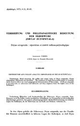
![4 C]-Polyethylenglykol bestimmt, der - HAL - INRIA](https://img.yumpu.com/22454280/1/177x260/4-c-polyethylenglykol-bestimmt-der-hal-inria.jpg?quality=85)