MEG/EEG in clinical practice
MEG/EEG in clinical practice
MEG/EEG in clinical practice

Create successful ePaper yourself
Turn your PDF publications into a flip-book with our unique Google optimized e-Paper software.
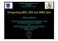
<strong>MEG</strong>/<strong>EEG</strong> Bra<strong>in</strong> Mapp<strong>in</strong>g Course, HBM2005<br />
June 12, 2005<br />
<strong>MEG</strong>/<strong>EEG</strong> <strong>in</strong> cl<strong>in</strong>ical <strong>practice</strong><br />
- State of the art – specific challenges of cl<strong>in</strong>ical studies<br />
- Technical challenge and caveats<br />
- Perspectives on <strong>MEG</strong>/<strong>EEG</strong> bra<strong>in</strong> mapp<strong>in</strong>g<br />
<strong>in</strong> cl<strong>in</strong>ical <strong>practice</strong><br />
Kyousuke Kamada M.D.<br />
Department of Neurosurgery,<br />
The University of Tokyo
Maximal resection of bra<strong>in</strong> tumors
Non-<strong>in</strong>vasive Bra<strong>in</strong> Mapp<strong>in</strong>g<br />
Vessels<br />
fMRI<br />
Bra<strong>in</strong> section<br />
Neurons<br />
<strong>MEG</strong>,<strong>EEG</strong><br />
Axons<br />
Tractography
Somatosensory Evoked Magnetic Fields (SEFs)<br />
Median N. Stim (4 mA, 5 Hz)<br />
M20<br />
M30<br />
M30<br />
M70<br />
<br />
<br />
Right Left<br />
: Central sulcus<br />
Bra<strong>in</strong> tumors
spike analysis by <strong>MEG</strong><br />
Ant.<br />
Post.<br />
Spike 1 Spike 2<br />
Left<br />
Right<br />
Spike 1<br />
Left<br />
<strong>EEG</strong><br />
(+)<br />
<strong>EEG</strong><br />
(-)<br />
Spike 2 Spike 1<br />
Left<br />
Right
Cl<strong>in</strong>ical applications of <strong>MEG</strong>
Investigations of language functions<br />
Neuropsycho. Exam. to evaluate symptoms<br />
Amytal test to identify the dom<strong>in</strong>ant hemisphere<br />
(angiography)<br />
Electrical stimulation, awake surgery Invasive<br />
to exam<strong>in</strong>e one side<br />
1, Preoperative non-<strong>in</strong>vasive functional mapp<strong>in</strong>g<br />
for language functions<br />
Comb<strong>in</strong>ation of <strong>MEG</strong> & fMRI<br />
2, Validation of the results by amytal test & electrical<br />
stimulation with subdural electrodes
<strong>MEG</strong><br />
(Abstract/Concrete discrim<strong>in</strong>ation (read<strong>in</strong>g) task)<br />
“”<br />
“”<br />
<br />
<br />
<br />
<br />
Left Key<br />
<br />
<br />
<br />
<br />
Right Key<br />
1.2s 1.2s<br />
<br />
<br />
Time Limit<br />
3.04.0s<br />
Time Limit<br />
3.04.0s
<strong>MEG</strong><br />
M250<br />
M400<br />
RMS<br />
250
<strong>MEG</strong><br />
M250<br />
M400<br />
RMS<br />
250
<strong>MEG</strong><br />
M250<br />
M400<br />
RMS<br />
250
verb-generation fMRI<br />
Inferior & middle frontal gyrus<br />
left hemisphere<br />
right-handed control subject
Left mesial temporal tumor<br />
<strong>in</strong> a left-handed patient<br />
verb-generation fMRI<br />
Right hemisphere<br />
right<br />
right
ead<strong>in</strong>g-task <strong>MEG</strong><br />
right<br />
No. of dipoles<br />
right left<br />
72 15<br />
<br />
Right hemisphere<br />
(amytal test : right)<br />
radical removal
K.H.<br />
a mesial temporal grade-2 astrocytoma<br />
pre op.<br />
residue<br />
post op.<br />
recurrence<br />
2 years after<br />
irradiation
J.K.<br />
Left mesial temporal tumor<br />
verb-genaration fMRI<br />
right-handed 35-y.o. man<br />
Bra<strong>in</strong> tumor<br />
left<br />
left<br />
left
ead<strong>in</strong>g-task <strong>MEG</strong><br />
No. of dipoles<br />
right left<br />
30 136<br />
<br />
dipole accumulation<br />
<strong>in</strong> the fusiform gyrus<br />
J.K.
LORETA<br />
(Low resolution tomography)<br />
250-450ms<br />
[nAm]<br />
0.20<br />
0.15<br />
0.10<br />
<br />
<br />
L<br />
R<br />
0.05<br />
0.00
pre op.<br />
post op.<br />
J.K.<br />
dyslexia<br />
<br />
<br />
<br />
<br />
<br />
<br />
<br />
<br />
<br />
<br />
<br />
<br />
<br />
N.T.; not tested
serial changes of <strong>MEG</strong><br />
Left fronto-temporal<br />
Pre OP.<br />
10 days after OP.<br />
3 months after OP.<br />
8 months after OP.<br />
Right fronto-temporal<br />
-500<br />
0 600<br />
msec<br />
-500<br />
0 600<br />
msec<br />
Left temporo-occipital<br />
Right temporo-occipital<br />
Case J.K<br />
-500<br />
0 600 -500 0 600<br />
msec
ead<strong>in</strong>g-task <strong>MEG</strong><br />
8 months after resection<br />
No. of dipoles<br />
right left<br />
23 102<br />
J.K.
A.M.<br />
Right <strong>in</strong>sular glioma, 34y.o. male<br />
Right-handed<br />
fMRI<br />
Verb<br />
generation<br />
glioma<br />
Read<strong>in</strong>g
<strong>MEG</strong> (Read<strong>in</strong>g-task)<br />
Right <strong>in</strong>sular glioma, 34y.o. Male<br />
No. of dipoles<br />
left right<br />
30 136<br />
dipole accumulation<br />
<strong>in</strong> the STG and PITG
Task Left <strong>in</strong>jection Right <strong>in</strong>jection<br />
paresis Right hemiparesis Left hemiparesis<br />
pictures &<br />
objects-nam<strong>in</strong>g<br />
60% (3/5) 100% (5/5)<br />
letter-read<strong>in</strong>g 71% (5/7) 14% (1/7)<br />
auditory<br />
comprehension<br />
75% (3/4) 25% (1/4)<br />
repeat<strong>in</strong>g<br />
7 numbers<br />
possible possible<br />
retriev<strong>in</strong>g 5 items<br />
(short memory)<br />
100% (5/5) 100% (5/5)<br />
General<br />
impression<br />
Amytal test<br />
impaired overt nam<strong>in</strong>g<br />
with severe dysarthria,<br />
but little dyslexia<br />
Right <strong>in</strong>sular glioma<br />
34 y.o. male<br />
severe dyslexia<br />
with mild dysarthria
Translocation of Broca's area to the contralateral hemisphere<br />
as the result of the growth of a left <strong>in</strong>ferior frontal glioma.<br />
Holodny AI, et al.<br />
Our case<br />
Read<strong>in</strong>g<br />
Verb generation<br />
glioma
Results of Amytal test, language-<strong>MEG</strong> and –fMRI<br />
<strong>in</strong> 91 cases with bra<strong>in</strong> tumors or epilepsy (controls)
Conclusion 1<br />
<strong>MEG</strong> &fMRIlocalized the receptive- & motor-language<br />
functions <strong>in</strong> STG and FuG, & IFG and MFG,<br />
respectively.<br />
The comb<strong>in</strong>ation of fMRI and <strong>MEG</strong> confidentially enabled<br />
us to identify the dom<strong>in</strong>ant hemisphere of the language<br />
functions.<br />
Serial <strong>MEG</strong> and fMRI <strong>in</strong>vestigations might possibly reflect<br />
the neurological recovery.
Pitfalls of functional mapp<strong>in</strong>g<br />
for cl<strong>in</strong>ical utility
Right f<strong>in</strong>ger tapp<strong>in</strong>g<br />
Cerebral Ischemia<br />
due to Right IC occlusion<br />
fMRI<br />
Left f<strong>in</strong>ger tapp<strong>in</strong>g<br />
49 y.o. male<br />
Right medial N. stim.<br />
<strong>MEG</strong><br />
Left medial N. stim.<br />
Left<br />
SEF<br />
Right<br />
SEF
SPECT with Diamox<br />
Cerebral Ischemia<br />
due to Right IC occlusion<br />
49 y.o. male<br />
Verb-generation fMRI<br />
fMRI raw images
fMRI on<br />
rest SPECT<br />
Verb<br />
generation<br />
Cerebral Ischemia<br />
due to left IC occlusion<br />
43 y.o. male<br />
Right-side<br />
dom<strong>in</strong>ance ?<br />
Right<br />
Left<br />
Right f<strong>in</strong>ger<br />
tapp<strong>in</strong>g<br />
Right-side<br />
activation ?<br />
Right<br />
Left
Conclusion 2<br />
BOLD-based fMRI is vulnerable to pathological bra<strong>in</strong><br />
conditions (ischemia, bra<strong>in</strong> edema).<br />
<strong>MEG</strong> can well reflect bra<strong>in</strong> functions, but there still<br />
rema<strong>in</strong> <strong>in</strong>verse problems.<br />
It is, therefore, important to comb<strong>in</strong>e multi image<br />
modalities with different basics to make functional<br />
bra<strong>in</strong> mapp<strong>in</strong>g more reliable for cl<strong>in</strong>ical purposes.
Validation of<br />
functional imag<strong>in</strong>g
age/sex handedness diagnosis<br />
spike location<br />
Lt; 81 dipoles R) Bil. fronto-temporal<br />
Verb generation fMRI<br />
Read<strong>in</strong>g-task <strong>MEG</strong><br />
Amytal test : Right
Comb<strong>in</strong>ed 3D-image
“<br />
“<br />
”
“”<br />
““
“<br />
”
Conclusion<br />
The validation of functional imag<strong>in</strong>g is <strong>in</strong>dispensable for<br />
further development <strong>in</strong> human bra<strong>in</strong> mapp<strong>in</strong>g.<br />
Cl<strong>in</strong>ical <strong>in</strong>stitutes should understand benefits and pitfalls<br />
of imag<strong>in</strong>g modalities and carefully use results of the<br />
bra<strong>in</strong> mapp<strong>in</strong>g for mak<strong>in</strong>g treatment strategy.
for future studies<br />
White matter mapp<strong>in</strong>g<br />
• Fiber Track<strong>in</strong>g based on<br />
Diffusion Tensor Imag<strong>in</strong>g<br />
D (x i+1 )<br />
x i+2 x i+1 = x i + ∆e 1 (x i )<br />
e 1 (x i+1 )<br />
∆<br />
Corticosp<strong>in</strong>al<br />
tract<br />
Arcuate<br />
fascicles<br />
e 1<br />
(x i<br />
) e 3<br />
(x i<br />
)<br />
e 2<br />
(x i<br />
)<br />
x i<br />
D(x i )<br />
Corpus<br />
callosum<br />
Optic<br />
radiation<br />
e 1 , e 2 , e 3 : eigenvectors of tensor<br />
|e 1 |= |e 2 | =|e 3 |=1