Dimercaptosuccinic Acid (DMSA) - Thorne Research
Dimercaptosuccinic Acid (DMSA) - Thorne Research
Dimercaptosuccinic Acid (DMSA) - Thorne Research
You also want an ePaper? Increase the reach of your titles
YUMPU automatically turns print PDFs into web optimized ePapers that Google loves.
<strong>Dimercaptosuccinic</strong> <strong>Acid</strong> (<strong>DMSA</strong>), A<br />
Non-Toxic, Water-Soluble Treatment For<br />
Heavy Metal Toxicity<br />
by Alan L. Miller, N.D.<br />
Abstract<br />
Heavy metals are, unfortunately, present in the air, water, and food supply. Cases<br />
of severe acute lead, mercury, arsenic, and cadmium poisoning are rare; however,<br />
when they do occur an effective, non-toxic treatment is essential. In addition, chronic,<br />
low-level exposure to lead in the soil and in residues of lead-based paint; to mercury in<br />
the atmosphere, in dental amalgams and in seafood; and to cadmium and arsenic in<br />
the environment and in cigarette smoke is much more common than acute exposure.<br />
Meso-2,3-dimercaptosuccinic acid (<strong>DMSA</strong>) is a sulfhydryl-containing, water-soluble,<br />
non-toxic, orally-administered metal chelator which has been in use as an antidote to<br />
heavy metal toxicity since the 1950s. More recent clinical use and research substantiates<br />
this compound’s efficacy and safety, and establishes it as the premier metal chelation<br />
compound, based on oral dosing, urinary excretion, and its safety characteristics<br />
compared to other chelating substances.<br />
(Altern Med Rev 1998;3(3):199-207)<br />
Introduction<br />
Contamination of water, air, and food by numerous chemicals and non-essential elements,<br />
such as heavy metals, is an unfortunate byproduct of a complex, industrialized, hightech<br />
society. The resultant accumulation of heavy metals in the human body poses a significant<br />
health risk, leading to a wide array of symptomatology, including anemia, learning deficits,<br />
reduced intelligence, behavioral and cognitive changes, tremor, gingivitis, hypertension, irritability,<br />
cancer, depression, memory loss, fatigue, headache, hyperuricemia, gout, chronic renal<br />
failure, male infertility, osteodystrophies, and possibly multiple sclerosis and Alzheimer’s disease.<br />
Although human lead toxicity has decreased in the United States since discontinuation<br />
of the use of lead as a gasoline additive, it continues to be a significant problem, especially in<br />
urban areas, where lead-based paint exposure is still an issue, and in areas where lead is mined<br />
and/or smelted. Chronic mercury toxicity from occupational, environmental, dental amalgam,<br />
and contaminated food exposure, is a significant threat to public health. Other heavy metals,<br />
including cadmium and arsenic, can also be found in the human body due to cigarette smoke,<br />
and occupational and environmental exposure. Diagnostic testing for the presence of heavy<br />
Alan L. Miller, N.D.—Technical Advisor, <strong>Thorne</strong> <strong>Research</strong>, Inc.; Senior Editor, Alternative Medicine Review.<br />
Correspondence address: P.O. Box 25, Dover, ID 83825. alan@thorne.com<br />
Alternative Medicine Review ◆ Volume 3, Number 3 ◆ 1998 Page 199<br />
Copyright©1998 <strong>Thorne</strong> <strong>Research</strong>, Inc. All Rights Reserved. No Reprint Without Written Permission
Figure 1.<br />
HO<br />
C<br />
O<br />
SH<br />
C<br />
H<br />
SH<br />
H<br />
OH<br />
meso - 2,3 - <strong>Dimercaptosuccinic</strong> <strong>Acid</strong> (<strong>DMSA</strong>)<br />
H<br />
SH<br />
C<br />
H<br />
SH<br />
C<br />
H<br />
C<br />
H<br />
H<br />
metals, and subsequently decreasing the<br />
body’s burden of these substances, should be<br />
an integral part of the overall treatment regimen<br />
for individuals with the above-mentioned<br />
symptomatology or a known exposure to these<br />
substances.<br />
It has long been acknowledged that<br />
sulfhydryl-containing compounds have the<br />
ability to chelate metals. The sulfur-containing<br />
amino acids methionine and cysteine,<br />
cysteine’s acetylated analogue N-<br />
acetylcysteine, the methionine metabolite S-<br />
adenosylmethionine, alpha-lipoic acid, and the<br />
tri-peptide glutathione (GSH) all contribute to<br />
the chelation and excretion of metals from the<br />
human body.<br />
Meso-2,3-dimercaptosuccinic acid<br />
(<strong>DMSA</strong>), is a water-soluble, sulfhydrylcontaining<br />
compound which is an effective<br />
oral chelator of heavy metals. Initial studies<br />
C<br />
O<br />
SO 3 H<br />
2,3 Dimercapto - 1 - propanesulfonic <strong>Acid</strong> (DMPS)<br />
H<br />
SH<br />
H<br />
SH<br />
H<br />
C<br />
H<br />
C C C OH<br />
2,3 - Dimercapto - 1 - propanol (Dimercaprol, BAL)<br />
H<br />
over forty years ago identified <strong>DMSA</strong> as an<br />
effective antidote to heavy metal poisoning.<br />
<strong>DMSA</strong> was subsequently studied for twenty<br />
years in the People’s Republic of China, Japan,<br />
and Russia before scientists in Europe and the<br />
United States “discovered” the substance and<br />
its potential usefulness in the mid-1970s. 1<br />
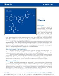
<strong>DMSA</strong> is a dithiol (containing two<br />
sulfhydryl, or S-H, groups) and an analogue<br />
of dimercaprol (BAL, British Anti-Lewisite),<br />
a lipid-soluble compound also used for metal<br />
chelation (see Figure 1). <strong>DMSA</strong>’s water solubility<br />
and oral dosing create a distinct advantage<br />
over BAL, which has a small therapeutic<br />
index and must be administered in an oil solution<br />
via painful, deep intramuscular injection. 2<br />
<strong>DMSA</strong>, on the other hand, has a large therapeutic<br />
window and is the least toxic of the<br />
dithiol compounds. 3<br />
Lead<br />
Lead exposure is still a public health<br />
problem in the United States, being found in<br />
approximately 21 million pre-1940 homes.<br />
Dust and soil lead, derived from flaking,<br />
weathering, and chalking paint, also contribute<br />
to chronic exposure.<br />
Lead competes in the body with calcium,<br />
causing numerous malfunctions in calcium-facilitated<br />
cellular metabolism and calcium<br />
uptake and usage, including inhibition<br />
of neurotransmitter release and blockade of<br />
calcium channels and calcium-sodium ATP<br />
pumps.<br />
The central nervous system (CNS)<br />
appears to be affected the greatest by lead.<br />
Children in particular are susceptible to its<br />
devastating effects on mental development and<br />
intelligence. Neurobehavioral deficits<br />
resembling attention deficit disorder have also<br />
been found in lead-exposed children. 4 Blood<br />
lead concentrations of 20-25 µg/100 ml can<br />
cause irreversible CNS damage in children. 5<br />
Page 200 Alternative Medicine Review ◆ Volume 3, Number 3 ◆ 1998<br />
Copyright©1998 <strong>Thorne</strong> <strong>Research</strong>, Inc. All Rights Reserved. No Reprint Without Written Permission
<strong>DMSA</strong><br />
Poor quality nutrition, including<br />
deficiencies in iron and calcium, are known<br />
to exacerbate the manifestations of lead<br />
exposure, including its CNS effects. Acute<br />
adult lead exposure leads to renal proximal<br />
tubular damage; chronic exposure causes renal<br />
dysfunction characterized by hypertension,<br />
hyperuricemia, gout, and chronic renal failure. 6<br />
Inorganic lead is absorbed, distributed,<br />
and excreted. Once in the blood, lead is distributed<br />
primarily among three compartments<br />
– blood, soft tissue (kidney, bone marrow, liver,<br />
and brain), and mineralizing tissue (bones and<br />
teeth). Mineralizing tissue contains about 95<br />
percent of the total body burden of lead in<br />
adults.<br />
After lead is absorbed in the human<br />
body, it reacts with thiol (sulfhydryl) groups<br />
on peptides and proteins, inhibiting enzymes<br />
involved in heme synthesis and interfering<br />
with normal neurotransmitter functions. 7 This<br />
natural reaction with thiols is also the body’s<br />
method of eliminating lead, especially from<br />
the liver. Hepatic glutathione attaches to lead<br />
and enhances its excretion in the feces. Unfortunately,<br />
hepatic glutathione can be depleted<br />
in this manner, resulting in less glutathione<br />
being available for conjugation of other toxic<br />
substances. In addition to these hepatic effects,<br />
individuals with higher concentrations of<br />
blood lead have been noted to have lower levels<br />
of erythrocyte reduced glutathione. 8 Decreased<br />
erythrocyte glutathione is due to the<br />
fact that 99 percent of lead in the blood is attached<br />
to red blood cells; the remaining one<br />
percent is in the plasma. Lead stored in bone<br />
has a half-life of 25 years, although lead in<br />
bone can be mobilized into the blood, and subsequently<br />
to other tissues.<br />
Mercury<br />
Humans are exposed to mercury primarily<br />
in two forms: mercury vapor and methyl<br />
mercury compounds. Unfortunately, mercury<br />
is a ubiquitous substance in our environment.<br />
Mercury vapor in the atmosphere makes<br />
its way into fresh and salt water by falling in<br />
precipitation. Methyl mercury compounds are<br />
created by bacterial conversion of inorganic<br />
mercury in water and soil, which subsequently<br />
concentrates in seafood and fish. Dietary fish<br />
intake has been found to have a direct correlation<br />
with methyl mercury levels in blood and<br />
hair. 9,10 “Silver” amalgam dental fillings are<br />
the major source of inorganic mercury exposure<br />
in humans. 11 This term, however, is a misnomer,<br />
as this compound is not predominately<br />
silver; the proper term should be “mercury<br />
amalgam.” The most common dental filling<br />
material, amalgams contain approximately 50<br />
percent liquid metallic mercury, 35 percent<br />
silver, 9 percent tin, 6 percent copper, and a<br />
trace of zinc. 12 As they are prepared and placed<br />
in the patient’s mouth, the dentist and the person<br />
preparing the amalgam, 13,14 as well as the<br />
patient are exposed to mercury vapor (HgO).<br />
Clinical studies indicate <strong>DMSA</strong> is a safe and<br />
effective means of decreasing the body burden of<br />
these metals.<br />
Lead<br />
Mercury<br />
Arsenic<br />
Cadmium<br />
The patient is further exposed to mercury vapor<br />
as the amalgam releases HgO when the<br />
individual chews, 15-17 brushes, 18 or drinks hot<br />
beverages. 16-18 Studies of mercury content in<br />
expired air of those with and without amalgams<br />
have found significantly higher baseline<br />
mercury levels in subjects with amalgams, and<br />
Alternative Medicine Review ◆ Volume 3, Number 3 ◆ 1998 Page 201<br />
Copyright©1998 <strong>Thorne</strong> <strong>Research</strong>, Inc. All Rights Reserved. No Reprint Without Written Permission
Laboratories Performing Hair and/or Urine Mercury Analyses<br />
Doctor’s Data 800-323-2784<br />
Great Smokies Diagnostic Lab 800-522-4762<br />
Meridian Valley Clinical Lab 800-234-6825<br />
MetaMetrix Clinical Lab 800-221-4640<br />
up to a 15.6-fold increase in mercury in expired<br />
air after chewing. 15,16 Mercury release<br />
was found to be greater in corroded amalgams<br />
compared to new, polished fillings. 18 Mercury<br />
vapor from amalgams enters the bloodstream<br />
after being inhaled into the lungs.<br />
Comparisons of blood levels of mercury<br />
and the number of amalgams show a direct<br />
correlation between number of amalgam<br />
fillings and concentration of blood 17,19 and<br />
urine mercury. 13,14,20 In addition, a statistically<br />
significant correlation was found between the<br />
number of dental amalgam fillings and mercury<br />
content of the kidney cortex (p
<strong>DMSA</strong><br />
whether this is an age-related phenomenon, or<br />
exactly what the significance of this finding<br />
might be. It is unknown if these pharmacokinetic<br />
parameters are different in other metal<br />
toxicities or concomitant disease processes.<br />
For instance, if the patient has increased gut<br />
permeability, does this increase <strong>DMSA</strong> absorption?<br />
Studies addressing the possibility that<br />
<strong>DMSA</strong> may chelate metal stored in the gut,<br />
because a significant percentage of an oral<br />
dose is not absorbed, have yet to be done. A<br />
study of whole body retention in mice of<br />
radioactively-tagged, orally-administered<br />
mercuric chloride revealed <strong>DMSA</strong> and its<br />
analogue DMPS (2,3-dimercaptopropanesulfonate)<br />
(see Figure 1), given orally<br />
at the same time as the mercury, significantly<br />
decreased the absorption and whole body<br />
retention of the metal. 33 This is an important<br />
finding which suggests administration of<br />
<strong>DMSA</strong> a short time after an acute ingestion of<br />
mercury will chelate the metal and decrease<br />
the amount absorbed. It does not, however,<br />
answer the question of whether <strong>DMSA</strong> will<br />
bind gut reservoirs of mercury or whether it<br />
will assist the liver in elimination of mercury<br />
due to chronic exposure.<br />
<strong>DMSA</strong> Treatment in Lead Toxicity<br />
<strong>DMSA</strong> has been used since the 1950s<br />
as an antidote for lead poisoning in Russia,<br />
Japan, and the Peoples Republic of China.<br />
<strong>DMSA</strong> has been shown in recent studies to be<br />
a safe and effective chelator of lead, reducing<br />
blood levels significantly. 1,34,35 At a dose of 10<br />
mg/kg for five days in adult males, <strong>DMSA</strong><br />
lowered blood lead levels 35.5 percent; a more<br />
aggressive approach utilizing a 30 mg/kg dose<br />
lowered blood lead 72.5 percent. Clinical<br />
symptoms and biochemical indices of lead<br />
toxicity also improved. 35<br />
An animal study indicated <strong>DMSA</strong> is<br />
an effective chelator of lead in soft tissue, but<br />
it may not chelate lead from bone. 36 Another<br />
found <strong>DMSA</strong> or calcium disodium ethylenediamine<br />
tetraacetic acid (CaNa 2<br />
EDTA) produced<br />
significant reductions in kidney, bone<br />
and brain lead levels, but <strong>DMSA</strong> produced<br />
greater reductions of bone lead. 37<br />
In a preliminary animal study,<br />
combination therapy with <strong>DMSA</strong> and<br />
CaNa 2<br />
EDTA was more effective than either<br />
individual chelator at increasing urinary and<br />
fecal elimination of lead, and reducing hepatic,<br />
renal, and femur lead concentrations in rats. 38<br />
It has been suggested that chelating<br />
agents, including BAL and CaNa 2<br />
EDTA, may<br />
mobilize and redistribute lead to soft tissue,<br />
including the brain. Lead-exposed rats given<br />
CaNa 2<br />
EDTA showed an initial decrease in<br />
bone and kidney lead, and an increase in hepatic<br />
and brain lead concentrations, indicating<br />
redistribution to these organs. 39 Rats administered<br />
<strong>DMSA</strong> in combination with<br />
CaNa 2<br />
EDTA showed increased urinary lead<br />
output and decreased tissue burden versus use<br />
of these therapeutic substances individually.<br />
No redistribution of lead to the brain was observed<br />
with the combined therapy. A decrease<br />
in blood zinc level was noted with the combination,<br />
as has been observed with CaNa 2<br />
EDTA<br />
monotherapy. 40<br />
In a study of lead’s pro-oxidant activity<br />
and the effect of thiol substances as antioxidants,<br />
five weeks of lead exposure in mice<br />
depleted hepatic and brain glutathione (GS)<br />
levels, and increased malondialdehyde<br />
(MDA), a marker of lipid peroxidation. <strong>DMSA</strong><br />
administration for seven days resulted in a reduction<br />
in blood, liver, and brain lead levels.<br />
N-acetylcysteine supplementation decreased<br />
MDA levels, indicating amelioration of oxidative<br />
stress by NAC, but it did not decrease<br />
lead levels. 41<br />
In an animal study, co-administration<br />
of ascorbic acid (vitamin C) with <strong>DMSA</strong> enhanced<br />
the urinary excretion of lead in rats<br />
compared to <strong>DMSA</strong> alone. 42<br />
Alternative Medicine Review ◆ Volume 3, Number 3 ◆ 1998 Page 203<br />
Copyright©1998 <strong>Thorne</strong> <strong>Research</strong>, Inc. All Rights Reserved. No Reprint Without Written Permission
A suggested protocol for lead toxicity<br />
is to identify and remove the environmental<br />
exposure, and use <strong>DMSA</strong> 10 mg/kg three times<br />
a day for the first five days, followed by 14<br />
days at 10 mg/kg twice a day.<br />
<strong>DMSA</strong> Treatment in Mercury<br />
Toxicity<br />
<strong>DMSA</strong> has been in recent use as a treatment<br />
for mercury poisoning since Friedheim<br />
reported on <strong>DMSA</strong> treatment of experimental<br />
toxicity in mice in 1975, noting its low toxicity<br />
and favorable efficacy compared to BAL<br />
and D-penicillamine. 43 Since that time, numerous<br />
animal and human studies have shown<br />
<strong>DMSA</strong> administration increases urinary mercury<br />
excretion and reduces blood and tissue<br />
mercury concentration. 3,33,44-47<br />
In a comparison study of chelating<br />
agents, eleven construction workers with acute<br />
mercury poisoning were treated with either<br />
<strong>DMSA</strong> or N-acetyl-D,L-penicillamine (NAP),<br />
another sulfhydryl-containing metal chelator.<br />
<strong>DMSA</strong> treatment resulted in greater urinary<br />
excretion of mercury than NAP. 48<br />
In a study of single-dose, <strong>DMSA</strong>-induced<br />
urinary excretion in occupationally-poisoned<br />
workers, a significant increase in urinary<br />
mercury excretion was noted, especially<br />
in the first 24 hours. Mercury excretion was<br />
greatest in the first eight hours after oral<br />
<strong>DMSA</strong> administration. 49<br />
After methylmercuric chloride administration<br />
in rats, <strong>DMSA</strong>, DMPS, and NAP were<br />
studied for their ability to remove mercury<br />
from blood and tissue. <strong>DMSA</strong> was the most<br />
effective at removing mercury from the blood,<br />
liver, brain, spleen, lungs, large intestine, skeletal<br />
muscle, and bone. DMPS was more effective<br />
at removing mercury from the kidneys.<br />
50<br />
Chelation of Mercury from the<br />
Brain<br />
In rats, following intravenous administration<br />
of methyl mercury, <strong>DMSA</strong> was found<br />
to be the “most efficient chelator for brain mercury.”<br />
51 In another animal study, <strong>DMSA</strong> was<br />
given four days after methyl mercury injection<br />
in mice, and continued for eight days.<br />
<strong>DMSA</strong> removed two-thirds of the brain mercury<br />
deposits, NAP removed approximately<br />
one-half, while DMPS did not remove significant<br />
amounts of mercury from the brain. 44<br />
<strong>DMSA</strong> has also been used effectively<br />
in arsenic and cadmium poisoning. 2,3,52<br />
Mercury Diagnostic and Treatment<br />
Protocol<br />
Hair analysis is an inexpensive and<br />
valuable tool for evaluating prior mercury exposure.<br />
10,53 An effective way to evaluate mercury<br />
toxicity quantitatively is to determine the<br />
amount of mercury excreted in the urine after<br />
a challenge dose of <strong>DMSA</strong>. A baseline 24-hour<br />
urine is collected before the challenge, then<br />
again on day three of a three-day dosing of<br />
200 mg three times a day.<br />
The therapeutic dosage of <strong>DMSA</strong> for<br />
mercury toxicity is not well defined in the literature.<br />
Doses as high as 30 mg/kg per day<br />
have been used, with no serious side effects<br />
noted. 34 One <strong>DMSA</strong> treatment protocol suggests<br />
10 mg/kg day taken in divided doses for<br />
three days. The patient then discontinues taking<br />
<strong>DMSA</strong> for 14 days, then takes it again for<br />
3 days. Five to 10 treatment cycles may be<br />
necessary. 54 Another protocol suggests 500 mg<br />
per day on an empty stomach, every other day<br />
for a minimum of five weeks. For very sensitive<br />
patients, 250 mg per day, every other day<br />
may be necessary, with an increase to 500 mg<br />
after two to three weeks, for a total of five<br />
weeks of therapy. 55 More studies need to be<br />
done to define optimal dosing strategies for<br />
Page 204 Alternative Medicine Review ◆ Volume 3, Number 3 ◆ 1998<br />
Copyright©1998 <strong>Thorne</strong> <strong>Research</strong>, Inc. All Rights Reserved. No Reprint Without Written Permission
<strong>DMSA</strong><br />
this substance. Be aware that sulfhydryl compounds<br />
in <strong>DMSA</strong> will make urine smell very<br />
sulfurous. Adequate communication with the<br />
patient regarding this issue is important, so<br />
they are not taken by surprise.<br />
Adjunctive nutrient therapy includes<br />
hydrolyzed whey protein, as it contains cysteine<br />
and cysteine residues which can be of<br />
benefit while using <strong>DMSA</strong>. Cysteine is the<br />
rate-limiting step in glutathione production,<br />
necessary for fecal heavy metal excretion and<br />
hepatoprotection. Whey also contains<br />
branched-chain amino acids, which will occupy<br />
transport sites at the blood-brain barrier,<br />
effectively keeping bound metals from being<br />
re-deposited in the brain. Supplemental dosing<br />
of N-acetylcysteine, 500 mg three times<br />
per day, can also be helpful. 54 A multi-mineral<br />
supplement can be taken between cycles.<br />
References<br />
1. Aposhian HV. <strong>DMSA</strong> and DMPS – Watersoluble<br />
antidotes for heavy metal poisoning.<br />
Ann Rev Pharmacol Toxicol 1983;23:193-215.<br />
2. Muckter H, Liebl B, Reichl FX, et al. Are we<br />
ready to replace dimercaprol (BAL) as an<br />
arsenic antidote? Hum Exp Toxicol<br />
1997;16:460-465.<br />
3. Graziano JH. Role of 2,3-dimercaptosuccinic<br />
acid in the treatment of heavy metal poisoning.<br />
Med Tox 1986;1:155-162.<br />
4. Winneke G, Kramer U. Neurobehavioral<br />
aspects of lead neurotoxicity in children. Cent<br />
Eur J Public Health 1997;5:65-69.<br />
5. Landrigan PJ, Baker EL. Exposure of children<br />
to heavy metals from smelters: epidemiology<br />
and toxic consequences. Environ Res<br />
1981;25:204-224.<br />
6. Perazella MA. Lead and the kidney:<br />
nephropathy, hypertension, and gout. Conn<br />
Med 1996;60:521-526.<br />
7. Clarkson TW. Metal toxicity in the central<br />
nervous system. Environ Health Perspect<br />
1987;75:59-64.<br />
8. Ignacak J, Brandys J, Danek M, Moniczewski<br />
A. [Selected biochemical indicators of the<br />
effect of environmental lead on humans]. Folia<br />
Med Cracov 1991;32:111-118. [Article in<br />
Polish].<br />
9. Turner MD, Marsh DO, Smith JC, et al.<br />
Methylmercury in populations eating large<br />
quantities of marine fish. Arch Environ Health<br />
1980;35:367-377.<br />
10. Wilhelm M, Muller F, Idel H. Biological<br />
monitoring of mercury vapour exposure by<br />
scalp hair analysis in comparison to blood and<br />
urine. Toxicol Lett 1996;88:221-226.<br />
11. Sandborgh-Englund G, Elinder CG,<br />
Langworth S, et al. Mercury in biological<br />
fluids after amalgam removal. J Dent Res<br />
1998;77:615-624.<br />
12. Lorscheider FL, Murray JV, Summers AO.<br />
Mercury exposure from “silver” tooth fillings:<br />
emerging evidence questions a traditional<br />
dental paradigm. FASEB J 1995;9:504-508.<br />
13. Jokstad A. Mercury excretion and occupational<br />
exposure of dental personnel. Community Dent<br />
Oral Epidemiol 1990;18:143-148.<br />
14. Nilsson B, Nilsson B. Mercury in dental<br />
practice. II. Urinary mercury excretion in<br />
dental personnel. Swed Dent J 1986;10:221-<br />
232.<br />
15. Vimy MJ, Lorscheider FL. Intra-oral air<br />
mercury released from dental amalgam. J Dent<br />
Res 1985;64:1069-1071.<br />
16. Svare C, Peterson L, Reinhardt J, et al. The<br />
effect of dental amalgams on mercury levels in<br />
expired air. J Dent Res 1981;60:1668-1671.<br />
17. Abraham JE, Svare CW, Frank CW. The effect<br />
of dental amalgam restorations on blood<br />
mercury levels. J Dent Res 1984;63:71-73.<br />
18. Derand T. Mercury vapor from dental amalgams,<br />
an in vitro study. Swed Dent J<br />
1989;13:169-175.<br />
19. Snapp KR, Boyer DB, Peterson LC, Svare<br />
CW. The contribution of dental amalgam to<br />
mercury in blood. J Dent Res 1989;68:780-<br />
785.<br />
20. Jokstad A, Thomassen Y, Bye E, et al. Dental<br />
amalgam and mercury. Pharmacol Toxicol<br />
1992;70:308-313.<br />
21. Maas C, Bruck W, Haffner HT, Schweinsberg<br />
F. [Study on the significance of mercury<br />
accumulation in the brain from dental amalgam<br />
fillings through direct mouth-nose-brain<br />
transport]. Zentralbl Hyg Umweltmed<br />
1996;198:275-291. [Article in German]<br />
22. Ekstrand J, Bjorkman L, Edlund C,<br />
Sandborgh-Englund G. Toxicological aspects<br />
on the release and systemic uptake of mercury<br />
from dental amalgam. Eur J Oral Sci<br />
1998;106:678-686.<br />
Alternative Medicine Review ◆ Volume 3, Number 3 ◆ 1998 Page 205<br />
Copyright©1998 <strong>Thorne</strong> <strong>Research</strong>, Inc. All Rights Reserved. No Reprint Without Written Permission
23. Bjorkman L, Sandborgh-Englund G, Ekstrand<br />
J. Mercury in saliva and feces after removal of<br />
amalgam fillings. Toxicol Appl Pharmacol<br />
1997;144:156-162.<br />
24. Ballatori N, Clarkson TW. Biliary secretion of<br />
glutathione-metal complexes. Fundam Appl<br />
Toxicol 1985;5:816-831.<br />
25. Ballatori N, Clarkson TW. Dependence of<br />
biliary secretion of inorganic mercury on the<br />
biliary transport of glutathione. Biochem<br />
Pharmacol 1984;33:1093-1098.<br />
26. Gregus Z, Varga F. Role of glutathione and<br />
hepatic glutathione S-transferase in the biliary<br />
excretion of methyl mercury, cadmium and<br />
zinc: a study with enzyme inducers and<br />
glutathione depletors. Acta Pharmacol Toxicol<br />
1985;56:398-403.<br />
27. Gregus Z, Klaassen CD. Disposition of metals<br />
in rats: a comparative study of fecal, urinary,<br />
and biliary excretion and tissue distribution of<br />
eighteen metals. Toxicol Appl Pharmacol<br />
1986;85:24-38.<br />
28. Ishihara N, Matsushiro T. Biliary and urinary<br />
excretion of metals in humans. Arch Environ<br />
Health 1986;41:324-330.<br />
29. Aposhian HV, Maiorino RM, Rivera M, et al.<br />
Human studies with the chelating agents,<br />
DMPS and <strong>DMSA</strong>. J Toxicol Clin Toxicol<br />
1992;30:505-528.<br />
30. Maiorino RM, Bruce DC, Aposhian HV.<br />
Determination and metabolism of dithiol<br />
chelating agents. VI. Isolation and identification<br />
of the mixed disulfides of meso-2,3-<br />
dimercaptosuccinic acid with L-cysteine in<br />
human urine. Toxicol Appl Pharmacol<br />
1989;97:338-349.<br />
31. Maiorino RM, Akins JM, Blaha K, et al.<br />
Determination and metabolism of dithiol<br />
chelating agents: X. In humans, meso-2,3-<br />
dimercaptosuccinic acid is bound to plasma<br />
proteins via mixed disulfide formation. J<br />
Pharmacol Exp Ther 1990;254:570-577.<br />
32. Aposhian HV, Maiorino RM, Dart RC, Perry<br />
DF. Urinary excretion of meso-2,3-<br />
dimercaptosuccinic acid in human subjects.<br />
Clin Pharmacol Ther 1989;45:520-526.<br />
33. Nielson JB, Andersen O. Effect of four thiolcontaining<br />
chelators on the disposition of<br />
orally administered mercuric chloride. Hum<br />
Exp Toxicol 1991;10:423-430.<br />
34. Fournier L, Thomas G, Garnier R, et al. 2,3-<br />
<strong>Dimercaptosuccinic</strong> acid treatment of heavy<br />
metal poisoning in humans. Med Toxicol<br />
Adverse Drug Exp 1988;3:499-504.<br />
35. Graziano JH, Siris ES, Lolacono N, et al. 2,3-<br />
dimercaptosuccinic acid as an antidote for lead<br />
intoxication. Clin Pharmacol Ther<br />
1985;37:432-438.<br />
36. Cory-Slechta DA. Mobilization of lead over<br />
the course of <strong>DMSA</strong> chelation therapy and<br />
long-term efficacy. J Pharmacol Exp Ther<br />
1988;246:84-91.<br />
37. Jones MM, Basinger MA, Gale GR, et al.<br />
Effect of chelate treatments on kidney, bone<br />
and brain lead levels of lead-intoxicated mice.<br />
Toxicology 1994;89:91-100.<br />
38. Tandon SK, Floras SJS. Therapeutic efficacy<br />
of dimercaptosuccinic acid and thiamine/<br />
ascorbic acid on lead intoxication in rats. Bull<br />
Environ Contam Toxicol 1989;43:705-712.<br />
39. Cory-Slechta DA, Weiss B, Cox C. Mobilization<br />
and redistribution of lead over the course<br />
of calcium disodium ethylenediamine<br />
tetraacetate chelation therapy. J Pharmacol<br />
Exp Ther 1987;243:804-813.<br />
40. Flora SJ, Bhattacharya R, Vijayaraghavan R.<br />
Combined therapeutic potential of meso-2,3-<br />
dimercaptosuccinic acid and calcium disodium<br />
edetate on the mobilization of lead in experimental<br />
lead intoxication in rats. Fundam Appl<br />
Toxicol 1995;25:233-240.<br />
41. Ercal N, Treeratphan P, Hammond TC, et al. In<br />
vivo indices of oxidative stress in leadexposed<br />
C57BL/6 mice are reduced by<br />
treatment with meso-2,3-dimercaptosuccinic<br />
acid or N-acetylcysteine. Free Radic Biol Med<br />
1996;21:157-161.<br />
42. Tandon SK, Singh S, Jain VK. Efficacy of<br />
combined chelation in lead intoxication. Chem<br />
Res Toxicol 1994;7:585-589.<br />
43. Friedheim E, Corvi C. Mesodimercaptosuccinic<br />
acid, a chelating agent for<br />
the treatment of mercury poisoning. J Pharm<br />
Pharmacol 1975;27:624-626.<br />
44. Aaseth J, Friedheim EA. Treatment of methyl<br />
mercury poisoning in mice with 2,3-<br />
dimercaptosuccinic acid and other complexing<br />
thiols. Acta Pharmacol Toxicol 1978;42:248-<br />
252.<br />
Page 206 Alternative Medicine Review ◆ Volume 3, Number 3 ◆ 1998<br />
Copyright©1998 <strong>Thorne</strong> <strong>Research</strong>, Inc. All Rights Reserved. No Reprint Without Written Permission
<strong>DMSA</strong><br />
45. Aaseth J, Alexander J, Raknerud N. Treatment<br />
of mercuric chloride poisoning with<br />
dimercaptosuccinic acid and diuretics:<br />
preliminary studies. J Toxicol Clin Toxicol<br />
1982;19:173-186.<br />
46. Grandjean P, Guldager B, Larsen IB, et al.<br />
Placebo response in environmental disease.<br />
Chelation therapy of patients with symptoms<br />
attributed to amalgam fillings. J Occup<br />
Environ Med 1997;39:707-714.<br />
47. Ramsey DT, Casteel SW, Faggella AM, et al.<br />
Use of orally administered succimer (meso-<br />
2,3-dimercaptosuccinic acid) for treatment of<br />
lead poisoning in dogs. J Am Vet Med Assoc<br />
1996;208:371-375.<br />
48. Bluhm RE, Bobbitt RG, Welch LG, et al.<br />
Elemental mercury vapour toxicity, treatment,<br />
and prognosis after acute, intensive exposure<br />
in chloralkali workers. Part I: History,<br />
neurophysical findings and chelator effects.<br />
Hum Exp Toxicol 1992;11:201-210.<br />
49. Roels HA, Boeckx M, Ceulemans E, Lauwerys<br />
RR. Urinary excretion of mercury after<br />
occupational exposure to mercury vapour and<br />
influence of the chelating agent meso-2,3-<br />
dimercaptosuccinic acid (<strong>DMSA</strong>). Br J Ind<br />
Med 1991;48:247-253.<br />
50. Planas-Bohne F. The influence of chelating<br />
agents on the distribution and biotransformation<br />
of methylmercuric chloride in rats. J<br />
Pharmacol Exp Ther 1981;217:500-504.<br />
51. Butterworth RF, Gonce M, Barbeau A.<br />
Accumulation and removal of Hg203 in<br />
different regions of the rat brain. Can J Neurol<br />
Sci 1978;5:397-400.<br />
52. Lenz K, Hruby K, Druml W, et al. 2,3-<br />
<strong>Dimercaptosuccinic</strong> acid in human arsenic<br />
poisoning. Arch Toxicol 1981;47:241-243.<br />
53. Schweinsberg F. Risk estimation of mercury<br />
intake from different sources. Toxicol Lett<br />
1994;72:345-351.<br />
54. Quig, David, PhD. Doctor’s Data, Chicago, IL.<br />
Personal communication. May 1998.<br />
55. Frackelton JP, Christensen RL. Mercury<br />
poisoning and its potential impact on hormone<br />
regulation and aging: preliminary clinical<br />
observations using a new therapeutic approach.<br />
J Adv Med 1998;11:9-25.<br />
Alternative Medicine Review ◆ Volume 3, Number 3 ◆ 1998 Page 207<br />
Copyright©1998 <strong>Thorne</strong> <strong>Research</strong>, Inc. All Rights Reserved. No Reprint Without Written Permission


