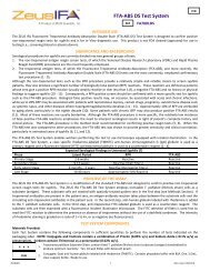
nDNA Test System - ZEUS Scientific
nDNA Test System - ZEUS Scientific
nDNA Test System - ZEUS Scientific

Create successful ePaper yourself
Turn your PDF publications into a flip-book with our unique Google optimized e-Paper software.
TM<br />
<strong>nDNA</strong> <strong>Test</strong> <strong>System</strong><br />
REF<br />
FA1001<br />
IVD<br />
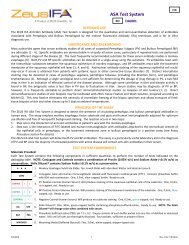
INTENDED USE<br />
The <strong>ZEUS</strong> IFA <strong>nDNA</strong> <strong>Test</strong> <strong>System</strong> is a pre-standardized assay designed for the qualitative and semi-quantitative detection of<br />
antibodies to native DNA by the Indirect Fluorescent Antibody (IFA) technique and is for In Vitro diagnostic use.<br />
SIGNIFICANCE AND BACKGROUND<br />
Anti-Native Deoxyribonucleic Acid (<strong>nDNA</strong>) antibodies are frequently found in sera from patients with active spontaneous systemic<br />
lupus erythematosus (SLE) and some drug-induced lupus syndromes (1 - 9). The presence of <strong>nDNA</strong> antibodies is indicative of active<br />
SLE and correlates closely with the onset of lupus nephritis (5, 10 - 13). The specificity of <strong>nDNA</strong> antibodies for SLE is much greater<br />
than antinuclear antibodies (5, 12). Therefore, detection of <strong>nDNA</strong> antibodies provides valuable diagnostic, as well as prognostic<br />
information for the differential diagnosis of SLE (5, 10 - 13). The presence of <strong>nDNA</strong> antibodies in known SLE sera is considered an<br />
indication of recurrent active disease or poor response to therapy (5, 13). Consequently, periodic monitoring of <strong>nDNA</strong> antibodies in<br />
SLE patients aids in evaluating the clinical course of the disease and its response to therapy (5, 10 - 13). DNA antibodies were<br />
discovered in sera of patients with SLE more than 15 years ago (1 - 4). Since then, DNA antibodies have been studied by a number of<br />
techniques, including gel diffusion (1, 14 - 15), complement fixation (2, 14, and 16), agglutination (17, 18), DNA spot tests (13, 19),<br />
radioimmuno-electrophoresis (20), counter-immunoelectrophoresis (21, 22), and ammonium sulfate precipitation (10, 23, and 24).<br />
Considerable effort has been made to determine the specificity of DNA antibodies. It is now apparent that antibodies have been<br />
found which react with either <strong>nDNA</strong> or denatured single stranded (sDNA) or both (8, 12, 14, and 20). <strong>nDNA</strong> antibodies are thought<br />
to correlate with the clinical activity of the disease (2, 5, 10, and 25). In addition, antibodies to DNA have been eluted from the<br />
kidneys of patients with SLE and one report demonstrated the presence of DNA-Anti-DNA complexes in sera from patients with<br />
active SLE (26). However, these antibodies have been found in patients with and without active lupus nepharitis (27, 28).<br />
The <strong>ZEUS</strong> IFA <strong>nDNA</strong> <strong>Test</strong> <strong>System</strong> is based on the use of the Crithidia luciliae kinetoplast substrate first described by Aarden, et al (29).<br />
Recent reports from a number of investigators have shown this method to be a useful laboratory test to detect <strong>nDNA</strong> antibodies in<br />
patients with systemic lupus erythematosus (30 - 33). These studies also indicate that the IFA <strong>nDNA</strong> <strong>Test</strong> <strong>System</strong> is comparable to<br />
the radioimmunoassay method for detecting <strong>nDNA</strong> antibodies.<br />
PRINCIPLE OF THE ASSAY<br />
The <strong>ZEUS</strong> IFA <strong>nDNA</strong> <strong>Test</strong> <strong>System</strong> is a pre-standardized indirect fluorescent antibody assay for the qualitative and semi-quantitative<br />
determination of <strong>nDNA</strong> antibodies in patient and control sera. The reaction occurs in two steps:<br />
1. Step one; If <strong>nDNA</strong> antibodies are present, a reaction between <strong>nDNA</strong> antibodies and the kinetoplast of the C. luciliae substrate<br />
takes place in the first step.<br />
2. Step two; goat anti-human immunoglobulin labeled with fluorescein isothiocyanate (FITC) is added to the substrate. If the<br />
patient’s sera contains <strong>nDNA</strong> antibody, a positive apple-green fluorescent antigen-antibody reaction will be observed when the<br />
Slides are examined with the fluorescence microscope. A positive reaction is recognized as an intense staining reaction in the<br />
small kinetoplasts of the C. luciliae.<br />
TEST SYSTEM COMPONENTS<br />
Materials Provided:<br />
Each <strong>Test</strong> <strong>System</strong> contains the following components in sufficient quantities to perform the number of tests indicated on the<br />
packaging label. NOTE: Conjugate and Controls contain a combination of Proclin (0.05% v/v) and Sodium Azide (
NOTES:<br />
1. The following components are not <strong>Test</strong> <strong>System</strong> Lot Number dependent and may be used interchangeably with the <strong>ZEUS</strong><br />
IFA <strong>Test</strong> <strong>System</strong>s, as long as the product numbers are identical: SAVe Diluent ® (Product #: FA005CC), Mounting Media<br />
(Product #: FA0009S), and PBS (Product #: 0008S).<br />
2. <strong>Test</strong> <strong>System</strong> also contains:<br />
a. Component Label containing lot specific information inside the <strong>Test</strong> <strong>System</strong> box.<br />
b. A CD containing all <strong>ZEUS</strong> IFA Product Inserts, providing instructions for use.<br />
PRECAUTIONS<br />
1. For In Vitro diagnostic use.<br />
2. Follow normal precautions exercised in handling laboratory reagents. In case of contact with eyes, rinse immediately with<br />
plenty of water and seek medical advice. Wear suitable protective clothing, gloves, and eye/face protection. Do not breathe<br />
vapor. Dispose of waste observing all local, state, and federal laws.<br />
3. The wells of the Slide do not contain viable organisms. However, consider the Slide potentially bio-hazardous materials and<br />
handle accordingly.<br />
4. The Controls are potentially bio-hazardous materials. Source materials from which these products were derived were found<br />
negative for HIV-1 antigen, HBsAg and for antibodies against HCV and HIV by approved test methods. However, since no test<br />
method can offer complete assurance that infectious agents are absent, these products should be handled at the Bio-safety<br />
Level 2 as recommended for any potentially infectious human serum or blood specimen in the Centers for Disease<br />
Control/National Institutes of Health manual “Biosafety in Microbiological and Biomedical Laboratories”: current edition; and<br />
OSHA’s Standard for Bloodborne Pathogens (20).<br />
5. Adherence to the specified time and temperature of incubations is essential for accurate results. All reagents must be allowed<br />
to reach room temperature (20 - 25°C) before starting the assay. Return unused reagents to their original containers<br />
immediately and follow storage requirements.<br />
6. Improper washing could cause false positive or false negative results. Be sure to minimize the amount of any residual PBS, by<br />
blotting, before adding Conjugate. Do not allow the wells to dry out between incubations.<br />
7. The SAVe Diluent ® , Conjugate, and Controls contain Sodium Azide at a concentration of
5. Staining dish: A large staining dish with a small magnetic mixing set-up provides an ideal mechanism for washing Slides between<br />
incubation steps.<br />
6. Cover slips, 24 x 60mm, thickness No. 1.<br />
7. Distilled or deionized water.<br />
8. Properly equipped fluorescence microscope.<br />
9. 1 Liter Graduated Cylinder.<br />
10. Laboratory timer to monitor incubation steps.<br />
11. Disposal basin and disinfectant (i.e.: 10% household bleach – 0.5% Sodium Hypochlorite).<br />
The following filter systems, or their equivalent, have been found to be satisfactory for routine use with transmitted or incident light<br />
darkfield assemblies:<br />
Transmitted Light<br />
Light Source: Mercury Vapor 200W or 50W<br />
Excitation Filter Barrier Filter Red Suppression Filter<br />
KP490 K510 or K530 BG38<br />
BG12 K510 or K530 BG38<br />
FITC K520 BG38<br />
Light Source: Tungsten – Halogen 100W<br />
KP490 K510 or K530 BG38<br />
Incident Light<br />
Light Source: Mercury Vapor 200, 100, 50 W<br />
Excitation Filter Dichroic Mirror Barrier Filter Red Suppression Filter<br />
KP500 TK510 K510 or K530 BG38<br />
FITC TK510 K530 BG38<br />
Light Source: Tungsten – Halogen 50 and 100 W<br />
KP500 TK510 K510 or K530 BG38<br />
FITC TK510 K530 BG38<br />
SPECIMEN COLLECTION<br />
1. <strong>ZEUS</strong> <strong>Scientific</strong> recommends that the user carry out specimen collection in accordance with CLSI document M29: Protection of<br />
Laboratory Workers from Occupationally Acquired Infectious Diseases. No known test method can offer complete assurance<br />
that human blood samples will not transmit infection. Therefore, all blood derivatives should be considered potentially<br />
infectious.<br />
2. Only freshly drawn and properly refrigerated sera obtained by approved aseptic venipuncture procedures with this assay (34,<br />
35). No anticoagulants or preservatives should be added. Avoid using hemolyzed, lipemic, or bacterially contaminated sera.<br />
3. Store sample at room temperature for no longer than 8 hours. If testing is not performed within 8 hours, sera may be stored<br />
between 2 - 8°C, for no longer than 48 hours. If delay in testing is anticipated, store test sera at –20°C or lower. Avoid multiple<br />
freeze/thaw cycles which may cause loss of antibody activity and give erroneous results.<br />
STORAGE CONDITIONS<br />
Unopened <strong>Test</strong> <strong>System</strong>.<br />
Mounting Media, Conjugate, SAVe Diluent ® , Slides, Positive and Negative Controls.<br />
Rehydrated PBS (Stable for 30 days).<br />
Phosphate-buffered-saline (PBS) Packets.<br />
ASSAY PROCEDURE<br />
1. Remove Slides from refrigerated storage and allow them to warm to room temperature (20 - 25°C). Tear open the protective<br />
envelope and remove Slides. Do not apply pressure to flat sides of protective envelope.<br />
2. Identify each well with the appropriate patient sera and Controls. NOTE: The Controls are intended to be used undiluted.<br />
Prepare a 1:10 dilution (e.g.: 10µL of serum + 90µL of SAVe Diluent ® or PBS) of each patient serum. The SAVe Diluent ® will<br />
undergo a color change confirming that the specimen has been combined with the Diluent.<br />
Dilution Options:<br />
a. Users may titrate the Positive Control to endpoint to serve as a semi-quantitative (1+ Minimally Reactive) Control. In such<br />
cases, the Control should be diluted two-fold in SAVe Diluent ® or PBS. When evaluated by <strong>ZEUS</strong> <strong>Scientific</strong>, an endpoint<br />
dilution is established and printed on the Positive Control vial (± one dilution). It should be noted that due to variations<br />
within the laboratory (equipment, etc.), each laboratory should establish its own expected end-point titer for each lot of<br />
Positive Control.<br />
b. When titrating patient specimens, initial, and all subsequent dilutions should be prepared in SAVe Diluent ® or PBS only.<br />
R2080EN 3 (Rev. Date 7/3/2013)
3. With suitable dispenser (listed above), dispense 20µL of each Control and each diluted patient sera in the appropriate wells.<br />
4. Incubate Slides at room temperature (20 - 25°C) for 30 minutes.<br />
5. Gently rinse Slides with PBS. Do not direct a stream of PBS into the test wells.<br />
6. Wash Slides for two, 5 minute intervals, changing PBS between washes.<br />
7. Remove Slides from PBS one at a time. Invert Slide and key wells to holes in blotters provided. Blot Slide by wiping the reverse<br />
side with an absorbent wipe. CAUTION: Position the blotter and Slide on a hard, flat surface. Blotting on paper towels may<br />
destroy the Slide matrix. Do not allow the Slides to dry during the test procedure.<br />
8. Add 20µL of Conjugate to each well.<br />
9. Repeat steps 4 through 7.<br />
10. Apply 3 - 5 drops of Mounting Media to each Slide (between the wells) and coverslip. Examine Slides immediately with an<br />
appropriate fluorescence microscope.<br />
NOTE: If delay in examining Slides is anticipated, seal coverslip with clear nail polish and store in refrigerator. It is recommended<br />
that Slides be examined on the same day as testing.<br />
QUALITY CONTROL<br />
1. Every time the assay is run, a Positive Control, a Negative Control and a Buffer Control must be included.<br />
2. It is recommended that one read the Positive and Negative Controls before evaluating test results. This will assist in establishing<br />
the references required to interpret the test sample. If Controls do not appear as described, results are invalid.<br />
a. Negative Control - characterized by the absence of fluorescent staining of the kinetoplast. Staining of the nucleus only<br />
and/or staining of the basal body should be interpreted as a negative test.<br />
b. Positive Control - characterized by any apple-green fluorescent staining of the kinetoplast. Staining of the basal body in<br />
conjunction with the kinetoplast should be considered a positive result.<br />
3. Additional Controls may be tested according to guidelines or requirements of local, state, and/or federal regulations or<br />
accrediting organizations.<br />
NOTES:<br />
a. The intensity of the observed fluorescence may vary with the microscope and filter system used.<br />
b. The kinetoplast is generally located closer to the basal body than the nucleus; however, because of the fluid nature of<br />
the endoplasm, the location of the kinetoplast may vary from cell to cell (36).<br />
c. Read only single, well-defined organisms in each field. Not all organisms will appear optimal; morphology may vary<br />
between organisms because of fixation, their stages of growth, and/or their orientation on the Slide as they dried (36).<br />
INTERPRETATION OF RESULTS<br />
1. Titers less than 1:10 are considered negative.<br />
2. Positive <strong>Test</strong>: Any observed apple-green staining of the small kinetoplast of the C. luciliae substrate organism, at a 1:10 dilution<br />
based on a 1+ to 4+ scale. 1+ is considered a weak reaction, and 4+ a strong reaction. All sera positive at 1:10 should be titered<br />
to endpoint dilution. This is accomplished by making a 1:10, 1:20, 1:40, etc., serial dilution of all positives. The endpoint is the<br />
highest dilution that produces a positive reaction.<br />
3. Staining of both the small kinetoplast and the adjacent larger C. luciliae nucleus simultaneously should be interpreted as a<br />
positive test.<br />
4. Polar staining at the base of the flagella is not significant.<br />
5. Staining of the nucleus only should not be interpreted as a positive test.<br />
NOTE: A prozone phenomenon may occur at low patient sera dilutions. It is suggested that retesting of the patient specimen be<br />
performed at a higher dilution (i.e., 1:40 or 1:80) when this phenomenon is suspected.<br />
LIMITATIONS OF THE ASSAY<br />
1. The <strong>ZEUS</strong> IFA <strong>nDNA</strong> <strong>Test</strong> <strong>System</strong> is a diagnostic aid. It is therefore imperative that the <strong>nDNA</strong> antibody results be interpreted in<br />
light of the patient’s clinical condition by a medical authority.<br />
2. SLE patients undergoing steroid therapy may have negative test results (5, 8, and 9).<br />
3. Some drugs, particularly hydralazine, may induce <strong>nDNA</strong> antibody production (5, 6, and 8).<br />
EXPECTED RESULTS<br />
Expected values in a normal population are negative at a 1:10 starting dilution. However, certain drugs may induce a positive <strong>nDNA</strong><br />
antibody test (5, 6).<br />
PERFORMANCE CHARACTERISTICS<br />
The <strong>ZEUS</strong> IFA <strong>nDNA</strong> <strong>Test</strong> <strong>System</strong> was evaluated in parallel with a reference ANA test procedure employing rat liver as the substrate.<br />
Fifty-two (52) ANA positive sera obtained from patients with various diagnoses, including SLE, were retested with the IFA <strong>nDNA</strong> <strong>Test</strong><br />
<strong>System</strong>. The following table summarizes the comparative ANA and <strong>nDNA</strong> results.<br />
R2080EN 4 (Rev. Date 7/3/2013)
Summary of <strong>ZEUS</strong> IFA <strong>nDNA</strong> <strong>Test</strong> <strong>System</strong> Comparative Study: ANA vs <strong>nDNA</strong> in Various Diseases<br />
Number of Patients Diagnosis Number of ANA Positives Number of <strong>nDNA</strong> Positives<br />
11 <strong>System</strong>ic lupus 11 9<br />
8 Hypertension or uremia 8 0<br />
3 Scleroderma 3 0<br />
4 Rheumatoid Arthritis 4 0<br />
1 Sjögren’s Syndrome 1 0<br />
5 Open Heart Surgery 5 0<br />
20 Other or No diagnosis 20 0<br />
52* 52* 9*<br />
*The data shown in the above table reveals the relative specificity of the <strong>nDNA</strong> test for systemic lupus erythematosus. Of the 11<br />
patients with a diagnosis of SLE, 9 were proven to be in acute stage lupus nephritis as determined by renal biopsy evaluation. The<br />
two SLE patients with negative <strong>nDNA</strong> antibody tests were absent of active kidney disease. None of the remaining patients with<br />
other diseases contained <strong>nDNA</strong> antibodies in their sera although all 41 had positive ANA titers ranging from 1:40 to 1:40,000.<br />
Specificity: The <strong>ZEUS</strong> IFA <strong>nDNA</strong> <strong>Test</strong> <strong>System</strong> is capable of detecting IgG, IgA, and IgM antibody classes based on gel diffusion and<br />
immunoelectrophoretic analysis of the FITC labeled anti-human immunoglobulin conjugate.<br />
REFERENCES<br />
1. Robbins WC, Holman HR, Deicher HRG, et al: Proc. Soc. Exp. Biol. Med. 96:575, 1957.<br />
2. Seligman M: Cr. Acad. Sci. (Paris), 245:243, 1957.<br />
3. Ceppelini R, Polli E, Celada F: Proc. Soc. Exp. Biol. Med. 96:572, 1957.<br />
4. Deicher HRG, Holman HR, Kunkel HG: J. Exp. Med. 109:97, 1959.<br />
5. Dubois EL: J. Rheumatol. 2:204, 1975.<br />
6. Epstein WV: J. Rheumatol. 2:215, 1975.<br />
7. Blomgren SE: Seminars Heamtol. 10:345, 1973.<br />
8. Alarcon-Segovia D, Fishbein E: J. Rheumatol. 2:167, 1975.<br />
9. Klajman A, Farkas R, Gold E, Ben-Efraim S: Clin. Immunol. Immunopath. 3:525, 1975.<br />
10. Pincus T, Schur PH, Rose JA: N. Engl. J. Med. 281:701, 1969.<br />
11. Koffler D, Carr RI, Agnello V, et al : Science 166:1648, 1969.<br />
12. Gershwin ME, Steinberg AD: Arthritis. Rheum. 17:947, 1974.<br />
13. Casals SP, Friou GJ, Myers LI: Arthritis. Rheum. 7:379, 1964.<br />
14. Arana R, Seligmann M: J. Clin. Invest. 46:1867, 1967.<br />
15. Tan EM, Schur PH, Carr RI, et al: J. Clin. Invest. 45:1732, 1966.<br />
16. Stollar D, Levine L, Leher HI, et al: Proc. Nat’l. Acad. Sci. USA 48:874, 1962.<br />
17. Bosicevich J, Nasou JP, Kayhoe DE: Proc. Soc. Exp. Biol. Med. 103:636, 1960.<br />
18. Jokinen EJ, Julkunen H: Ann. Rheum.Dis. 24:477, 1965.<br />
19. Matrer R, Helgeland SM, Tonder O: J. Immunol. Methods 5:345, 1974.<br />
20. Bickel YB, Barnett EV, Pearson CM: Clin. Exp. Immunol. 3:641, 1968.<br />
21. Davis JS, IV: Arthritis. Rheum. 14:377, 1971.<br />
22. Johnson GD, Edmonds JP, Holbrow EJ: Lancet 2:883, 1973.<br />
23. Wold RT, Young FE, Tan EM, et al: Science 161:806, 1968.<br />
24. Farr Rd: J. Infect. Dis. 103:239, 1958.<br />
25. Koffler D, Schur PH, Kunkel HG: J. Exp. Med. 126:607, 1967.<br />
26. Harbeck RJ, Bardana EJ, Kohler PF, et al: J. Clin. Invest. 52:789, 1973.<br />
27. Cohen SA, Hughes GRV, Noel GL, et al: Clin. Exp. Immunol. 8:551, 1971.<br />
28. Natali PG, Tan EM: J. Clin. Invest. 51:345, 1972.<br />
29. Aarden LA, deGroot ER, Feltkemp EW: Ann. NY Acad. Sci. 254:505, 1975.<br />
30. Slater NGP, Cameron JS, and Lessof MH: Clin. Exp. Immunol. 25:480, 1976.<br />
31. Stingl G, Meingassner JG, Swelty P, et al: Clin. Immunol. Immunopath. 6:131, 1976.<br />
32. Davis P, Christian B, and Russel AS: J. Rheumatol. 4:15, 1977.<br />
33. Tourville DR, and Benn V: Microbiological Proceeding, 1977.<br />
34. Procedures for the collection of diagnostic blood specimens by venipuncture. Second Edition: Approved Standard (1984). Published by National Committee for<br />
Clinical Laboratory Standards.<br />
35. Procedures for the Handling and Processing of Blood Specimens. NCCLS Document H18-A, Vol. 10, No. 12, Approved Guideline, 1990.<br />
36. U.S. Department of Labor, Occupational Safety and Health Administration: Occupational Exposure to Bloodborne Pathogens, Final Rule. Fed. Register 56:64175-<br />
64182, 1991.<br />
<strong>ZEUS</strong> <strong>Scientific</strong>, Inc.<br />
200 Evans Way, Branchburg, New Jersey, 08876, USA<br />
Toll Free (U.S.): 1-800-286-2111<br />
International: +1 908-526-3744<br />
Fax: +1 908-526-2058<br />
<strong>ZEUS</strong> IFA and SAVe Diluent ® are trademarks of <strong>ZEUS</strong> <strong>Scientific</strong>, Inc.<br />
Customer Service E-mail: orders@zeusscientific.com<br />
Technical Support E-mail: support@zeusscientific.com<br />
Website: www.zeusscientific.com<br />
© 2013 <strong>ZEUS</strong> <strong>Scientific</strong>, Inc. All Rights Reserved.<br />
R2080EN 5 (Rev. Date 7/3/2013)