AXIOM Innovations - Siemens Healthcare
AXIOM Innovations - Siemens Healthcare
AXIOM Innovations - Siemens Healthcare

Create successful ePaper yourself
Turn your PDF publications into a flip-book with our unique Google optimized e-Paper software.
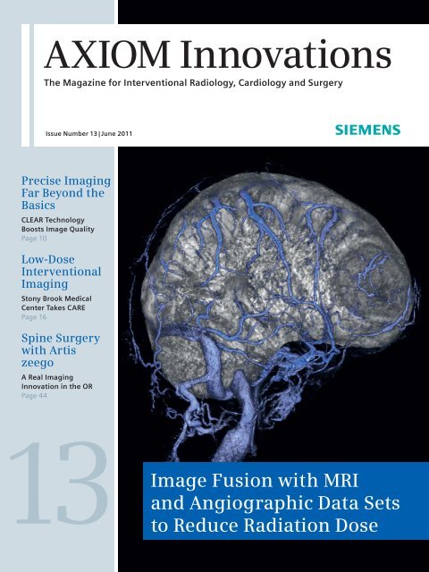
<strong>AXIOM</strong> <strong>Innovations</strong><br />
The Magazine for Interventional Radiology, Cardiology and Surgery<br />
Issue Number 13 | June 2011<br />
Precise Imaging<br />
Far Beyond the<br />
Basics<br />
CLEAR Technology<br />
Boosts Image Quality<br />
Page 10<br />
Low-Dose<br />
Interventional<br />
Imaging<br />
Stony Brook Medical<br />
Center Takes CARE<br />
Page 16<br />
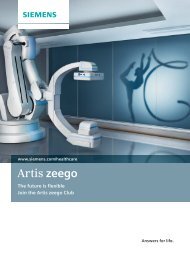
Spine Surgery<br />
with Artis<br />
zeego<br />
A Real Imaging<br />
Innovation in the OR<br />
Page 44<br />
3<br />
to<br />
Image Fusion with MRI<br />
and Angiographic Data Sets<br />
Reduce Radiation Dose
Editorial<br />
“Pioneering into new<br />
fields and exploring<br />
new applications for<br />
interventional imaging<br />
is the driving force<br />
for our innovations –<br />
for our customers,<br />
their patients and<br />
advancements in<br />
medicine.”<br />
Dr. Heinrich Kolem,<br />
CEO of the Angiography & Interventional X-Ray Business Unit (AX)<br />
at <strong>Siemens</strong> <strong>Healthcare</strong><br />
2 <strong>AXIOM</strong> <strong>Innovations</strong> · June 2011 · www.siemens.com/healthcare-magazine
Editorial<br />
Dear Reader,<br />
Dr. Heinrich Kolem<br />
CEO AX Division<br />
One of our customers once said: “What<br />
I can see, I can do.” Short and to the<br />
point, this describes exactly the major<br />
challenges in interventional imaging<br />
today. Doctors need excellent image<br />
quality for correct diagnosis and treatment<br />
of their patients. They rely on the<br />
images prepared by medical imaging<br />
equipment for their daily work.<br />
This is especially true when they treat<br />
patients minimally invasively by catheter.<br />
With the patient on the examination<br />
table, the catheter inserted and in<br />
place, it is vital for the medical team to<br />
reach a fast diagnosis and a fast treatment<br />
decision. Sometimes a patient’s<br />
life depends on it.<br />
Keeping this in mind, we at <strong>Siemens</strong><br />
<strong>Healthcare</strong> want our customers to have<br />
the best possible image quality and<br />
guidance to make their treatment<br />
decisions with confidence. For imaging<br />
challenges during interventional procedures<br />
we have introduced CLEAR – an<br />
imaging technology to enhance the<br />
contrast of vessel edges, improve stent<br />
visibility and to correct noise and motion<br />
artefacts, but without increasing X-ray<br />
dose for the patient or staff.<br />
Over the last years, 3D imaging has<br />
become equally important in the interventional<br />
suite. 3D applications like<br />
syngo DynaCT have improved greatly,<br />
providing excellent guidance and orientation<br />
during interventions. And not<br />
only radiologists and cardiologists can<br />
benefit in their labs. In hybrid surgery,<br />
too, fixed C-arm systems and 3D imaging<br />
have become more than just a<br />
trend, especially in cardiac and vascular<br />
surgery. But there are more clinical<br />
disciplines that benefit tremendously<br />
from using 3D imaging. For instance<br />
neurosurgeons and thoracic surgeons<br />
see a lot of potential in our solutions for<br />
their ORs. Thoracic surgery is a field that<br />
could benefit greatly from angiographic<br />
2D and 3D image guidance for the<br />
biopsy and treatment of lung nodes.<br />
In addition, the ability to guide the catheter<br />
to peripheral lesions and immediately<br />
start therapy could be very useful<br />
in treating lung cancer.<br />
Learn more about CLEAR technology,<br />
new approaches in neuro and thoracic<br />
surgery and many other innovative topics<br />
in this issue of <strong>AXIOM</strong> <strong>Innovations</strong>.<br />
I hope you enjoy reading it.<br />
Dr. Heinrich Kolem<br />
<strong>AXIOM</strong> <strong>Innovations</strong> · June 2011 · www.siemens.com/healthcare-magazine 3
Content<br />
Content<br />
16<br />
Stony Brook Medical Center CAREs –<br />
With Low Dose Interventional Imaging<br />
30<br />
Catheterization in<br />
Congenital Heart Disease<br />
10 Outstanding Image Quality<br />
at Lowest Possible Dose<br />
Physicians have to be able to visualize<br />
the state of fine vessels with<br />
ultimate precision for diagnostic<br />
purposes and during interventions.<br />
<strong>Siemens</strong> has developed technologies<br />
to achieve that. With CLEAR<br />
and CARE, outstanding image<br />
quality at the lowest possible dose<br />
has become a reality.<br />
2 Editorial<br />
6 News<br />
10 Cover Story<br />
Precise Imaging<br />
Far Beyond the Basics<br />
Stony Brook Medical Center<br />
CAREs – With Low Dose<br />
Interventional Imaging<br />
59 Imprint<br />
Cover<br />
Image fusion of a<br />
meningioma using MR<br />
images and angiographic<br />
data.<br />
Courtesy of Akira Iishii,<br />
M.D., Department of<br />
Neurosurgery, Kyoto<br />
University Graduate<br />
School of Medicine,<br />
Kyoto, Japan<br />
4 <strong>AXIOM</strong> <strong>Innovations</strong> · June 2011 · www.siemens.com/healthcare-magazine
Content<br />
40<br />
On-Site Navigation<br />
in the Lung<br />
54<br />
Fellowship Program<br />
Angiography<br />
20 Splenic Steal Syndrome<br />
Clinical case<br />
22 Stent Placement Across<br />
Cerebral Aneurysm<br />
Clinical case<br />
24 Stenting Innovation<br />
Made in China<br />
Clinical case<br />
26 Airway Stenting<br />
Clinical case<br />
28 Interventional Therapy<br />
of Vertebral Artery Dissection<br />
Clinical case<br />
Cardiology<br />
30 Catheterization in<br />
Congenital Heart Disease<br />
syngo DynaCT at work in the<br />
Pediatric Cardiology Department<br />
in Erlangen, Germany<br />
34 Treating Atrial Fibrillation,<br />
Preventing Stroke<br />
New treatment possibilities<br />
with 3D imaging<br />
36 Left Atrial Appendage Occlusion<br />
Clinical case<br />
38 Closure of Paravalvular<br />
Mitral Valve Leakage<br />
Clinical case<br />
Surgery<br />
40 A New Tool for Onsite<br />
Navigation in the Lung<br />
Dr. Hohenforst-Schmidt pioneers<br />
new navigation techniques<br />
for biopsies of pulmonary nodules<br />
44 A Real Imaging Innovation<br />
in the OR<br />
Spine surgery with Artis zeego<br />
50 Minimally Invasive Spinal<br />
Osteosynthesis<br />
Clinical case<br />
Customer Care<br />
52 High System Availability through<br />
High-End Service<br />
Proactive and remote service<br />
offerings are essential for hospitals<br />
54 Clinical Education is Key:<br />
Interventional Fellowship<br />
Program<br />
An on-site experience with Artis zee<br />
56 Discover. Try. Get a Quote.<br />
A new website to individually<br />
expand your system’s clinical<br />
capabilities<br />
57 Upcoming Congresses &<br />
Workshops<br />
58 Subscription & Information<br />
<strong>AXIOM</strong> <strong>Innovations</strong> · June 2011 · www.siemens.com/healthcare-magazine 5
News<br />
CARE Analytics Provides Optimized<br />
Dose Reporting<br />
Exposure) applications, which help<br />
medical staff to reduce X-ray dosages.<br />
The dosages received by each individual<br />
patient can be logged on computed<br />
tomography systems, radiography and<br />
angiography systems from <strong>Siemens</strong><br />
after the examination. The data, such<br />
as CT dose index (CTDI), dose length<br />
product (DLP), total recording time or<br />
the dose surface product during an<br />
intervention are imported into DICOM<br />
dose structured reports – DICOM SR.<br />
CARE Analytics extracts these data from<br />
the DICOM SR, exports and prepares<br />
them for analyses. The medical staff<br />
is now able to compare dosages given<br />
during different examinations with one<br />
another in order to further optimize the<br />
examination protocols. In addition, it is<br />
possible to ascertain the dose a patient<br />
has received on different systems over<br />
a series of examinations. Dose reporting<br />
between multiple hospitals is also<br />
possible. The increased transparency<br />
lets clinicians improve their working<br />
practices and be more sparing than in<br />
the past with the dosages given.<br />
CARE Analytics can be installed on any<br />
PC connected to the hospital network to<br />
retrieve the radiation dose values from<br />
the DICOM reports for analysis, making<br />
it easy for hospitals to analyze and monitor<br />
their monthly applied dose levels<br />
for specific body regions and promptly<br />
take corrective actions, if necessary.<br />
The measurement and calculation of<br />
radiation dose during interventions is<br />
an important topic for efficient dose<br />
management. Now <strong>Siemens</strong> takes dose<br />
management to a new level by providing<br />
tools such as DICOM Dose Structured<br />
Reports (DICOM SR) and CARE Analytics.<br />
The DICOM SR contains comprehensive<br />
data for each irradiation event, the data<br />
is provided in DICOM standard format<br />
that can be sent to any system which<br />
receives stores or processes dose information,<br />
such as conventional PACS or<br />
workstations.<br />
In order to evaluate and analyze the<br />
information contained in the DICOM SR<br />
files <strong>Siemens</strong> provides a new free tool,<br />
CARE Analytics. It is available for a multiple<br />
of radiology imaging modalities.<br />
This dose analysis application is part<br />
of a comprehensive portfolio of CARE<br />
(Combined Applications to Reduce<br />
Benefits at a glance<br />
• Enhanced in-house dose<br />
reporting and assessment<br />
• More transparency regarding<br />
dose per case<br />
• Improved reporting on patient<br />
dose history<br />
• Better cross-institutional<br />
reporting<br />
6 <strong>AXIOM</strong> <strong>Innovations</strong> · June 2011 · www.siemens.com/healthcare-magazine
News<br />
Robotic System Traveling<br />
Around the Patient<br />
The Department of Neuroradiology at the University Hospital<br />
in Erlangen is the first neurological hospital in Europe<br />
where an Artis zeego was installed for cerebro-vascular<br />
examinations. The system is used for a variety of diagnostic<br />
and interventional procedures, such as e.g. intracranial<br />
stent follow-up exams, cochlea implant studies and<br />
endovascular stroke treatment. The flexible C-arm includes<br />
a performance-oriented robot traveling around the patient’s<br />
body. “This allows our medical personnel to check their<br />
patient-protective treatments more directly and accurately,”<br />
said Professor Arnd Dörfler, head of the Department of<br />
Neuroradiology at the University Hospital. Due to the<br />
excellent spatial resolution of syngo DynaCT, the fine<br />
details of the inner ear can be visualized which is extremely<br />
important for cochlea implant imaging.<br />
Also follow-up exams to check the patency of intracranial<br />
stents, which were formerly scheduled for conventional CT<br />
imaging, are now performed with Artis zeego and syngo<br />
DynaCT along with a non-invasive intra-venous contrast<br />
administration. “With this acquisition technique we can<br />
see the fine details and can check if the stent is patent,”<br />
said Professor Arnd Dörfler. The Artis zeego system is also<br />
equipped with the unique application to visualize the per-<br />
Prof. Arnd Dörfler, M.D. and his assistant during endovascular treatment.<br />
(Photo: University Hospital, Erlangen)<br />
fused brain volume directly in the angio room based on a<br />
special syngo DynaCT protocol.<br />
Especially for endovascular stroke treatment it is extremely<br />
important to have this information available without the need<br />
to transfer the patient to another acquisition system like CT<br />
or MR. With this application, syngo Neuro PBV IR*, it is possible<br />
to visualize the perfusion status of the entire brain in a<br />
color-coded display mode in order to immediately decide the<br />
further treatment plan.<br />
Expanded Navigation in the EP Lab<br />
Proven compatibility of <strong>Siemens</strong> cath lab solution<br />
and Hansen Medical’s Sensei® X Robotic Catheter System.<br />
In January 2010 <strong>Siemens</strong> <strong>Healthcare</strong> signed a collaboration<br />
agreement with Hansen Medical, Inc. The joint integration<br />
tests have been successful and we now can declare the compatibility<br />
of all our <strong>AXIOM</strong> Artis/Artis zee* systems, <strong>AXIOM</strong><br />
Sensis XP and the syngo X Workplace with Hansen Medical’s<br />
Sensei X Robotic Catheter System.<br />
The combination of the excellent 2D and 3D imaging capabilities<br />
of Artis zee systems and the Sensei X System’s 3D robotic<br />
catheter control allows the electrophysiologist to navigate<br />
more efficiently in complex catheter-based procedures. Less<br />
exposure to radiation for user and patient is another benefit of<br />
the integrated solution with the <strong>Siemens</strong> Artis CARE Features<br />
(Combined Applications to Reduce Exposure) and the precise<br />
remote-controlled navigation of the Hansen Artisan control<br />
catheter.<br />
* For Artis zeego (VC14). This information about this product is preliminary. The<br />
product is under development and not commercially available in the U.S., and its<br />
future availability cannot be ensured.<br />
<strong>AXIOM</strong> <strong>Innovations</strong> · June 2011 · www.siemens.com/healthcare-magazine 7
News<br />
Uterine Fibroid<br />
Embolization<br />
An Investment in<br />
Top-Notch Medicine<br />
syngo Embolization Guidance helps to increase<br />
efficiency of uterine fibroid embolization procedures<br />
and reduces exposure to dose and contrast<br />
medium. The new version of syngo Embolization<br />
Guidance has been advanced to simplify and<br />
accelerate the minimally invasive embolization<br />
of benign tumors such as uterine myomas. The<br />
imaging software assists with treatment planning<br />
and delivery, and supports checking treatment outcomes.<br />
syngo Embolization Guidance enables physicians<br />
to mark the fibroid and its feeding arteries<br />
on pre-interventional MR* or CT* images. Planning<br />
the intervention on MR images drastically reduces<br />
patient exposure to dose. As with the previous<br />
version of syngo Embolization Guidance, syngo<br />
DynaCT images can be also used for pre-interventional<br />
planning. The software calculates the volume<br />
of the fibroid as well as the access path for catheter<br />
navigation and shows both in the 3D image.<br />
Superimposing this planning information on 2D<br />
live fluoroscopy during the intervention speeds up<br />
catheter navigation and largely removes the need<br />
for contrast medium, as the planned access path is<br />
overlaid on live fluoroscopy. Treating myomas with<br />
syngo Embolization Guidance is thus easier on the<br />
patient, as both the amount of contrast medium<br />
and radiation dose can be reduced as compared<br />
with existing embolization procedures.<br />
* The product/function described here is not yet available for sale. No<br />
assertions can be made as to its future availability due to the special<br />
legal provisions governing medical devices.<br />
Embolization path planning on a pre-procedural MR image.<br />
Dr. Konrad Göhl performs the procedure remotely from the control room.<br />
With its new Cardiac Vascular Center, Nuremberg Hospital is reaching<br />
for the stars in the treatment of patients with cardiovascular<br />
disease. The three mainstays of the new center are the departments<br />
of cardiology, vascular surgery, and cardiac surgery. State-of-the-art<br />
medical technology, combined with a mature process structure,<br />
allow an annual throughput of approx. 4,000 patients.<br />
The departments are located right next door to each other. “The<br />
patients here expect wait times and walking distances to be short,”<br />
emphasizes hospital director Dr. Alfred Estelmann, “and that’s especially<br />
important for our aging patient population,” adds Prof. Theodor<br />
Fischlein, head physician of the cardiac surgery clinic. Lastly, modern<br />
surgical technology is increasingly enabling gentle, minimally invasive<br />
cardiac interventions on patients well advanced in years. Dr. Eric<br />
Verhoeven, head physician of the vascular surgery clinic, affirms,<br />
“We’ll be working more closely than ever and treating more patients<br />
as a team.”<br />
The top floor of the new building boasts three new operating<br />
rooms, including a hybrid OR with the modern, robot-assisted Artis<br />
zeego angiography system, which opens up new possibilities for<br />
minimally invasive procedures thanks to its high image quality and<br />
3D imaging capability.<br />
Cardiac arrhythmias are treated using an Artis zee Magnetic Navigation<br />
biplane angiography system, allowing physicians to perform<br />
often complex procedures with greater confidence and less radiation.<br />
Whether the pathway to diagnosis and treatment is interventional,<br />
endovascular, or open surgery, patients now enjoy everything under<br />
one roof. “Nuremberg Hospital offers state-of-the-art medicine, and<br />
can hold its head high in comparison with other treatment centers,”<br />
explains Professor Matthias Pauschinger, head physician of the<br />
cardiology clinic.<br />
8 <strong>AXIOM</strong> <strong>Innovations</strong> · June 2011 · www.siemens.com/healthcare-magazine
News<br />
It’s Time to Clear up Your EP Control Room<br />
Since January 2011 third-party vendor<br />
systems can also be connected to the<br />
Artis zee Cockpit, the <strong>Siemens</strong> solution<br />
to declutter the control room by combining<br />
various images from different systems<br />
into one workplace. Even the latest<br />
versions of electroanatomical mapping<br />
systems like the Ensite Velocity from St.<br />
Jude Medical and Carto3 from Biosense<br />
Webster can now be displayed with full<br />
HD resolution of 1920 x 1200 in real size<br />
on the medical-grade display of the Artis<br />
zee Cockpit. Additionally, the signals<br />
can be integrated in the Artis zee Large<br />
Display in the examination room.<br />
Complex procedures require a number<br />
of dedicated systems in addition to<br />
the <strong>Siemens</strong> Artis zee imaging system<br />
to support patient treatment. These<br />
systems need to be operated from the<br />
control room desk, every system with its<br />
own monitor, with its own mouse and<br />
keyboard. The result: multiple displays<br />
and keyboards on one desk. Artis zee<br />
Cockpit combines them all in one workplace<br />
and creates more free space to<br />
work efficiently in the control room.<br />
Taking CARE of the Installed Base<br />
The Angiography and Interventional<br />
X-Ray Unit (AX) of <strong>Siemens</strong> <strong>Healthcare</strong><br />
is currently undergoing its largest field<br />
update in history. Over 2,000 Artis zee<br />
systems installed worldwide will be<br />
updated to the latest software platform<br />
VC14 H. Additionally all Artis zee customers<br />
will benefit from image quality<br />
enhancements and receive all of the AX<br />
CARE features for improved dose saving,<br />
dose monitoring and dose reporting. In<br />
addition the update opens the door to<br />
new advanced applications and innovations<br />
such as the Large Display or the<br />
Cockpit to be installed on the systems.<br />
One of the first updated customers<br />
since the roll-out started in December<br />
2010 was Hacettepe University in<br />
Ankara, Turkey. The neuroradiologist<br />
and head of the department, Professor<br />
Cekirge, now enjoys better image quality<br />
than ever before. “Since <strong>Siemens</strong><br />
updated my Artis zee biplane system to<br />
the latest software platform, the image<br />
quality for 2D and 3D is excellent at the<br />
lowest possible dose. I have never seen<br />
the fine structures of a stent that well,”<br />
states Cekirge after the update. Artis<br />
zee systems around the world are getting<br />
updated during this year, and Dr.<br />
David Lord from Westmead Children’s<br />
Hospital in Sydney, Australia is also very<br />
satisfied with his system performance:<br />
“I am very impressed with the VC14H<br />
update. Originally I was skeptical that<br />
there would be any difference from a<br />
system upgrade but I was totally surprised<br />
by the improvement in image<br />
quality both in contrast and spatial resolution,<br />
and with lower dose. The biggest<br />
surprise was that <strong>Siemens</strong> is making<br />
this update available gratis across the<br />
whole world. This initiative tells me<br />
that <strong>Siemens</strong> are a company unafraid to<br />
show leadership in a critical area. Dose<br />
reduction in pediatric interventional<br />
radiology protects the most vulnerable<br />
members of our society: sick children<br />
and babies. Thank you!”<br />
Prof. Cekirge is impressed with the performance<br />
of his new updated Artis zee system.<br />
<strong>AXIOM</strong> <strong>Innovations</strong> · June 2011 · www.siemens.com/healthcare-magazine 9
Cover Story CLEAR<br />
Precise Imaging<br />
Far Beyond the Basics<br />
Every second, and often every millimeter<br />
counts in the cardiac catheter laboratory.<br />
Physicians have to be able to visualize the<br />
state of the fine vessels with ultimate<br />
precision for diagnostic purposes and during<br />
interventions. And, as surgical invasiveness is<br />
reduced to a minimum, the necessary devices<br />
are being progressively miniaturized. The<br />
demands placed on modern angiography<br />
systems have increased in line with these<br />
developments. Professor Stefan Sack, a<br />
cardiovascular specialist and interventionalist<br />
based in Munich, relates his experiences of<br />
and plans for Artis zee.<br />
A rotablator is sometimes the only<br />
answer. Coronary vessel constrictions<br />
occasionally display such high-grade<br />
calcification that it is impossible to<br />
enlarge them with an angiography balloon.<br />
In these cases, a special guidewire<br />
is the only way to thread a diamondtipped<br />
rotational catheter (with a diameter<br />
of a mere 1.2 to 2.5 millimeters)<br />
along the artery to the stenosis, which<br />
is subsequently opened at a speed of<br />
up to 280,000 revolutions per minute.<br />
Professor Stefan Sack comments: “This<br />
is just one of the instruments we use<br />
where excellent imaging is essential.”<br />
The physician heads the Department<br />
for Cardiology, Pneumology and Internistic<br />
Intensive Care Medicine at the<br />
10 <strong>AXIOM</strong> <strong>Innovations</strong> · June 2011 · www.siemens.com/healthcare-magazine
Titel Kategorie Topic<br />
<strong>AXIOM</strong> <strong>Innovations</strong> · June 2011 · www.siemens.com/healthcare-magazine 11
Kategorie Titel<br />
Schwabing Clinic in Munich, Germany.<br />
Approximately 1,600 patients are<br />
treated in the clinic’s two cardiac cath<br />
labs each year, which operate 24 hours<br />
a day to ensure that patients with acute<br />
myocardial infarctions receive immediate<br />
attention. This is normally necessary<br />
10 to 15 times a week.<br />
The angiography system selected for<br />
one of the two cardiac cath labs, a<br />
newly-built hybrid room, had to satisfy<br />
diverse requirements. On the one hand,<br />
physicians need a high-performance,<br />
flexible imaging instrument for acute<br />
clinical practice. On the other, the<br />
device must facilitate the further development<br />
of modern interventional procedures<br />
and completely new methods.<br />
Particular emphasis was placed on the<br />
following criteria: optimal image acquisition<br />
and post-processing; the variable,<br />
precise control of C-arm and examination<br />
table; customization options in line<br />
with physicians’ specific requirements<br />
and the full technical integration of<br />
additional procedures. The choice fell<br />
on Artis zee, which was commissioned<br />
by Professor Sack and his team in<br />
August 2010.<br />
Improved Images with Reduced<br />
Radiation Dose<br />
Stefan Sack on working with Artis zee:<br />
“The resolution is outstanding and<br />
the image quality excellent, despite<br />
the smaller detector.” This flat-panel<br />
detector, which measures just 20 by<br />
20 centimeters, was an important<br />
selection criterion, as space for treating<br />
acute patients with resuscitation<br />
technology is limited in the new cardiac<br />
cath lab. Here, a larger detector would<br />
impair the chances of achieving specific<br />
camera angles with the C-arm. The<br />
new, more powerful X-ray tubes have<br />
already proved that higher-resolution<br />
images are possible. This also pays off<br />
when it comes to screening adipose<br />
patients. According to Professor Sack,<br />
it is theoreti cally possible to operate<br />
Artis zee in high-energy mode, but this<br />
has proved unnecessary to date. On the<br />
contrary, the latest generation of hightech<br />
C-arm systems is equipped with<br />
12 <strong>AXIOM</strong> <strong>Innovations</strong> · June 2011 · www.siemens.com/healthcare-magazine
CLEAR Cover Story<br />
CLEAR and CARE technologies, which<br />
facilitate optimal image quality even in<br />
low-dose conditions.<br />
These technical advances provide interventionalist<br />
Stefan Sack and his team<br />
with a broad spectrum of possible applications.<br />
CLEAR, a software package for<br />
improved image quality, further reduces<br />
image noise via filter algorithms. Additional<br />
algorithms provide for effectively<br />
compensated movement artifacts –<br />
a prerequisite for improving images of<br />
regions including the beating heart.<br />
It’s all in the name with CARE, too. This<br />
software reduces exposure for examiners<br />
and patients avoiding unnecessarily<br />
high radiation doses. If, for example, a<br />
slim patient is screened, a corresponding<br />
low-dose program can be selected.<br />
Alternatively, the radiation dose is<br />
optimized by decreasing the number<br />
of X-ray pulses. Stefan Sack explains:<br />
“This is suitable for ventriculographic<br />
applications, that is images of the cardiac<br />
chambers, or for normal coronary<br />
angio graphy.”<br />
Precision from all Perspectives<br />
He is impressed by his achievements<br />
with Artis zee. Firstly, progress is evidenced<br />
by sharper image definition and,<br />
secondly, by an overall quality improvement<br />
with simultaneously increased<br />
flexibility. As Stefan Sack reports, the<br />
individual technologies can be selected<br />
to meet specific requirements: “We<br />
have a special program for the coronary<br />
vessels and another for ventriculography.<br />
On the other hand, we can also<br />
customize device programming so that<br />
the projections run automatically, while<br />
the equipment’s designated algorithms<br />
simultaneously provide optimal image<br />
quality and radiation reduction.”<br />
This can be termed the personalization<br />
of image quality. In addition, the ergonomics<br />
– controlling the entire system<br />
via the touch screen, joystick and foot<br />
<strong>AXIOM</strong> <strong>Innovations</strong> · June 2011 · www.siemens.com/healthcare-magazine 13
Cover Story CLEAR<br />
switch for example – have improved<br />
considerably, even enabling the rapid,<br />
precise positioning of the C-arm and<br />
examination table. In the new cardiac<br />
cath lab, full-body images are no longer<br />
a problem.<br />
Stefan Sack says: “It’s necessary to<br />
observe the heart from different angles<br />
in order to view and fully evaluate a<br />
stenosis.” This is where another special<br />
Artis zee feature comes into play: syngo<br />
DynaCT Cardiac, which enables CT-like<br />
3D reconstructions. After an injection of<br />
contrast media, the heart is displayed<br />
from all angles with a single rotation<br />
of the C-arm. The rotation takes just<br />
five seconds, and the 3D results can<br />
be superimposed over the fluoroscopy<br />
images in less than a minute.<br />
Does the precision of Artis zee measurements<br />
bear comparison with another<br />
procedure whose accuracy is generally<br />
acknowledged? The modern C-arm facilitates<br />
vessel measurement via quantitative<br />
coronary analyses (QCA). Prof. Sack<br />
has compared the results with those of<br />
the intravascular ultrasound (IVUS), in<br />
which a tiny ultrasonic probe measures<br />
the vessel from the inside.<br />
The results of both procedures tally<br />
very well. Examiners can see this on the<br />
system monitors, which display highdefinition<br />
images of vessel edges and<br />
instruments in the sub-millimeter range<br />
in real time.<br />
Faster Diagnosis and<br />
Therapeutic Decisions<br />
The coronary angiography of a woman<br />
in her early seventies demonstrates<br />
the success of the progressive Artis zee<br />
system during a supposedly routine<br />
examination, in which the dispersion<br />
of the contrast agents aroused the<br />
surgeons’ suspicions. Standard projections<br />
have just revealed a tiny deviation<br />
in the flow of contrast agent in the<br />
branch of the right coronary artery as it<br />
exits the aorta, whereupon Stefan Sack<br />
selects a 90 degree display angle. He<br />
rapidly identifies a high-grade stenosis<br />
and decides to treat it immediately.<br />
The vital coronary vessel is narrowed to<br />
such an extent that it initially requires<br />
14 <strong>AXIOM</strong> <strong>Innovations</strong> · June 2011 · www.siemens.com/healthcare-magazine
CLEAR Cover Story<br />
“The resolution is outstanding<br />
and the image quality excellent,<br />
despite the smaller detector.”<br />
Prof. Stefan Sack, M.D., Head of the Department for Cardiology, Pneumology and<br />
Internistic Intensive Care Medicine, Schwabing Clinic Munich, Germany<br />
enlargement via a balloon. When the<br />
stent is subsequently inserted, every<br />
millimeter counts. Focusing intently on<br />
the task at hand, the interventionalists<br />
follow their progress on the monitors.<br />
The small stent must be positioned<br />
in such a way that it extends 2 to 3<br />
millimeters into the aorta from the<br />
artery and remains securely in place.<br />
After the intervention is concluded via<br />
radiographic monitoring, Professor Sack<br />
uses the fully-integrated IVUS system to<br />
check the results. Of the three screens<br />
opposite the examination table, the<br />
right-hand monitor clearly shows that<br />
both the size of the stent and its position<br />
are correct.<br />
Summing up the intervention in the<br />
control room afterward, the physician<br />
comments: “The stenosis was located<br />
in a position which is easy to overlook.”<br />
The case effectively demonstrates the<br />
extent to which diagnostic accuracy and<br />
the reliability of therapeutic decisions<br />
have increased with Artis zee. According<br />
to Stefan Sack, the combination<br />
of improved imaging quality, sharper<br />
contrasts and higher resolution is like<br />
driving out of the mist into sunlight.<br />
Innovative Basis<br />
The central importance of imaging in<br />
the cardiac cath lab is emphasized once<br />
again in Professor Sack’s department.<br />
With its high-definition images and<br />
advanced image processing abilities,<br />
Artis zee optimizes standard examinations<br />
and interventions. However, this<br />
latest C-arm generation simultaneously<br />
constitutes the basis of further and<br />
even new interventional developments.<br />
Stefan Sack and his team are already<br />
using the system for percutaneous aortic<br />
valve replacement and mitral valve<br />
reconstruction. At the same time, the<br />
demands placed on imaging are increasing<br />
in line with the growing complexity<br />
of interventions. In a collaboration<br />
with <strong>Siemens</strong>, the reconstruction of the<br />
mitral valve function is being used to<br />
define what imaging still has to achieve.<br />
In Munich 3D imaging with syngo<br />
DynaCT is already being used successfully<br />
in order to examine neck vessels.<br />
And Stefan Sack is planning a series of<br />
new projects with this feature: implantation<br />
of atrial appendage occlusion<br />
systems and therapeutic arteriovenous<br />
fistulas to treat patients with severe<br />
chronic obstructive pulmonary disease<br />
(COPD).<br />
Matthias Manych is a biologist, freelance science<br />
journalist and editor specializing in medicine.<br />
Among other topics, he writes about imaging<br />
procedures on a regular basis.<br />
Contact<br />
dirk.sunderbrink@siemens.com<br />
<strong>AXIOM</strong> <strong>Innovations</strong> · June 2011 · www.siemens.com/healthcare-magazine 15
Cover Story CARE<br />
Stony Brook Medical Center CAREs –<br />
With Low Dose Interventional Imaging<br />
16 <strong>AXIOM</strong> <strong>Innovations</strong> · June 2011 · www.siemens.com/healthcare-magazine
CARE Cover Story<br />
No question, radiation dose management is<br />
serious business in diagnostic and interventional<br />
imaging. There is growing awareness and urgency<br />
about the need for safeguards to protect both<br />
patients and medical staff from unnecessary<br />
radiation or overexposure. As a pioneer in dose<br />
management strategies, <strong>Siemens</strong> has been at the<br />
forefront of innovation, developing dose reduction<br />
technologies since 1994. Today, <strong>Siemens</strong> offers a<br />
comprehensive range of technologies for devices<br />
that work with X-rays or radioactive tracers; CARE<br />
(Combined Applications to Reduce Exposure)<br />
enables dose to be significantly reduced without<br />
compromising image quality.<br />
CARE combines a variety of advanced<br />
applications that optimize outcomes<br />
while significantly reducing radiation<br />
exposure for both patients and physicians<br />
and medical staff. CARE is fully<br />
integrated with the Artis zee family of<br />
C-arm systems, making dose reduction a<br />
seamless aspect of diagnostic and interventional<br />
care.<br />
“syngo DynaCT and the<br />
integrated CARE features<br />
make the task of<br />
keeping the dose low<br />
virtually effortless”<br />
Charles Mazzarese, Medical Radiographer,<br />
Cerebrovascular Center, Stony Brook Medical<br />
Center, New York, NY, USA<br />
The Cerebrovascular and Stroke Center<br />
at Stony Brook University Medical Center<br />
(SBUMC) has been using CARE since<br />
early 2008 with the installation of their<br />
first Artis zee biplane, and the Center<br />
now boasts two new Artis zee biplane<br />
rooms, each equipped with syngo<br />
DynaCT and integrated CARE features.<br />
The technologies allow physicians<br />
and staff to work with peace of mind,<br />
knowing that procedures can be managed<br />
with the lowest doses of radiation<br />
possible. Charles Mazzarese, R.T.(R)<br />
(CT), LRT, medical radiographer in the<br />
Cerebrovascular Center at Stony Brook<br />
Medical Center (SBUMC), New York,<br />
says the facility is using low dose acquisition<br />
to merge with already acquired CT<br />
and angiographic sets to navigate and<br />
reduce both radiation dose and contrast<br />
agent. “It’s integrated seamlessly with<br />
an easy-to-use interface and can be<br />
accessed right from the footswitch. It<br />
makes the task of keeping the dose low<br />
virtually effortless.”<br />
Mazzarese, who has been the technical<br />
point-person for technology implementations<br />
in the Center, says all of the<br />
CARE features are implemented and in<br />
use, with the exception of CAREguard<br />
for skin dose management, which is<br />
anticipated with future upgrades.<br />
<strong>AXIOM</strong> <strong>Innovations</strong> · June 2011 · www.siemens.com/healthcare-magazine 17
Cover Story CARE<br />
Stony Brook also uses the optional Low<br />
Dose syngo DynaCT to acquire real-time<br />
3D CT-like images which incorporates<br />
CARE for lower dose radiation.<br />
Dr. Henry Woo, cerebrovascular and<br />
endovascular neurosurgeon, and<br />
director of the Cerebrovascular and<br />
Stroke Center at SBUMC, says CARE has<br />
been an important feature. Dr. Woo’s<br />
interdisciplinary team of neurologists,<br />
interventional neuroradiologists, and<br />
cerebrovascular neurosurgeons diagnoses<br />
and treats cerebrovascular diseases<br />
from arteriovenous malformations, arteriovenous<br />
fistulas, hemorrhagic strokes,<br />
ischemic strokes, atherosclerosis, and<br />
carotid stenosis. The center also is home<br />
to an active clinical trials unit with four<br />
clinical research coordinators and an<br />
NIH-funded hemodynamics lab.<br />
“As a high volume center, we handle<br />
many cases.” explains Dr. Woo. “With<br />
certain modalities, such as angiography,<br />
you can potentially deliver large<br />
amounts of radiation without even<br />
realizing it. In emergency situations,<br />
for example, and especially in circumstances<br />
when you are managing a<br />
complex, delicate procedure, you may<br />
Dr. Henry Woo appreciates CARE as<br />
he handles several cases per day.<br />
“syngo iPilot and syngo<br />
iGuide allow us to focus<br />
our attention above all<br />
on patient care”<br />
Henry Woo, M.D., Director of the<br />
Cerebrovascular and Stroke Center,<br />
New York, NY, USA<br />
not be able to pay as close attention<br />
to radiation exposure and dose as you<br />
would like. Having the technology to<br />
manage it for you automatically puts a<br />
lot of minds at ease.”<br />
Dr. Woo says for certain procedures,<br />
new processes could potentially lead<br />
to more exposure, so the technology<br />
is essential. “In particular, for arteriovenous<br />
malformations where we are<br />
using Onyx, the new liquid embolic,<br />
injections can last for hours, as opposed<br />
Dr. Woo’s interdisciplinary team.<br />
18 <strong>AXIOM</strong> <strong>Innovations</strong> · June 2011 · www.siemens.com/healthcare-magazine
CARE Cover Story<br />
Combined Applications to Reduce Dose:<br />
An Integrated Answer to Dose Reduction<br />
CAREvision: pulsed fluoroscopy application that provides extremely low frequencies<br />
to meet individual dose saving targets. Pulses can drop from a range of 30 p/s<br />
to only 0.5 p/s. When dropped to 7.5 p/s, a 75% dose reduction is achieved.<br />
CAREfilter: a variable lead filter (0.2 mm-0.9 mm) is automatically set according to<br />
current transparency of the object/C-arm angulation, without any necessary interaction<br />
from the user. Dose reduction: up to 50 %.<br />
CAREposition: positioning without repeated fluoroscopy. The feature is especially<br />
needed during long-lasting neurointerventions that can take several hours as the<br />
provider can control patient positioning without the need for additional fluoroscopy.<br />
Dose reduction: up to 5%.<br />
CAREprofile: radiation-free adjustment of collimators as well as radiation-free<br />
semitransparent filter parameter setting. Dose reduction: Up to 9%<br />
CAREguard: a new real-time application that monitors skin dose exposure and<br />
allows for effective skin dose control. Three separate thresholds can be defined<br />
with warning indicators that alert to length of exposure time. The feature reduces<br />
exposure for radiologists, technicians, and patients.<br />
Low Dose Acquisition: additional low dose protocols that can be accessed hands<br />
free, directly from a footswitch. These tools can reduce radiation dose by 67%.<br />
Low Dose syngo DynaCT: an optional feature, offers CT-like 3D imaging for<br />
radiosensitive patients and others. As an example, 5 sec protocol can be done at<br />
0.1 µGy/frame instead of 0.36 µGy/frame which results in a 72% reduction.<br />
CAREreport: a structured dose report that contains all patient demographics,<br />
procedure, and dose information. Using commercially available programs or inhouse<br />
software, this information can be filtered for further processing, such as<br />
dose analysis.<br />
to just the several minutes the old nBCa<br />
injections would take. You’re talking<br />
about a large, large difference in dose<br />
you would be delivering, so dose saving<br />
features are absolutely essential.”<br />
Mazzarese says today’s educated public<br />
is also driving home the need for more<br />
attention to dose control in radiation<br />
interventions. “We’re in a world where<br />
patients are more educated and savvy<br />
about radiation dose and risk. It’s comforting<br />
as providers to know – and to be<br />
able to tell patients – that our system<br />
has the ability to help us keep radiation<br />
doses as low as possible. This is<br />
especially important for outpatients and<br />
those with follow ups, who require a lot<br />
of serial imaging. The exposure is definitely<br />
a factor in the long term. It puts<br />
patients at ease when they understand<br />
we use the lowest dose possible to get<br />
the information we need.”<br />
But what about image quality? Dr. Woo<br />
says the image quality remains excellent,<br />
despite reductions in dose. “Image<br />
quality is critically important for us.<br />
These sophisticated tools allow us to<br />
see what we need to see without delivering<br />
as much radiation.”<br />
Stony Brook is also using syngo DynaCT,<br />
which can produce CT-like images using<br />
a <strong>Siemens</strong> C-arm system with flat detector<br />
technology. “The flat technology<br />
allows you to produce traditional fluoroscopic,<br />
subtracted roadmap, and digital<br />
subtraction angiographic images as well<br />
as CT and CTA images,” says Mazzarese.<br />
“The fact we can provide cutting-edge<br />
technologies (like syngo DynaCT, angiography,<br />
or CT perfusion) and deliver<br />
real-time advanced data sets right on<br />
the table, contributes greatly to the<br />
quality of care we provide to patients.<br />
Despite the dose reduction, we aren’t<br />
compromising image quality.”<br />
Reporting is also made easier with<br />
the use of CAREreport. “We report on<br />
every case, including the dose that was<br />
received by the patient and reported in<br />
dictation; if we reach a certain threshold,<br />
we document it and follow up with<br />
a medical physicist,” says Mazzarese.<br />
“We use CAREreport extensively. Instead<br />
of having to print a report and scan it<br />
into the system, we can send a direct<br />
DICOM image as part of the study. That<br />
makes the image readily available and<br />
easier to look at, which speeds up dictation<br />
as well,” Dr. Woo agrees. “We use<br />
CAREreport to dictate directly into our<br />
final reports, not only in DICOM, but<br />
also into the final procedural reports.”<br />
According to Mazzarese, dose exposure<br />
reduction bears out in daily TLD (thermoluminescent<br />
dosimeter) readings.<br />
He says the staff’s TLD readings are<br />
consistently quite low for interventional<br />
radiology and explains that he and the<br />
other Stony Brook radiology staff are<br />
consistently at the lower end of the<br />
acceptable spectrum of dose exposure.<br />
“Managing radiation exposure and dose<br />
– as well as contrast administration – is<br />
particularly important for me and my<br />
staff, because we are in those rooms all<br />
the time. It has a direct effect on us and<br />
on the type of care we can deliver to our<br />
patients. Radiation dose management<br />
is absolutely crucial; <strong>Siemens</strong>’ attention<br />
to this is one of the key reasons we use<br />
<strong>Siemens</strong> equipment. The built-in dose<br />
reduction applications, the ability to<br />
use tools like syngo DynaCT, and the<br />
more sophisticated software packages<br />
like syngo iPilot and syngo iGuide allow<br />
us to focus our attention above all on<br />
patient care,” says Dr. Woo.<br />
Contact<br />
ericssmith@siemens.com<br />
<strong>AXIOM</strong> <strong>Innovations</strong> · June 2011 · www.siemens.com/healthcare-magazine 19
Angiography syngo iFlow<br />
Diagnosis of Splenic Steal Syndrome<br />
in Liver Transplant Recipients<br />
Supported by syngo iFlow<br />
Courtesy of Wael E. Saad, M.D., FSIR, Curtis Anderson, M.D., Ph.D., John F. Angle, M.D.<br />
Division of Vascular and Interventional Radiology, Department of Radiology,<br />
University of Virginia Health System, USA<br />
Wael E. Saad, M.D., FSIR,<br />
Division of Vascular and Interventional<br />
Radiology, Department<br />
of Radiology, University of<br />
Virginia Health System, USA<br />
Splenic steal syndrome<br />
is a controversial diagnosis<br />
of hepatic artery<br />
hypoperfusion that is an<br />
under-recognized cause<br />
of graft ischemia. There<br />
is no objective imaging<br />
criteria for the diagnosis.<br />
It is diagnosed subjectively<br />
by angiography<br />
where there is “sluggish”<br />
flow in the hepatic artery<br />
compared to the splenic<br />
artery.<br />
Patient history<br />
50-year-old female liver transplant recipient<br />
with hepatic graft dysfunction 14<br />
days after transplantation and a biopsy<br />
suspicious of ischemia. A conventional<br />
angiogram of the celiac axis demonstrated<br />
reduced flow in the hepatic<br />
artery relative to the splenic artery.<br />
Diagnosis<br />
Celiac angiographic images are obtained<br />
pre- and post-balloon test<br />
occlusion of the splenic artery. Quantification<br />
of potential increase in the<br />
hepatic arterial flow was obtained by<br />
quantitative digitally subtracted angiography<br />
(Q-DSA) utilizing syngo iFlow.<br />
The upstroke (rate) of the hepatic arterial<br />
flow, total arterial flow, and peak<br />
contrast density are compared pre- and<br />
post-test balloon occlusion of the<br />
splenic artery.<br />
In this particular patient, the rate of<br />
hepatic arterial flow (upslope of the<br />
arterial flow), the peak contrast density,<br />
and the amount of contrast in the arterial<br />
phase of the angiogram (AUC: Area<br />
Under Curve during upslope) increased<br />
by 12.8-fold, 6.3-fold, and 7.6-fold,<br />
respectively after the splenic artery<br />
was test-occluded. These are encouraging<br />
results that would suggest that a<br />
permanent embolization of the splenic<br />
artery would increase the hepatic arterial<br />
flow and potentially improve the<br />
ischemic complications associated with<br />
splenic steal syndrome.<br />
Treatment<br />
The patient subsequently underwent<br />
a splenic artery embolization and has<br />
done well for at least 6 months postembolization.<br />
The hepatic graft dysfunction<br />
was reversed.<br />
Comments<br />
The excellent detail resolution of syngo<br />
DynaCT helped to check the correct<br />
deployment of the Neuroform EZ stent<br />
and to further analyze the aneurysm<br />
architecture before coiling.<br />
Contact<br />
simone.prummer@siemens.com<br />
20 <strong>AXIOM</strong> <strong>Innovations</strong> · June 2011 · www.siemens.com/healthcare-magazine
syngo iFlow Angiography<br />
1 2<br />
3<br />
spleen<br />
*<br />
*<br />
*<br />
1 Celiac angiogram demonstrating a prominent<br />
left gastric artery (white arrow) and<br />
delayed filling (sluggish flow) in the hepatic<br />
artery (black arrow) compared to the splenic<br />
artery where the contrast has reached the<br />
splenic parenchyma (spleen).<br />
2 Fluoroscopic image demonstrating the catheter<br />
technique for the test balloon-occlusion<br />
angiogram. The balloon-occlusion catheter is inflated<br />
with diluted contrast (asterisk). From an<br />
additional femoral access, an adjacent 5-French<br />
catheter has been advanced into the celiac axis.<br />
It is this catheter that will be used to perform<br />
the post-test occlusion celiac angiogram.<br />
3 Celiac angiogram with test balloon occlusion<br />
(asterisk, denotes site of balloon occlusion<br />
of splenic artery) of the splenic artery<br />
demonstrating the considerably improved<br />
flow (albeit subjective) in the hepatic artery<br />
(black arrow). Again noted is the prominent<br />
left gastric artery (white arrow).<br />
4<br />
5<br />
Pre-test occlusion<br />
Post-test occlusion<br />
Spleen<br />
HA<br />
HA<br />
Spleen<br />
Pre-test occlusion<br />
Post-test occlusion<br />
6<br />
4 syngo iFlow image showing pre- and post-test occlusion.<br />
5 Fluoroscopic image after blockage (definitive treatment<br />
of splenic steal) of the splenic artery utilizing coils (between<br />
black arrows) and Amplatzer Vascular Plugs (between white<br />
arrows).<br />
6 Celiac angiogram after proximal splenic artery<br />
embolization demonstrating the considerably improved flow<br />
in the hepatic artery (black arrow). Again noted is the<br />
prominent left gastric artery (white arrow) which helps<br />
reconstitute the distal splenic artery (yellow arrow) and thus<br />
vitalizes the spleen.<br />
<strong>AXIOM</strong> <strong>Innovations</strong> · June 2011 · www.siemens.com/healthcare-magazine 21
Angiography syngo DynaCT<br />
Stent Placement across Cerebral Aneurysm<br />
Supported by syngo DynaCT<br />
Courtesy of Demetrius Lopes, M.D.<br />
Rush University Medical Center, Chicago, IL, USA<br />
Demetrius Lopes, M.D.<br />
Director Endovascular Surgery<br />
Rush University Medical Center,<br />
IL, USA<br />
Patient history<br />
65-year-old male with hypertension<br />
and history of myocardial infarction and<br />
coronary stenting with incidental left<br />
internal carotid artery aneurysm.<br />
Diagnosis<br />
The patient had a wide-necked aneurysm<br />
of the supraclinoid portion of the<br />
internal carotid artery. The patient was<br />
on antiplatelet medication due to the<br />
coronary stent increasing the risk of<br />
surgery, and the wide-neck of the aneurysm<br />
made simple coiling a poor option.<br />
Treatment<br />
The patient was brought to the<br />
angiography suite and placed under<br />
general anesthesia and neurological<br />
monitoring. We proceeded to catheterize<br />
the left internal carotid artery<br />
with a Neuron guide catheter and with<br />
a Berenstein Select catheter. Once the<br />
guide catheter was in position in the<br />
left ICA, the patient was heparinized<br />
to an activating clotted time of 250<br />
or greater for the remainder of the<br />
procedure. The left middle cerebral<br />
artery was catheterized with the<br />
Hi-Flo Neuro Renegade system, and<br />
a Neuroform EZ 4.5 x 30 mm stent<br />
was deployed across the wide-necked<br />
aneurysm from left M1 segment<br />
to left internal carotid artery just<br />
beyond the ophthalmic artery takeoff.<br />
Subsequently, the deployed stent<br />
was recrossed, and a second stent,<br />
Neuroform EZ 4.5 x 20 mm, was<br />
placed across the aneurysm. Following<br />
stent placement, syngo DynaCT was<br />
performed to verify 3-dimensional<br />
conformation of stents across the<br />
aneurysm. An SL-10 microcatheter and<br />
a Synchro micro-guidewire were used to<br />
gain access to the aneurysm, before the<br />
aneurysm was coiled with Matrix coils.<br />
Comments<br />
The excellent detail resolution of syngo<br />
DynaCT helped to check the correct<br />
deployment of the Neuroform EZ stent<br />
and to further analyze the aneurysm<br />
architecture before coiling.<br />
Examination Protocol<br />
Imaging Protocol<br />
Contrast Injection<br />
Injection rate<br />
Volume<br />
Injection dilution<br />
X-ray delay<br />
20 s DR<br />
2 cc/sec.<br />
40 cc<br />
20 % contrast<br />
2 sec.<br />
VOI size<br />
Medium<br />
Slice Matrix 512 x 512<br />
Kernel Type<br />
Bone<br />
Image Characteristics Normal<br />
Automatic Visualization<br />
Yes<br />
Reconstruction Mode<br />
Native<br />
Viewing Preset DynaCT Soft Tissue<br />
Secondary Reconstruction<br />
MIP thin (10 mm) slices<br />
Images obtained with parallel<br />
and radial ranges<br />
Contact<br />
sigrid.ferschel@siemens.com<br />
22 <strong>AXIOM</strong> <strong>Innovations</strong> · June 2011 · www.siemens.com/healthcare-magazine
syngo DynaCT Angiography<br />
1 2 3<br />
1 AP digital subtraction angiography of widenecked<br />
left internal carotid artery aneurysm.<br />
2 Lateral DSA of left ICA aneurysm.<br />
3 Advancement of microcatheter through<br />
first stent for deployment of second stent.<br />
The radio-opaque tines demonstrate the<br />
limits of the first stent.<br />
4 5 6<br />
4 Slices through the aneurysm after stent<br />
placement using syngo DynaCT.<br />
5 Rotation about the axis of the Neuroform EZ<br />
stents using syngo DynaCT, demonstrating the<br />
architecture of the aneurysm and the exact<br />
placement of the stents prior to coiling.<br />
6 Final AP angiography showing occlusion<br />
of the aneurysm.<br />
<strong>AXIOM</strong> <strong>Innovations</strong> · June 2011 · www.siemens.com/healthcare-magazine 23
Angiography syngo DynaCT<br />
Stenting <strong>Innovations</strong> Made in China<br />
The First Affiliated Hospital of Zhengzhou University and<br />
Interventional Therapy Center will soon become one of the<br />
largest hospitals in China. With an annual throughput of about<br />
3,200 procedures, they need imaging technologies that are<br />
reliable and precise.<br />
Bullet stent<br />
Y type stent<br />
Y type stent<br />
The First Affiliated Hospital of Zhengzhou<br />
University in China was built in<br />
1928. More than 80 years of development<br />
has now made it the biggest<br />
hospital in the He Nan province. The<br />
hospital recently grew to 4,000 beds<br />
and two new buildings are currently<br />
under construction. After construction<br />
work will be finished, the hospital<br />
will increase the total amount of beds<br />
to 8,000 making it one of the largest<br />
hospitals in China.<br />
The Interventional Radiology Department<br />
is a key department for the<br />
hospital. There are 25 doctors managing<br />
180 beds for their daily work. The<br />
department performs various kinds<br />
of interventional procedures such as<br />
interventional neuroradiology procedures<br />
and interventional peripheral<br />
vascular procedures. But its specialty<br />
is the interventional treatment of airway<br />
and esophageal lesions. Professor<br />
Han, the director of the department, is<br />
renowned for this kind of procedure.<br />
He has also invented numbers of airway<br />
and esophageal stents and is holding<br />
the patent for a bullet stent for a<br />
residual segment fistula of the bronchus<br />
and a Y-type stent for carina lesions or<br />
complex lesions with both left and right<br />
main bronchus (see image on the left).<br />
They were all invented by Professor<br />
Han’s team.<br />
In August 2009, Artis zeego, the multiaxis<br />
system based on robotic technology<br />
from <strong>Siemens</strong> was introduced into<br />
the department. With its high end<br />
technology, 3,200 procedures were<br />
24 <strong>AXIOM</strong> <strong>Innovations</strong> · June 2011 · www.siemens.com/healthcare-magazine
syngo DynaCT Angiography<br />
Prof. Dr. Han and his team in front of the Artis zeego imaging system, which is very well integrated in their daily routine work<br />
at the interventional radiology department.<br />
performed in the first year after installation.<br />
The high image quality and its<br />
advanced applications such as syngo<br />
DynaCT, syngo iPilot and syngo iGuide<br />
help to improve the workflow and<br />
enable such high patient throughput<br />
for the department.<br />
The following clinical case example<br />
shows how the stents are being used<br />
in the daily case load of the department.<br />
In this case a 7-year-old boy was<br />
referred to the interventional radiology<br />
department because he suffered from<br />
severe dyspnea. Prof. Han´s airway<br />
stent was able to drastically improve the<br />
condition of the young boy.<br />
Contact<br />
hui.ye@siemens.com<br />
janina.beilner@siemens.com<br />
<strong>AXIOM</strong> <strong>Innovations</strong> · June 2011 · www.siemens.com/healthcare-magazine 25
Angiography syngo DynaCT<br />
Airway Stenting<br />
Supported by syngo DynaCT<br />
Courtesy of Prof. Xinwei Han, M.D.<br />
Director of Interventional Radiology Department, Hospital of Zhengzhou University, Henan, China<br />
Patient history<br />
A 7-year-old male was admitted due to<br />
moderate to severe dyspnea.<br />
The patient received tracheotomy one<br />
year ago because of laryngeal edema.<br />
After intervention, he felt gradually<br />
short of breath. CT scan showed a<br />
severe stenosis 5 cm cranial to the<br />
carina with the narrowest area being<br />
2.9 x 4.5 mm*.<br />
Diagnosis<br />
Tracheal stenosis, post-tracheotomy.<br />
Treatment<br />
The patient underwent a pre-procedural<br />
syngo DynaCT scan to assess the degree<br />
of the stenosis. Both MPR images and<br />
VRT image were reconstructed with<br />
respective advantages. MPR images<br />
provide accurate measurements<br />
comparable to the CT scan. From the<br />
3D reconstructed image, the overview<br />
of tracheobronchial tree could be<br />
shown and the area of stenosis could be<br />
appraised in different angles.<br />
The patient was under general anesthesia<br />
for the airway stenting procedure.<br />
A 5 F angiographic catheter (Cordis,<br />
USA) was navigated into the trachea<br />
with the aid of a 0.035" guidewire<br />
(Terumo, Japan) orally. When the<br />
catheter was in the proper position, the<br />
0.035" guidewire was exchanged for an<br />
Amplatzer extra-stiff wire guide (Cook,<br />
USA). Then a covered airway stent<br />
system (5.0 x 12 mm, Micro-Tech, China)<br />
was precisely positioned in the center of<br />
the stenosis covering its whole length.<br />
After stenting, a post-procedure balloon<br />
dilation was performed.<br />
During the procedure, the 3D reconstructed<br />
tracheobronchial tree was overlaid<br />
onto live fluoroscopy for guidance.<br />
This technique provided better anatomic<br />
Prof. Dr. Xinwei Han is known for complicated procedures such as airway stenting<br />
using his own innovative stents and <strong>Siemens</strong> equipment for graft implantation.<br />
orientation, resulting in the enhanced<br />
confidence of the interventionalists.<br />
After stenting and post-procedure<br />
balloon dilation, syngo DynaCT was<br />
performed to check the position and<br />
morphology of the stent. Compared<br />
to the pre-operative syngo DynaCT,<br />
the diameter of the trachea enlarged<br />
significantly (pre-operative diameter:<br />
3.0 x 4.9 mm*, post-operative diameter:<br />
3.9 x 4.9 mm).<br />
Comments<br />
After stenting, the minor axis of the<br />
stenosis changed from 3.0 mm to<br />
3.9 mm, with a 0.9 mm increase. The<br />
patient’s symptoms improved immediately.<br />
However, doctors pay more attention<br />
to the percentage increased and the<br />
result at of 3 - 5 days after the procedure<br />
than the immediate increased number<br />
and the result.<br />
It usually takes 3 - 5 days for the stent<br />
to expand to its maximum of 90 % -<br />
100 % of the normal diameter. For this<br />
case, there was 0.9 mm increase and<br />
the enlarged diameter accounted for<br />
about 80 % (3.9/4.9) of the normal one<br />
immediately.<br />
Six days later, the stent was pulled out,<br />
with the airway diameter assumedly<br />
enlarged to more than 90 %. The patient<br />
was discharged showing no symptoms<br />
of dyspnea. No complications such as<br />
stent migration or bleeding occurred.<br />
* 2.9 x 4.5 mm in the first paragraph is the value measured<br />
by CT, 3.0 x 4.9 mm is the value measured by<br />
syngo DynaCT. There is a minor difference between<br />
them because of the window center and window<br />
width. This shows that syngo DynaCT measured<br />
are comparable if not more precise compared to CT<br />
measures.<br />
Contact<br />
hui.ye@siemens.com<br />
janina.beilner@siemens.com<br />
26 <strong>AXIOM</strong> <strong>Innovations</strong> · June 2011 · www.siemens.com/healthcare-magazine
syngo DynaCT Angiography<br />
2a 2b 3a 3b<br />
2 syngo DynaCT images in sagittal (a) and coronal (b)<br />
plane by MPR mode of airway stenosis before procedure.<br />
3 syngo DynaCT images in sagittal (a) and coronal (b) plane<br />
by MPR mode of airway stenosis after procedure.<br />
4a<br />
4b<br />
4 syngo iPilot shows stent system introducing (a) and post-stenting balloon dilation (b).<br />
<strong>AXIOM</strong> <strong>Innovations</strong> · June 2011 · www.siemens.com/healthcare-magazine 27
Angiography syngo DynaCT<br />
Interventional Therapy<br />
of Vertebral Artery Dissection<br />
Supported by syngo DynaCT<br />
Courtesy of Prof. Jianmin Liu, M.D.<br />
Director of Neurology Medical Center, Interventional Radiology Department,<br />
The Affiliated Changhai Hospital of Second Military Medical University, Shanghai, China<br />
1a<br />
1b<br />
2a<br />
2b<br />
1 Pre-procedural view of C3 and C7 with DSA<br />
in frontal (a) and lateral view (b).<br />
2 Pre-procedural syngo DynaCT images<br />
in coronal view (a) and sagittal view (b), MPR mode.<br />
Patient history<br />
37-year-old male presented with<br />
headache and nausea associated with<br />
vertigo already lasting one week.<br />
Patient was admitted into the hospital.<br />
Immediate CT scan showed cerebellar<br />
infarction, and CTA revealed dissection<br />
of the left vertebral artery.<br />
Diagnosis<br />
Cerebellar infarction, left vertebral<br />
artery dissection<br />
Treatment<br />
The patient underwent a pre-procedural<br />
syngo DynaCT scan to assess the range<br />
and morphology of the dissection. The<br />
comparison of syngo DynaCT data and<br />
DSA provided more detailed information.<br />
The dissection located at V2 segment<br />
of left vertebral artery, covering<br />
an area from C7 to C3 (C stands for<br />
cervical vertebra). The diameter of<br />
the dissection is 5.81 mm, while the<br />
diameter of the distal healthy artery is<br />
5.1 mm. Sagittal and coronal plane of<br />
VA dissection shows similar images in<br />
syngo DynaCT and DSA, however the<br />
dissection is much more vivid. Moreover<br />
it was possible to get the actual<br />
morphology of the VA dissection in the<br />
longitudinal axis from the axial plane<br />
which could not be assessed in DSA.<br />
A transfemoral access was chosen to<br />
navigate the microcatheter (VASCO Balt<br />
Extrusion, France) under magnified<br />
fluoroscopy and digital biplane road<br />
mapping into the true lumen of the dissection<br />
with the aid of a guidewire.<br />
Then two stent systems (Leo 5.5/50 and<br />
Leo 5.5/50, Balt Extrusion, France) were<br />
introduced in the distal and proximal<br />
segment of the dissection respectively.<br />
In order to reinforce the occlusion<br />
of the intimal tear, a third stent (Leo<br />
5.5/35 Balt Extrusion, France) was<br />
placed in the proximal segment of the<br />
dissection. The procedure was successfully<br />
done without any complication.<br />
After stenting, a post-procedural syngo<br />
DynaCT was performed to check the<br />
position and morphology of the stent.<br />
Furthermore a comparison between<br />
the pre- and post-syngo DynaCT was<br />
done to assess the outcome of the<br />
procedure. From the post-operative<br />
syngo DynaCT, the structure of stents<br />
could be seen very clearly. The false<br />
lumen still existed, however, it shrank<br />
to some extent, with the true lumen<br />
enlarged significantly compared to the<br />
pre-operative syngo DynaCT.<br />
Comments<br />
Patient’s symptoms disappeared at the<br />
second day after the operation. Eight<br />
days postprocedure the patient was<br />
discharged free of symptoms.<br />
Contact<br />
hui.ye@siemens.com<br />
janina.beilner@siemens.com<br />
28 <strong>AXIOM</strong> <strong>Innovations</strong> · June 2011 · www.siemens.com/healthcare-magazine
syngo DynaCT Angiography<br />
3a<br />
3b<br />
3c<br />
3 Post-procedure DSA in lateral view (a), post-procedure syngo DynaCT in coronal view (b) and sagittal view (c), MIP mode.<br />
4<br />
True lumen<br />
False lumen<br />
True lumen<br />
False lumen<br />
4 Pre- and post-comparison in axial plane, MPR mode.<br />
<strong>AXIOM</strong> <strong>Innovations</strong> · June 2011 · www.siemens.com/healthcare-magazine 29
Cardiology syngo DynaCT Cardiac<br />
Catheterization<br />
in Congenital Heart Disease<br />
with syngo iPilot<br />
For about one year now, the Pediatric Cardiology department of<br />
the University Hospital Erlangen in Germany has been using one<br />
of the most modern imaging systems in Europe to treat children<br />
with congenital heart defects. Compared to the previous system,<br />
the new Artis zee system features syngo DynaCT Cardiac with low<br />
dose protocols for 3D imaging in the cathlab.<br />
By Dr. Martin Glöckler<br />
“In recent years, the number of minimally<br />
invasive percutaneous interventions<br />
for congenital heart diseases has<br />
grown constantly. Enhanced imaging<br />
capabilities with less radiation are<br />
essential for therapy. But especially<br />
the new intra-procedural 3D imaging<br />
capabilities with syngo DynaCT provide<br />
a new perspective and help us to perform<br />
complex procedures with more<br />
efficiency,” says Prof. Dittrich, Head<br />
of Pediatric Cardiology Department at<br />
the University of Erlangen. With syngo<br />
DynaCT Cardiac, 3D images of the heart<br />
and great vessels can be acquired using<br />
rotational angiography within 5 seconds<br />
while the patient is on the table. The<br />
images are automatically reconstructed<br />
and presented as a CT-like dataset at the<br />
workstation monitor in the examination<br />
room. Relevant structures are segmented<br />
out of the dataset and the result is<br />
then finally superimposed with the live<br />
fluoroscopy data using syngo iPilot.<br />
“The overlay of the 3D structures onto<br />
the live fluoroscopy image saves procedure<br />
time and contrast media. For our<br />
young patients, this means gentler and<br />
more efficient treatment,” explains Prof.<br />
Dittrich.<br />
More information<br />
for the right decision<br />
The main advantage of syngo DynaCT<br />
Cardiac is the availability of 3D imaging<br />
within one procedure directly in the<br />
cath lab. For young patients this means<br />
only one procedure instead of an additional<br />
scan in a CT or MRI scanner. Using<br />
syngo DynaCT Cardiac with administration<br />
of contrast media directly in the<br />
region of interest enables angiographic<br />
imaging with high resolution.<br />
Dr. Martin Glöckler, Assistant Medical<br />
Director, has used syngo DynaCT Cardiac<br />
in more than 80 cases (23 percent<br />
of total cases performed in one year<br />
with 40 of them being interventional<br />
catheterization cases). In addition, he<br />
also uses the syngo iPilot application to<br />
overlay the 3D image onto fluoroscopy<br />
in all of the cases he performs. For<br />
him the results are very positive, as he<br />
explains: “The 3D reconstruction and<br />
the live fluoro overlay make it easier for<br />
us to understand a complex anatomic<br />
relationship. We can start the procedure<br />
with the right C-arm angulation, choose<br />
the ideal catheter or implantation materials,<br />
navigate easily through difficult<br />
vascular crossings and find the ideal<br />
implant position for stents, plugs, coils,<br />
or valves.”<br />
Better treatment possibilities<br />
Using syngo DynaCT Cardiac as a new<br />
imaging tool requires experience.<br />
The University of Erlangen optimized<br />
application protocols to visualize the<br />
pulmonary artery to the far periphery,<br />
the cavo-pulmonary connections, the<br />
pulmonary veins, the aorta and the coronary<br />
arteries accurately. An axial view<br />
reveals much more information than<br />
with normal biplane angiographies.<br />
30 <strong>AXIOM</strong> <strong>Innovations</strong> · June 2011 · www.siemens.com/healthcare-magazine
syngo DynaCT Cardiac Cardiology<br />
“The overlay of the<br />
3D structures onto<br />
the live fluoro image<br />
saves procedure<br />
time and contrast<br />
media. For our<br />
young patients this<br />
means gentler and<br />
more efficient<br />
treatment.”<br />
Prof. Sven Dittrich, M.D., Head of the Department of<br />
Pediatric Cardiology, University of Erlangen, Germany<br />
Before starting the procedure Prof. Dittrich checks<br />
the placed catheter monitored at the flat detector.<br />
<strong>AXIOM</strong> <strong>Innovations</strong> · June 2011 · www.siemens.com/healthcare-magazine 31
Cardiology syngo DynaCT Cardiac<br />
In the case of a nine-year-old boy with<br />
pulmonary atresia and ventricular septal<br />
defect, after unifocalization on a main<br />
pulmonary artery conduit, stenosis of<br />
the right pulmonary branch can be seen<br />
by a view from head end. The stenosis is<br />
marked by an arrow (Fig. 1).<br />
For visualizing the aorta or the coronary<br />
arteries, image quality might be<br />
slightly compromised because of moving<br />
artifacts and the 5 sec imaging time<br />
and the fast drainage of the contrast<br />
volume (Fig. 2).<br />
But good results can be achieved by<br />
blocking the aorta with a balloon catheter<br />
in neonates or by the use of rapid<br />
pacing in schoolchildren. In this way even<br />
the coronary arteries and the aortic valve<br />
can be visualized sufficiently (Fig. 2).<br />
syngo iPilot is essential<br />
Another benefit of syngo DynaCT<br />
Cardiac is the 3D image overlaid onto<br />
the live fluoroscopy image using syngo<br />
iPilot. This three-dimensional landscape<br />
follows the C-arm movement and vice<br />
versa. With this technique the ideal<br />
C-arm angle can be easily found to accurately<br />
visualize e.g. a vascular crossing<br />
that is difficult to reach by a catheter or<br />
wire. The optimal implant position for<br />
coils, plugs, stents and valves can be<br />
shown, especially their spatial relationship<br />
to critical surrounding anatomical<br />
structures. For example, by implanting<br />
a pulmonary valve the coronary arteries<br />
can be spatialized (Fig. 2). “We regard<br />
the impact of overlaid 3D images in 60 %<br />
of all our cases as very useful.”<br />
Image integration from previous examinations<br />
(MRI, MDCT) is also possible. The<br />
registration on the imaging system can<br />
be done with a low dose syngo DynaCT<br />
Cardiac run without contrast medium.<br />
After registration a large region of the<br />
body, such as the whole aorta from an<br />
MRI angiography, can be overlaid onto<br />
fluoroscopy (Fig. 3 + 4) and can be used<br />
for 3D roadmapping.<br />
Dr. Glöckler prepares segmentation at the workstation.<br />
A worthwhile investment<br />
“After one year experience with the new<br />
imaging technique, we found that syngo<br />
DynaCT Cardiac during catheterization of<br />
congenital heart disease offers new possibilities<br />
in preoperative diagnostics and<br />
in catheter interventions. The visualization<br />
of the spatial relationship to specific<br />
surrounding structures, for example<br />
coronaries, trachea or bronchi seems to<br />
be very important, just as the possibility<br />
to view the 3D model in a position like,<br />
lets say the ‘surgical view’, or to have a<br />
look from every angle of the 3D model.<br />
With dedicated application protocols the<br />
image quality is useful in about 90 %,”<br />
says Dr. Glöckler. The use of syngo<br />
DynaCT Cardiac with overlay onto live<br />
fluoroscopy is a promising aid in complex<br />
interventions; a 3D angiographic<br />
image from other imaging modalities<br />
like MRI or MDCT can be used by image<br />
fusion in the same way (Fig. 5). Prof.<br />
Dittrich explains confidently: “It is not<br />
only possible to obtain immediate CT-like<br />
images during interventions, but also if<br />
there are complications like a bleed, the<br />
patient does not have to be transported<br />
in a critical situation and can be imaged<br />
right in the cath lab.”<br />
Contact<br />
dirk.sunderbrink@siemens.com<br />
32 <strong>AXIOM</strong> <strong>Innovations</strong> · June 2011 · www.siemens.com/healthcare-magazine
syngo DynaCT Cardiac Cardiology<br />
1 2<br />
3<br />
1 Pulmonary artery conduit, the stenosis of the right<br />
pulmonary branch can be seen via a view from the head<br />
end. The stenosis is marked by an arrow.<br />
2 Imaging during transcatheter valve implantation in a<br />
16-year-old boy with Tetralogy of Fallot. A balloon is<br />
inflated with contrast medium positioned in the old<br />
pulmonary conduit for sizing. Simultaneously, contrast<br />
agent is introduced into the aortic root to display the<br />
coronary arteries and to visualize their spatial relationship<br />
to the new valve.<br />
3 + 4 Stenting of the aortic arch, 3D MRI image<br />
integrated in the live fluoroscopy picture; positions of<br />
the stenosis, vascular crossings and the stent are always<br />
displayed. 3D image follows the C-arm.<br />
5 To visualize the spatial relationship between bronchi<br />
and bronchial arteries in a 9-month-old girl with<br />
pulmonary atresia and ventricular septal defect, stented<br />
pulmonary branches.<br />
4 5<br />
<strong>AXIOM</strong> <strong>Innovations</strong> · June 2011 · www.siemens.com/healthcare-magazine 33
Cardiology syngo DynaCT Cardiac<br />
34 <strong>AXIOM</strong> <strong>Innovations</strong> · June 2011 · www.siemens.com/healthcare-magazine
syngo DynaCT Cardiac Cardiology<br />
Treating Atrial Fibrillation,<br />
Preventing Stroke<br />
Atrial fibrillation (AF) is the most common cardiac<br />
arrhythmia with a prevalence of 0.9 % in the<br />
general population, increasing to over 13 % in<br />
people over the age of 80. As the population ages,<br />
the prevalence of AF will continue to rise. The most<br />
catastrophic complication resulting from AF can be<br />
a stroke.<br />
Proven therapies to reduce AF-associated<br />
stroke include oral anticoagulation<br />
such as Warfarin. However, Warfarin<br />
side effects are frequent, often severe<br />
and include significant bleeding complications.<br />
As a result, many people are<br />
unable to take Warfarin. Studies estimate<br />
that up to 50 % of patients at risk<br />
for AF-related stroke may be unable to<br />
use the drug.<br />
Therefore several surgical and percutaneous<br />
endovascular techniques have<br />
been explored to occlude the left atrium<br />
appendage (LAA). As an alternative<br />
of the surgical closure, percutaneous<br />
exclusion of the LAA is a new approach<br />
used to prevent strokes in high-risk<br />
patients with AF and contra-indication<br />
to long-term oral anticoagulant therapy.<br />
Although the Amplatzer septal occluder<br />
device was not originally intended<br />
to occlude the LAA it has been used<br />
with success in the hospital in Coburg,<br />
Germany for this purpose.<br />
According to Dr. Harald Johannes<br />
Rittger, cardiologist at the Klinikum<br />
Coburg in Germany, the reconstruction<br />
of the left atrial appendage using syngo<br />
DynaCT Cardiac is feasible and enhances<br />
preprocedural diagnostic information<br />
for proper device selection.<br />
syngo DynaCT Cardiac can be used for<br />
imaging patients during LAA occluder<br />
implantation and allows visualization<br />
of the LAA. It adds important information<br />
regarding the selection of the<br />
optimal LAA occluder size and might be<br />
an effective adjunct to LAA-occlusion<br />
device selection.<br />
Using rotational angiography and<br />
syngo DynaCT Cardiac it is possible to<br />
create CT-like 3D images of the heart<br />
directly in the cath lab in less than one<br />
minute. The unique one-click segmentation<br />
of syngo InSpace EP allows quick<br />
and convenient segmentation of the<br />
cardiac chambers, thus reducing timeconsuming<br />
manual interactions. The 3D<br />
segmentation image can be explored<br />
via endoscopic view.<br />
Additionally, catheter guidance during<br />
device implantation can be enhanced<br />
by using syngo iPilot, which provides an<br />
overlay of 3D segmentation results and<br />
live fluoroscopy. Angulations, even table<br />
movements, are synchronized between<br />
the 3D view and the Artis zee imaging<br />
system. Quantitative measuring tools<br />
are also available to determine, for<br />
example the proper occluder size. These<br />
innovations in 3D imaging during interventional<br />
cardiology procedures make it<br />
possible to explore such new treatment<br />
methods and help physicians to make<br />
the right decision for their patients.<br />
The clinical cases on the following pages<br />
show two left atrial appendage occlusion<br />
treatments using syngo DynaCT<br />
and other 3D imaging applications.<br />
“Reconstruction of the left atrial<br />
appendage using syngo DynaCT<br />
Cardiac is feasible and enhances<br />
preprocedural diagnostic information<br />
for proper device selection.”<br />
Harald Johannes Rittger, M.D., Department of Cardiology, Klinikum Coburg, Germany<br />
<strong>AXIOM</strong> <strong>Innovations</strong> · June 2011 · www.siemens.com/healthcare-magazine 35
Cardiology syngo DynaCT Cardiac<br />
Left Atrial Appendage Occlusion<br />
Supported by syngo DynaCT<br />
Courtesy of Harald Johannes Rittger, M.D.,<br />
II. Medizinische Klinik, Klinikum Coburg, Germany<br />
Case 1<br />
1<br />
Patient history<br />
79-year-old female with coronary artery<br />
disease. The patient has a history of<br />
coronary artery disease and a reduced<br />
ejection fraction of 35 %, diabetes mellitus,<br />
hypertension, atrial fibrillation<br />
since November 2005. Ten months later<br />
the patient developed ischemic stroke<br />
and she underwent coumadin therapy.<br />
In the following month she suffered<br />
from an acute brain hemorrhage and<br />
therefore oral anticoagulation was<br />
contraindicated.<br />
2<br />
1 Side by side display of angiography and segmented left atrium.<br />
Diagnosis<br />
Her overall condition forced the decision<br />
for closure of left atrial appendage.<br />
Treatment<br />
• Estimation of the pulmonary transition<br />
time with test injection of 20 cc<br />
CM (flow 20 cc/sec.) into the main<br />
pulmonary artery measuring the time<br />
to the appearance of CM in the LA by<br />
overview acquisition angiography.<br />
• Rotational angiography in inspiration<br />
breath hold along a circular 198° arc<br />
(99° RAO to 99° LAO) in 5 sec. with<br />
the C-arm system.<br />
• Image acquisition with administration<br />
of 80 cc of CM into the main PA<br />
with a standard pigtail catheter (flow<br />
15 cc/sec.) using a large focus with<br />
preferred 70 kVp, automatic exposure<br />
control, system dose 0.54 µG/pulse.<br />
Collection of about 250 projection<br />
images data at 60 frames/sec.<br />
• Device implantation: Successful closure<br />
of the LAA with an ACP Occluder<br />
(Amplatzer Cardiac Plug, 28 mm) after<br />
transseptal puncture.<br />
3<br />
2 Quantitative measurement of the size of the left atrial appendage.<br />
3 Mesh clipped inside view of the left atrium<br />
towards the ostia of the left atrial appendage.<br />
36 <strong>AXIOM</strong> <strong>Innovations</strong> · June 2011 · www.siemens.com/healthcare-magazine
syngo DynaCT Cardiac Cardiology<br />
1<br />
1 Quantitative measurement of the<br />
size of the left atrial appendage.<br />
Case 2<br />
Patient history<br />
81-year-old male. The patient has a<br />
history of atrial fibrillation since September<br />
2008. In the following year he<br />
developed ischemic stroke and allergy<br />
to coumadin therapy (tongue swelling)<br />
and hematuria<br />
2a<br />
2b<br />
Diagnosis<br />
Decision for closure of left atrial<br />
appendage.<br />
3<br />
2 Side-by-side display of angiography (a) and segmented left atrium (b).<br />
3 Mesh clipped inside<br />
view of the left atrium<br />
towards the ostia of the<br />
left atrial appendage.<br />
Treatment<br />
• Test injection with 10 cc CM<br />
(transit time).<br />
• Injection of 70 cc contrast<br />
(flow 15 cc/sec.) into the pulmonary<br />
artery.<br />
• Rotational angiography (syngo DynaCT<br />
Cardiac run) 5 sec. with radiation delay<br />
of 7 sec.<br />
• Device implantation: After transseptal<br />
puncture, successful closure of the<br />
LAA with an ACP Occluder (Amplatzer<br />
Cardiac Plug, 24 mm).<br />
Contact<br />
johann-gerhard.kreft<br />
@siemens.com<br />
<strong>AXIOM</strong> <strong>Innovations</strong> · June 2011 · www.siemens.com/healthcare-magazine 37
Cardiology syngo DynaCT Cardiac and syngo iGuide Toolbox<br />
Closure of Paravalvular Mitral Valve Leakage<br />
Supported by syngo DynaCT<br />
Courtesy of Samir Kapadia, M.D.,<br />
Heart and Vascular Institute, The Cleveland Clinic Foundation, Cleveland, OH, USA<br />
Patient history<br />
72-year-old, male patient with previously<br />
placed bioprosthetic mitral valve.<br />
Diagnosis<br />
The patient had a wide-necked aneurysm<br />
of the supraclinoid portion of the<br />
internal carotid artery. The patient was<br />
on antiplatelet medication due to the<br />
coronary stent increasing the risk of<br />
surgery, and the wide-neck of the aneurysm<br />
made simple coiling a poor option.<br />
Treatment<br />
Mitral regurgitation (grade 3+) due to<br />
periprosthetic leak (see Fig. 1).<br />
Closure of the paravalvular leak with<br />
an 8 mm VSD closure device under TEE<br />
(transesophageal echo) and fluoroscopic<br />
guidance. syngo iGuide Toolbox<br />
graphics were overlaid as additional<br />
guidance. Pre-procedural cardiac CT<br />
data was retrieved from the PACS<br />
(Fig. 2). The following structures were<br />
marked with syngo iGuide Toolbox<br />
graphics:<br />
• Mitral valve plane<br />
• Positions of the paravalvular leaks<br />
(based on the information of the<br />
diagnostic TEE, (Fig. 3)<br />
• Aortic valve plane, (Fig. 4)<br />
• Area for transseptal<br />
puncture, (Fig. 5)<br />
The patient was positioned on the table<br />
with arms up to match the patient’s<br />
position during the pre-procedural cardiac<br />
CT. Arms were kept up during the<br />
whole procedure.<br />
A 5-second syngo DynaCT Cardiac<br />
run was performed without contrast<br />
(Fig. 6). During this rotational acquisition<br />
a pigtail catheter was positioned<br />
in the aortic root to visualize the position<br />
of the aorta and the aortic root as<br />
landmarks for the syngo InSpace 3D/3D<br />
registration (Fig. 7). Patient was asked<br />
just to hold his breath, i.e., no deep<br />
inspiration or expiration in order to be<br />
in a similar breathing situation as during<br />
the remaining procedure. This helps<br />
increasing the overlay accuracy.<br />
3D/3D registration of syngo DynaCT<br />
Cardiac image and CT was performed in<br />
syngo InSpace with the pigtail catheter<br />
and the mitral valve serving as landmarks.<br />
(Fig. 9).<br />
The overlaid 3D markers helped as additional<br />
guidance during the procedure<br />
for:<br />
a) the puncture of the atrial septum<br />
b) the progression of a guidewire<br />
through a leak, (Fig. 8)<br />
c) the adjustment of the closure<br />
devices, (Fig. 9)<br />
Comments<br />
The excellent detail resolution of syngo<br />
DynaCT helped to check the correct<br />
deployment of the Neuroform EZ stent<br />
and to further analyze the aneurysm<br />
architecture before coiling.<br />
Contact<br />
andrea.beulcke@siemens.com<br />
38 <strong>AXIOM</strong> <strong>Innovations</strong> · June 2011 · www.siemens.com/healthcare-magazine
syngo DynaCT Cardiac and syngo iGuide Toolbox Cardiology<br />
1 2 3<br />
1 Diagnostic TEE showing<br />
the paravalvular leak.<br />
2 Pre-procedural cardiac CT.<br />
3 The positions of the paravalvular<br />
leaks from TEE.<br />
4 5 6<br />
4 The plane of the aortic valve.<br />
5 The desired region of the transseptal<br />
puncture. Rotation about the axis of the<br />
Neuroform EZ stents using syngo DynaCT,<br />
demonstrating the architecture of the<br />
aneurysm and the exact placement of the<br />
stents prior to coiling.<br />
6 5s syngo DynaCT Cardiac acquisition;<br />
Protocol: 5sDRc. Final AP angiography<br />
showing occlusion of the aneurysm.<br />
7 8 9<br />
7 3D/3D registration of cardiac CT<br />
and syngo DynaCT Cardiac.<br />
8 Live fluoroscopic image with overlaid<br />
syngo iGuide Toolbox graphics. It shows the<br />
alignment of a closure device (two radiopaque<br />
markers).<br />
9 Live fluoroscopic image with overlaid<br />
syngo iGuide Toolbox graphics. It shows the<br />
guidewire through a leak.<br />
<strong>AXIOM</strong> <strong>Innovations</strong> · June 2011 · www.siemens.com/healthcare-magazine 39
Surgery Thoracic Surgery<br />
syngo DynaCT –<br />
A New Tool<br />
for Onsite<br />
Navigation<br />
in the Lung<br />
40 <strong>AXIOM</strong> <strong>Innovations</strong> · June 2011 · www.siemens.com/healthcare-magazine
Thoracic Surgery Surgery<br />
syngo DynaCT 3D image datasets not only allow<br />
physicians to navigate the blood vessel system,<br />
but through the bronchial tree as well. This<br />
permits minimally invasive access to even<br />
peripheral solitary pulmonary nodules with<br />
obscure malignancy.<br />
By Hildegard Kaulen, PhD<br />
The lung is a ramified system consisting<br />
of large bronchial tubes and smaller<br />
bronchioles. Each mainstem bronchus<br />
divides 22 times before terminating in<br />
the alveoli. As bronchoscopes are virtually<br />
impossible to control, conventional<br />
bronchoscopies only show the major<br />
branches of the bronchial tree. Large<br />
sections of the lung are thus only accessible<br />
via invasive procedures. When<br />
asked whether a navigation system for<br />
minimally invasive access to peripheral<br />
lesions is required, Dr. Wolfgang<br />
Hohenforst-Schmidt, Senior Physician<br />
at the Coburg Hospital in Germany,<br />
answers emphatically in the affirmative.<br />
He attributes this need to the growing<br />
significance of bronchial carcinomas.<br />
This tumor group is often diagnosed in<br />
the advanced stages, resulting in a poor<br />
prognosis. The median survival period<br />
is six months in the case of grade III and<br />
IV tumors. If the tumor is discovered<br />
at grade I, 88 percent of patients are<br />
still alive after a decade has passed.<br />
Dr. Hohenforst-Schmidt explains: “The<br />
close correlation between grading and<br />
survival rate means that smokers and<br />
former smokers benefit highly from<br />
screening procedures. If the lesions are<br />
discovered at a very early pre-clinical<br />
stage, the 10-year survival rate could<br />
even increase to 92 percent. However,<br />
a less invasive procedure for the clarification<br />
of abnormal findings is required<br />
in order to balance the risk-benefit ratio.<br />
Additionally, the accuracy of biopsies<br />
obtained during bronchoscopies must<br />
be high in order to prevent too many<br />
false-negative results. If possible, these<br />
should be taken from lung sections<br />
which have proved inaccessible to date.<br />
This is only feasible via navigation.”<br />
A few months ago, the National Lung<br />
Screening Trial in the USA was abandoned<br />
prematurely after eight years.<br />
It underlines the importance of screening<br />
for smokers and former smokers<br />
and is guaranteed to increase interest<br />
in the procedure. During this mass<br />
screening, which was sponsored by the<br />
National Cancer Institute, participants<br />
were offered either a low-dose CT or a<br />
conventional X-ray image for the early<br />
detection of lung cancer. The analysis<br />
revealed that participants who received<br />
low-dose CT scans were 20 percent less<br />
likely to die of lung cancer than those<br />
offered conventional X-ray examinations.<br />
Problem: Incidental Diagnosis<br />
However, many solitary pulmonary nodules<br />
are discovered purely by chance.<br />
It would appear that one in every<br />
two hundred thorax CT scans reveals<br />
a lung lesion which requires further<br />
clarification. This is a substantial figure<br />
in light of the many scans performed<br />
nowadays. This also places diagnostics<br />
in a dilemma, particularly as far as the<br />
very small peripheral solitary pulmonary<br />
nodules are concerned. The likelihood<br />
of a lesion being malignant depends<br />
not only on age and smoking behavior,<br />
<strong>AXIOM</strong> <strong>Innovations</strong> · June 2011 · www.siemens.com/healthcare-magazine 41
Surgery Thoracic Surgery<br />
but also on its size. Lesions measuring<br />
less than 4 millimeters found in lowrisk<br />
individuals only prove malignant<br />
in one percent of cases, those measuring<br />
4 to 8 millimeters have an 8 to 10<br />
percent chance of being malignant<br />
and 50 percent of lesions over 20 millimeters<br />
are cancerous. It follows that<br />
lesions measuring between 10 and 20<br />
millimeters are frequently critical, with<br />
10 to 20 percent of solitary pulmonary<br />
nodules within this interval found to be<br />
malignant, making clarification essential.<br />
However, the accuracy of tissue<br />
samples taken from lesions of this size<br />
is relatively low: 33 percent accuracy<br />
in the case of lesions under 20 mm as<br />
opposed to 62 percent for lesions over<br />
20 millimeters. Dr. Hohenforst-Schmidt<br />
says: “This poses a real problem.<br />
Although very small solitary pulmonary<br />
nodules tend to be benign rather than<br />
malignant, they require more invasive<br />
treatment because conventional bronchoscopies<br />
on lesions of this size result<br />
in a low accuracy rate and tissue samples<br />
can be false-negative. The German<br />
S3 guideline proposes the surgical clarification<br />
of solitary pulmonary nodules<br />
under three centimeters via transthoracic<br />
needle aspiration or wedge resection.<br />
However, in plain language, this<br />
means that we subject patients with<br />
very small lesions to potentially useless<br />
interventions instead of giving them the<br />
necessary treatment. The alternative<br />
would be watchful waiting. Unfortunately,<br />
many individuals find this too<br />
difficult to endure. Besides, small-celled<br />
bronchial carcinomas can grow rapidly<br />
in the meantime, which can worsen the<br />
prognosis substantially. As a result, we<br />
urgently require an enhanced solution<br />
for the diagnostic clarification of small,<br />
peripherally located lung lesions.”<br />
department at the Coburg Hospital is<br />
located in the cardiology department<br />
headed by Professor Dr. Johannes<br />
Brachmann. The syngo DynaCT application<br />
generates a CT-like dataset within<br />
just a few seconds, and real-time fluoroscopic<br />
images are superimposed on this<br />
during the bronchoscopy, facilitating<br />
navigation within the bronchial tree.<br />
Hohenforst-Schmidt uses no contrast<br />
agents, but ensures that the diaphragm<br />
remains in a fixed position via jet ventilation<br />
during deep sedation. According<br />
to the lung specialist, the major benefit<br />
of this approach is that it is a real-time<br />
procedure. Both stages, namely the<br />
acquisition of the syngo DynaCT dataset<br />
and the bronchoscopy under fluoroscopic<br />
guidance, are performed at the<br />
same time, in the same place and in<br />
the same position in the diaphragm. By<br />
contrast, electromagnetic navigation<br />
involves two stages. A CT scan is initially<br />
performed in one room, followed by<br />
the bronchoscopy in another room.<br />
During the intervention, the computer<br />
indicates the position of the electromagnetic<br />
probe’s tip on the CT image.<br />
Dr. Hohenforst-Schmidt: “The position<br />
of the target volume is variable<br />
within limits under these conditions.<br />
A solitary pulmonary nodule can shift<br />
by several centimeters via breathing<br />
alone. This reduces the accuracy of twophase<br />
navigation.” To date, the lung<br />
specialist has taken biopsies from close<br />
to thirty patients via syngo DynaCTguided<br />
bronchoscopies. In lesions with<br />
an average diameter of 24 x 23 x 23mm,<br />
the hit rate was 78%. Even in very small<br />
nodules with an average diameter of<br />
13 x 12 x 14 mm the hit rate was 50%<br />
- and this during the proof-of-concept<br />
period. He also demonstrated that<br />
it is possible to dye small lesions so<br />
that they are more visible and easier<br />
to remove during subsequent surgical<br />
interventions.<br />
Navigation under<br />
Jet Ventilation<br />
Hohenforst-Schmidt believes that the<br />
solution lies in guided navigation with<br />
syngo DynaCT. He has access to an<br />
angio graphy suite equipped with a<br />
ceiling-mounted angiography system,<br />
because the pulmonary medicine<br />
The tumor and the path for the biopsy tool through the bronchia<br />
are clearly visible in the syngo DynaCT image<br />
42 <strong>AXIOM</strong> <strong>Innovations</strong> · June 2011 · www.siemens.com/healthcare-magazine
Thoracic Surgery Surgery<br />
Hybrid Room for Pulmonary<br />
Medicine<br />
So which departments could benefit<br />
from an angiography suite? As the navigation<br />
is particularly effective for the<br />
clarification of very small solitary pulmonary<br />
nodules with obscure malignancy,<br />
Dr. Hohenforst-Schmidt believes that the<br />
procedure is chiefly suited to hospitals<br />
with high patient circulation, in which<br />
at least 800 to 1,000 bronchoscopies<br />
are performed annually. The physician<br />
says: “Around 10 percent of bronchoscopies<br />
are performed as a result of small<br />
lesions. So a certain volume is required<br />
to legitimate the investment. Large solitary<br />
pulmonary nodules will continue to<br />
be treated with conventional bronchoscopies<br />
or surgical interventions. On the<br />
one hand, the accuracy rate in lesions of<br />
this size is high, while on the other, they<br />
are more likely to be malignant, which<br />
then justifies an operation.”<br />
Dr. Hohenforst-Schmidt also points to<br />
the fact that hybrid rooms could soon<br />
be used in pulmonary medicine. This<br />
concept is already well established in<br />
such fields as cardiology and cardiac<br />
surgery, where angiographies and openheart<br />
surgery take place on the same<br />
table. Hybrid rooms are used in the<br />
event that a combination of minimally<br />
invasive procedures and open surgery<br />
is required, or when diagnostic angiographies<br />
are necessary prior to, during<br />
or right after surgery. Dr. Hohenforst-<br />
Schmidt is convinced that a similar<br />
collaboration is also conceivable for<br />
pulmonary medicine. Because pathologists<br />
can now determine whether tissue<br />
samples are benign or malignant after<br />
just thirty minutes, it would be possible<br />
to operate directly in the case of positive<br />
results, and patients would only<br />
require a single anesthetic.<br />
Which complications are possible with<br />
syngo DynaCT-supported bronchoscopies?<br />
According to Dr. Hohenforst-<br />
Schmidt, they are comparable with<br />
those which could arise as a result<br />
of conventional bronchoscopy under<br />
fluoroscopic guidance: bleeding, pneumothorax<br />
and problems which could<br />
develop during deep sedation. Although<br />
Dr. med. Wolfgang Hohenforst-Schmidt<br />
Studied medicine at the University of Marburg, Germany,<br />
internist specializing in pneumology, advanced training<br />
in internistic intensive care medicine and interventional<br />
cardiology, additional qualifications in medicinal tumor<br />
therapy and sleep medicine. Training completed at Imperial<br />
College London, Trinity College of Dublin and several<br />
institutions in Germany (Neuss, Düsseldorf, Immenhausen).<br />
Previous posts include that of Senior Physician at the<br />
Evangelical Specialist Hospital for Respiratory Diseases in<br />
Neustadt and at hospitals in Pirna and Bayreuth. Senior<br />
Physician at the Coburg Hospital since April 2010.<br />
the procedure involves additional radiation<br />
exposure of between three and<br />
four millisieverts, which corresponds<br />
to that of a normal CT scan, the CARE<br />
programs, which are a standard feature<br />
of every <strong>Siemens</strong> Artis zee angiography<br />
system, help to minimize this as far as<br />
possible. Ultimately, Dr. Hohenforst-<br />
Schmidt feels this level of radiation<br />
exposure is justified in the light of the<br />
improved access to smaller peripheral<br />
lesions and the increased accuracy rate<br />
in terms of biopsy diagnostics.<br />
Dr. Hildegard Kaulen is a molecular biologist.<br />
After stints at the Rockefeller University in New<br />
York and the Harvard Medical School in Boston,<br />
she moved to the field of freelance science<br />
journalism in the mid-1990s, and contributes<br />
to numerous reputable daily newspapers and<br />
scientific journals.<br />
Contact<br />
anne.figel@siemens.com<br />
<strong>AXIOM</strong> <strong>Innovations</strong> · June 2011 · www.siemens.com/healthcare-magazine 43
Kategorie Titel<br />
44 <strong>AXIOM</strong> <strong>Innovations</strong> · June 2011 · www.siemens.com/healthcare-magazine
Spine Surgery Surgery<br />
Artis zeego –<br />
A Real Imaging Innovation<br />
in the OR<br />
Since minimally invasive therapy has<br />
conquered spinal surgery, the target of the<br />
intervention is no longer visible to the<br />
surgeon. A challenge that can be elegantly<br />
met using the Artis zeego. Spinal implants<br />
for patients with unstable vertebrae are one<br />
example. But that is only the beginning.<br />
By Wiebke Kathmann<br />
<strong>AXIOM</strong> <strong>Innovations</strong> · June 2011 · www.siemens.com/healthcare-magazine 45
Kategorie Titel<br />
For Christian Raftopoulos, MD, PhD,<br />
Head of the Neurosurgery Department<br />
at the University Hospital Saint-Luc in<br />
Brussels, Belgium, these are exciting<br />
times in spinal surgery. He has finally<br />
found a way to improve the outcome<br />
in patients undergoing osteosynthesis<br />
of vertebrae, a delicate procedure that<br />
requires utmost precision. The vertebrae<br />
first have to be fixed in place with<br />
screws through the pedicles of each vertebra.<br />
The angle and depth of insertion<br />
have to be exactly right in all pinnacles<br />
in order to achieve maximal stability<br />
when attaching the screws to the metal<br />
rod on either side of the vertebrae. In<br />
the era of percutaneous spinal surgery<br />
improved image guidance is needed in<br />
these patients. It supports pre-operative<br />
planning, intra-operative navigation,<br />
confirmation of the accurate localization<br />
of the pinnacle screws and visualization<br />
of special anatomical structures<br />
or artifacts from prior interventions.<br />
In general, neurosurgeons around the<br />
world use 3D imaging only before and<br />
48 hours after the surgery. For the<br />
insertion of the pedicle screws they rely<br />
on 2D fluoroscopy systems. With the 2D<br />
approach clinical screw misplacements<br />
can occur in up to 7%.<br />
Quality Control Right<br />
on the Table<br />
In his search for ways to spare patients,<br />
surgeons and hospitals avoidable<br />
and costly reoperations, Dr. Christian<br />
Raftopoulos has come to use the unique<br />
robot-supported, fixed-mounted Artis<br />
zeego C-arm system in the OR. It provides<br />
exceptional 3D image quality right<br />
in the OR, allowing for quality control<br />
during the procedure: “If we want to be<br />
better tomorrow than today, we have<br />
to introduce quality control into the<br />
OR. We build cars and motorcycles very<br />
precisely and check the quality of what<br />
we are doing during the set-up of the<br />
machine. We should do the same in the<br />
OR. So far surgeons control what they<br />
are doing 48 hours after the intervention.<br />
With Artis zeego we can do it right<br />
there on the operating table.”<br />
In his experience integrated intraoperative<br />
imaging in the OR not only<br />
provides comprehensive diagnostic and<br />
therapeutic support. It also saves time<br />
46 <strong>AXIOM</strong> <strong>Innovations</strong> · June 2011 · www.siemens.com/healthcare-magazine
Titel Kategorie<br />
and costs, he states. “In spinal implants,<br />
using Artis zeego saves money because<br />
we avoid second operations. That way<br />
the patient is happy, the surgeon is<br />
happy and the hospital is happy. Avoiding<br />
second surgeries and medical-legal<br />
problems is a big plus. Especially here<br />
in Brussels, being the capital of Europe,<br />
patients are very demanding. They want<br />
the best result immediately.”<br />
More Flexibility in the OR<br />
So far Dr. Raftopoulos has used intraoperative<br />
MRI in spinal osteosynthesis<br />
procedures. “Artis zeego, however, is<br />
the future in minimally invasive spine<br />
surgery,” he states. “The patient does<br />
not have to go into the tube. You have<br />
full access to the patient and you can<br />
move the C-arm out of the surgical field<br />
if required. That gives you maximal<br />
space in the OR.”<br />
For Dr. Raftopoulos the most important<br />
feature of the system is its integrated<br />
robotic technology. “You can program<br />
it to do the same movement ten times<br />
without errors. Mobile C-arms require<br />
manual positioning with less reproducibility.<br />
With zeego it is always the same<br />
move with the precision of a robot.”<br />
Another argument in favor of Artis<br />
zeego is its high quality 3D imaging,<br />
which is essential for spatial orientation<br />
during the procedure. “The pedicle<br />
screw must be implanted with the best<br />
precision possible if you want to achieve<br />
the best possible stability.”<br />
Asked why he prefers Artis zeego to<br />
O-arm systems in spinal surgery Dr.<br />
Raftopolous states that the O-arm was<br />
too bulky for him. He was on the verge of<br />
getting the Artis zee system when he discovered<br />
Artis zeego. “The standard Artis<br />
zee system does not use robotic technology.<br />
To find the perfect position for the<br />
pedicle screw you need to carefully reposition<br />
the machine to find the optimal<br />
fluoroscopic view. If you have to fixate<br />
three vertebrae that means you have<br />
to find six views for the pedicle screws.<br />
Sometimes you need to repeatedly<br />
move the machine to the same views<br />
to correct the position of the pedicle<br />
perforator. With Artis zeego we store the<br />
fluoroscopic views once and the system<br />
travels to these views fully automatically<br />
just by pressing the control.”<br />
<strong>AXIOM</strong> <strong>Innovations</strong> · June 2011 · www.siemens.com/healthcare-magazine 47
Surgery Spine Surgery<br />
New Indications<br />
So far Dr. Raftopoulos has a limited<br />
access to Artis zeego. In spinal surgery<br />
Dr. Raftopoulos uses it mainly on<br />
elderly patients and those with complex<br />
spines. “For example this morning we<br />
implanted four pedicle screws. One of<br />
them had to be implanted in a compact<br />
dense pedicle. So in order to enter<br />
into it I had to hammer on my pedicle<br />
perforator with more force than usual.<br />
Intraoperative 3D imaging reconfirmed<br />
the special situation created by a prior<br />
intervention and kept the stress level<br />
down during the intervention.” Dr. Raftopoulos<br />
believes he is faster when using<br />
3D imaging before and during surgery<br />
because he has a better 3D perception<br />
of the patient’s spine.<br />
He sees the future of Artis zeego less<br />
in vertebroplasty and kyphoplasty<br />
but more in the minimally invasive<br />
placement of stabilizing implants, not<br />
just for the spine but also of the head<br />
and in neuromodulation technologies<br />
for essential tremor, Parkinson’s or<br />
psychiatric diseases.”<br />
“The future of Artis zeego can be very<br />
bright and the potential is much greater<br />
than it appears even now.”<br />
Artis zeego in the Hybrid Room<br />
It takes about 10 to 20 procedures to<br />
familiarize oneself fully with the system<br />
in order to get most out of robotassisted<br />
intraoperative 3D imaging<br />
using the Artis zeego. All surgeons at<br />
the table need to be fully trained and<br />
have to familiarize themselves with the<br />
workflow, according to Dr. Raftopoulos.<br />
“Doing six cases per month we have a<br />
satisfactory experience with this intervention.<br />
And we already have a backlog<br />
of four months.” Dr. Raftopoulos shares<br />
the OR with the vascular surgery department<br />
in his hospital, giving him access<br />
to Artis zeego one day every two weeks.<br />
Therefore, he is eager to get his own<br />
OR with an Artis zeego. Several treated<br />
patients were so conscious of the need<br />
for such a system they made donations<br />
for a new system in order to give more<br />
patients access to the same quality<br />
intervention. By 2012 this pioneer of 3D<br />
intraopertive imaging in spinal surgery<br />
hopes to have his own Artis zeego,<br />
under full control of his neurosurgical<br />
team. His goal is to maximize its use by<br />
designing a new double OR. “I am pretty<br />
sure that in one or two years, using<br />
Artis zeego full-time, we should be able<br />
to recoup our investment.”<br />
Medical writer Wiebke Kathmann is a frequent<br />
contributor to medical publications. She holds a<br />
Master in biology and a PhD in theoretical medicine<br />
and worked as an editor for many years<br />
before she became a freelancer in 1999. She is<br />
based in Munich, Germany.<br />
Contact<br />
thomas.hartkens@siemens.com<br />
48 <strong>AXIOM</strong> <strong>Innovations</strong> · June 2011 · www.siemens.com/healthcare-magazine
Spine Surgery Surgery<br />
“Surgeons do things and<br />
verify what they have done<br />
48 hours after the intervention.<br />
In up to 7% of the<br />
cases they have to tell the<br />
patient: ‘It’s not perfect.<br />
We have to operate on you<br />
again.’ We want to reduce<br />
those numbers through<br />
intraoperative 3D imaging<br />
with the Artis zeego.”<br />
Christian Raftopoulos, MD, Head of Neurosurgery Department,<br />
University Hospital Saint-Luc, Brussels, Belgium<br />
Advantages of the<br />
Fixed C-Arm System<br />
• CT-like images (syngo DynaCT) due to high<br />
precision and speed of detector movement<br />
around the patient<br />
• Fluoroscopic views can be stored and the<br />
robotic C-arm travels into these exact<br />
positions, reducing human interaction,<br />
therefore speeding up the procedure.<br />
• Imaging of the whole spine and from both<br />
sides of the patient<br />
• Exceptional image quality<br />
• Fast 3D reconstruction<br />
• Roadmapping by merging 3D images with<br />
2D fluoroscopic live images (syngo iPilot)<br />
• Large display<br />
• Support of the workflow due to<br />
synchronized movements of C-arm<br />
and table<br />
• Flexibility during the procedure<br />
(park positions)<br />
<strong>AXIOM</strong> <strong>Innovations</strong> · June 2011 · www.siemens.com/healthcare-magazine 49
Surgery Spine Surgery<br />
Minimally Invasive Spinal Osteosynthesis<br />
Using robot-assisted 3D imaging<br />
with Artis zeego<br />
Courtesy of Christian Raftopoulos, M.D.,<br />
University Hospital Saint-Luc in Brussels, Belgium<br />
Patient history<br />
An 88-year-old female patient suffered<br />
from lower back pain. She was operated<br />
on before due to a traumatic fracture of<br />
L5 which required cementoplasty.<br />
Diagnosis<br />
Spinal instability in spinal cord from<br />
L4 – L5 – S1<br />
Treatment<br />
A spondylodesis of L4, L5 and S1 was<br />
planed for fixation of the spine under<br />
fluoroscopic and 3D image guidance.<br />
Intraoperatively, the robot-assisted<br />
C-arm is moved interactively to six<br />
preferred fluoroscopy positions in order<br />
to place, under 2D image guidance, two<br />
screws in each vertebra at L4, L5, and<br />
S1. These six C-arm positions are stored<br />
in the Artis zeego system and can be<br />
recalled and automatically revisited<br />
by the robot during the procedure.<br />
A frontal and lateral view is taken as<br />
a reference image (Fig 1a, b). The 6<br />
needles are placed under fluoroscopic<br />
guidance from the stored C-arm positions.<br />
The exact position of the needles<br />
is reconfirmed by a 3D syngo DynaCT<br />
image using an 8-second run. 3D<br />
reconstruction of the spine and MPR<br />
slices visualize the depth and direction<br />
of the 6 placed needles. In this case, the<br />
cement in L5 poses a problem, thus, the<br />
needles need repositioning in order to<br />
put the trajectory for the screw more<br />
inward and create better stability of the<br />
implant. Re-positioning of the selected<br />
needle was performed by recalling the<br />
stored C-arm position for this vertebra<br />
which drives automatically the robot<br />
into the exact position of the preferred<br />
fluroscopy view. After optimizing<br />
the location of the K-wire and final<br />
confirmation with 3D syngo DynaCT,<br />
the pedicle screws are implanted along<br />
the needles. The final positions of the<br />
pedicle screws are again verified by a<br />
syngo DynaCT acquisition before the<br />
metal bars on both sides of the spine<br />
are attached.<br />
Comments<br />
Intraoperative 3D imaging with syngo<br />
DynaCT can help to confirm the accurate<br />
position of the pedicale screws<br />
and to visualize special anatomical<br />
structures or artifacts due to prior interventions.<br />
It can re-ensure the correct<br />
progress of the intervention and might<br />
indicate necessary adjustments right in<br />
the OR.<br />
The unique robotic drive of Artis zeego<br />
allows surgeons to store specific fluroscopic<br />
views which can be re-called<br />
during the procedure. This reduces<br />
both manual interaction and error and<br />
speeds up the workflow in the OR without<br />
losing flexibility.<br />
Contact<br />
thomas.hartkens@siemens.com<br />
50 <strong>AXIOM</strong> <strong>Innovations</strong> · June 2011 · www.siemens.com/healthcare-magazine
Spine Surgery Surgery<br />
Dr. Raftopoulos verifies the progression of the procedure on the live<br />
fluoro monitors. The left monitor displays a reference image which was<br />
acquired at the beginning of the intervention. Besides the image itself,<br />
the exact position in which this image was taken is stored by the system,<br />
as well. During the procedure, Dr. Raftopoulos recalls this position and<br />
the robotic system automatically moves the C-arm to the same<br />
fluoroscopic angulation. In this way, Dr. Raftopoulos can directly<br />
compare the anatomy at the beginning of the procedure in the reference<br />
image with the actual anatomy in the live fluoro image on the right<br />
monitor. Note, that the angulation of the C-arm and, thus, the<br />
fluoroscopic views are exactly the same for the images on both<br />
monitors.<br />
1a<br />
1 Intra-operative 3D syngo DynaCT images showing a<br />
lateral (a), frontal (b), and axial (c) view of the spine after<br />
the K-wires are placed. Such 3D reconstruction of the spine<br />
reconfirms visually the depth and the direction of the<br />
K-wires and might indicate necessary adjustments of the<br />
screw trajectories.<br />
1b<br />
1c<br />
<strong>AXIOM</strong> <strong>Innovations</strong> · June 2011 · www.siemens.com/healthcare-magazine 51
Customer Care Customer Services<br />
High System Availability<br />
through High-End Service<br />
*Subject to the terms and conditions<br />
of the service agreement.<br />
The availability of medical equipment is<br />
one of the most crucial factors affecting<br />
economic efficiency at any clinical facility.<br />
Unexpected failures and downtimes<br />
can quickly lead to gaps in the provision<br />
of care and significantly affect the facility’s<br />
income. With the Guardian Program,<br />
<strong>Siemens</strong> <strong>Healthcare</strong> has put together an<br />
extensive service package that continuously<br />
remotely analyzes equipment in<br />
real time, making it possible to predict<br />
failures and downtimes, and thereby,<br />
helps to ensure system availability. The<br />
number of patient examinations at the<br />
University Hospital Brussels (UZ Brussels)<br />
increased about 25 percent over<br />
the last three years and the systems<br />
operate at full capacity. Any unplanned<br />
system downtime not only comprises<br />
patient comfort but also damages<br />
the clinic reputation. That’s why UZ<br />
Brussels opted for the <strong>Siemens</strong> Guardian<br />
Program.<br />
As an advanced service offering, the<br />
Guardian Program provides proactive<br />
online monitoring of medical system<br />
performance on an ongoing real-time<br />
basis – making it possible to detect and<br />
resolve system errors before malfunctions<br />
occur.<br />
Rapid service support<br />
Professor Danny Schoors, head of the<br />
Cardiology Department, describes how<br />
the Guardian Program prevented a system<br />
failure on an angiography system:<br />
“One day we were treating a patient<br />
on the <strong>AXIOM</strong> Artis dBC and were just<br />
about to place a stent when we received<br />
a call from the <strong>Siemens</strong> service center.”<br />
The <strong>Siemens</strong> expert informed Professor<br />
Schoors that the electronic cabinet had<br />
exceeded its normal temperature limit<br />
and recommended a system shutdown in<br />
order to prevent damage on the system.<br />
“This call took us by surprise since we<br />
had not determined any inconsistency<br />
in the system’s operation. After the<br />
call, one of our engineers went to the<br />
room where the electric cabinet stands.<br />
Opening the door, we quickly realized<br />
that the room air conditioning was<br />
not working properly. Fortunately, just<br />
opening the door of the room helped<br />
to lower the temperature of the system<br />
below the critical level and we could<br />
continue the treatment while a technician<br />
fixed the cooling,” reports Schoors.<br />
Customer transparency –<br />
a pillar of the Guardian<br />
service level<br />
The Nuremberg Hospital, Germany, also<br />
decided on Guardian service as part of<br />
a facility-wide service and maintenance<br />
agreement. The service is not limited to<br />
a specific kind of equipment or subject<br />
to different rules: “The different kinds of<br />
service have been harmonized.<br />
52 <strong>AXIOM</strong> <strong>Innovations</strong> · June 2011 · www.siemens.com/healthcare-magazine
Customer Services Customer Care<br />
Professor Danny Schoors,<br />
Head of Cardiology Department,<br />
University Hospital Brussels, Belgium<br />
Left to right: Roland Simmler, IT Manager; Reinhard Loose, MD, head of the Diagnostic and<br />
Interventional Radiology Department and Michael Wucherer, PhD, head of the Department<br />
of Medical Physics at the Nuremberg Hospital, Germany<br />
There is a standing rule how to reach<br />
service technicians. This way, we feel<br />
that we are a kind of premium customer<br />
for <strong>Siemens</strong> <strong>Healthcare</strong>,” says Reinhard<br />
Loose, MD, a university lecturer with<br />
a double doctorate and head of the<br />
Diagnostic and Interventional Radiology<br />
Department at Nuremberg Hospital<br />
North.<br />
Segment<br />
Interventional Radiology and Cardiology<br />
System<br />
Family Artis zee Artis zeego <strong>AXIOM</strong> Artis* <strong>AXIOM</strong> Sensis ACOM.Net<br />
Service<br />
<strong>Siemens</strong><br />
Guardian Program<br />
*Except <strong>AXIOM</strong> Artis U<br />
Service Availability X-Ray Systems<br />
Our Challenge<br />
Further information:<br />
www.siemens.com/<br />
guardian-program<br />
www.siemens.com/<br />
remote-service<br />
• Keeping imaging equipment up and running with<br />
minimum downtime despite heavy wear and tear<br />
• Maintaining patient satisfaction and comfort level<br />
• Tight appointment scheduling of angiography systems<br />
• System failures impede workflow<br />
<strong>AXIOM</strong> <strong>Innovations</strong> · June 2011 · www.siemens.com/healthcare-magazine 53
Customer Care Education<br />
Customers from Tehran, Iran took the opportunity to learn about the<br />
Artis zeego imaging system at University of Munich, Grosshadern Campus<br />
in Germany for one week during our Fellowship Program.<br />
Clinical Education is Key:<br />
Interventional Fellowship Program<br />
Two medicals physicists from Teheran<br />
took part in the <strong>Siemens</strong> Clinical Fellowship<br />
program and visited the Department<br />
of Radiology at the University of<br />
Munich at Grosshadern, Germany for<br />
one week. During their stay they had<br />
the chance to observe clinical procedures<br />
performed with Artis zeego and to<br />
exchange experiences and knowledge<br />
with the German physicians.<br />
The Fellowship Program offers <strong>Siemens</strong><br />
customers from all around the world<br />
the opportunity to spend a few days in a<br />
department of a <strong>Siemens</strong> partner hospital.<br />
Customers can use this opportunity<br />
to further improve their knowledge and<br />
skills by observing clinical workflows and<br />
seeing interventional systems in clinical<br />
operation outside their own institution.<br />
So they can learn through sharing knowledge,<br />
experience, and best practices<br />
with other clinical experts in their field<br />
during a three- to five-day period.<br />
Looking over the Shoulder of<br />
Clinical Experts<br />
Mohammad Reza Salmani and his colleague<br />
Shahryar ShahrAshoob Risheh,<br />
two medical physicists from Pars Hospital<br />
in Tehran came to Munich for one<br />
week to see Dr. Waggershauser and<br />
his team, who have been working with<br />
the Artis zeego for a few years now.<br />
They were especially interested in the<br />
settings and workflows configured into<br />
the system by the colleagues in Gros-<br />
54 <strong>AXIOM</strong> <strong>Innovations</strong> · June 2011 · www.siemens.com/healthcare-magazine
Education Customer Care<br />
Clinical Field Institution System<br />
Interventional Neuroradiology<br />
Interventional Radiology<br />
Interventional Cardiology<br />
Vivantes Hospital Berlin<br />
University Hospital Erlangen, Germany<br />
Vivantes Hospital Berlin<br />
Städtisches Klinikum Karlsruhe, Germany<br />
University of Munich Grosshadern, Germany<br />
Martha Maria Hospital Nuremberg, Germany<br />
University Hospital Frankfurt, Germany<br />
University Hospital Frankfurt, Germany<br />
Leopoldina Hospital Schweinfurt, Germany<br />
Artis zee biplane (mixed detectors)<br />
Artis zee biplane<br />
Artis zee ceiling-mounted system<br />
Artis zee multi-purpose system<br />
<strong>AXIOM</strong> Artis dMP<br />
Artis zeego with syngo DynaCT<br />
<strong>AXIOM</strong> Artis MP<br />
Artis zeego<br />
Artis zee ceiling-mounted system<br />
Artis zee floor-mounted system<br />
Artis zee biplane system<br />
<strong>AXIOM</strong> Artis dBC with <strong>AXIOM</strong> Sensis<br />
Artis zee floor-mounted system<br />
University of Munich, Campus Grosshadern, Germany<br />
The LMU Munich is one of the world’s largest hospitals with<br />
a total of 2,000 beds and two different clinics and several<br />
departments. The Grosshadern Campus is specialized in:<br />
• Surgery (liver / heart)<br />
• Transplants<br />
• Internal medicine<br />
• Gastroenterology<br />
• Oncology<br />
Dr. Waggershauser has been working at the University of<br />
Munich for over 20 years and started with <strong>Siemens</strong> Multistar<br />
Top imaging system in 1994. Today, he is working with one<br />
of the most innovative systems in the arena of interventional<br />
imaging: Artis zeego.<br />
shadern and welcomed the opportunity<br />
to see how they use the syngo DynaCT<br />
imaging application in their daily work.<br />
One of the first procedures the guests<br />
from Tehran were able to follow was a<br />
chemoembolization and their interest<br />
was high when the rotational angiography<br />
was performed using syngo<br />
DynaCT. The visitors were able to view<br />
the procedure from the control room<br />
and could also watch in detail right<br />
beside the table. Dr. Waggershauser<br />
explained the cases to the customers,<br />
showed them how the images could<br />
easily be reconstructed and visualized<br />
on the syngo X Workplace and shared<br />
the latest tips and tricks on how to work<br />
with the system efficiently. During the<br />
course of their visit, the guests from<br />
Tehran had the opportunity to see many<br />
different kinds of interventional procedures<br />
largely in the field of interventional<br />
oncology, especially procedures<br />
for:<br />
• Port implantation and explantation<br />
• Chemoembolization<br />
• Technetium angiography<br />
• Selective Internal Radiation<br />
Therapy (SIRT)<br />
• Transjugular intrahepatic<br />
portosystemic shunt (TIPSS)<br />
• Leg-pelvic angiography<br />
An Outstanding Learning<br />
Experience<br />
After a busy week in Grosshadern,<br />
everyone had an outstanding learning<br />
experience. According to Mohammed<br />
Reza Salmani the program was a great<br />
success. “I am so glad to cooperate with<br />
<strong>Siemens</strong> in this way; the fellowship gave<br />
me the opportunity to acquire clinical<br />
and application knowledge and I am<br />
proud to share this to my colleagues in<br />
my country,” he states.<br />
Not only the visiting customers but Dr.<br />
Waggershauser was also very impressed<br />
and positive about the program and<br />
adds: “What I really like is the exchange<br />
of experience between doctors, technicians<br />
and <strong>Siemens</strong> engineers, since I am<br />
not just trained as a doctor but also have<br />
an engineering background.”<br />
Contact<br />
margarete.eibert@siemens.com<br />
<strong>AXIOM</strong> <strong>Innovations</strong> · June 2011 · www.siemens.com/healthcare-magazine 55
Customer Care Customer Service<br />
<strong>Siemens</strong> <strong>Healthcare</strong> presents a newly<br />
designed website that facilitates navigation<br />
through interventional cardiology and<br />
interventional radiology solutions.<br />
Discover. Try. Get a Quote.<br />
A New Website to Individually Expand Your System’s Clinical Capabilities.<br />
The new <strong>Siemens</strong> <strong>Healthcare</strong> Website<br />
for clinical applications in interventional<br />
radiology and cardiology is now online.<br />
On this online portal, customers can<br />
discover clinical applications via key<br />
feature descriptions, demo videos<br />
and clinical image galleries. Case<br />
studies describe the experience of<br />
users in order to illustrate how these<br />
applications can improve clinical<br />
routine. These new online pages offer<br />
comprehensive information – easily<br />
accessible, all in one place.<br />
One of the highlight applications is<br />
syngo iFlow. For the first time in the<br />
interventional suite, syngo iFlow allows<br />
for the visualization of a complete<br />
digital subtraction angiography (DSA)<br />
run in a full color single image, all<br />
at the press of a button. syngo iFlow<br />
provides a greater understanding of<br />
the contrast flow within the pathology,<br />
greater ease in visualizing the success<br />
of a procedure and assists the clinician<br />
in image review.<br />
On the angiography Website, a clinical<br />
case book shows the advantages of<br />
syngo iFlow when comparing pre- and<br />
post-treatment acquisitions to visualize<br />
contrast flow within pathologic blood<br />
vessels.<br />
<strong>Siemens</strong> <strong>Healthcare</strong> exclusively offers<br />
customers the opportunity to discover<br />
and try syngo iFlow for a 90-day period<br />
at no cost. The free trial license program<br />
is available for many more clinical<br />
applications<br />
More Information<br />
www.siemens.com/<br />
discoverInterventionalImaging<br />
56 <strong>AXIOM</strong> <strong>Innovations</strong> · June 2011 · www.siemens.com/healthcare-magazine
Congresses and Events Customer Care<br />
Upcoming Congresses 2010 / 2011<br />
We always would like to give you the<br />
opportunity to get in “touch” with the<br />
real system and learn more about system<br />
handling to keep you in step with<br />
the latest technological advances. You<br />
will have the chance to experience our<br />
technology at international congresses,<br />
trade fairs, and workshops. In the list<br />
below you will find information on<br />
various events where we offer you<br />
the opportunity to meet AX.<br />
Title Location Short Description Date Contact<br />
SIRM Torino, Italy Società Italiana di Radiologia June 1 - 5, 2011 www.congresso.sirm.org/<br />
UKRC Manchester, UK UK Radiological Congress Jun 6 - 8, 2011 www.ukrc.org.uk/<br />
ISMICS<br />
WCIO<br />
Washington DC, USA<br />
New York, USA<br />
Interventional Society for Minimally<br />
Invasive Cardiothoracic Surgery June 8 - 11, 2011 www.ismics.org/<br />
World Conference on Interventional<br />
Oncology June 9 - 12, 2011 www.wcio2011.com/<br />
SVS<br />
Chicago, Illionois,<br />
USA Society for Vascular Surgery June 16 - 18, 2011 www.vascularweb.org/<br />
CSI Frankfurt, Germany Congenital & Structural Interventions June 23 - 25, 2011 www.csi-congress.org/<br />
PICS-AICS Boston, MA, USA Pediatric & Adult Interventional Symposium July 24 - 27, 2011 www.picsymposium.com/<br />
SNIS<br />
Colorado Springs,<br />
Colorado, USA Society of Neurointerventional Surgery July 25 - 28, 2011 www.snisonline.org/<br />
ESC Paris, France European Society of Cardiology Congress Aug 27 - 31, 2011 www.escardio.org/<br />
ESMINT<br />
CIRSE<br />
Nice, France<br />
Munich, Germany<br />
European Society of Minimally Invasive<br />
Neurological Therapy Sept 8 - 10, 2011 www.esmint.com/<br />
Cardiovascular and Interventional<br />
Radiological Society of Europe Sept 10 - 14, 2011 www.cirse.org/<br />
ESVS Athens, Greece European Society for Vascular Surgery Sept 22 - 25, 2011 www.esvs.org/<br />
ESNR Antwerp, Belgium European Society of Neuroradiology Sept 22 - 25, 2011 www.esnr.org/<br />
EACTS<br />
Lisbon, Portugal<br />
European Association for<br />
Cardio-Thoracic Surgery Oct 1 - 5, 2011 www.eacts.org/<br />
DGNR Köln, Germany Deutsche Gesellschaft für Neuroradiologie Oct 6 - 8, 2011 www.dgnr.org/<br />
Radiologie<br />
Kongress Ruhr Bochum, Germany Radiologie und Nuklearmedizin Oct 13 - 15, 2011 http://radiologiekongressruhr.de/<br />
ESPR Newcastle, UK European Society for Paediatric Radiology Oct 14 - 17, 2011<br />
www2.kenes.com/espr2011/<br />
pages/home.aspx<br />
JFR Paris, France Journées Francaise de Radiologie Oct 21 - 25, 2011 www.sfrnet.org/<br />
TCT<br />
San Francisco, CA,<br />
USA Transcatheter Cardiovascular Therapeutics Nov 7- 11, 2011 www.tctconference.com/<br />
Medica Düsseldorf, Germany Trade Show Nov 16 - 19, 2011 www.medica.de/<br />
RSNA Chicago, IL, USA Annual Meeting Nov 27 - Dec 2, 2011 www.rsna.org/<br />
LINC<br />
Houston, TX, USA<br />
Live Interventional<br />
Neuroradiology Conference Dec 5 - 7, 2011 www.linccourse.org/<br />
<strong>AXIOM</strong> <strong>Innovations</strong> · June 2011 · www.siemens.com/healthcare-magazine 57
Subscription<br />
<strong>Siemens</strong> <strong>Healthcare</strong> – Customer Magazines<br />
Our customer magazine family offers the latest information and background for every<br />
healthcare field. From the hospital director to the radiological assistant – here, you can<br />
quickly find information relevant to your needs.<br />
Medical Solutions<br />
Innovation and trends<br />
in healthcare. The<br />
magazine, published<br />
three times a year, is<br />
designed especially<br />
for members of hospital<br />
management,<br />
administration personnel,<br />
and heads of<br />
medical departments.<br />
eNews<br />
Our latest topics<br />
such as product<br />
news, reference<br />
stories, reports,<br />
and general<br />
interest topics are<br />
always available at:<br />
www.siemens.com/<br />
healthcare-news<br />
<strong>AXIOM</strong> <strong>Innovations</strong><br />
Everything from the worlds<br />
of interventional radiology,<br />
cardiology, and surgery.<br />
This semi-annual magazine<br />
is primarily designed for<br />
physicians, physicists,<br />
researchers, and technical<br />
personnel.<br />
MAGNETOM Flash<br />
Everything from the world<br />
of magnetic resonance<br />
imaging. The magazine<br />
presents case reports,<br />
technology, product news,<br />
and how-to’s. It is primarily<br />
designed for physicians,<br />
physicists, and technical<br />
personnel.<br />
SOMATOM Sessions<br />
Everything from the world<br />
of computed tomography.<br />
With its innovations, clinical<br />
applications, and vision,<br />
this semiannual magazine<br />
is primarily designed for<br />
physicians, physicists,<br />
researchers, and technical<br />
personnel.<br />
Perspectives<br />
Everything from the world<br />
of clinical diagnostics. This<br />
semi-annual publication<br />
provides clinical labs with<br />
diagnostic trends, technical<br />
innovations, and case studies.<br />
It is primarily designed<br />
for laboratorians, clinicians<br />
and technical personnel.<br />
For current and older issues and to order the magazines, please visit www.siemens.com/healthcare-magazine<br />
58 <strong>AXIOM</strong> <strong>Innovations</strong> · June 2011 · www.siemens.com/healthcare-magazine
Imprint<br />
<strong>AXIOM</strong> <strong>Innovations</strong><br />
© 2011 by <strong>Siemens</strong> AG, Berlin and Munich, All Rights Reserved<br />
Publisher<br />
<strong>Siemens</strong> AG<br />
Medical Solutions<br />
Angiography & Interventional<br />
X-Ray Systems<br />
<strong>Siemens</strong>str. 1,<br />
91301 Forchheim, Germany<br />
Responsible for contents<br />
Heinrich Kolem, Ph.D<br />
Chief editor<br />
Sabine Wich<br />
sabine.wich@siemens.com<br />
Co-editor<br />
Nadine Meru, Ph.D.<br />
nadine.meru@siemens.com<br />
Editorial board<br />
Anne Figel, Vera Jünnemann,<br />
Simone Prummer, Dirk Sunderbrink, Andrea<br />
Beulcke, Johann-Gerhard Kreft,<br />
Thomas Hartkens<br />
Contributors to this issue<br />
Michaela Kandolf, Raphaela Putz,<br />
Eric Smith, Martin von Roden,<br />
Margarete Eibert, Janina Beilner.<br />
All at <strong>Siemens</strong> <strong>Healthcare</strong><br />
Division of Vascular and Interventional<br />
Radiology<br />
Sven Dittrich, M.D.,<br />
Martin Glöckler, M.D.,<br />
Department of Pediatric Cardiology,<br />
University of Erlangen, Erlangen,<br />
Germany<br />
Xinwei Han, M.D.,<br />
Director of Interventional Radiology,<br />
Hospital of Zhengzhou University,<br />
Henan, China<br />
Wolfgang Hohenforst-Schmidt, M.D.,<br />
Department of Internal Medicine,<br />
Klinikum Coburg, Coburg, Germany<br />
Samir Kapadia, M.D.,<br />
Heart and Vascular Institute,<br />
The Cleveland Clinic Foundation,<br />
Cleveland, OH, USA<br />
Jianmin Liu, M.D.,<br />
Director of Neurology, Interventional Radiology<br />
Department, The Affiliated<br />
Changhai Hospital of Second Military<br />
Medical University, Shanghai, China<br />
Reinhard Loose, M.D.,<br />
Michael Wucherer, Ph.D.,<br />
Roland Simmler,<br />
Department of Interventional Radiology<br />
Klinikum Nürnberg-Nord<br />
Nuremberg, Germany<br />
Demetrius Lopes, M.D.,<br />
Director Endovascular Surgery,<br />
Rush University Medical Center,<br />
Chicago, IL, USA<br />
Christian Raftopoulos, M.D.,<br />
Head of Neurosurgery Department,<br />
University Hospital Saint-Luc, Brussels,<br />
Belgium<br />
Harald Johannes Rittger, M.D.,<br />
Department of Cardiology, Klinikum Coburg,<br />
Coburg, Germany<br />
Wael E. Saad, M.D.,<br />
Curtis Anderson, M.D, Ph.D.,<br />
Department of Radiology,<br />
University of Virginia Health System,<br />
Charlottesville, VA, USA<br />
Stefan Sack, M.D.,<br />
Head of the Department for Cardiology,<br />
Pneumology and Internistic Intensive<br />
Care Medicine, Schwabing Clinic Munich,<br />
Munich, Germany<br />
Danny Schoors, M.D.,<br />
Head of Cardiology Department,<br />
University Hospital Brussels, Belgium.<br />
Henry Woo, M.D.,<br />
Charles Mazzarese,<br />
Cerebrovascular and Stroke Center,<br />
Stony Brook Medical Center New York,<br />
NY, USA<br />
Production<br />
Michael Brumme, <strong>Siemens</strong> <strong>Healthcare</strong><br />
Layout and editorial staff<br />
Satzwerker – Jäger & Tuppi<br />
Nuremberg/Karlsruhe, Germany<br />
Printer<br />
Farbendruck Hofmann<br />
Gewerbestraße 5<br />
90579 Langenzenn, Germany<br />
<strong>AXIOM</strong> <strong>Innovations</strong> on the net:<br />
www.siemens.com/<br />
healthcare-magazine<br />
Note in accordance with § 33 Para.1 of the Federal<br />
Data Protection Law: Dispatch is made using an address<br />
file which is maintained with the aid of an automated<br />
data processing system. We remind our<br />
readers that, when printed, X-ray films never disclose<br />
all the information content of the original. Artifacts<br />
in X-ray, CT, MR and ultrasound images are<br />
recognizable by their typical features and are generally<br />
distinguishable from existing pathology. As<br />
referenced above, healthcare practition ers are expected<br />
to utilize their own learning, training and<br />
expertise in evaluating images.<br />
Partial reproduction in printed form of individual<br />
contributions is permitted, provided the customary<br />
bibliographical data such as author’s name and title<br />
of the contribution as well as date and pages of<br />
“<strong>AXIOM</strong> <strong>Innovations</strong>” are cited. The editors request<br />
that two copies be sent to their attention. The consent<br />
of the authors and editors is required for the<br />
complete reprint of an article. Manuscripts submitted<br />
without prior agreement as well as suggestions,<br />
proposals and information are always welcome;<br />
they will be carefully assessed and submitted to the<br />
editorial board for review.<br />
<strong>AXIOM</strong> <strong>Innovations</strong> · June 2011 · www.siemens.com/healthcare-magazine 59
Global <strong>Siemens</strong> Headquarters<br />
<strong>Siemens</strong> AG<br />
Wittelsbacherplatz 2<br />
80333 Muenchen<br />
Germany<br />
www.siemens.com/healthcare-magazine<br />
On account of certain regional limitations of<br />
sales rights and service availability, we cannot<br />
guarantee that all products included in this<br />
brochure are available through the <strong>Siemens</strong><br />
sales organization worldwide. Availability and<br />
packaging may vary by country and are subject<br />
to change without prior notice. Some / All of<br />
the features and products described herein may<br />
not be available in the United States or other<br />
countries.<br />
Global <strong>Siemens</strong><br />
<strong>Healthcare</strong> Headquarters<br />
<strong>Siemens</strong> AG<br />
<strong>Healthcare</strong> Sector<br />
Henkestr. 127<br />
91052 Erlangen<br />
Germany<br />
Phone: +49 9131 84-0<br />
www.siemens.com/healthcare<br />
Order No. A91AX-81101-12C1-7600 | Printed in Germany | CC AX ZS 061120. | © 6.2011, <strong>Siemens</strong> AG<br />
Global Business Unit<br />
<strong>Siemens</strong> AG, Medical Solutions<br />
Angiography & Interventional<br />
X-Ray Systems<br />
<strong>Siemens</strong>str. 1<br />
DE-91301 Forchheim<br />
Germany<br />
Phone: +49 9191 18-0<br />
www.siemens.com/healthcare<br />
Legal Manufacturer<br />
<strong>Siemens</strong> AG<br />
Wittelsbacherplatz 2<br />
DE-80333 Muenchen<br />
Germany<br />
USA:<br />
<strong>Siemens</strong> Medical Solutions U.S.A., Inc.<br />
51 Valley Stream Parkway<br />
Malvern, PA 19355-1406<br />
USA<br />
Phone: +1-888-826-9702<br />
www.siemens.com/healthcare<br />
Issue Number 13 | Juni 2011 13 <strong>AXIOM</strong> <strong>Innovations</strong><br />
The information in this document contains<br />
general technical descriptions of specifications<br />
and options as well as standard and optional<br />
features that do not always have to be present<br />
in individual cases.<br />
<strong>Siemens</strong> reserves the right to modify the design,<br />
packaging, specifications and options<br />
described herein without prior notice. Please<br />
contact your local <strong>Siemens</strong> sales representative<br />
for the most current information.<br />
In the interest of complying with legal requirements<br />
concerning the environmental compatibility<br />
of our products (protection of natural<br />
resources and waste conservation), we recycle<br />
certain components. Using the same extensive<br />
quality assurance measures as for factory-new<br />
components, we guarantee the quality of these<br />
recycled components.<br />
Note: Any technical data contained in this<br />
document may vary within defined tolerances.<br />
Original images always lose a certain amount<br />
of detail when reproduced.<br />
For product accessories see:<br />
www.siemens.com/medical-accessories<br />
Local Contact Information<br />
Asia/Pacific:<br />
<strong>Siemens</strong> Medical Solutions<br />
Asia Pacific Headquarters<br />
The <strong>Siemens</strong> Center<br />
60 MacPherson Road<br />
Singapore 348615<br />
Phone: +65 9622-2026<br />
www.siemens.com/healthcare<br />
Canada:<br />
<strong>Siemens</strong> Canada Limited<br />
Medical Solutions<br />
2185 Derry Road West<br />
Mississauga ON L5N 7A6<br />
Canada<br />
Phone: +1 905 819-5800<br />
www.siemens.com/healthcare<br />
Europe/Africa/Middle East:<br />
<strong>Siemens</strong> AG, Medical Solutions<br />
Henkestr. 127,<br />
DE-91052 Erlangen<br />
Germany<br />
Phone: +49 9131 84-0<br />
www.siemens.com/healthcare<br />
Latin America:<br />
<strong>Siemens</strong> S.A., Medical Solutions<br />
Avenida de Pte. Julio A. Roca No 516, Piso 7<br />
C1067ABN Buenos Aires<br />
Argentina<br />
Phone: +54 11 4340 8400<br />
www.siemens.com/healthcare<br />
1

![WalkAway plus Technical Specifications [41 KB] - Siemens Healthcare](https://img.yumpu.com/51018135/1/190x253/walkaway-plus-technical-specifications-41-kb-siemens-healthcare.jpg?quality=85)