cephalic shield
cephalic shield
cephalic shield

You also want an ePaper? Increase the reach of your titles
YUMPU automatically turns print PDFs into web optimized ePapers that Google loves.
314 Sid STAUBACH & Annette KLUSSMANN-KOLB: Cephalic sensory organ in Aceton<br />
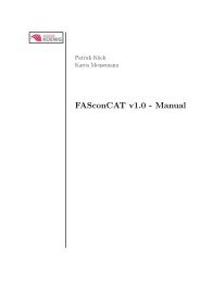
Fig. 2. A. Lateral SEM photography of the groove at the ventral surface of the <strong>cephalic</strong> <strong>shield</strong> of Acteon tornatilis. cs <strong>cephalic</strong><br />
<strong>shield</strong>, gr groove. B. Frontal SEM photography of the mouth region of Acteon tornatilis. cs – <strong>cephalic</strong> <strong>shield</strong>, mo – mouth, f – foot.<br />
4. DISCUSSION<br />
The present study demonstrates the constancy of nervous<br />
structures in the opisthobranch mollusc. Throughout our<br />
investigation of several individuals of the acteonid, A c t e o n<br />
tornatilis we found uniform innervation patterns of the<br />
head region via four cerebral nerves, which can be attributed<br />
to characteristic neuronal cell clusters in the CNS.<br />
These cellular innervation patterns in A. tornatilis show<br />
an extremely high congruence with the cellular innervation<br />
patterns described for the four cerebral nerves of<br />
Haminoea hydatis (STAUBACH et al. in press).<br />
In the N1, the number of cerebral clusters as well as the<br />
position of these clusters to each other is the same in A.<br />
tornatilis and H. hydatis. However, we found some differences<br />
in the size and number of somata when comparing<br />
both species. A d d i t i o n a l l y, we could not detect a pleural,<br />
a parietal and a pedal cluster in A. tornatilis, which<br />
were described for H. hydatis. This may be due to the differences<br />
in the peripheral innervation area of the N1. In<br />
A. tornatilis it only provides for the lip and very small parts<br />
of the median <strong>cephalic</strong> <strong>shield</strong> whereas in H. hydatis, it innervates<br />
the lip and large parts of the anterior <strong>cephalic</strong><br />
<strong>shield</strong>. For the second nerve, the N2 (Nervus labialis), we<br />
nearly found no differences between the presence and distributions<br />
of the cell clusters for both species. The only<br />
ostentatious difference was the lack of a single pedal soma<br />
and its contra-lateral analogue in A. tornatilis. In the<br />
Nclc (Nervus clypei capitis), the difference between the<br />
two species was also reduced to the presence of a single<br />
cerebral soma in A. tornatilis. In contrast to the three<br />
nerves described above, we found a prominent difference<br />
in the structure of the N3 when comparing Acteon and<br />
Haminoea. On the other hand, in H. hydatis the N3 terminates<br />
in a rhinophoral ganglion. Such a ganglion is<br />
missing in A. tornatilis. Hence, we expected considerable<br />
differences in the cellular innervation patterns for the N3<br />
of these species. However, these differences were marg i n-<br />
al and only amounted to the lack of one single cell soma<br />
in the cerebral ganglion of A. tornatilis. This implies that<br />
basic innervation patterns of the N3 are probably the same<br />
in both species. Additional functions of the N3 processed<br />
in the rhinophoral ganglion can be proposed for H. hydatis.<br />
These functions are probably related to the Hancock´s organ,<br />
which is innervated by nerves originating in the<br />
rhinophoral ganglion (STA U B A C H et al. in pre s s). We were<br />
unable to locate such an organ in A. tornatilis in contrast<br />
to earlier descriptions (EDLINGER 1980).<br />
Upon comparing the innervation patterns presented here<br />
for A. tornatilis with those for H. hydatis (STAUBACH et<br />
al. in press) we find constant features of these patterns<br />
across species. This is congruent with other findings that<br />
neuronal structures in the central nervous system of molluscs<br />
and other invertebrates seem to be highly conserved<br />
(CROLL 1987; ARBAS 1991; HAYMAN-PAUL 1991; KUTSCH<br />
& BREIDBACH 1994; NEWCOMB et al. 2006). Hence, we<br />
postulate the N1 of A. tornatilis to be homologous to the<br />
N1 (Nervus oralis) described by HU B E R ( 1 9 9 3 ) for Cephalaspideans.<br />
Additionally, we postulate homologies of the<br />
N2 and the N3 of A. tornatilis to the N2 (Nervus labialis)<br />
and N3 (Nervus rhinophoralis) of Chepalaspideans. This<br />
is congruent to the assumption of HOFFMANN (1939) that<br />
the c3 (after VAYSSIÈRE 1880) of H. hydatis represents the<br />
Nervus labialis and the c4 represents the Nervus tentacularis,<br />
here a synonym for the Nervus rhinophoralis (HU-<br />
B E R 1 9 9 3 ). Our data cannot support ED L I N G E R’S ( 1 9 8 0 ) d e-<br />
scription of independent nerves for the lip organ (N1 after<br />
Edlinger 1980) and the anterior Hancock`s organ (N2<br />
after Edlinger 1980). The Nclc of A. tornatilis also seems<br />
to be homologous to the Nclc of Cephalaspideans (Huber