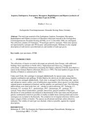
cephalic shield
cephalic shield
cephalic shield

Create successful ePaper yourself
Turn your PDF publications into a flip-book with our unique Google optimized e-Paper software.
Bonner zoologische Beiträge Band 55 (2006) Heft 3/4 Seiten 311–318 Bonn, November 2007<br />
The <strong>cephalic</strong> sensory organs of Acteon tornatilis<br />
(Linnaeus, 1758) (Gastropoda Opisthobranchia) –<br />
cellular innervation patterns as a tool for homologisation*<br />
Sid STAUBACH & Annette KLUSSMANN-KOLB 1)<br />
1) Institute for Ecology, Evolution and Diversity – Phylogeny and Systematics,<br />
J. W. Goethe-University, Frankfurt am Main, Germany<br />
*Paper presented to the 2nd International Workshop on Opisthobranchia, ZFMK, Bonn, Germany, September 20th to 22nd, 2006<br />
Abstract. Gastropoda are guided by several sensory organs in the head region, referred to as <strong>cephalic</strong> sensory organs<br />
(CSOs). This study investigates the CSO structure in the opisthobranch, Acteon tornatilis whereby the innervation patterns<br />
of these organs are described using macroscopic preparations and axonal tracing techniques.<br />
A bipartite <strong>cephalic</strong> <strong>shield</strong> and a lateral groove along the ventral side of the <strong>cephalic</strong> <strong>shield</strong> was found in A. tornatilis.<br />
Four cerebral nerves can be described innervating different CSOs: N1: lip, N2: anterior <strong>cephalic</strong> <strong>shield</strong> and lateral groove,<br />
N3 and Nclc: posterior <strong>cephalic</strong> <strong>shield</strong>. Cellular innervation patterns of the cerebral nerves show characteristic and constant<br />
cell clusters in the CNS for each nerve.<br />
We compare these innervation patterns of A. tornatilis with those described earlier for Haminoea hydatis (STAUBACH et<br />
al. in press). Previously established homologisation criteria are used in order to homologise cerebral nerves as well as<br />
the organs innervated by these nerves. Evolutionary implications of this homologisation are discussed.<br />
Keywords. Haminoea hydatis, Cephalaspidea, axonal tracing, homology, innervation patterns, lip organ, Hancock´s<br />
organ.<br />
1. INTRODUCTION<br />
Gastropoda are guided by several organs in the head region<br />
which are assumed to have primarily chemo- and<br />
mechanosensory functions (AUDESIRK 1979; DAVIS &<br />
MATERA 1982; BICKER et al.1982; EMERY 1992; CHASE<br />
2000; CROLL et al. 2003). In Opisthobranchia, these<br />
<strong>cephalic</strong> sensory organs (CSOs) present an assortment of<br />
forms including rhinophores, labial tentacles, oral veils,<br />
Hancock´s organs and <strong>cephalic</strong> <strong>shield</strong>s. Recent investigations<br />
of CSOs in Opisthobranchia have focussed primarily<br />
on functional aspects (CROLL 1983; BOUDKO et al.<br />
1999; CROLL et al. 2003) while homology of the different<br />
types of CSOs in different taxa has never been investigated<br />
in detail. We want to clarify their homology in separate<br />
evolutionary lineages so as to elucidate key questions<br />
regarding character evolution and phylogeny.<br />
Acteon tornatilis belongs to the subgroup Acteonoidea,<br />
formerly ascribed to the basal Cephalaspidea (ODHNER<br />
1939, BURN & THOMPSON 1998). However, recent investigations<br />
have either excluded the Acteonoidea from the<br />
Opisthobranchia (MIKKELSEN 1996) or proposed a sister<br />
group relationship of Acteonoidea and the highly derived<br />
Nudipleura (VONNEMANN et al. 2005) thus, rendering the<br />
phylogenetic position of Acteonoidea within Opisthobranchia<br />
unsettled. Acteonoidea are characterised by the<br />
presence of a prominent <strong>cephalic</strong> <strong>shield</strong>. This structure is<br />
also present in Cephalaspidea and has been considered to<br />
be an apomorphie of the Cephalaspidea (SC H M E K E L 1 9 8 5 ) .<br />
H o w e v e r, the structure of the <strong>cephalic</strong> <strong>shield</strong>s differs considerably<br />
in Cephalaspidea and Acteonoidea with the latter<br />
possessing two distinct hemispheres while the <strong>cephalic</strong><br />
<strong>shield</strong> in the Cephalaspidea possesses uniform structure.<br />
Therefore, common origin of both types of <strong>cephalic</strong><br />
<strong>shield</strong>s and thus homology is questionable. Further CSOs<br />
have been described in Acteonoidea and Cephalaspidea<br />
such as lip organ and Hancock´s organ (RUDMAN 1971A;<br />
RUDMAN 1971B; RUDMAN 1972a, b; RUDMAN 1972c;<br />
EDLINGER 1980).<br />
Since the presence of these organs in members of the<br />
genus Acteon has been disputed by different authors<br />
(EDLINGER 1980; SCHMEKEL 1985), absolute clarification<br />
is certainly necessary . The intention of this study is to describe<br />
the structure emphasizing the innervation of the<br />
CSOs in the acteonid A. tornatilis. Our descriptions focus<br />
on the cellular innervation patterns reconstructed for
312 Sid STAUBACH & Annette KLUSSMANN-KOLB: Cephalic sensory organ in Aceton<br />
the cerebral nerves using axonal tracing. In an earlier study<br />
(STAUBACH et al. in press) these cellular innervation patterns<br />
were shown to be more adequate in homologising<br />
cerebral nerves than ganglionic origins of nerves (HUBER<br />
1993). By comparising the innervation patterns in A. tor -<br />
n a t i l i s to previously published data on H. hydatis<br />
(STA U B A C H et al. in pre s s), we want to survey whether the<br />
preliminary characteristic cell clusters in the central nervous<br />
system (CNS) of both taxa can be identified by homologising<br />
cerebral nerves across taxa. Based on the homologisation<br />
of the nerves innervating the CSOs we want<br />
to clarify if A. tornatilis has homologous structures to the<br />
CSOs of Cephalaspideans. It is our intent interest that we<br />
shed light on the phylogenetic position and evolutionary<br />
history of Acteonoidea within the Opisthobranchia for future<br />
studies.<br />
2. MATERIALS AND METHODS<br />
2.1. Specimens<br />
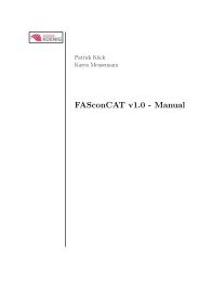
A. tornatilis (Fig. 1A) were collected in the wild at St.<br />
Michel en Greve (Brittany, France). They were then stored<br />
alive at our lab in Frankfurt. Fourty specimens measuring<br />
a shell length between 15 and 20 mm were traced directly<br />
(5 to 15 days after collecting) and five were fixed<br />
for SEM.<br />
2.2. Tracing studies<br />
Animals were relaxed with an injection of 7 % magnesium<br />
chloride. The central nervous system consisting of<br />
the cerebral, pleural and pedal ganglia was removed and<br />
placed in a small Petri dish containing filtered artificial<br />
seawater (ASW; Tropic Marin, Rebie-Bielefeld; GER-<br />
MANY). We then followed the procedures from CROLL<br />
& BAKER (1990) for Ni 2+ -lysine (Ni-Lys) tracing of axons.<br />
Briefly, the nerves of the right cerebral ganglion were<br />
dissected free from the connective tissue. The nerves were<br />
cut and the distal tip was gently drawn into a glass micropipette<br />
using suction provided by an attached 2.5 ml<br />
syringe. Subsequently, the saline in the micropipette was<br />
replaced by a Ni-Lys solution (1.9g NiCl-6H 2 O, 3,5 g L-<br />
Lysine freebase in 20 ml double distilled H 2 O). The preparation<br />
was then incubated for 12–24 hours at 8º C to allow<br />
transport of the tracer. The micropipette was then removed<br />
and the ganglia were washed in ASW three times.<br />
The Ni-Lys was precipitated by the addition of five to ten<br />
drops of a saturated rubeanic acid solution in absolute Dimethylsulfoxide<br />
(DMSO). After 45 minutes the ganglia<br />
were transferred to 4 % paraformaldehyde (PFA) and fixed<br />
for 4–12 hours at 4º C. Thereafter the ganglia were dehydrated<br />
in an increasing ethanol series (70/80/90/99/99%<br />
10 minutes each), cleared in methylsalicylate and mounted<br />
on an objective slide dorsal side up in Entellan (VWR<br />
International) and covered with a cover slip. Ten replicates<br />
were prepared for each cerebral nerve of A. tornatilis.<br />
Samples with only a partial staining of the nerve were not<br />
used because of possible incomplete innervation patterns.<br />
Our criterion for a well-stained preparation was a dark blue<br />
stained nerve indicating intact axons ( FR E D M A N 1 9 8 7 ). T h e<br />
Ni-Lys tracings were analysed by light microscopy (Leica<br />
TCS 4D). Camera lucida drawings were digitalised following<br />
the method of CO L E M A N ( 2 0 0 3 ) adapted for Corel-<br />
DRAW 11. The somata in the innervation scemes occurs<br />
in all replicates. Somata only occurring in single samples<br />
are not considered part of the schematics. The axonal pathways<br />
are estimated over all replicates. Additionally, we<br />
tested for asymmetries making axonal tracings (n = 2 to<br />
3) for each cerebral nerve of the left cerebral ganglion.<br />
2.3. Scanning electron microscopy studies<br />
The specimens were relaxed by an injection of 7 % Mg-<br />
C l 2 in the foot. T h e r e a f t e r, the entire head region was dissected<br />
from the rest of the animal. The CSOs were fixed<br />
in 2,5 % glutaraldehyde, 1 % paraformaldehyde in 0,1M<br />
phosphate buffer (pH 7,2) at room temperature. For the<br />
SEM, the fixed CSOs were dehydrated through a graded<br />
acetone series followed by critical point drying (CPD 030,<br />
BAL-TEC). Finally, they were spattered with gold (Sputter-Coater,<br />
Agar Scientific) and examined with a Hitachi<br />
S4500 SEM. All photographs were taken using DISS (Digital<br />
Image Scanning System – Point Electronic) and subsequently<br />
adjusted for brightness and contrast with Corel<br />
PHOTO-PAINT 11.<br />
3. RESULTS<br />
3.1. Organisation and innervation of the <strong>cephalic</strong> sensory<br />
organs<br />
A. tornatilis possesses a prominent bipartite <strong>cephalic</strong> <strong>shield</strong><br />
(cs) in which each hemisphere of this <strong>shield</strong> is divided into<br />
an anterior and a posterior lobe (Figs. 1A and B). Eyes<br />
are embedded deeply within the tissue of the <strong>shield</strong>. A l o n g<br />
the lateral margin of the anterior lobe of the <strong>cephalic</strong> <strong>shield</strong><br />
a groove is present (Fig. 1B, 2A). Hidden under the cs and<br />
above the foot, the mouth opening is situated at the median<br />
frontal edge (Fig. 2B) surrounded by the lip (not visible<br />
in Figure 2B). We found four nerves innervating the<br />
CSOs (Fig.1B). The N1 (Nervus oralis) provides innervation<br />
to the lip and small median parts of the anterior<br />
<strong>cephalic</strong> <strong>shield</strong>. The bifurcated N2 (Nervus labialis/labiotentacularis)<br />
innervates the complete anterior <strong>cephalic</strong><br />
<strong>shield</strong> whereby the groove at the ventral anterior lobe of<br />
the <strong>cephalic</strong> <strong>shield</strong> is especially innervated. The small N3<br />
(Nervus tentacularis/rhinophoralis) innervates a little re-
Bonner zoologische Beiträge 55 (2006)<br />
313<br />
gion of the posterior <strong>cephalic</strong> <strong>shield</strong>. The Nclc (Nervus<br />
clypei capitis) innervates the largest hind part of the posterior<br />
<strong>cephalic</strong> <strong>shield</strong>. We could not detect a lip organ (Fig.<br />
2B), which according to ED L I N G E R (1980) should comprise<br />
two small lobes on the <strong>cephalic</strong> <strong>shield</strong> above the mouth.<br />
A Hancock´s organ described by EDLINGER (1980) for A.<br />
Tornatilis, here a folded structure separated from the<br />
<strong>cephalic</strong> <strong>shield</strong> was likewise not found in the present study.<br />
3.2. Tracing studies<br />
By conducting the axonal tracing studies we were able to<br />
reconstruct cellular innervation patterns for the four cerebral<br />
nerves of A. tornatilis. Ten replicate tracings were performed<br />
each for the N1 N2, N3 and Nclc using only the<br />
nerves of the right cerebral ganglion. The characteristic<br />
patterns of labelled somata for all nerves are shown in Figure<br />
3A-D, including the approximate pathways of the<br />
stained axons. The identified clusters were named with abbreviations<br />
signifying the ganglion in which they are located,<br />
the nerve filled and a number indicating the order<br />
of their description (for example, Cnlc3: Cerebral Nervus<br />
labialis cluster 3). Nerve cells are grouped in clusters on<br />
the basis of their close positioning in the ganglia and the<br />
tight fasciculation of their axons projecting into the filled<br />
nerve. Asymmetries for tracings of the left nerves could<br />
not be detected.<br />
For the N1 (n = 10) we identified six cerebral clusters<br />
(Cnoc1-6) and one pedal cluster (Pdnoc1) in each sample<br />
(Fig. 3A). The variation between the samples was restricted<br />
to very few somata in some clusters. The cerebral<br />
clusters were distributed over the whole cerebral ganglion.<br />
The pedal cluster Pdnoc1 was located on the anterior margin<br />
of the pedal ganglion above the pedal commissure. T h e<br />
innervation pattern of the N2 (n=10) consists of five cerebral<br />
clusters (Cnlc1-5) and three pedal clusters (Pdnlc1-<br />
3) (Fig. 3B). The cerebral clusters show distinct spatial<br />
separations and are easy to identify. The third traced cerebral<br />
nerve (n = 10) was the N3. Six cerebral (Cnrc1-6) and<br />
three pedal clusters (Pdnrc1-3) were identified (Fig. 3C).<br />
We found an additional single cluster (C c l n rc 1) and a single<br />
soma in the left cerebral ganglion (see arrows in Fig.<br />
3C). The contralateral cluster was located at the base of<br />
the N2 whereas the single soma was found at the root of<br />
the cerebral commissure. We observed slight intraspecific<br />
variability between the ten samples which amounted only<br />
to very few somata in some clusters. In the Nclc, the<br />
innervation (n = 10) pattern consisted of five cerebral clusters<br />
(Cncc1-5) and a single soma at the lateral margin of<br />
the cerebral ganglion above the pedal connective (Fig.<br />
3D). Additionally we found four pedal clusters (Pdncc1-<br />
4). The Nclc had the highest amount of pedal clusters in<br />
all investigated nerves. The number of pedal somata, however,<br />
was comparable to the number of pedal somata for<br />
the N2 innervation pattern (Fig 3B).<br />
Fig. 1. A: Photograph of Acteon tornatilis with the <strong>cephalic</strong> <strong>shield</strong> visible. B: Schematic illustration of the CNS, the four cerebral<br />
nerves (excluding the optical nerve) and the <strong>cephalic</strong> sensory organs of Haminoea hydatis and Acteon tornatilis. Only the right<br />
cerebral nerves are shown. N1 Nervus oralis, N2 Nervus labialis, N3 Nervus rhinophoralis, Nclc Nervus clypei capitis, ey eye, gr<br />
groove, al anterior lobe, pl posterior lobe, sh shell, cs <strong>cephalic</strong> <strong>shield</strong>, f foot.
314 Sid STAUBACH & Annette KLUSSMANN-KOLB: Cephalic sensory organ in Aceton<br />
Fig. 2. A. Lateral SEM photography of the groove at the ventral surface of the <strong>cephalic</strong> <strong>shield</strong> of Acteon tornatilis. cs <strong>cephalic</strong><br />
<strong>shield</strong>, gr groove. B. Frontal SEM photography of the mouth region of Acteon tornatilis. cs – <strong>cephalic</strong> <strong>shield</strong>, mo – mouth, f – foot.<br />
4. DISCUSSION<br />
The present study demonstrates the constancy of nervous<br />
structures in the opisthobranch mollusc. Throughout our<br />
investigation of several individuals of the acteonid, A c t e o n<br />
tornatilis we found uniform innervation patterns of the<br />
head region via four cerebral nerves, which can be attributed<br />
to characteristic neuronal cell clusters in the CNS.<br />
These cellular innervation patterns in A. tornatilis show<br />
an extremely high congruence with the cellular innervation<br />
patterns described for the four cerebral nerves of<br />
Haminoea hydatis (STAUBACH et al. in press).<br />
In the N1, the number of cerebral clusters as well as the<br />
position of these clusters to each other is the same in A.<br />
tornatilis and H. hydatis. However, we found some differences<br />
in the size and number of somata when comparing<br />
both species. A d d i t i o n a l l y, we could not detect a pleural,<br />
a parietal and a pedal cluster in A. tornatilis, which<br />
were described for H. hydatis. This may be due to the differences<br />
in the peripheral innervation area of the N1. In<br />
A. tornatilis it only provides for the lip and very small parts<br />
of the median <strong>cephalic</strong> <strong>shield</strong> whereas in H. hydatis, it innervates<br />
the lip and large parts of the anterior <strong>cephalic</strong><br />
<strong>shield</strong>. For the second nerve, the N2 (Nervus labialis), we<br />
nearly found no differences between the presence and distributions<br />
of the cell clusters for both species. The only<br />
ostentatious difference was the lack of a single pedal soma<br />
and its contra-lateral analogue in A. tornatilis. In the<br />
Nclc (Nervus clypei capitis), the difference between the<br />
two species was also reduced to the presence of a single<br />
cerebral soma in A. tornatilis. In contrast to the three<br />
nerves described above, we found a prominent difference<br />
in the structure of the N3 when comparing Acteon and<br />
Haminoea. On the other hand, in H. hydatis the N3 terminates<br />
in a rhinophoral ganglion. Such a ganglion is<br />
missing in A. tornatilis. Hence, we expected considerable<br />
differences in the cellular innervation patterns for the N3<br />
of these species. However, these differences were marg i n-<br />
al and only amounted to the lack of one single cell soma<br />
in the cerebral ganglion of A. tornatilis. This implies that<br />
basic innervation patterns of the N3 are probably the same<br />
in both species. Additional functions of the N3 processed<br />
in the rhinophoral ganglion can be proposed for H. hydatis.<br />
These functions are probably related to the Hancock´s organ,<br />
which is innervated by nerves originating in the<br />
rhinophoral ganglion (STA U B A C H et al. in pre s s). We were<br />
unable to locate such an organ in A. tornatilis in contrast<br />
to earlier descriptions (EDLINGER 1980).<br />
Upon comparing the innervation patterns presented here<br />
for A. tornatilis with those for H. hydatis (STAUBACH et<br />
al. in press) we find constant features of these patterns<br />
across species. This is congruent with other findings that<br />
neuronal structures in the central nervous system of molluscs<br />
and other invertebrates seem to be highly conserved<br />
(CROLL 1987; ARBAS 1991; HAYMAN-PAUL 1991; KUTSCH<br />
& BREIDBACH 1994; NEWCOMB et al. 2006). Hence, we<br />
postulate the N1 of A. tornatilis to be homologous to the<br />
N1 (Nervus oralis) described by HU B E R ( 1 9 9 3 ) for Cephalaspideans.<br />
Additionally, we postulate homologies of the<br />
N2 and the N3 of A. tornatilis to the N2 (Nervus labialis)<br />
and N3 (Nervus rhinophoralis) of Chepalaspideans. This<br />
is congruent to the assumption of HOFFMANN (1939) that<br />
the c3 (after VAYSSIÈRE 1880) of H. hydatis represents the<br />
Nervus labialis and the c4 represents the Nervus tentacularis,<br />
here a synonym for the Nervus rhinophoralis (HU-<br />
B E R 1 9 9 3 ). Our data cannot support ED L I N G E R’S ( 1 9 8 0 ) d e-<br />
scription of independent nerves for the lip organ (N1 after<br />
Edlinger 1980) and the anterior Hancock`s organ (N2<br />
after Edlinger 1980). The Nclc of A. tornatilis also seems<br />
to be homologous to the Nclc of Cephalaspideans (Huber
Bonner zoologische Beiträge 55 (2006)<br />
315<br />
Fig. 3. Schematic outline of cell clusters providing the N1 (A), N2 (B), N3 (C) and Nclc (D) of Acteon tornatilis. The size and<br />
position of the somata were digitalized from a camera lucida drawing, the distribution of the axons are averaged from all replicates.<br />
N1 Nervus oralis, N2 Nervus labialis, N3 Nervus rhinophoralis, Nclc Nervus clypei capitis, N. opt. Nervus opticus, CG cerebral<br />
ganglia, RhG rhinophoral ganglia, PlG pleural ganglia, PdG pedal ganglia.
316 Sid STAUBACH & Annette KLUSSMANN-KOLB: Cephalic sensory organ in Aceton<br />
1993). HO F F M A N N (1939) described the same nerve as the<br />
Nervus proboscidis. We define this nerve however, as<br />
Nervus clypei capitis according to HUBER (1993).<br />
Considering the homologisation of the cerebral nerves in<br />
light of their neurological origin, neuro-anatomics and<br />
nervous innervation patterns, we postulate hypotheses of<br />
homologies respective of the organs innervated by these<br />
nerves. Thus, we consider the lip of A. tornatilis to be homologous<br />
to the lip of Cephalaspideans (HUBER 1993)<br />
since both organs are innervated by the N1. The same<br />
holds true for the small median parts of the <strong>cephalic</strong> <strong>shield</strong><br />
in Acteon and the anterior <strong>cephalic</strong> <strong>shield</strong> of Haminoea.<br />
We could not find a lip organ in A. tornatilis as described<br />
by EDLINGER (1980), but we detected a groove at the ventral<br />
side of the anterior <strong>cephalic</strong> <strong>shield</strong>. This groove is innervated<br />
by the N2 as is the lip organ of Cephalaspideans<br />
(HUBER 1993). Therefore, we postulate this groove to be<br />
homologous to the lip organ. This hypothesis is also supported<br />
by data on immunoreactivity against several neurotransmitters.<br />
In the groove of A. tornatilis as well as in<br />
the lip organ of H. hydatis, characteristic sub-epidermal<br />
sensory neurons containing catecholamines could be found<br />
in high density indicating that both organs are involved<br />
in contact chemoreception (S. FALLER, Frankfurt, pers.<br />
comm. 2007).<br />
The N2 of Haminoea is divided into two branches which<br />
are described as two single nerves by EDLINGER (1980).<br />
The first or inner branch provides the lip organ as described<br />
earlier. The second, outer branch is related to the<br />
anterior Hancock´s organ ( ED L I N G E R 1980; HU B E R 1 9 9 3 ).<br />
In Acteon we also found two branches of the N2: the inner<br />
one providing the largest part of the groove whereas<br />
the outer branch is restricted to a small region between<br />
the anterior and posterior lobe of the <strong>cephalic</strong> <strong>shield</strong>.<br />
Therefore, this latter region may be homologous to the anterior<br />
Hancock´s organ of H. hydatis and not to the posterior<br />
Hancock´s organ as described by EDLINGER (1980).<br />
The N3 of A. tornatilis provides a large part of the posterior<br />
<strong>cephalic</strong> <strong>shield</strong> but no identifiable posterior Hancock´s<br />
organ. Additional immunohistochemical and ultrastructural<br />
investigations could also not detect a posterior<br />
Hancock´s organ in A. tornatilis (S. FALLER, Frankfurt,<br />
pers. comm. 2007; GÖBBELER & KLUSSMANN-KOLB in<br />
p re s s). The posterior parts of the <strong>cephalic</strong> <strong>shield</strong>s in A c t e o n<br />
and Haminoea are probably equally homologous as both<br />
where innervated by the Nclc.<br />
The lack of a posterior Hancock´s organ in A. tornatilis<br />
might be due to three different reasons: 1. the ancestor of<br />
A. tornatilis never had a posterior Hancock´s organ; 2. the<br />
posterior <strong>cephalic</strong> <strong>shield</strong> of A. tornatilis may be a homologous<br />
structure to the posterior Hancock´s organ of H. hy -<br />
datis; and 3. the posterior Hancock´s organ has secondarily<br />
been reduced in A. tornatilis.<br />
The first hypothesis is rather implausible since we found<br />
a distinct N3 with conserved cellular innervation patterns<br />
in the central nervous system. If the ancestor of A. tornatilis<br />
never had a posterior Hancock´s organ, this nerve<br />
and associated neural structures should be lacking. Moreover,<br />
a Hancock´s organ has been described for other<br />
Acteonoidea (RU D M A N 1 9 7 1a, b; RU D M A N 1972a, b; RU D-<br />
MAN 1972c). If we consider the second explanation for<br />
lack of a posterior Hancock´s organ in A. tornatilis, we<br />
imply that the posterior <strong>cephalic</strong> <strong>shield</strong> in this species, innervated<br />
by the N3, presents a sensory organ as the Hancock´s<br />
organ in Cephalaspidea. However, immunohistochemical<br />
and ultrastructural investigations of the respective<br />
epithelia in A. tornatilis do not indicate a sensory function<br />
at all (S. FALLER, Frankfurt, pers. comm. 2007;<br />
GÖBBELER & KLUSSMANN-KOLB in press). We reject this<br />
hypothesis of homology of the posterior <strong>cephalic</strong> <strong>shield</strong><br />
in A. tornatilis and posterior Hancock´s organ in H. hy -<br />
d a t i s since we found no evidence for a function of the posterior<br />
<strong>cephalic</strong> <strong>shield</strong> as an olfactory sensory organ. Moreo<br />
v e r, the posterior <strong>cephalic</strong> <strong>shield</strong> is mostly innervated by<br />
the Nclc and not by the N3. The third hypothesis regarding<br />
the reduction of a Hancock´s organ seems to be the<br />
most plausible when the habitat and the food sources of<br />
A. tornatilis in comparison to H. hydatis are considered.<br />
The posterior Hancock´s organ is believed to be an olfactory<br />
sensory organ (AU D E S I R K 1979; EM E RY 1 9 9 2). H. hy -<br />
datis feeds on green algae which occur in patches in open<br />
water whereas A. tornatilis is a predator of soft invertebrates<br />
living up to ten centimeters in solid sand (FRETTER<br />
1939; YONOW 1989; own investigations). In such an environment,<br />
an olfactory sensory organ is not plausible<br />
since olfaction or distance chemoreception is generally associated<br />
with water currents, which are not substantial in<br />
a sandy substrate habitat. Here, a contact chemoreceptor,<br />
which is located near the edge of the <strong>cephalic</strong> <strong>shield</strong> is<br />
more plausible. This we witnessed in Acteon tornatilis v i a<br />
its display of a potentially chemoreceptive groove along<br />
the lateral margin of the anterior <strong>cephalic</strong> <strong>shield</strong>.<br />
This assumption of secondary reduction of the Hancock´s<br />
o rgan in the endobenthic A. tornatilis is also supported by<br />
the fact that a Hancock´s organ has been described for other<br />
epibenthic Acteonoidea (e. g. Bullina, Micromelo, Hy -<br />
datina) (RUDMAN 1971a, b; RUDMAN 1972a, b; RUDMAN<br />
1972a,b,c).<br />
Despite all discussion, homology of the described Hancock´s<br />
organs to those in Cephalaspidea cannot undoubtedly<br />
be proposed at this stage, particularly since data on<br />
innervation patterns in these acteonids are lacking to date.<br />
M o r e o v e r, current phylogenetic hypotheses (GR A N D E et al.
Bonner zoologische Beiträge 55 (2006)<br />
317<br />
2004; VO N N E M A N N et al. 2005) regarding Opisthobranchia<br />
propose an independent origin of Acteonoidea<br />
and Cephalaspidea, indicating convergent development of<br />
these sensory organs in both evolutionary lineages. Further<br />
studies will have us utilizing cellular innervation patterns<br />
for CSOs in order to compare several taxa while homologising<br />
the different types of CSOs in Opisthobranchia.<br />
This procedure will enable us to glean a better<br />
understanding of the evolution of these organs.<br />
Acknowledgements. Marc Hasenbank, Patrick Schultheiss and<br />
Christiane Weydig were constructive in locating the Acteon tor -<br />
natilis population at St. Michel en Greve. Thanks to Katrin<br />
Göbbeler and Simone Faller for their personal commitments.<br />
Roger Croll was always very helpful in discussing tracing patterns<br />
and CSOs. We are grateful to Angela Dinapoli, Adrienne<br />
Jochum and Alen Kristof for their comments on an earlier version<br />
of this manuscript.<br />
This study was supported by the German Science Foundation,<br />
KL 1303/3-1 and by the Verein der Freunde und Förderer der<br />
Johann-Wolfgang-Goethe Universität.<br />
Also thanks to an unknown referee who provided valuable comments<br />
on the manuscript.<br />
REFERENCES<br />
ARBAS, E. A. 1991. Evolution in nervous systems. Annual Review<br />
in Neuroscience 14: 9–38.<br />
AUDESIRK, T. E. 1979. Oral mechanoreceptors in Tritonia<br />
diomedea. Journal of Comparative Physiology 130: 71–78.<br />
BICKER, G., DAVIS, W. J. & MATERA, E. M. 1982. Chemoreception<br />
and mechanoreception in the gastropod mollusc Pleuro -<br />
branchea californica. Journal of Comparative Physiology 1 4 9:<br />
235–250.<br />
BO C K, W. J. 1989. The homology concept: its philosophical foundation<br />
and practical methodology. Zoologische Beiträge (Neue<br />
Folge) 32: 327–353.<br />
BOUDKO, D. Y., SWITZER-DUNLAP, M. & HADFIELD, M. G. 1999.<br />
Cellular and subcellular structure of anterior sensory pathways<br />
in Phestilla sibogae (Gastropda, Nudibranchia). Journal of<br />
Comparative Neurology 403: 39–52.<br />
BURN, R. & THOMPSON, T. 1998. Order Cephalaspidea. Pp<br />
943–959 in: BE E S L E Y, P. L., RO S S, G. J. B. & WE L L S, A . ( e d s . )<br />
Mollusca: The Southern Synthesis. Fauna of Australia. Vol.<br />
5, Part B. CSIRO Publishing, Melbourne.<br />
CHASE, R. 2000. Behavior and its neural control in gastropod<br />
molluscs. Oxford University Press, New York.<br />
COLEMAN, C. O. 2003. “Digital inking”: how to make perfect<br />
line drawings on computers.<br />
O rganisms, Diversity and Evolution 3: Electronical Supplement<br />
14: 1–14.<br />
CROLL, R. P. 1983. Gastropod chemoreception. Biological Reviews,<br />
Supplement 3, 58: 293–319.<br />
CROLL, R. P. 1987. Identified neurons and cellular homologies.<br />
Pp. 41–59 in: ALI, M.A. (ed.) Nervous Systems in Invertebrates.<br />
CR O L L, R. P. & BA K E R, M. 1990. Axonal regeneration and sprouting<br />
following injury to the cerebral-buccal connective in the<br />
snail Achatina fulica. The Journal of Comparative Neurology<br />
300: 273–286.<br />
CR O L L, R. P., BO U D K O, D. Y., PI R E S, A. & HA D F I E L D, M. G. 2003.<br />
Transmitter content of cells and fibers in the <strong>cephalic</strong> sensory<br />
organs of the gastropod mollusc Phestilla sibogae. Cell Ti s-<br />
sue Research 314: 437–448.<br />
DAV I S, W. J. & MAT E R A, E. M. 1982. Chemoreception in gastropod<br />
molluscs: electron microscopy of putative receptor cells.<br />
Journal of Neurobiology 13(1): 79–84.<br />
EDLINGER, K. 1980. Zur Phylogenie der chemischen Sinnesorgane<br />
einiger Cephalaspidea<br />
(Mollusca, Opisthobranchia). Zeitschrift für Zoologie, Systematik<br />
und Evolutionsforschung. 18: 241–256.<br />
EMERY, D. G. 1992. Fine structure of olfactory epithelia of gastropod<br />
molluscs. Microscopy Research and Technique 22:<br />
307–324.<br />
FREDMAN, S. M. 1987. Intracellular staining of neurons with<br />
nickel-lysine. Journal of Neuroscience Methods 2 0( 3 ) :<br />
181–194.<br />
FRETTER, V. 1939. The structure and function of the alimentary<br />
canal of some tectibranch molluscs, with a note on extraction.<br />
Transactions of the Royal Society of Edinborough 5 9:<br />
599–646.<br />
GÖBBELER, K. & KLUSSMAN-KOLB, A. 2007. A comparative ultrastructural<br />
investigation of the <strong>cephalic</strong> sensory organs in<br />
Opisthobranchia (Mollusca, Gastropoda). Tissue Cell, doi<br />
10.1016/j.tice.2007.07.02.<br />
GR A N D E, C., TE M P L A D O, J., CE RV E R A, J.L. & ZA R D O YA, R. 2004.<br />
Phylogenetic relationships among Opisthobranchia (Mollusca<br />
: Gastropoda) based on mitochondrial cox 1, trnV, and rrnL<br />
genes. Molecular Phylogenetics and Evolution 3 3( 2 ) :<br />
378–388.<br />
HANCOCK, A. 1852. Observations on the olfactory apparatus in<br />
the Bullidae. Annual Magazine of Natural History 2: 9.<br />
HAYMAN-PAUL, D. 1991. Pedigrees of neurobehavioral circuits:<br />
tracing the evolution of novel behaviours by comparing motor<br />
patterns, muscles and neurons in members of related taxa.<br />
Brain Behavioural Evolution 38: 226–239.<br />
HOFFMANN, H. 1939. Mollusca. I Opisthobranchia. Pp. 1–1248<br />
in: BR O N N S, H. G. (ed.) Klassen und Ordnungen des Ti e r r e i c h s<br />
III (1). Akademische-Verlagsgesellschaft, Leipzig.<br />
HUBER, G. 1993. On the cerebal nervous system of marine Heterobranchia<br />
(Gastropoda). Journal of Molluscan Studies 59:<br />
381–420.<br />
KUTSCH, W. & BREIDBACH, O. 1994. Homologous structures in<br />
the nervous systems of arthropoda. Advances in Insect Physiology<br />
24: 2–113.<br />
NEWCOMB, J.M., FICKBOHM, D.J. & KATZ, P.S. 2006. Comparative<br />
mapping of serotonin-immunoreactive neurons in the central<br />
nervous system of nudibranch molluscs. The Journal of<br />
Comparative Neurology 499: 485–505.<br />
MI K K E L S E N, P. M . 1996. The evolutionary relationships of<br />
Cephalaspidea s.l. (Gastropoda; Opisthobranchia): a phylogenetic<br />
analysis. Malacologia 37: 375–442.<br />
ODHNER, N.H. 1939. Opisthobranchiate Mollusca from the western<br />
and northern coasts of Norway. Kongelige Norske Videnskabernes<br />
Selskabs Skrifter NR No. 1: 1–93.<br />
RUDMAN, W.B. 1971a. The genus Bullina (Opisthobranchia) in<br />
New Zealand. Journal of the Malacological Society of Australia<br />
2(2): 195–203.<br />
RU D M A N, W. B. 1971b. The family Acteonidae (Opisthobranchia,<br />
Gastropoda) in New Zealand. Journal of the Malacological Society<br />
of Australia 2(2): 205–214.<br />
RUDMAN, W.B. 1972a. The anatomy of the opisthobranch genus<br />
H y d a t i n a and the functioning of the mantle cavity and alimentary<br />
canal. Zoological Journal of the Linnean Society 51:<br />
121–139.
318 Sid STAUBACH & Annette KLUSSMANN-KOLB: Cephalic sensory organ in Aceton<br />
RUDMAN, W.B. 1972b. Studies on the primitive opisthobranch<br />
genera Bullina Fèrussac and Micromelo Pilsbry. Zoological<br />
Journal of the Linnean Society 51: 105–119.<br />
RU D M A N, W. B. 1972c. A study of the anatomy of P u p a and M a x -<br />
a c t e o n (Acteonidae, Opisthobranchia), with an account of the<br />
breeding cycle of Pupa kirki. Journal of Natural History 6:<br />
603–619.<br />
SCHMEKEL, L. 1985. Aspects of evolution within the opisthobranchs.<br />
Pp. 221–267 in: TRUEMAN, E.R. & CLARKE, M.R.<br />
(eds.) The Mollusca. Vol. 1 0 Evolution. Academic Press, London.<br />
STAUBACH, S.E., SCHÜTZNER, P., CROLL, R.P. & KLUSSMANN-<br />
KOLB, A.Innervation patterns of the cerebral nerves in Hami -<br />
noea hydatis (Linnaeus 1758) (Gastropoda, Opisthobranchia)<br />
– A test for intraspecific variability. In press, Zoomorphology<br />
(2007).<br />
VAY S S I È R E, A . 1980. Recherches anatomiques sur les Mollusques<br />
de la famille des Bullidés Annales des Sciences Naturelle Zoologie<br />
6: 9.<br />
VO N N E M A N N, V., SC H R Ö D L, M., KL U S S M A N N- KO L B, A. &<br />
WÄGELE, H. 2005. Reconstruction of the phylogeny of the<br />
Opisthobranchia (Mollusca, Gastropoda) by means of 18S and<br />
28S rDNA sequences. Journal of Molluscan Studies 71:<br />
113–125.<br />
YO N O W, N. 1989. Feeding observations on Acteon tornatilis ( L i n-<br />
naeus) (Opisthobranchia, Acteonoidae). Journal of Molluscan<br />
Studies 55(1): 97–102.<br />
Author´s adresses: Sid STA U B A C H (corresponding author)<br />
and Annette KL U S S M A N N- KO L B, Institute for Ecology, Evolution<br />
and Diversity – Phylogeny and Systematics, J. W.<br />
G o e t h e - U n i v e r s i t y, Siesmayerstraße 70, 60054, Frankfurt<br />
am Main, Germany. E-mail: Staubach@bio.unifrankfurt.de;<br />
Klussmann-Kolb@bio.uni-frankfurt.de.