Sample Chapter 10 from the Textbook (35559.0K) - McGraw-Hill
Sample Chapter 10 from the Textbook (35559.0K) - McGraw-Hill
Sample Chapter 10 from the Textbook (35559.0K) - McGraw-Hill

Create successful ePaper yourself
Turn your PDF publications into a flip-book with our unique Google optimized e-Paper software.
350 PART 2 Support and Movement<br />
Table <strong>10</strong>.21 Muscles of <strong>the</strong> Thigh (see figures <strong>10</strong>.30 and <strong>10</strong>.32)<br />
Muscle Origin Insertion Nerve Action<br />
Anterior Compartment<br />
Quadriceps femoris<br />
(kwah′dri-seps fem′ŏ-ris)<br />
Sartorius<br />
(sar-tōr′ē-ū s)<br />
Medial Compartment<br />
Adductor brevis<br />
(a-dŭk′ter, a-dŭk′tōr<br />
brev′is)<br />
Adductor longus<br />
(a-dŭk′ter, a-dŭk′tōr<br />
lon′gŭs)<br />
Adductor magnus<br />
(a-dŭk′ter, a-dŭk′tōr<br />
mag′nŭs)<br />
Rectus femoris—anterior inferior<br />
iliac spine<br />
Vastus lateralis—greater trochanter<br />
and linea aspera of femur<br />
Vastus intermedius—body of femur<br />
Vastus medialis—linea aspera of femur<br />
Anterior superior iliac spine<br />
Pubis<br />
Patella and onto tibial<br />
tuberosity through<br />
patellar ligament<br />
Medial side of tibial<br />
tuberosity<br />
Pectineal line and linea<br />
aspera of femur<br />
Femoral<br />
Femoral<br />
Obturator<br />
Extends knee; rectus femoris<br />
also flexes hip<br />
Flexes hip and knee; rotates<br />
thigh laterally and leg medially<br />
Adducts and laterally rotates<br />
thigh; flexes hip<br />
Pubis Linea aspera of femur Obturator Adducts and laterally rotates<br />
thigh; flexes hip<br />
Adductor part: pubis and ischium<br />
Hamstring part: ischial tuberosity<br />
Adductor part: linea<br />
aspera of femur<br />
Hamstring part: adductor<br />
tubercle of femur<br />
Adductor part: obturator<br />
Hamstring part: tibial<br />
Adductor part: adducts thigh<br />
and flexes hip<br />
Hamstring part: extends hip<br />
and adducts thigh<br />
Gracilis (gras′i-lis) Pubis near symphysis Tibia Obturator Adducts thigh; flexes knee<br />
Pectineus (pek′ti-nē′ŭs) Pubic crest Pectineal line of femur Femoral and obturator Adducts thigh; flexes hip<br />
Posterior Compartment<br />
Biceps femoris<br />
(bī ′seps fem′ŏ-ris)<br />
Semimembranosus<br />
(sem′ē-mem-bră-nō′sŭs)<br />
Semitendinosus<br />
(sem′ē-ten-di-nō′sŭs)<br />
Long head—ischial tuberosity Head of fibula Long head—tibial Flexes knee; laterally rotates leg;<br />
extends hip<br />
Short head—femur<br />
Short head—<br />
common fibular<br />
Ischial tuberosity<br />
Medial condyle of tibia<br />
and collateral ligament<br />
Tibial<br />
Flexes knee; medially rotates<br />
leg; tenses capsule of knee joint;<br />
extends hip<br />
Ischial tuberosity Tibia Tibial Flexes knee; medially rotates leg;<br />
extends hip<br />
been identified based on <strong>the</strong>ir location in <strong>the</strong> thigh and are organized<br />
into compartments: The muscles of <strong>the</strong> anterior compartment<br />
flex <strong>the</strong> hip and/or extend <strong>the</strong> knee (see figure <strong>10</strong>.28a); <strong>the</strong><br />
muscles of <strong>the</strong> medial compartment adduct <strong>the</strong> thigh (figure <strong>10</strong>.30);<br />
and <strong>the</strong> muscles of <strong>the</strong> posterior compartment extend <strong>the</strong> hip and<br />
flex <strong>the</strong> knee (figure <strong>10</strong>.31).<br />
The anterior thigh muscles are <strong>the</strong> quadriceps femoris<br />
(fem′ŏ-ris) and <strong>the</strong> sartorius (sar-tōr′ē-ŭs; see table <strong>10</strong>.20 and<br />
figure <strong>10</strong>.28a). The quadriceps femoris is actually four muscles:<br />
<strong>the</strong> rectus femoris, <strong>the</strong> vastus lateralis, <strong>the</strong> vastus medialis, and <strong>the</strong><br />
vastus intermedius. The quadriceps group extends <strong>the</strong> knee. The<br />
rectus femoris also flexes <strong>the</strong> hip because it crosses both <strong>the</strong> hip<br />
and knee joints.<br />
The quadriceps femoris makes up <strong>the</strong> large mass on <strong>the</strong><br />
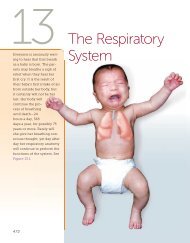
anterior thigh (see figure <strong>10</strong>.28c). The vastus lateralis is sometimes<br />
used as an injection site, especially in infants who do not have<br />
well-developed deltoid or gluteal muscles. The muscles of <strong>the</strong><br />
quadriceps femoris have a common insertion, <strong>the</strong> patellar tendon,<br />
on and around <strong>the</strong> patella. The patellar ligament is an extension of<br />
<strong>the</strong> patellar tendon onto <strong>the</strong> tibial tuberosity. The patellar ligament<br />
is <strong>the</strong> point that is tapped with a rubber hammer when testing<br />
<strong>the</strong> knee-jerk reflex in a physical examination.<br />
The sartorius is <strong>the</strong> longest muscle of <strong>the</strong> body, crossing <strong>from</strong><br />
<strong>the</strong> lateral side of <strong>the</strong> hip to <strong>the</strong> medial side of <strong>the</strong> knee. As <strong>the</strong><br />
muscle contracts, it flexes <strong>the</strong> hip and knee and laterally rotates <strong>the</strong><br />
thigh. This is <strong>the</strong> action required for crossing <strong>the</strong> legs.<br />
The medial thigh muscles (see figure <strong>10</strong>.30) are involved primarily<br />
in adduction of <strong>the</strong> thigh. Some of <strong>the</strong>se muscles also laterally<br />
rotate <strong>the</strong> thigh and/or flex or extend <strong>the</strong> hip. The gracilis also<br />
flexes <strong>the</strong> knee.