Intraoral Radiography: Positioning and Radiation ... - IneedCE.com
Intraoral Radiography: Positioning and Radiation ... - IneedCE.com
Intraoral Radiography: Positioning and Radiation ... - IneedCE.com
Create successful ePaper yourself
Turn your PDF publications into a flip-book with our unique Google optimized e-Paper software.
Educational Objectives<br />
Upon <strong>com</strong>pletion of this course, the clinician will be able to<br />
do the following:<br />
1. Underst<strong>and</strong> the various types of intraoral radiographs<br />
that can be taken <strong>and</strong> what these are used for<br />
2. Know how to correctly use the paralleling <strong>and</strong> bisecting<br />
techniques to take intraoral radiographs<br />
3. Know <strong>com</strong>mon errors that occur when taking intra-oral<br />
radiographs <strong>and</strong> how to avoid these<br />
4. Know how to minimize radiation exposure for patients<br />
<strong>and</strong> the operator<br />
Abstract<br />
Several types of intraoral radiographs can be taken. An underst<strong>and</strong>ing<br />
of both the paralleling <strong>and</strong> bisecting techniques<br />
<strong>and</strong> when to use these is necessary. Avoiding <strong>com</strong>mon errors<br />
when taking intraoral radiographs reduces the need for retakes.<br />
Minimizing radiation exposure for patients <strong>and</strong> the<br />
operator is an essential <strong>com</strong>ponent of intraoral radiography.<br />
Introduction<br />
X-rays were discovered in 1895 by Professor Wilhelm Conrad<br />
Roentgen, <strong>and</strong> Dr. Otto Walkhoff is credited with the first<br />
dental radiograph. Until the 1980s, dental radiographs were<br />
typically captured using film. Dr. Frances Mouyens invented<br />
direct digital radiography to take intraoral dental radiographs<br />
in 1984, <strong>and</strong> this technology was introduced into the U.S. in<br />
1989. While the use of digital radiography in dentistry continues<br />
to gain strength, film-based radiographs are still more<br />
<strong>com</strong>mon. The <strong>com</strong>plete transition to digital radiography is<br />
just a matter time.<br />
<strong>Intraoral</strong> dental radiographs fall into two main categories:<br />
bite-wings <strong>and</strong> periapicals. Bite-wing radiographs are the<br />
best diagnostic tool available for the detection of interproximal<br />
caries <strong>and</strong> assessment of alveolar bone levels. Bite-wings<br />
are usually taken in the posterior regions of the mouth.<br />
However, size 1 bite-wings can be taken of the anterior<br />
teeth to assess anterior bone levels. Periapical radiographs<br />
record the entire tooth <strong>and</strong> supporting bone <strong>and</strong> are used to<br />
evaluate the extent of caries <strong>and</strong> periodontal bone loss <strong>and</strong><br />
aid in the diagnosis <strong>and</strong> treatment of root <strong>and</strong> bony pathoses.<br />
Periapicals <strong>and</strong> bite-wings can be <strong>com</strong>bined to form surveys<br />
of varying configurations, for a <strong>com</strong>prehensive view of the<br />
entire dentition. <strong>Intraoral</strong> radiographs can be captured using<br />
film or digital receptors. Digital receptors are available<br />
as wired <strong>and</strong> wireless rigid sensors (CCD — charge-coupled<br />
device; CMOS — <strong>com</strong>plementary metal oxide semiconductor)<br />
<strong>and</strong> photostimulable phosphor plates. Both systems are<br />
<strong>com</strong>puter-based technologies that require specific hardware<br />
<strong>and</strong> software <strong>com</strong>ponents for operation. Digital receptors are<br />
available in sizes <strong>com</strong>parable to film, mostly typically sizes 0,<br />
1, <strong>and</strong> 2.<br />
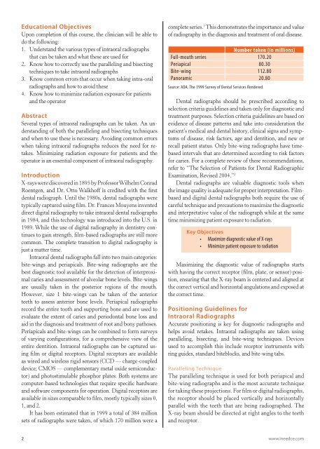
It has been estimated that in 1999 a total of 384 million<br />
sets of radiographs were taken, of which 170 million were a<br />
<strong>com</strong>plete series. 1 This demonstrates the importance <strong>and</strong> value<br />
of radiography in the diagnosis <strong>and</strong> treatment of oral disease.<br />
Number taken (in millions)<br />
Full-mouth series 170.20<br />
Periapical 80.30<br />
Bite-wing 112.80<br />
Panoramic 20.80<br />
Source: ADA. The 1999 Survey of Dental Services Rendered.<br />
Dental radiographs should be prescribed according to<br />
selection criteria guidelines <strong>and</strong> taken only for diagnostic <strong>and</strong><br />
treatment purposes. Selection criteria guidelines are based on<br />
evidence of disease patterns <strong>and</strong> take into consideration the<br />
patient’s medical <strong>and</strong> dental history, clinical signs <strong>and</strong> symptoms<br />
of disease, risk factors, age <strong>and</strong> dentition, <strong>and</strong> new or<br />
recall patient status. Only bite-wing radiographs have timebased<br />
intervals that are determined according to risk factors<br />
for caries. For a <strong>com</strong>plete review of these re<strong>com</strong>mendations,<br />
refer to “The Selection of Patients for Dental Radiographic<br />
Examination, Revised 2004.” 2<br />
Dental radiographs are valuable diagnostic tools when<br />
the image quality is adequate for proper interpretation. Filmbased<br />
<strong>and</strong> digital dental radiographs both require the use of<br />
careful technique <strong>and</strong> precautions to maximize the diagnostic<br />
<strong>and</strong> interpretative value of the radiograph while at the same<br />
time minimizing patient exposure to radiation.<br />
Key Objectives<br />
• Maximize diagnostic value of X-rays<br />
• Minimize patient exposure to radiation<br />
Maximizing the diagnostic value of radiographs starts<br />
with having the correct receptor (film, plate, or sensor) position,<br />
ensuring that the X-ray beam is centered <strong>and</strong> aligned at<br />
the correct vertical <strong>and</strong> horizontal angulations <strong>and</strong> exposed at<br />
the correct time.<br />
<strong>Positioning</strong> Guidelines for<br />
<strong>Intraoral</strong> Radiographs<br />
Accurate positioning is key for diagnostic radiographs <strong>and</strong><br />
helps avoid retakes. <strong>Intraoral</strong> radiographs are taken using<br />
paralleling, bisecting, <strong>and</strong> bite-wing techniques. Devices<br />
used to ac<strong>com</strong>plish this include receptor instruments with<br />
ring guides, st<strong>and</strong>ard biteblocks, <strong>and</strong> bite-wing tabs.<br />
Paralleling Technique<br />
The paralleling technique is used for both periapical <strong>and</strong><br />
bite-wing radiographs <strong>and</strong> is the most accurate technique<br />
for taking these projections. For film or digital radiographs,<br />
the receptor should be placed vertically <strong>and</strong> horizontally<br />
parallel with the teeth that are being radiographed. The<br />
X-ray beam should be directed at right angles to the teeth<br />
<strong>and</strong> receptor.<br />
2 www.ineedce.<strong>com</strong>


